Production, stability and degradation of Trichoderma gliotoxin in growth medium, irrigation water and agricultural soil
←
→
Page content transcription
If your browser does not render page correctly, please read the page content below
www.nature.com/scientificreports
OPEN Production, stability
and degradation of Trichoderma
gliotoxin in growth medium,
irrigation water and agricultural
soil
R. Jayalakshmi1,6, R. Oviya1,6, K. Premalatha1,6, S. T. Mehetre2, M. Paramasivam3,
R. Kannan5, M. Theradimani1, M. S. Pallavi4, Prasun K. Mukherjee2 & V. Ramamoorthy1*
Gliotoxin produced by Trichoderma virens is inhibitory against various phytopathogenic fungi and
bacteria. However, its stability in soil-ecosystem has not yet been well-defined. This study aimed
to decipher its persistence and behaviour in growth media, irrigation water and soil ecosystems.
Gliotoxin production was noticed at logarithmic growth phase and converted into bis-thiomethyl
gliotoxin at late stationary growth phase of T. virens in acidic growth medium. But, no gliotoxin
production was observed in neutral and alkaline growth medium. Gliotoxin was stable for several
days in acidic water but degraded in alkaline water. Degradation of gliotoxin was more in unsterile
soil than sterile soil and also that was higher under wet soil than dry soil. Degradation of gliotoxin
was hastened by alkaline pH in wet soil but not in dry soil. Under unsterile soil conditions, high soil
moisture increased the degradation of gliotoxin and the degradation of gliotoxin occurred quickly
in alkaline soil (in 5 days) compared to acidic soil (in 10 days). Under sterile soil conditions, high soil
moisture also enhanced the degradation of gliotoxin but level of degradation was less compared to
unsterile conditions. Thus, gliotoxin stability is influenced mainly by the soil wetness, soil microbial
community and pH conditions.
Abbreviations
TLC Thin layer chromatography
LC-TandemMS Liquid chromatography tandem mass spectrometry
ESI Electro spray ionization
TSM Trichoderma selective medium
PDA Potato dextrose agar medium
PGPF Plant growth promoting fungus
Trichoderma species mainly survive as soil saprophyte and can be isolated from agricultural soil, decaying
woods, spent mushroom bed, tree bark and agricultural waste using TSM medium1. Among the various species
of Trichoderma, only “Q” strains of T. virens produce g liotoxin2–7. Besides T. virens, gliotoxin is also produced
by a few other soil-dwelling fungi such as Eurotium chevalieri, Neosartorya pseudofischeri, Aspergillus fumigatus,
some Penicillium and Acremonium species3,8.
Gliotoxin is the most forestanding member of the epipolythiopiperazines, a large class of natural products
featuring a diketopiperazine having di- or polysulfide linkage with potent antimicrobial activity. Gliotoxin is
1
Department of Plant Pathology, Agricultural College and Research Institute, Tamil Nadu Agricultural University,
Madurai, Tamil Nadu, India. 2Nuclear Agriculture and Biotechnology Division, Bhabha Atomic Research Centre,
Mumbai, India. 3Pesticide Toxicology Laboratory, Department of Agricultural Entomology, Tamil Nadu Agricultural
University, Coimbatore, Tamil Nadu, India. 4Pesticide Residue and Food Quality Analysis Laboratory, University
of Agricultural Sciences, Raichur, Karnataka, India. 5Department of Plant Pathology, Agricultural College and
Research Institute, Tamil Nadu Agricultural University, Killikulam, Tamil Nadu, India. 6These authors contributed
equally: R. Jayalakshmi, R. Oviya and K. Premalatha. *email: rvrmoorthy@yahoo.com
Scientific Reports | (2021) 11:16536 | https://doi.org/10.1038/s41598-021-95907-6 1
Vol.:(0123456789)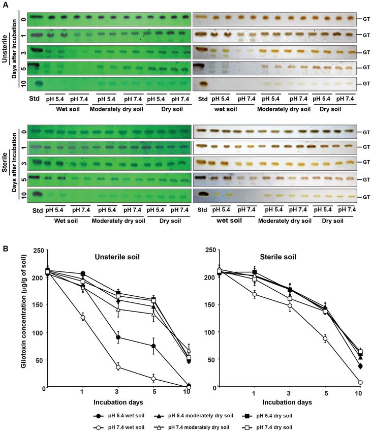
www.nature.com/scientificreports/
the second antibiotic discovered next to penicillin. It has broad spectrum antifungal and antibacterial activity.
Either purified gliotoxin or gliotoxin producing T. virens are effective in inhibition of several phyto-pathogenic
fungi such as Rhizoctonia solani, Botrytis cinerea, Colletotrichum spp., Pythium ultimum, Fusarium spp. etc.5,9–13.
Antibiosis by gliotoxin production was proved as an important mechanism in biocontrol activity of T. virens. The
suppression of soil-borne diseases was positively correlated with the accumulation of gliotoxin produced by T.
virens14,15. Mutant strains of T. virens lacking gliotoxin production were less effective for the control of damping
off disease caused by Pythium spp. and R. solani in cotton and zinnia plants compared to its isogenic wild-type
strain16,17. Gliotoxin producing T. virens strain G-20 was the first microbial antagonist commercially formulated
and marketed as a bio-pesticide in the name of SoilGard (Certis, USA)14,15. In addition to crop protection, T.
virens also enhances the plant growth and yield potential by producing plant growth promoting substances such
as indole acetic a cids18–21. Thus, it is called as plant growth promoting fungus.
T. virens produced enormous amount of gliotoxin in liquid10,22 and solid media23,24. T. virens also produced
gliotoxin when it was applied in soil containing lignocellulosic materials such as paddy s traw13–15,25. Gliotoxin
presence was noticed in the soil sown with T. virens treated s eeds13. Application of T. virens in soil followed
by planting cotton seed resulted in accumulation of gliotoxin in cotton spermosphere and surrounding s oil21.
Among the gliotoxin producing fungi, A. fumigatus is an opportunistic human and animal pathogen and
gliotoxin production in A. fumigatus has been found as one of the virulence factors in causing a spergillosis26,27.
Several strains of clinical and non-clinical isolates of A. fumigatus produce gliotoxin. Gliotoxin of T. virens func-
tions as antibiotic and biopesticide and thus it is considered as beneficial factor in agricultural system. Whereas
gliotoxin of A. fumigatus is considered as detrimental factor and mycotoxin in human and animal health system8.
Though gliotoxin production by T. virens has been well documented for suppression of soil-borne pathogens,
its persistence and behaviour in soil ecosystem have not been well described. There is an urgent need to study
its stability in soil- ecosystem for judging its bio-efficacy and biosafety in agriculture system. Thus, the present
study was carried out with the following objectives. Firstly, to study the metabolism and stability of gliotoxin
in growth media compared with its stability in water samples used for irrigation of crop plants. Secondly, to
decipher its stability and behaviour in acidic and alkaline garden land soil under different soil moisture regime
and sterile and unsterile status.
Results
Gliotoxin metabolism in T. virens. Using gliotoxin amended medium (selective medium for isolation of
gliotoxin producing Trichoderma strains), 10 strains of gliotoxin producing Trichoderma species were isolated
from soil (Fig. 1A,B). They were identified as T. virens based on Gliocladium type of conidiophore and ITS
sequence analysis (data not shown). Among these strains, T. virens strain TK1 produced the highest amount of
gliotoxin as that of T. virens strain MTCC 2977, gliotoxin producing strain used as reference strain. Thus, in this
study, T. virens strain TK1 was used for the analysis of gliotoxin metabolism.
We tested the production, persistence and dissipation levels of gliotoxin in the growth medium during its
various growth stages. Gliotoxin production started from the second day after inoculation of T. virens and its
accumulation was maximum on the sixth day of incubation in Weindling medium and there after its accumula-
tion declined sharply. On the tenth day of incubation, its level of accumulation reached to the minimum and no
visible gliotoxin band was observed. A fluorescent compound with lower Rf value (0.44) appeared from the sixth
day onwards in the medium as revealed from an extra band irrespective to the standard gliotoxin (Rf value 0.65).
Since, this new compound started to appear at the time point when the amount of accumulated gliotoxin started
to decline (6th day of incubation) and also the new compound reached to the maximum level when gliotoxin
totally disappeared in the medium, we assumed that gliotoxin was converted to modified gliotoxin (Fig. 2A,B).
Both gliotoxin and modified gliotoxin from the 4th day and 10th day old growth medium were separated and
purified using preparative TLC and they were subjected to LC-Tandem MS analysis, which showed that glio-
toxin and modified gliotoxin were eluted at the retention time of 3.924 and 3.942 min, respectively. The selected
MRM transitions for gliotoxin were m/z 327 > 263.15 as the quantitation transition and m/z 327 > 227.10 as the
qualitative transition. For modified gliotoxin, m/z 356.60 was selected as the precursor ion, and its quantitative
and qualitative product ions were m/z 309.05 and m/z 243.05, respectively. LC-Tandem MS analysis revealed that
modified gliotoxin had molecular weight of 357 Dalton corresponding to bis-thiomethyl gliotoxin. Gliotoxin
had molecular weight of 327 Dalton as expected (Fig. 3).
Mycelial dry weight reached the highest level on the fourth day, and there after its biomass remained at the
same level indicating the growth stage reached to stationary phase on the fourth day and the logarithmic phase
occurred between the second and fourth day of incubation (Fig. 2C). This indicated that T. virens produced
gliotoxin at the early logarithmic growth phase and converted it into bis-thiomethyl gliotoxin at late growth
phase. Since, gliotoxin stability is influenced by pH status, the same was measured in the growth medium. pH
of the buffered Weindling medium remained around four and there was no significant change in pH conditions
of the medium from the time of inoculation to 15 days of incubation (Fig. 2C).
When the mycelia of T. virens were analysed for cellular gliotoxin and bis-thiomethyl gliotoxin contents,
gliotoxin was present at logarithmic growth phase and converted into bis-thiomethyl gliotoxin in late growth
phase. Thus, gliotoxin is converted into bis-thiomethyl gliotoxin in cells of T. virens at stationary growth phase
and released into culture medium (Fig. S1).
T. virens produces and accumulates gliotoxin in acidic media but not in basic media. Earlier
studies indicated that gliotoxin was stable in acidic conditions but unstable in basic conditions. At the begin-
ning, we assessed the metabolism and stability of gliotoxin in acidic Weindling medium (pH 4). Thus, we again
assessed its metabolism in growth medium at different pH levels for a period of 30 days.
Scientific Reports | (2021) 11:16536 | https://doi.org/10.1038/s41598-021-95907-6 2
Vol:.(1234567890)
www.nature.com/scientificreports/

                                  Figure 1.  Isolation of gliotoxin producing T. virens strains from soil. (A) Gliotoxin producing T. virens strains
                                  were isolated and cultured on PDA medium. (B) Analysis of gliotoxin production by T. virens strains using TLC.
                                  Four days old culture filtrates were extracted with chloroform. Extracts were spotted on TLC plates and sprayed
                                  with silver nitrate. Presence of brown colour band indicates the gliotoxin. GT gliotoxin, Std standard gliotoxin.

                                      The production of gliotoxin started in two days after inoculation of T. virens in Weindling medium at pH 3,
                                  4 and 5 whereas its production was noticed in the third day at pH 6 and its stability lasted for five to seven days
                                  of culturing period. Seven days after incubation, the gliotoxin was converted into bis-thiomethyl gliotoxin (Rf
                                  value of 0.44). The lower the pH of the growth medium, the higher would be the gliotoxin production. No intact
                                  gliotoxin was noticed when T. virens was cultured in the medium at pH 7 but a smear fluorescent band, with
                                  a reduced Rf value (0.60), was observed indicating gliotoxin might have been produced but degraded (Fig. 4).
                                  Gliotoxin production was noticed in liquid molasses yeast medium and potato dextrose medium as in Weindling
                                  medium. Among these media, modified molasses yeast medium and Weindling medium favoured the higher
                                  level gliotoxin production compared to potato dextrose medium (Fig. S2).

Scientific Reports |   (2021) 11:16536 |               https://doi.org/10.1038/s41598-021-95907-6                                                  3

                                                                                                                                              Vol.:(0123456789)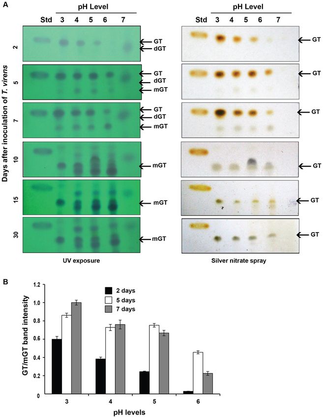
www.nature.com/scientificreports/

                                            Figure 2.  Gliotoxin metabolism in T. virens. (A) Gliotoxin production by T. virens. Gliotoxin was extracted
                                            from culture filtrate at different time points with chloroform and spotted on TLC plate. Upper panel: TLC
                                            plate viewed under UV light 254 nm. Blue colour band indicates the gliotoxin. Lower panel: TLC plate
                                            sprayed with silver nitrate. The presence of brown colour band indicates the gliotoxin. GT gliotoxin, mGT
                                            bis-thiomethyl gliotoxin, Std standard gliotoxin. (B) The levels of gliotoxin (GT) and bis-thiomethyl gliotoxin
                                            (mGT) production by T. virens were measured by densitometry analysis. Gliotoxin band intensity values were
                                            normalized with respect to the highest intensity considered as 1. (C) Biomass production and pH conditions
                                            during the growth of T. virens. Fungal mycelial mat was collected at different time points and weighed. pH levels
                                            were measured.

                                            Gliotoxin, but not bis‑thiomethyl gliotoxin, inhibited M. phaseolina. Though gliotoxin is anti-
                                            fungal against several fungi, its antifungal activity has not been tested against M. phaseolina. Thus, the antifun-
                                            gal effect of gliotoxin and bis-thiomethyl gliotoxin was assessed. Strong growth inhibition was noticed in the
                                            PDA medium amended with the fourth day old culture filtrates containing high amount of gliotoxin. When the
                                            medium was amended with 2% levels of culture filtrate, the stronger growth inhibition was noticed compared
                                            to the 1% level of culture filtrate indicating growth inhibition was positively correlated with concentration of
                                            gliotoxin. Conversely, the 10th day old culture filtrates containing bis-thiomethyl gliotoxin showed weak growth
                                            inhibition. Thus, this experiment clearly indicated that gliotoxin showed antifungal effect against M. phaseolina
                                            and bis-thiomethyl gliotoxin did not (Fig. 5 and Table 1).

                                            Gliotoxin is highly stable in acidic water. Our preliminary experiments clearly showed that T. virens
                                            accumulated gliotoxin from early logarithmic growth phase to stationary growth phase. Later, gliotoxin was

          Scientific Reports |   (2021) 11:16536 |               https://doi.org/10.1038/s41598-021-95907-6                                                  4

Vol:.(1234567890)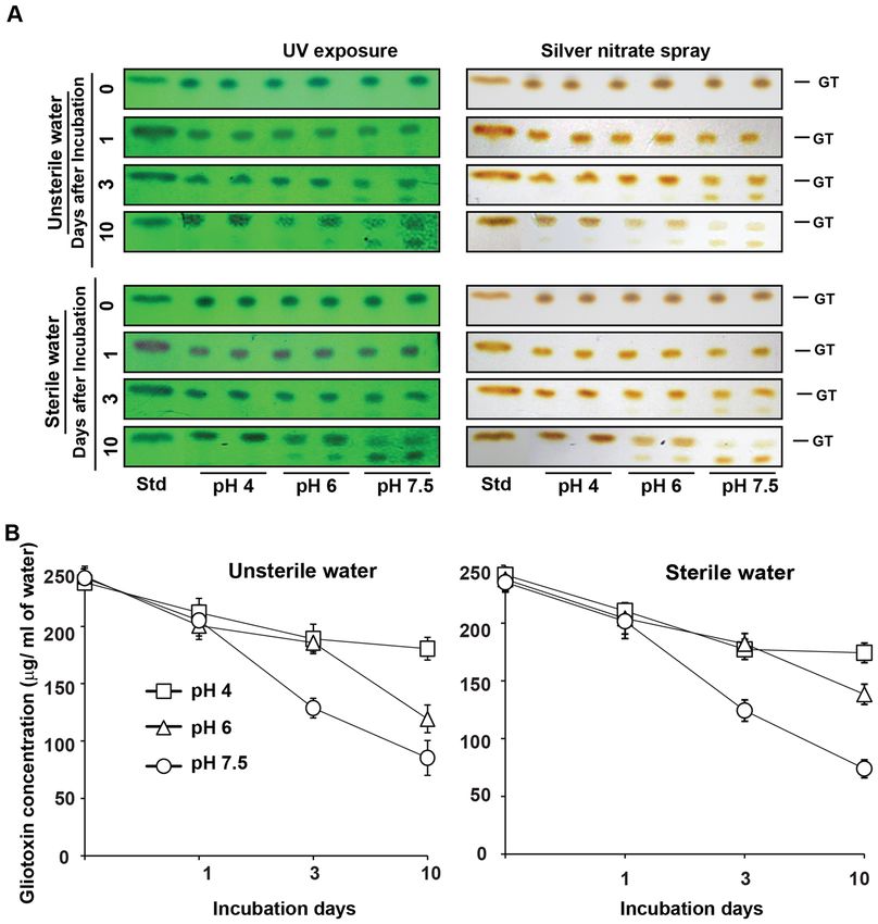
www.nature.com/scientificreports/
Figure 3. LC-Tandem MS analysis of gliotoxin and bis-thiomethyl gliotoxin. (A) gliotoxin; (B) bis-thiomethyl
gliotoxin (mGT)
converted completely into bis-thiomethyl gliotoxin in late stationary growth phase by T. virens itself. Next, we
analysed its stability in water under acidic and alkaline conditions.
Stability and intactness of gliotoxin in the sterile and unsterile water appeared in similar pattern. In acidic
water (pH 4), gliotoxin was stable throughout the experimental period of 10 days (Fig. 6; Table 2) and also more
than 30 days. Whereas in alkaline water (pH 7.5), 53% of gliotoxin was stable on the 3rd day and 31–35% of
gliotoxin was stable on the 10th day of incubation. This experiment indicates that gliotoxin is stable in acidic
water (Fig. 6 and Table 2).
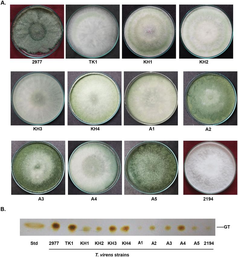
Unsterile status, wet soil conditions and alkaline pH enhance degradation of gliotoxin. Sta-
bility of secondary metabolites may be influenced by soil environmental factors such as soil moisture, pH and
soil microorganisms. Thus, we assessed the effect of soil moisture, pH and their combined effect on the stability
of gliotoxin in unsterile soil (soil having complex microbiome) and also in sterile soil (soil without microbiome).
Using chloroform as extraction solvent, 82 to 86% of gliotoxin can be extracted from the total gliotoxin
applied in soil as revealed by the gliotoxin recovery (recovered gliotoxin was 206 to 213 µg/g from the soil spiked
with 250 µg of gliotoxin/g of soil on the 0 day of incubation indicating 82–86% gliotoxin extraction efficiency by
chloroform). Gliotoxin content in soil gradually decreased over the incubation period of 10 days both in unsterile
soil and sterile soil. In unsterile soil, stability of gliotoxin was higher under dry conditions than wet conditions
in acidic and alkaline soil on the third, the fifth and the tenth day of incubation period. Under dry and semi dry
conditions, the stability of gliotoxin was 68 to 81% on the third day; 63 to 75% on the fifth day and 23 to 32% on
the tenth day in both acidic and alkaline soil types. Whereas in wet soil, the stability of gliotoxin was 17–44% on
the third day; 8 to 37% on the fifth day and 0- 2% on the tenth day (Fig. 7; Table 3).
Soil pH does not affect gliotoxin stability in dry soil under unsterile conditions. pH of the soil
(acidic and alkaline soil) did not significantly influence the stability of gliotoxin under dry conditions whereas
that drastically reduced the gliotoxin in wet conditions. Stability of gliotoxin was 75 and 68% in acidic and alka-
line soil respectively under semi dry conditions and 81 and 79% in acidic and alkaline soil respectively under
dry conditions on the third day of incubation. Stability of gliotoxin was 69 and 63% in acidic and alkaline soil
respectively under semi dry conditions and 75% in both acidic and alkaline soil under dry conditions on the
fifth day of incubation. Stability of gliotoxin was 23 and 32% in acidic and alkaline soil respectively under semi
dry conditions and 25 and 26% in acidic and alkaline soil respectively under dry conditions on the tenth day of
incubation. However, pH of the soil significantly affected the stability of gliotoxin under wet conditions. Stability
of gliotoxin was 44 and 17% in acidic and alkaline soil respectively on the third day of incubation; 37 and 8% in
acidic and alkaline soil respectively on the fifth day of incubation. However, gliotoxin was completely degraded
on the tenth day in both acidic and alkaline soil (Fig. 7; Table 3).
These experiments clearly indicate that gliotoxin is more stable in dry conditions than wet conditions of the
soil. pH of the soil does not affect the stability of gliotoxin under dry conditions. But pH levels of soil greatly
influenced the gliotoxin stability under wet conditions. Higher pH levels reduce the stability of gliotoxin in wet
soil. The more the pH of the soil, the lower the stability of gliotoxin and vice versa under wet and unsterile soil
conditions.
Gliotoxin is more stable in sterile soil compared to unsterile soil. When compared to gliotoxin
stability in unsterile soil conditions, gliotoxin stability in sterile soil was higher. With regard to moisture levels
Scientific Reports | (2021) 11:16536 | https://doi.org/10.1038/s41598-021-95907-6 5
Vol.:(0123456789)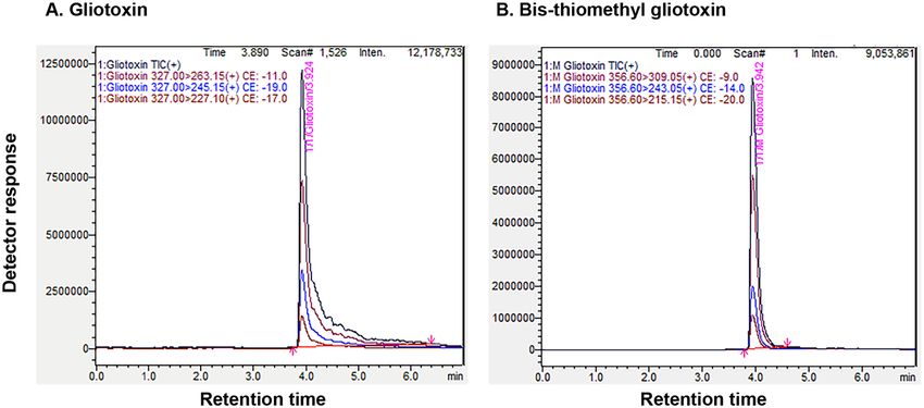
www.nature.com/scientificreports/

                                            Figure 4.  Stability of gliotoxin at different pH conditions. (A) T. virens culture was inoculated in Weindling
                                            medium buffered at different pH levels. The production and stability of gliotoxin were analysed at different time
                                            of incubation as described in Fig. 2A. GT gliotoxin, mGT Bis-thiomethyl gliotoxin; dGT-degraded gliotoxin,
                                            Std standard gliotoxin. (B) The levels of gliotoxin (GT) production by T. virens were measured by densitometry
                                            analysis. Gliotoxin band intensity values were normalized with respect to the highest intensity considered as 1.

                                            on the gliotoxin stability, gliotoxin levels were higher in dry conditions compared to wet conditions of the soil.
                                            Under dry and semi dry conditions, the stability of gliotoxin was 76–86% on the third day; 65 to 69% on the fifth
                                            day and 25 to 30% on the tenth day in both the acidic and neural soil types. Whereas under wet conditions, the
                                            stability of gliotoxin was 69 to 83% on the third day; 41 to 66% on the fifth day and 3–17% on the tenth day of
                                            incubation (Fig. 7; Table 4).

                                            Soil pH does not affect gliotoxin stability in dry soil under sterile conditions. pH of the soil
                                            (acidic soil and alkaline soil) did not significantly influence the stability of gliotoxin in dry soil under sterile
                                            status as noticed in unsterile status of the soil. Stability of gliotoxin was 84 and 85% in acidic and alkaline soil
                                            respectively under semi dry conditions and 85 and 76% in acidic and alkaline soil respectively under dry condi-
                                            tions on the third day. Stability of gliotoxin was 69 and 66% in acidic and alkaline soil respectively under semi
                                            dry conditions and 67and 65% in acidic and alkaline soil respectively under dry conditions on the fifth day.
                                            Stability of gliotoxin was 25 and 29% in acidic and alkaline soil respectively under semi dry conditions and 28

          Scientific Reports |   (2021) 11:16536 |               https://doi.org/10.1038/s41598-021-95907-6                                                   6

Vol:.(1234567890)
www.nature.com/scientificreports/
Figure 5. Antifungal activity of gliotoxin and bis-thiomethyl gliotoxin. PDA medium was amended with 4th
and 6th day old culture filtrate (containing gliotoxin) and 10th day old culture filtrate (containing bis-thiomethyl
gliotoxin) of T. virens and inoculated with M. phaseolina. Control consists of PDA amended with Weindling
medium. The mycelial growth was measured.
and 30% in acidic and alkaline soil respectively under dry conditions on the tenth day. However, pH of the soil
significantly affected the stability of gliotoxin under wet conditions in both acidic and alkaline soil as noticed in
unsterile soil. Stability of gliotoxin was 83 and 69% in acidic and alkaline soil respectively on the third day; 66
and 41% in acidic and alkaline soil respectively on the fifth day and 17 and 3% on the tenth day of incubation in
acidic and alkaline soil (Fig. 7; Table 4).
Discussion
In the present study, gliotoxin production was noticed on the second day (logarithmic growth phase) and contin-
ued to accumulate until the sixth day of incubation (stationary growth phase) as reported in earlier s tudies9,22,28.
During late stationary growth phase, gliotoxin was converted into bis-thiomethyl gliotoxin in the mycelial cell
(intracellularly) and also in the culture medium (extracellularly) under acidic conditions. Similarly, A. fumigatus
Scientific Reports | (2021) 11:16536 | https://doi.org/10.1038/s41598-021-95907-6 7
Vol.:(0123456789)www.nature.com/scientificreports/
1% culture filtrate (n = 4)* 2% culture filtrate (n = 4)*
Sl. no. Days after inoculation Mycelial growth (mm) Growth reduction (%) Mycelial growth (mm) Growth reduction (%)
1 2 47.8b 19.7b 41.3b 30.7b
2 4 40.3a 32.4a 29.3a 50.8a
a a a
3 6 39.0 34.5 29.0 51.3a
c c c
4 10 50.8 14.7 52.0 12.6c
d d
5 Control 59.5 – 59.5 –
Table 1. Efficacy of culture filtrates of T. virens on the mycelial growth inhibition of M. phaseolina. In the
column, mean values followed by a common letter are not significantly different (p ≤ 0.05, DMRT analysis).
*Values are means of four replications.
also converts gliotoxin into bis-thiomethyl gliotoxin during stationary growth phase and this conversion is medi-
ated enzymatically through bis-thiomethyltransferase29–31. When the purified gliotoxin was incubated in water,
the gliotoxin was stable for longer period and not converted into non-toxic bis-thiomethyl gliotoxin. However,
gliotoxin was degraded in alkaline culture medium and culture free alkaline water. Thus, T. virens itself converts
toxic gliotoxin into non-toxic bis-thiomethyl gliotoxin enzymatically under acidic conditions and gliotoxin
degrades chemically under alkaline conditions.
Gliotoxin is produced specifically by a few fungi such as T. virens, A. fumigatus and certain species of Penicil-
lium and these fungi have soil ecosystem as their main habitat. A few studies described that upon seed treatment
and soil application, T. virens produced gliotoxin in soil and its stability was affected by soil pH as in liquid growth
medium13,25. However, stability of gliotoxin in soil ecosystem is influenced by several factors. In the present study,
we assessed the influence of soil moisture, pH reactions and native microorganisms on soil gliotoxin stability.
We noticed that gliotoxin stability was more in sterile soil than unsterile soil indicating native soil microbes
could utilize gliotoxin as a carbon and nitrogen source for their growth and multiplication or they could produce
extracellular bis-thiomethyltransferase that converts gliotoxin into bis-thiomethyl gliotoxin.
Comparing the gliotoxin stability in water and soil, gliotoxin is stable for more than 10 days in acidic water
whereas gliotoxin was completely degraded in wet soil. Since agricultural soil contains carbon sources and several
nutrients that support growth and multiplication of microorganisms that can utilize or degrade the gliotoxin and
thus gliotoxin is less stable in wet soil. Gliotoxin is highly degraded in unsterile soil than sterile soil confirm-
ing again that native soil microbes could degrade the soil gliotoxin. With regard to soil wetness, degradation
of gliotoxin is more in wet soil conditions compared to dry soil. Soil pH does not have any influence on the
gliotoxin stability under dry soil whereas that greatly affects its stability in wet soil. Taken together, both soil pH
and native microorganisms do not have influence on the stability of gliotoxin in dry soil whereas both factors
greatly enhance the gliotoxin degradation in wet soil. Comparison of influence of soil microbes (unsterile vs
sterile soil conditions) on the stability of gliotoxin under wet conditions revealed that the stability of gliotoxin
was two-fold (acidic soil) and four-fold higher (alkaline soil) on the 3rd and 5th day, respectively, in sterile soil
than unsterile soil indicating soil microorganisms enhances the gliotoxin degradation. Usually, microbial growth
and activities are higher under wet conditions and that could be the reason for the higher degradation levels in
wet soil conditions. Since, chemical degradation of gliotoxin by pH (hydrogen ion concentration) occurs more
in the presence of water (wet soil), higher soil pH enhances the degradation in wet soil and not in dry soil. In
a nutshell, wet soil conditions and native soil microbes had additive effects on the degradation of gliotoxin in
addition to higher soil pH conditions.
In conclusion, purified gliotoxin is stable in acidic water samples for longer period and it is degraded when the
pH of the water sample is raised. Stability of gliotoxin is influenced by various soil factors. Soil moisture, native
microorganisms and higher soil hydroxyl ion concentrations (alkaline pH) enhance gliotoxin degradation. Since
gliotoxin is produced by T. virens at early growth phase, seed treatment and soil application of T. virens would
effectively protect the germinating seeds from infection by soil-borne pathogens such as Pythium and R solani
in acidic soil under moderately wet conditions.
Methods
Culture, media and growth conditions. T. virens strains were isolated from soil using gliotoxin amended
PDA medium and morphologically identified based on the conidiophore morphology under light microscope.
M. phaseolina was isolated from dry root rot infected black gram plants cultivated from our research field at
Agricultural College and Research Institute, Madurai, India. PDA medium was used for culturing and mainte-
nance of the fungi. For conidial collection, T. virens colonies were grown for 10 days on the PDA medium and
the conidia were harvested using sterile water containing 0.1% tween 20 and stored under refrigeration for fur-
ther use. T. virens strain MTCC 2977 and MTCC 2194 were obtained from the microbial type culture collection
(MTCC), Chandigarh, India and used as reference strains.
Genomic DNA isolation and analysis. Total genomic DNA was isolated from fungal mycelium using
CTAB (hexadecyltrimethylammonium bromide) p rotocol32. Trichoderma strains were identified at species level
by internal transcribed spacer sequence analysis (ITS) by amplifying the ITS region with ITS 1 [5′TCCGTAGGT
GAACCTGCGG3′(F)] and ITS 4 [5′ TCCTCCGCTTATTGATATGC3′(R)] primer pair and ITS PCR fragment
Scientific Reports | (2021) 11:16536 | https://doi.org/10.1038/s41598-021-95907-6 8
Vol:.(1234567890)www.nature.com/scientificreports/
Figure 6. Stability of gliotoxin in irrigation water. (A) Gliotoxin was added to sterilized and unsterilized water
samples (pH 4, 6 and 7.5) @ 250 µg/ml of water and incubated at 30° C for 10 days. Water samples were taken at
0, 1, 3 and 10 days after incubation and levels of gliotoxin was analysed as explained in Fig. 2A. GT gliotoxin, Std
standard gliotoxin. (B) Stability of gliotoxin was semi-quantitatively measured by densitometry analysis. Blue
band intensity and density was measured using Gel Quant NET software and concentration was calculated using
standard curve as explained in “Methods”.
was sequenced and BLAST searched in the nucleotide database of National Centre for Biotechnology Informa-
tion (NCBI). Gel electrophoresis and PCR were performed using standard procedures33.
Analysis of gliotoxin in growth medium. Analysis of production, accumulation and stability of glio-
toxin at various growth stages of T. virens strain TK1 was carried out using thin layer chromatography (TLC).
One hundred microlitre of the conidial suspension (containing 1 × 10⁵ conidia/ml) of T. virens was inoculated in
conical flask containing 100 ml of autoclaved Weindling medium (pH 4) and incubated at 25 °C, 150 rpm. One
millilitre of culture filtrate was collected on 1, 2, 3, 4, 6, 8, 10, 12 and 15 days after inoculation and extracted with
0.5 ml of chloroform. Ten microliter of chloroform extracts was spotted on TLC silica gel 60 F 254 plate (Merck,
USA). Gliotoxin (purity ≥ 98%) purchased from Sigma, USA was used as standard for comparison. The extracts
Scientific Reports | (2021) 11:16536 | https://doi.org/10.1038/s41598-021-95907-6 9
Vol.:(0123456789)www.nature.com/scientificreports/
Available gliotoxin in water (µg/ml) (n = 3)* Stability of gliotoxin (%) (n = 3)
Sterile condition Unsterile condition Sterile condition Unsterile condition
Days of
incubation pH 4 pH 6 pH 7.5 pH 4 pH 6 pH 7.5 pH 4 pH 6 pH 7.5 pH 4 pH 6 pH 7.5
0 241.67** 237.88** 235.25** 238.20** 243.49** 242.17** – – – – – –
210.42** 204.02** 201.57** 211.92** 200.61** 205.21**
1 86.95** 85.72** 85.77** 89.04** 82.38** 84.79**
(13.04) (14.27) (14.22) (10.95) (17.61) (15.20)
a a b a a b a a b a a
3 177.24 (26.75) 182.10 (23.48) 124.25 (47.12) 189.01 (20.58) 185.70 (23.73) 128.77 (46.78) 73.24 76.51 52.87 79.41 76.26 53.21b
10 174.21a (28.01) 135.26b (43.16) 73.87c (68.56) 180.55a (24.13) 119.32b (50.99) 85.26c (64.76) 71.98a 56.83b 31.43c 75.86a 49.00b 35.23c
Table 2. Stability of gliotoxin in irrigation water. In each row, the mean values followed by a common letter or
** are not significantly different (p ≤ 0.05, DMRT analysis). Values in the parentheses are percent degradation
of gliotoxin. *Values are means of three replications.
were resolved using chloroform: acetone (70:30 v/v) solvent mixture. The presence of gliotoxin was visualized
under UV light 254 nm (laminar flow chamber) and again confirmed by spraying TLC plate (after baking at
80 °C for 10 min) with 2% silver nitrate dissolved in 50% aqueous acetone. The levels of production and accumu-
lation of gliotoxin at different time of incubation were semi-quantitatively assessed by quantifying the gliotoxin
band intensity using Gel Quant.NET software. Mycelial dry weight and pH of the medium were recorded at dif-
ferent time points. Each treatment was replicated three times and the experiment was repeated twice.
Study on the effect of pH on the production and stability of gliotoxin at various growth stages of T. virens was
carried out by culturing T. virens in buffered Weindling medium, with different pH levels viz., 3, 4, 5, 6 and 7 pre-
pared using citrate-sodium phosphate buffer (McIlvaine buffer). The medium was inoculated with 100 µl of the
conidial suspension (containing 1 × 10⁵ conidia/ml) of T. virens and incubated at 25 °C, 150 rpm. One milli litre
of culture filtrate was taken on 2, 5, 7, 10, 15 and 30 days after incubation and presence of gliotoxin was analysed
using TLC as described above. The levels of gliotoxin production and its stability were also assessed at acidic (pH
4) and neutral pH conditions on 2, 5, 7, 10, 15 and 30 days after incubation in commonly used growth media.
Analysis of cellular gliotoxin and bis‑thiomethyl gliotoxin in T. virens. The mycelial mat grown
in the Weindling medium was collected at different growth phases and dried at 40° C for 2 h. The dried mycelial
mat was broken using pestle and mortor and 100 mg of the mycelial powder was extracted using methanol and
analysed for the presence of intracellular gliotoxin and bis-thiomethyl gliotoxin on TLC.
In vitro antifungal assay. In vitro antifungal activity of culture filtrate containing gliotoxin and bis-thi-
omethyl gliotoxin was carried out by poisoned food technique. PDA medium was incorporated with culture
filtrates of T. virens grown for 4 th, 6th and 1 0th day of incubation at 1 and 2% levels. PDA medium without culture
filtrate served as control. Actively growing culture discs of M. phaseolina was inoculated on the medium and
incubated at 30 °C.
Large scale purification of gliotoxin and bis‑thiomethyl gliotoxin. T. virens strain TK1 was cul-
tured in one litre Weindling medium for four and ten days for gliotoxin and bis-thiomethyl gliotoxin production
respectively. Gliotoxin and bis-thiomethyl gliotoxin were extracted separately using half the volume of chlo-
roform and dried using vacuum evaporator. The dried chloroform extracts were dissolved in five millilitre of
chloroform and purified using preparative TLC plate (PLC Silica gel 60 F 254, 2 mm thickness). The extracts were
separated and visualized under UV as described above. The fluorescent blue colored region on the preparative
TLC plate was marked with pencil and the silica gel from that region was scrapped. The scrapped silica was
extracted with 5 volumes of chloroform (w/v) for two to three times or until there is no left over gliotoxin from
the last chloroform extracts. All the extracts were pooled, dried and finally dissolved in methanol and the con-
centration of the extracted gliotoxin was measured using the standard gliotoxin in analytical TLC. The identity
and molecular weight of gliotoxin and bis-thiomethyl gliotoxin was confirmed by LC-Tandem MS.
LC‑tandem MS analysis. The confirmation of gliotoxin and bis-thiomethyl gliotoxin was performed in
Shimadzu LC-Tandem MS system (LC–MS-8040 Triple quadrupole series) with a Shimpack ODS C18 column
(150 mm × 2 mm id), which was equipped with an electrospray ionization (ESI) source. The mobile phases
were composed of solvent A: 0.0314 g ammonium formate (5 mM) + 2 ml MeOH + 10 µl formic acid (0.01%)
made-up the volume to 100 ml with HPLC water. Solvent B: 0.0314 g ammonium formate (5 mM) + 10 µl for-
mic acid (0.01%) made-up the volume to 100 ml with methanol and were pumped at a flow rate of 0.3 ml/min.
The injection volume was 2 µl, and the column temperature was maintained at 40 °C. The gradient elution was:
0.01–1.20 min, 95% A; 1.20–4.0 min, 5% A; 4.00–4.50 min, 95% A; maintained for 2.5 min. The analysis was per-
formed in the positive multiple reaction monitoring (MRM) mode. The MS source conditions were as follows:
collision energy was − 11.0 V and − 19.0 V for gliotoxin and − 9.0 V and − 14.0 V for bis-thiomethyl gliotoxin;
drying gas was 15 L/min, nebulizing gas was 2.7 l/min; desolvation line and injection block temperature was
250 °C and heat block temperature was 400 °C.
Scientific Reports | (2021) 11:16536 | https://doi.org/10.1038/s41598-021-95907-6 10
Vol:.(1234567890)www.nature.com/scientificreports/
Figure 7. Stability of gliotoxin in agricultural soil. (A) Gliotoxin was added to sterilized and unsterilized acidic
sandy loam soil (pH 5.4) and alkaline sand loam soil (pH 7.4) @ 250 µg/g of soil and incubated at 30 °C for
10 days. Soil samples were taken at 0, 1, 3, 5 and 10 days after incubation and levels of gliotoxin was analysed
as explained in Fig. 2A. GT gliotoxin, Std standard gliotoxin. (B) Stability of gliotoxin was semi-quantitatively
measured by densitometry analysis. Blue band intensity and density was measured using Gel Quant NET
software and concentration was calculated using standard curve as explained in “Methods”.
Assessment of gliotoxin persistence in water. Water samples used for irrigating crop plants were
adjusted to different pH levels viz., pH 4, 6 and 7.5 using citrate sodium buffer. Gliotoxin was added to the water
samples at 250 µg/ml of water and incubated for 10 days at 25 °C. 500 µl of water samples were withdrawn at 0, 1,
Scientific Reports | (2021) 11:16536 | https://doi.org/10.1038/s41598-021-95907-6 11
Vol.:(0123456789)www.nature.com/scientificreports/
Available gliotoxin in soil (µg/g of soil) (n = 3)* Stability of gliotoxin (%) (n = 3)*
Wet conditions Semi dry conditions Dry conditions Wet conditions Semi dry conditions Dry conditions
Days of Alkaline Alkaline Alkaline Alkaline Alkaline Alkaline
incubation Acidic soil soil Acidic soil soil Acidic soil soil Acidic soil soil Acidic soil soil Acidic soil soil
0 205.89** 210.94** 211.45** 210.03** 212.24** 209.60** – – – – – –
183.61b 127.11c 196.88ab 181.62b 206.02a 192.31ab ab c ab b a
1 89.17 60.24 93.08 86.48 97.09 91.57 ab
(10.82) (39.75) (6.91) (13.51) (2.90) (8.42)
91.03c 36.81d 158.67a 141.79b 170.99a 165.77a
3 44.21c 17.44d 75.02ab 67.52b 80.58a 78.93a
(55.78) (82.55) (24.97) (32.47) (19.41) (21.06)
75.19c 16.06d 146.70ab 132.99b 159.59a 157.38a
5 36.51c 7.61d 69.36ab 63.33b 75.20a 74.94a
(63.48) (92.38) (30.63) (36.66) (24.79) (25.05)
b a b ab
49.21 67.53 52.21 54.24
10 3.17c (98.45) 0.18c (99.92) 1.54c 0.08d 23.27b 32.15a 24.60b 25.83ab
(76.72) (67.84) (75.39) (74.16)
Table 3. Stability of gliotoxin in unsterile soil. In each row, the mean values followed by a common letter or
** are not significantly different (p ≤ 0.05, DMRT analysis). Statistical analysis was carried out separately for
available gliotoxin and stability of gliotoxin. Values in the parentheses are percent degradation of gliotoxin.
*Values are means of three replications.
Available gliotoxin in soil (µg/g of soil) (n = 3)* Stability of gliotoxin (%) (n = 3)*
Wet condition Semi dry condition Dry condition Wet condition Semi dry condition Dry condition
Days of Alkaline Alkaline Alkaline Alkaline Alkaline
incubation Acidic soil soil Acidic soil soil Acidic soil soil Acidic soil Alkaline soil Acidic soil soil Acidic soil soil
0 213.93** 212.75** 209.49** 207.51** 208.60** 210.72** – – – – – –
201.00a 168.20b 202.19a 202.79a 208.03a 195.90a b c ab ab a
1 93.94 78.96 96.51 97.49 99.72 92.84b
(6.05) (21.03) (3.99) (2.50) (0.04) (7.15)
176.89ab 147.44c 177.99ab 178.87a 176.66ab 160.35bc
3 82.66ab 69.22c 84.96a 85.99a 84.69a 75.99bc
(17.33) (30.77) (15.04) (14.00) (15.46) (24.00)
141.74a 87.324b 145.15a 137.81a 140.3a 137.02a
5 66.23a 40.99b 69.28a 66.25a 67.29a 64.94a
(33.76) (59.00) (30.71) (33.74) (32.83) (35.05)
36.91c 7.06d 52.86b 60.27ab 59.47ab 63.53a
10 17.24 a 3.31b 25.23a 28.97a 28.51a 22.61a
(82.75) (96.68) (74.77) (71.02) (71.54) (69.88)
Table 4. Stability of gliotoxin in sterile soil. In each row, the mean values followed by a common letter or **
are not significantly different (p ≤ 0.05). Statistical analysis was carried out separately for available gliotoxin and
stability of gliotoxin. Values in the parentheses are percent degradation of gliotoxin. *Values are means of three
replications.
3 and 10 days of incubation period and extracted using half of the volume of chloroform and re-extracted again
for two times. The extracts were pooled, dried and finally dissolved in one millilitre of chloroform. Thirty micro-
litre of gliotoxin extracts were spotted on TLC plate and the levels of gliotoxin was visualized under UV light
(254 nm) and again confirmed by spraying TLC plate with 2% silver nitrate dissolved in 50% aqueous acetone.
The amount of gliotoxin in water sample was measured semi-quantitatively by measuring the fluorescent band
intensity using Gel Quant.NET software. Standard curve was plotted using the standard gliotoxin band intensity
versus concentration. The gliotoxin concentration in water sample was calculated using the standard curve. The
same experiment was conducted with sterile water samples. Each treatment was replicated three times and the
experiments were repeated three times.
Assessment of gliotoxin persistence in soil. Two soil types viz., acidic sandy loam and alkaline sandy
loam soil were collected from garden land in two locations. Soil samples were sieved with 20 mesh sieve to
remove the gravels and dried. Soil samples were spiked with gliotoxin at 250 µg gliotoxin/g of soil. The moisture
level of soil was adjusted to 90–100% (wet soil conditions), 40–50% (semi dry soil conditions) and 10–20% water
holding capacity (dry soil conditions). Five grams of each soil sample was taken in test tube and incubated for
10 days at 30 °C. 500 mg of soil were withdrawn at 0, 1, 3, 5 and 10 days of incubation period and extracted using
one milliliter of chloroform and again re-extracted two times with chloroform and extracts were pooled, dried
and finally dissolved in one millilitre of chloroform. Thirty microlitre of chloroform extracts were spotted on
TLC. The presence of gliotoxin in each soil sample was visualized under UV light (254 nm) and again confirmed
by spraying TLC plate with 2% silver nitrate dissolved in 50% aqueous acetone. The amount of gliotoxin in soil
sample was measured semi-quantitatively using Gel Quant.NET software. The gliotoxin concentration was cal-
culated in the experimental soil sample using the standard curve as explained earlier. The same experiment was
conducted with autoclave-sterilized acidic sandy loam and alkaline sandy loam soil samples. Each treatment was
replicated three times and the experiments were repeated three times.
Scientific Reports | (2021) 11:16536 | https://doi.org/10.1038/s41598-021-95907-6 12
Vol:.(1234567890)www.nature.com/scientificreports/
Statistical analysis. All the experiments were conducted with a minimum of three replicates and the
results were expressed as the mean ± standard deviation. For the statistical analysis, one‐way and two‐way analy-
sis of variance (ANOVA) followed by Duncan’s Multiple Range test (DMRT) were used. Data in percentage were
subjected to arcsine transformation in statistical analysis.
Ethics declarations. Experimental research and field studies on plants were conducted as per the guideline
of Tamil Nadu Agricultural University, India. This study did not involve human participants or animals.
Received: 25 February 2021; Accepted: 15 July 2021
References
1. Elad, Y. & Chet, I. Improved selective media for isolation of Trichoderma spp. or Fusarium spp. Phytoparasitica 11, 55 (1983).
2. Atanasova, L. et al. Comparative transcriptomics reveals different strategies of Trichoderma mycoparasitism. BMC Genom. 14, 121
(2013).
3. Bulgari, D., Fiorinim, L., Gianoncelli, A., Bertuzzi, M. & Gobbi, E. Enlightening gliotoxin biological system in agriculturally relevant
Trichoderma spp. Front. Microbiol. 11, 200 (2020).
4. Dennis, C. & Webster, J. Antagonistic properties of species-groups of Trichoderma: I. Production of non-volatile antibiotics. Trans.
Br. Mycol. Soc. 57, 25–39 (1971).
5. Howell, C. R. Selective isolation from soil and separation in vitro of P and Q strains of Trichoderma virens with differential media.
Mycologia 91, 930–934 (1999).
6. Park, Y. H., Stack, J. P. & Kenerley, C. M. Selective isolation and enumeration of Gliocladium virens and G. roseum from soil. Plant
Dis. 76, 230–235 (1992).
7. Webster, J. & Lomas, N. Does Trichodema viride produce gliotoxin and viridin?. Trans. Br. Mycol. Soc. 47, 535–540 (1964).
8. Scharf, D. H., Brakhage, A. A. & Mukherjee, P. K. Gliotoxin–bane or boon?. Environ. Microbiol. 18, 1096–1109 (2016).
9. Brian, P. & Hemming, H. J. Gliotoxin, a fungistatic metabolic product of Trichoderma viride. Ann. Appl. Biol. 32, 214–220 (1945).
10. Weindling, R. Studies on a lethal principle effective in the parasitic action of Trichoderma lignorum on Rhizoctonia solani and other
soil fungi. Phytopathology 24, 151–153 (1934).
11. Harris, A. R. & Lumsden, R. D. Interactions of Gliocladium virens with Rhizoctonia solani and Pythium ultimum in non-sterile
potting medium. BioControl. Sci. Technol. 7, 37–48 (1997).
12. Tomah, A. A., Alamer, I. S. A., Li, B. & Zhanga, J. Z. A new species of Trichoderma and gliotoxin role: A new observation in
enhancing biocontrol potential of T. virens against Phytophthora capsici on chili pepper. Biol. Control 145, 104261 (2020).
13. Wright, J. M. Biological control of a soil-borne Pythium infection by seed inoculation. Plant Soil 8, 132–140 (1956).
14. Lumsden, R. D., Locke, J. C., Adkins, S. T., Walter, J. F. & Ridout, C. J. Isolation and localization of the antibiotics gliotoxin produced
by Gliocladium virens from alginate prill in soil and soilless media. Phytopathology 82, 230–235 (1992).
15. Lumsden, R. D. et al. Characterization of major secondary metabolites produced in soilless mix by a formulated strain of the
biocontrol fungus Gliocladium virens. Can. J. Microbiol. 38, 1274–1280 (1992).
16. Wilhite, S. E., Lumsden, R. D. & Straney, D. C. Mutational analysis of gliotoxin production by the biocontrol fungus Gliocladium
virens in relation to suppression of Pythium damping-off. Phytopathology 84, 816–821 (1994).
17. Vargas, W. A. et al. Role of gliotoxin in the symbiotic and pathogenic interactions of Trichoderma virens. Microbiology 160,
2319–2330 (2014).
18. Contreras-Cornejo, H. A., Macías-Rodríguez, L., Cortés-Penagos, C. & López-Bucio, J. Trichoderma virens, a plant beneficial
fungus enhances biomass production and promotes lateral root growth through an auxin dependent mechanism in Arabidopsis.
Plant Physiol. 149, 1579–1592 (2009).
19. Fiorentino, N. et al. Trichoderma-Based biostimulants modulate rhizosphere microbial populations and improve N uptake effi-
ciency, yield, and nutritional quality of leafy vegetables. Front. Plant Sci. 9, 743 (2018).
20. Halifu, S., Deng, X., Song, X. & Song, R. Effects of two Trichoderma strains on plant growth, rhizosphere soil nutrients, and fungal
community of Pinus sylvestris var. mongolica annual seedlings. Forests 10, 758 (2019).
21. Mukherjee, P. K. et al. A novel seed-dressing formulation based on an improved mutant strain of Trichoderma virens, and its field
evaluation. Front. Microbiol. 10, 1910 (2019).
22. Park, Y. H., Stack, J. P. & Kenerley, C. M. Production of gliotoxin by Gliocladium virens as a function of source and concentration
of carbon and nitrogen. Mycol. Res. 95, 1242–1248 (1991).
23. Anitha, R. & Murugesan, K. Production of gliotoxin on natural substrates by Trichoderma virens. J. Basic Microbiol. 45, 12–19
(2005).
24. Howell, C. R. Biological control of Pythium damping-off of cotton with seed-coating preparations of Gliocladium virens. Phytopa-
thology 81, 738–741 (1991).
25. Howell, C. R. & Stipanovic, R. D. Mechanisms in the biocontrol of Rhizoctonia solani-induced cotton seedling disease by Gliocla-
dium virens: antibiosis. Phytopathology 85, 469–472 (1995).
26. Reeves, E. P., Messina, C. G. M., Doyle, S. & Kavanagh, K. Correlation between gliotoxin production and virulence of Aspergillus
fumigatus in Galleria mellonella. Mycopathologia 158, 73–79 (2004).
27. Bok, J. W. et al. Genomic mining for Aspergillus natural products. Chem. Biol. 13, 31–37 (2006).
28. Wilhite, S. E. & Straney, D. C. Timing of gliotoxin biosynthesis in the fungal biological control agent Gliocladium virens (Tricho-
derma virens). Appl. Microbiol. Biotechnol. 45, 513–518 (1996).
29. Dolan, S. K. et al. Structural, mechanistic and functional insight into gliotoxin bis-thiomethylation in Aspergillus fumigatus. Open
Biol. 7, 160292 (2017).
30. Doyle, S., Jones, G. W. & Dolan, S. K. Dysregulated gliotoxin biosynthesis attenuates the production of unrelated biosynthetic gene
cluster-encoded metabolites in Aspergillus fumigatus. Fungal Biol. 122, 214–221 (2018).
31. Scharf, D. H., Habel, A., Heinekamp, T., Brakhage, A. A. & Hertweck, C. Opposed effects of enzymatic gliotoxin N-and S-meth-
ylations. J. Am. Chem. Soc. 136, 11674–11679 (2014).
32. Xu, J. R. & Leslie, J. F. A genetic map of Gibberella fujikuroi mating population A (Fusarium moniliforme). Genetics 143, 175–189
(1996).
33. Sambrook, J. & Russell, D. Molecular Cloning: A Laboratory Manual 3rd edn. (Cold Spring Harbor Press, 2001).
Scientific Reports | (2021) 11:16536 | https://doi.org/10.1038/s41598-021-95907-6 13
Vol.:(0123456789)www.nature.com/scientificreports/
Acknowledgements
We thank the Board of Research in Nuclear Sciences (BRNS), Mumbai Government of India, No. 35/14/14/2018-
BRNS/10392 for providing financial assistance to carry out this research. We also thank Tamil Nadu Agricultural
University for providing research facility.
Author contributions
R.J., R.O., K.P. and M.S.P. performed the research work. S.T.M., P.K.M. and V.R. conceived and designed the
experiments. M.P., R.K., M.T. revised the manuscript and gave constructive suggestions in carrying out the
experiments. V.R. prepared the manuscript. All authors read and approved the final manuscript.
Funding
Financial assistance provided by DAE-BRNS, Mumbai, India was highly acknowledged.
Competing interests
The authors declare no competing interests.
Additional information
Supplementary Information The online version contains supplementary material available at https://doi.org/
10.1038/s41598-021-95907-6.
Correspondence and requests for materials should be addressed to V.R.
Reprints and permissions information is available at www.nature.com/reprints.
Publisher’s note Springer Nature remains neutral with regard to jurisdictional claims in published maps and
institutional affiliations.
Open Access This article is licensed under a Creative Commons Attribution 4.0 International
License, which permits use, sharing, adaptation, distribution and reproduction in any medium or
format, as long as you give appropriate credit to the original author(s) and the source, provide a link to the
Creative Commons licence, and indicate if changes were made. The images or other third party material in this
article are included in the article’s Creative Commons licence, unless indicated otherwise in a credit line to the
material. If material is not included in the article’s Creative Commons licence and your intended use is not
permitted by statutory regulation or exceeds the permitted use, you will need to obtain permission directly from
the copyright holder. To view a copy of this licence, visit http://creativecommons.org/licenses/by/4.0/.
© The Author(s) 2021
Scientific Reports | (2021) 11:16536 | https://doi.org/10.1038/s41598-021-95907-6 14
Vol:.(1234567890)You can also read