Proteins Expression Proling of Rat Uteruses with Primary Dysmenorrhea Syndrome
←
→
Page content transcription
If your browser does not render page correctly, please read the page content below
Proteins Expression Profiling of Rat Uteruses with
Primary Dysmenorrhea Syndrome
Yazhen Xie ( xyzmore@163.com )
Nanjing University of Chinese Medicine https://orcid.org/0000-0001-8198-2349
Jianqiang Qian
Taicang Traditional Chinese Medicine Hospital Affiliated to Nanjing University of Chinese Medicine
Mingmei Wu
Taicang Traditional Chinese Medicine Hospital Affiliated to Nanjing University of Chinese Medicine
Research Article
Keywords: Primary dysmenorrhea, Proteomics, Differentially expressed proteins
Posted Date: April 29th, 2021
DOI: https://doi.org/10.21203/rs.3.rs-383474/v1
License: This work is licensed under a Creative Commons Attribution 4.0 International License.
Read Full License
Page 1/14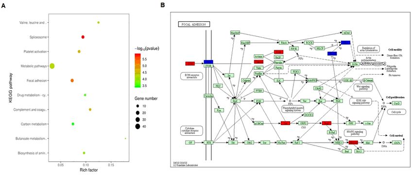
Abstract
Purpose The aim of this study was to investigate differentially expressed proteins (DEPs) and their
functions in the uteruses of primary dysmenorrhea (PD) rats by using label-free quantitative proteomics
analysis.

Methods The PD rat model was induced by injecting both estradiol benzoate and oxytocin. Twenty rats
were equally divided into two groups: a control group (normal rats), a PD model group (PD rats). Writhing
scores and serum levels of Prostaglandin E2 (PGE2) and Prostaglandin F2α (PGF2α) were used to
evaluate the success of the rat PD model. The DEPs were identified and analyzed by label-free
quantitative proteomics and bioinformatics analyses.

Results A total of 276 DEPs were identified, including 119 up-regulated DEPs and 157 down-regulated
DEPs. Bioinformatics revealed that the DEPs were mainly associated with ‘protein binding’, ‘metabolism’,
‘signal conduction’ and ‘focal adhesion’. The proteomic findings were verified by western blot analysis,
which confirmed that myosin light chain kinase (MLCK), heat shock protein 90 AB1 (HSP90AB1),
apolipoprotein A1 (Apoa1), p38 MAP kinase, c-Jun N-terminal kinase (JNK), and extracellular signal-
related kinase 1/2 (ERK1/2) were significantly differentially expressed in between the control and PD
samples.

Conclusions These results provide a deeper understanding the molecular pathogenesis of PD. The DEPs
found in the present study may provide new ideas for further study of the mechanism of PD and aid the
search for biomarkers for early diagnosis and treatment.

Introduction
Primary dysmenorrhea (PD) refers to recurrent menstrual cramps without identifiable pelvic pathology [1,
2]. It is estimated that the prevalence of PD ranges from 45–95% [1, 2]. The type of syndrome for PD is
pain which causes extreme physical and mental suffering to patients [3, 4]. Although previous studies
focused on the excessive secretion of uterine prostaglandins (PGs) for the understanding etiology of PD
[5–7], the pathogenesis of PD remains largely unknown. Therefore, identification of novel therapeutic
targets for PD will be beneficial for a large group of patients.

Label‑free quantitative proteomics is a novel tool used for etiological study and biomarker identification
in various diseases [8, 9]. Although the mRNA expression profile of uterus from PD syndrome rats was
reported previously [10], the protein expression profile of PD remains unknown so far. In this study,
label‑free quantitative proteomics and bioinformatics analyses were adopted to explore the differentially
expressed proteins (DEPs) and their functions in the uteri of PD rats. Our findings provide a deeper
understanding the molecular pathogenesis of PD and aid the search for biomarkers for early diagnosis
and treatment.

Materials And Methods
                                                 Page 2/14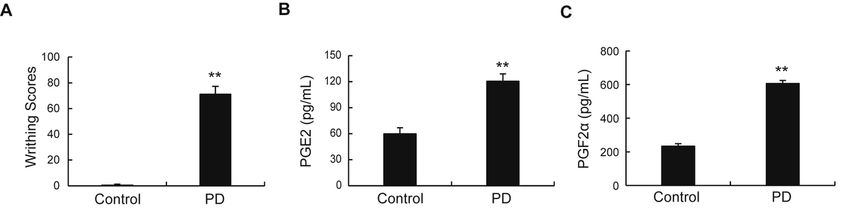
Animal model establishment

The rat model of PD was established as previously described by us [11]. Then Wistar female rats were
used in each group. The animal experiment was approved by Ethics Committee of Taicang Hospital of
traditional Chinese Medicine.

Writhing test

The writhing reaction of rats in response to pain was evaluate by writhing scores and calculated as
previously described by us [11].

Enzyme-linked immunosorbent assay (ELISA)

Serum levels of PGE2, PGF2α, TNF-α and IL-8 were measured using specific ELISA kits according to the
manufacturer’s guidelines.

Proteomics analysis

The uteri of three rats per group were collected. Sample preparation, protein digestion, LC-MS/MS
analysis, and identification of differentially expressed proteins (DEPs) were performed according to our
previous study [11]. Gene Ontology (GO) and Kyoto Encyclopedia of Genes and Genomes (KEGG)
analyses were adopted to investigate significantly enriched function and signaling pathways of the DEPs.
The interaction network of the DEPs was constructed using Search Tool for the Retrieval of Interacting
Genes/Proteins (STRING).

Western blotting

Western blotting assays were performed as previously described by us [12]. The following primary
antibodies were obtained from Cell Signaling Technology Inc. and used : anti- Apoa1(dilution:1/2000),
anti-MLCK (dilution:1/2000), anti-P-JNK (dilution:1/1000), anti-JNK (dilution:1/1000), anti-P-ERK
(dilution:1/2000) , anti-ERK (dilution:1/1000), anti-P-P38 (dilution:1/1000), anti-P38 (dilution:1/2000),
anti-HSP90AA (dilution:1/1000), anti-HSP90AB (dilution:1/1000), and anti-β-actin (dilution:1/1000). β-
actin was used as a loading control.

Statistical analysis

SPSS version 19.0 software (IBM Corp., NY) was used for all statistical analyses. Differences between the
two groups were analyzed by the Student’s t-test. One-way ANOVA was used to make comparisons
between multiple groups. Results are expressed as the mean ± standard deviation. Statistical significance
was assumed at P
As shown in Fig.1a, compared with control group, the PD rats exhibited high writhing scores (Fig.1a).
ELISA assay showed that the serum levels of PGE2 and PGF2α were significantly higher than those in the
control group (Fig.1b). These data suggested that the PD rat model had been established successfully.

Identification of DEPs in uterine tissue of PD rats

The protein expression profiles from the uterine tissue of PD rats were investigated by Lab-free based
quantitative proteomic method. The proteins expression pattern between the control and PD groups was
demonstrated using hierarchical clustering (Fig.2a). Statistically significant alterations in proteins
between the control and PD groups were identified using a volcano plot (Fig.2b). A total of 276
differentially expressed proteins [DEPs, fold change (FC) ≥1.5 and P
Discussion
Recently, Label‑free quantitative proteomics becomes a popular method in the search for
disease‑associated proteins [8, 9]. To the best of our knowledge, the present study firstly used label-free
quantitative proteomics in a rat model of PD to identify the key proteins directly, rather than mRNAs. The
in vivo rat model managed with estradiol benzoate and oxytocin is frequently used in PD studies [13–15].
The rat model of PD in the present study was successfully established by using this method.

A total of 379 DEPs were identified in the present proteomics experiments. GO and KEGG analyses
indicated that the DEPs were involved in various BPs and signaling pathways, which may play important
roles in the occurrence and development of PD. Protein-protein interaction (PPI) analysis further revealed
that the DEPs were involved in several signaling pathways or BPs and performed their functional roles
collectively in specific networks.

The uterus is an organ where lipid distribution plays a critical role for its function. Previous studies
showed that increased cholesterol could decrease uterine activity [16]. Cholesterol has been to be
enriched in microdomains of the plasma membrane known as rafts and caveolae, which have been
implicated in cellular signaling cascades [17]. Apolipoprotein A1 (Apoa1), which is the major protein
component of high density lipoprotein (HDL) in plasma, plays an important role in cholesterol transport
[18–21]. In the present study, the expression level of Apoa1 was significantly increased in the uterus of
PD rats. Based on the above findings, we speculate that the increased uterine contractions may due to up-
regulated Apoa1, which led to change of cholesterol content in the uterus of PD.

It is demonstrated that oxytocin signaling and the inflammatory response play critical roles in the
occurrence of PD [22–24]. MAP kinases, including p38MAP kinase, c-Jun N-terminal kinase (JNK/SAPK),
and extracellular signal-related kinase (ERK) were shown to mediate oxytocin signaling and regulate the
production of inflammatory cytokines [25–28]. In the present study, the phosphorylation levels of p38,
ERK, and JNK, were all significantly up-regulated in the uterus of PD rats, while the total expression of
these MAP kinases remained unchanged. It is will be interesting whether these activated MAP kinases
play roles in the occurrence of PD by regulating oxytocin signaling and the inflammatory response.

Heat shock protein 90 (HSP90) is a critical molecular chaperone protein that acts on a wide variety of
different proteins and cellular processes [29, 30]. HSP90 overexpression was demonstrated to be closely
related to the occurrence and development of many diseases, such as bronchopulmonary dysplasia,
cystic fibrosis and cancer [31]. HSP90AA1 and HSP90AB1, the two major isoforms of HSP90, has been
shown to exert multitude roles in many human diseases due to their interaction with different proteins (its
client proteins) [32–35]. In the present study, the expression level of HSP90AB1 was significantly
increased in the uterus of PD rats, while the expression level of HSP90AA1 remained unchanged.
Previous studies revealed that HSP90 is involved in the regulation of estrogen signaling, which is
essential for the progression of PD [36, 37]. Therefore, it is reasonable to speculate that up-regulated
HSP90AB1 promotes the progression of PD by regulating estrogen signaling.

                                                   Page 5/14
The increased uterine smooth muscle contraction has been considered as a main cause of PD [38–41].
Myosin light chain kinase (MLCK) I, a Ca(2+)-calmodulin-activated kinase, regulates smooth muscle
contraction by phosphorylation of myosin and is found in many tissues [42, 43]. MLCK was shown to be
important for regulating uterine smooth muscle contraction [44, 45]. In the present study, the expression
level of MLCK was significantly decreased in the uterus of PD rats, which contrary to we expected.
Whether the decreased MLCK plays a role in uterine smooth muscle contraction of PD remains unknown.
Myosin light-chain kinase (MLCK) of smooth muscle consists of an actin-binding domain at the N-
terminal, the catalytic domain in the central portion, and the myosin-binding domain at the C-terminal [42,
43]. Previous work has suggested that in addition to its kinase activity, MLCK exhibits non-kinase
properties within its N-terminus that could influence cytoskeletal organization of smooth muscle cells
[46].
In conclusion, our study firstly analyzed the protein expression profile of uterus from PD rats by using
label-free quantitative proteomics. The identified proteins and related signaling pathways might play
crucial roles in the development of PD. The identified DEPs in the present study may be utilized as
candidate biomarkers for PD.
Declarations
Funding
This research was funded by the Project of Taicang Science and Technology (Reference number:
TC2018JCYL21).
Conflicts of interest
The authors declare that they have no conflict of interest.
Availability of data and material
All of the data reported in this article are available from the corresponding author upon reasonable
request.
Code availability
Not applicable
Authors’ contributions
YX: Project development, Funding acquisition, Data analysis, Manuscript editing. JQ: Data collection,
Data analysis, Manuscript writing. MW: Data collection, Data analysis.
Ethical approval
Page 6/14The animal experiment was approved by Ethics Committee of Taicang Hospital of traditional Chinese
Medicine.
Consent to participate
Not applicable
Consent for publication
Not applicable
References
1. Tu F, Hellman K (2021) Primary Dysmenorrhea: Diagnosis and Therapy. Obstet Gynecol 137:752.
doi:10.1097/AOG.0000000000004341
2. Ferries-Rowe E, Corey E, Archer JS (2020) Primary Dysmenorrhea: Diagnosis and Therapy. Obstet
Gynecol 136:1047–1058. doi:10.1097/AOG.0000000000004096
3. Arik MI, Kiloatar H, Aslan B, Icelli M (2020) The effect of tens for pain relief in women with primary
dysmenorrhea: A systematic review and meta-analysis. Explore (NY):2541.
doi:10.1016/j.explore.2020.08.005
4. Myszko O, Al-Husayni N, Talib HJ (2020) Painful Periods in the Adolescent Girl. Pediatr Ann
49:e176–e182. doi:10.3928/19382359-20200318-01
5. Bottcher B, Laterza RM, Wildt L, Seufert RJ, Buhling KJ, Singer CF, Hill W, Griffin P, Jilma B, Schulz M,
Smith RP (2014) A first-in-human study of PDC31 (prostaglandin F2alpha receptor inhibitor) in
primary dysmenorrhea. Hum Reprod 29:2465–2473. doi:10.1093/humrep/deu205
6. Shi GX, Liu CZ, Zhu J, Guan LP, Wang DJ, Wu MM (2011) Effects of acupuncture at Sanyinjiao (SP6)
on prostaglandin levels in primary dysmenorrhea patients. Clin J Pain 27:258–261.
doi:10.1097/AJP.0b013e3181fb27ae
7. Fajrin I, Alam G, Usman AN (2020) Prostaglandin level of primary dysmenorrhea pain sufferers.
Enferm Clin 30 Suppl 2:5–9. doi:10.1016/j.enfcli.2019.07.016
8. Aly KA, Moutaoufik MT, Phanse S, Zhang Q, Babu M (2021) From fuzziness to precision medicine: on
the rapidly evolving proteomics with implications in mitochondrial connectivity to rare human
disease. iScience 24:102030. doi:10.1016/j.isci.2020.102030
9. Li N, Zhan X (2020) Mass Spectrometry-Based Mitochondrial Proteomics in Human Ovarian Cancers.
Mass Spectrom Rev 39:471–498. doi:10.1002/mas.21618
10. Fan P, Lin QH, Guo Y, Zhao LL, Ning H, Liu MY, Wei DQ (2018) The PPI network analysis of mRNA
expression profile of uterus from primary dysmenorrheal rats. Sci Rep 8:351. doi:10.1038/s41598-
017-18748-2
11. Xie Y, Qian J, Lu Q (2020) The Therapeutic Effect of Ge-Gen Decoction on a Rat Model of Primary
Dysmenorrhea: Label-Free Quantitative Proteomics and Bioinformatic Analyses. Biomed Res Int
Page 7/142020:5840967. doi:10.1155/2020/5840967
12. Xie Y, Lu Q (2020) Proteomic Analysis of Differentially Expressed Proteins in the Placenta of
Anticardiolipin Antibody- (ACA-) Positive Pregnant Mice after Anzi Heji Treatment. Evid Based
Complement Alternat Med 2020:1967698. doi:10.1155/2020/1967698
13. Huang X, Su S, Duan JA, Sha X, Zhu KY, Guo J, Yu L, Liu P, Shang E, Qian D (2016) Effects and
mechanisms of Shaofu-Zhuyu decoction and its major bioactive component for Cold - Stagnation
and Blood - Stasis primary dysmenorrhea rats. J Ethnopharmacol 186:234–243.
doi:10.1016/j.jep.2016.03.067
14. Cheng Y, Chu Y, Su X, Zhang K, Zhang Y, Wang Z, Xiao W, Zhao L, Chen X (2018) Pharmacokinetic-
pharmacodynamic modeling to study the anti-dysmenorrhea effect of Guizhi Fuling capsule on
primary dysmenorrhea rats. Phytomedicine 48:141–151. doi:10.1016/j.phymed.2018.04.041
15. Zhang N, Kong F, Zhao L, Yang X, Wu W, Zhang L, Ji B, Zhou F (2021) Essential oil, juice, and ethanol
extract from bergamot confer improving effects against primary dysmenorrhea in rats. J Food
Biochem 45:e13614. doi:10.1111/jfbc.13614
16. Smith RD, Babiychuk EB, Noble K, Draeger A, Wray S (2005) Increased cholesterol decreases uterine
activity: functional effects of cholesterol alteration in pregnant rat myometrium. Am J Physiol Cell
Physiol 288:C982–C988. doi:10.1152/ajpcell.00120.2004
17. Luo J, Yang H, Song BL (2020) Mechanisms and regulation of cholesterol homeostasis. Nat Rev Mol
Cell Biol 21:225–245. doi:10.1038/s41580-019-0190-7
18. Rosenblat M, Volkova N, Khatib S, Mahmood S, Vaya J, Aviram M (2014) Reduced glutathione
increases quercetin stimulatory effects on HDL- or apoA1-mediated cholesterol efflux from J774A.1
macrophages. Free Radic Res 48:1462–1472. doi:10.3109/10715762.2014.963574
19. Block RC, Holub A, Abdolahi A, Tu XM, Mousa SA, Oda MN (2017) Effects of aspirin in combination
with EPA and DHA on HDL-C cholesterol and ApoA1 exchange in individuals with type 2 diabetes
mellitus. Prostaglandins Leukot Essent Fatty Acids 126:25–31. doi:10.1016/j.plefa.2017.08.016
20. Flores R, Jin X, Chang J, Zhang C, Cogan DG, Schaefer EJ, Kruth HS (2019) LCAT, ApoD, and ApoA1
Expression and Review of Cholesterol Deposition in the Cornea. Biomolecules 9.
doi:10.3390/biom9120785
21. Lorkowski SW, Brubaker G, Rotroff DM, Kashyap SR, Bhatt DL, Nissen SE, Schauer PR, Aminian A,
Smith JD (2020) Bariatric Surgery Improves HDL Function Examined by ApoA1 Exchange Rate and
Cholesterol Efflux Capacity in Patients with Obesity and Type 2 Diabetes. Biomolecules 10.
doi:10.3390/biom10040551
22. Kannan P, Cheung KK, Lau BW (2019) Does aerobic exercise induced-analgesia occur through
hormone and inflammatory cytokine-mediated mechanisms in primary dysmenorrhea? Med
Hypotheses 123:50–54. doi:10.1016/j.mehy.2018.12.011
23. Feng X, Wang X (2018) Comparison of the efficacy and safety of non-steroidal anti-inflammatory
drugs for patients with primary dysmenorrhea: A network meta-analysis. Mol Pain
14:1744806918770320. doi:10.1177/1744806918770320
Page 8/1424. Akerlund M (2002) Involvement of oxytocin and vasopressin in the pathophysiology of preterm labor
and primary dysmenorrhea. Prog Brain Res 139:359–365. doi:10.1016/s0079-6123(02)39030-7
25. Cheng SC, Huang WC, JH SP, Wu YH, Cheng CY (2019) Quercetin Inhibits the Production of IL-1beta-
Induced Inflammatory Cytokines and Chemokines in ARPE-19 Cells via the MAPK and NF-kappaB
Signaling Pathways. Int J Mol Sci 20. doi:10.3390/ijms20122957
26. Sun M, Ji Y, Li Z, Chen R, Zhou S, Liu C, Du M (2020) Ginsenoside Rb3 Inhibits Pro-Inflammatory
Cytokines via MAPK/AKT/NF-kappaB Pathways and Attenuates Rat Alveolar Bone Resorption in
Response to Porphyromonas gingivalis LPS. Molecules 25. doi:10.3390/molecules25204815
27. Wei J, Ma L, Ju P, Yang B, Wang YX, Chen J (2020) Involvement of Oxytocin Receptor/Erk/MAPK
Signaling in the mPFC in Early Life Stress-Induced Autistic-Like Behaviors. Front Cell Dev Biol
8:564485. doi:10.3389/fcell.2020.564485
28. Rashed LA, Hashem RM, Soliman HM (2011) Oxytocin inhibits NADPH oxidase and P38 MAPK in
cisplatin-induced nephrotoxicity. Biomed Pharmacother 65:474–480.
doi:10.1016/j.biopha.2011.07.001
29. Prodromou C (2016) Mechanisms of Hsp90 regulation. Biochem J 473:2439–2452.
doi:10.1042/BCJ20160005
30. Schopf FH, Biebl MM, Buchner J (2017) The HSP90 chaperone machinery. Nat Rev Mol Cell Biol
18:345–360. doi:10.1038/nrm.2017.20
31. Hoter A, El-Sabban ME, Naim HY (2018) The HSP90 Family: Structure, Regulation, Function, and
Implications in Health and Disease. Int J Mol Sci 19. doi:10.3390/ijms19092560
32. Jing E, Sundararajan P, Majumdar ID, Hazarika S, Fowler S, Szeto A, Gesta S, Mendez AJ, Vishnudas
VK, Sarangarajan R, Narain NR (2018) Hsp90beta knockdown in DIO mice reverses insulin resistance
and improves glucose tolerance. Nutr Metab (Lond) 15:11. doi:10.1186/s12986-018-0242-6
33. Haase M, Fitze G (2016) HSP90AB1: Helping the good and the bad. Gene 575:171 – 86. doi:
10.1016/j.gene.2015.08.063
34. Jia L, Ge X, Du C, Chen L, Zhou Y, Xiong W, Xiang J, Li G, Xiao G, Fang L, Li Z (2021) EEF1A2 interacts
with HSP90AB1 to promote lung adenocarcinoma metastasis via enhancing TGF-beta/SMAD
signalling. Br J Cancer. doi:10.1038/s41416-020-01250-4
35. Chen W, Li G, Peng J, Dai W, Su Q, He Y (2020) Transcriptomic analysis reveals that heat shock
protein 90alpha is a potential diagnostic and prognostic biomarker for cancer. Eur J Cancer Prev
29:357–364. doi:10.1097/CEJ.0000000000000549
36. Chang Z, Lu M, Kim SS, Park JS (2014) Potential role of HSP90 in mediating the interactions
between estrogen receptor (ER) and aryl hydrocarbon receptor (AhR) signaling pathways. Toxicol Lett
226:6–13. doi:10.1016/j.toxlet.2014.01.032
37. Obermann WMJ (2018) A motif in HSP90 and P23 that links molecular chaperones to efficient
estrogen receptor alpha methylation by the lysine methyltransferase SMYD2. J Biol Chem
293:16479–16487. doi:10.1074/jbc.RA118.003578
Page 9/1438. Sen E, Ozdemir O, Ozdemir S, Atalay CR (2020) The Relationship between Serum Ischemia-Modified
Albumin Levels and Uterine Artery Doppler Parameters in Patients with Primary Dysmenorrhea. Rev
Bras Ginecol Obstet 42:630–633. doi:10.1055/s-0040-1715141
39. Senturk S (2020) Relation between uterine morphology and severity of primary dysmenorrhea. Turk J
Obstet Gynecol 17:84–89. doi:10.4274/tjod.galenos.2020.26032
40. Yang L, Chai CZ, Yue XY, Yan Y, Kou JP, Cao ZY, Yu BY (2016) Ge-Gen Decoction attenuates oxytocin-
induced uterine contraction and writhing response: potential application in primary dysmenorrhea
therapy. Chin J Nat Med 14:124–132. doi:10.1016/S1875-5364(16)60005-5
41. Quan S, Yang J, Dun W, Wang K, Liu H, Liu J (2020) Prediction of pain intensity with uterine
morphological features and brain microstructural and functional properties in women with primary
dysmenorrhea. Brain Imaging Behav. doi:10.1007/s11682-020-00356-w
42. He WQ, Wang J, Sheng JY, Zha JM, Graham WV, Turner JR (2020) Contributions of Myosin Light
Chain Kinase to Regulation of Epithelial Paracellular Permeability and Mucosal Homeostasis. Int J
Mol Sci 21. doi:10.3390/ijms21030993
43. Xiong Y, Wang C, Shi L, Wang L, Zhou Z, Chen D, Wang J, Guo H (2017) Myosin Light Chain Kinase: A
Potential Target for Treatment of Inflammatory Diseases. Front Pharmacol 8:292.
doi:10.3389/fphar.2017.00292
44. Mitchell BF, Chi M, Surgent E, Sorochan BM, Tracey CN, Aguilar HN, Mongin M, Zielnik B (2019)
Differential Regulation of Myosin Regulatory Light Chain Phosphorylation by Protein Kinase C
Isozymes in Human Uterine Myocytes. Reprod Sci 26:988–996. doi:10.1177/1933719118802062
45. Aguilar HN, Tracey CN, Zielnik B, Mitchell BF (2012) Rho-kinase mediates diphosphorylation of
myosin regulatory light chain in cultured uterine, but not vascular smooth muscle cells. J Cell Mol
Med 16:2978–2989. doi:10.1111/j.1582-4934.2012.01625.x
46. Xie C, Zhang Y, Wang HH, Matsumoto A, Nakamura A, Ishikawa R, Yoshiyama S, Hayakawa K,
Kohama K, Gao Y (2009) Calcium regulation of non-kinase and kinase activities of recombinant
myosin light-chain kinase and its mutants. IUBMB Life 61:1092–1098. doi:10.1002/iub.266
Figures
Figure 1
Page 10/14Up-regulation of writhing scores and the serum levels of PGF2α and PGE2 in PD rats. Data represent
mean ± SD (n = 10); **p < 0.01 versus normal control group.
Figure 2
Identification and analysis of differentially expressed proteins (DEPs) between control and PD groups. (A)
Clustering analysis of DEPs between control and PD groups. Red represents up-regulation of DEPs and
green represents down-regulation of DEPs. (B) Volcano plots represented all the genes in control and PD
groups according to P‑value and fold changes; black dots represent genes that were not differentially
expressed, while red dots and blue dots represent DEPs. (C) DEPs between control and PD groups. Up-
regulated proteins are indicated by red, while down-regulated proteins are indicated by green.
Page 11/14Figure 3
Functional annotation and categories of DEPs. (A) Bioinformatics analysis of the DEPs identified
between control and PD groups using BP, CC, MF, and KEGG pathway analysis. (B-D) The top ten
significantly enriched GO terms for the DEPs identified between control and PD groups.
Page 12/14Figure 4
KEGG pathway analysis of the DEPs identified between control and PD groups. (A) Distribution of the
enriched KEGG pathways. The right side of the column shows the number of proteins involved in a
specific pathway along with corresponding p-values. (B) The map of the DEPs that participate in the focal
adhesion pathway in KEGG database. Red indicates that the protein expression level is up-regulated, and
blue indicates that the protein expression level is down regulated.
Figure 5
PPI analysis of DEPs. A protein association network was constructed for the DEPs according to the fold
change of genes/proteins, KEGG pathway enrichment, protein-protein interaction, and biological process
enrichment. Circle nodes with gradient colors (green, down-regulation; red, up-regulation) represent
proteins. Protein-protein interactions are connected by solid lines. Dotted lines indicate the KEGG
pathways or BPs which the DEPs were involved in. The circular box represents the relevant KEGG/BP
term.
Page 13/14Figure 6
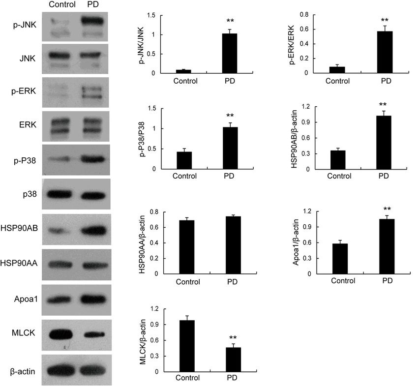
Western blotting validation of the DEPs identified by proteomics analysis. Levels of Apoa1, MLCK, P-JNK,
P-ERK, P-p38, HSP90AA and HSP90AB, in the uteruses of rats in control and PD groups were determined
by western blotting. **p < 0.01 versus control group.
Page 14/14You can also read