Relevance of 3D virtual planning in predicting bony interferences between distal and proximal fragments after sagittal split osteotomy
←
→
Page content transcription
If your browser does not render page correctly, please read the page content below
YIJOM-4335; No of Pages 9

Int. J. Oral Maxillofac. Surg. 2019; xxx: xxx–xxx
https://doi.org/10.1016/j.ijom.2019.12.001, available online at https://www.sciencedirect.com

                                                                                                                               Clinical Paper
                                                                                                                        Orthognathic Surgery

Relevance of 3D virtual                                                                                        A. Valls-Ontañón1,2,
                                                                                                               R. D. J. Ascencio-Padilla1,
                                                                                                               A. Vela-Lasagabaster1,
                                                                                                               A. Sada-Malumbres1,
planning in predicting bony                                                                                    O. L. Haas-Junior1,
                                                                                                               J. Masià-Gridilla1,2,
                                                                                                               F. Hernández-Alfaro1,2
interferences between distal                                                                                   1
                                                                                                                Institute of Maxillofacial Surgery, Teknon
                                                                                                               Medical Centre Barcelona, Barcelona, Spain;
                                                                                                               2
                                                                                                                Department of Oral and Maxillofacial

and proximal fragments after                                                                                   Surgery, Universitat Internacional de
                                                                                                               Catalunya, Sant Cugat del Vallès, Barcelona,
                                                                                                               Spain

sagittal split osteotomy
A. Valls-Ontañón, R. D. J. Ascencio-Padilla, A. Vela-Lasagabaster, A. Sada-
Malumbres, O. L. Haas-Junior, J. Masià-Gridilla, F. Hernández-Alfaro: Relevance of
3D virtual planning in predicting bony interferences between distal and proximal
fragments after sagittal split osteotomy. Int. J. Oral Maxillofac. Surg. 2019; xxx: xxx–
xxx. ã 2019 International Association of Oral and Maxillofacial Surgeons. Published
by Elsevier Ltd. All rights reserved.

Abstract. After sagittal split osteotomy, the mandibular distal and proximal fragments
do not always align themselves passively to one another, resulting in bony
interferences and subsequent anomalous settlement of the condyles. Predicting these
interferences could be an important ancillary procedure for avoiding intra- and
postoperative surgical complications, rendering orthognathic surgery more effective
and safer. This study evaluated the relevance of virtual surgical planning in assessing
the displacement of the proximal segments after virtual distal segment repositioning,
for predicting bony interferences between the segments and thus avoiding related
intra- and postoperative surgical complications. The presence of interferences
between the distal and proximal segments was compared between virtually predicted
(computer-assisted simulation surgery, Dolphin software) and real cases in 100
consecutive patients diagnosed with dentofacial deformities who underwent
orthognathic surgery with mandibular repositioning (using a short lingual osteotomy
(SLO)). The results indicated that clockwise rotation of the mandible was the
mandibular movement most prone to segment interference. Furthermore, virtual
planning was sensitive (100%) but had low specificity (51.6%) in predicting proximal
                                                                                                               Key words: orthognathic surgery; interfer-
and distal segment interferences. This low specificity was due to the software-based
                                                                                                               ences; distal segment; proximal segment; con-
automated design of the mandibular osteotomy, where the length of the distal segment                           dylar settlement; accuracy; three dimensions;
was longer than the real SLO, and the mandibular ramus sagittal split was located just                         virtual planning; CBCT.
behind Spix’s spine. Thus, more precise simulated osteotomies are needed to further
validate the accuracy of virtual planning for this purpose.                                                    Accepted for publication

0901-5027/000001+09                          ã 2019 International Association of Oral and Maxillofacial Surgeons. Published by Elsevier Ltd. All rights reserved.

  Please cite this article in press as: Valls-Ontañón A, et al. Relevance of 3D virtual planning in predicting bony interferences between
  distal and proximal fragments after sagittal split osteotomy, Int J Oral Maxillofac Surg (2020), https://doi.org/10.1016/j.
YIJOM-4335; No of Pages 9

2       Valls-Ontañón et al.

The bilateral sagittal split ramus osteot-
omy (BSSRO) is a technique used wide-
ly in orthognathic surgery for the
correction of mandibular deformities.
It has advantages compared to other
mandibular osteotomies due to the wide
overlapping cutting surface, which
enables a variable range of three-dimen-
sional (3D) movements of the distal seg-
ment (DS), ensuring broad enough
surface contact for osteosynthesis and
bone healing1.
   Unfortunately, the distal and proximal
fragments do not always align themselves
passively to one another, resulting in
                                                  Fig. 1. Marking the sagittal split in virtual planning with Dolphin software.
interferences between them: (1) forward
or backward movement of the DS of the
mandible may lead to external rotation of         Materials and methods                           Virtual planning work-up
the proximal segments (PS) in the pitch
and roll axes; (2) lateral shifting of the        Sample selection                                Computer-assisted simulation surgery was
midline causes one DS to rotate laterally                                                         conducted using specific software (Dolphin
                                                  This prospective study included 100 con-
while the other is rotated medially in the                                                        3D Orthognathic Surgery Planning Software
                                                  secutive patients diagnosed with a dento-
roll and yaw axes; (3) occlusal cant mod-                                                         version 11.8; Dolphin Imaging & Manage-
                                                  facial deformity and subjected to
ifications can produce a gap at the lower                                                         ment Solutions, Chatsworth, U.S.A.)1,2. The
                                                  orthognathic surgery with BSSRO at
margin on one side and at the upper                                                               BSSRO design was generated according to
                                                  Teknon Medical Centre Barcelona be-
margin on the other, in the vertical and                                                          the standardized Dolphin protocol, where
                                                  tween February 2017 and January 2018.
transverse axes; and (4) occlusal plane                                                           the clinician only needs to mark the follow-
                                                  All surgeries were virtually planned and
changes may require pitch adaptation of                                                           ing landmarks (Fig. 1): (1) in the lingual
                                                  performed by the same surgeon (FHA).
the PS2.                                             The patients were selected on the basis
                                                                                                  view, two landmarks are placed parallel to
   Thus, inadequate accommodation of the                                                          the occlusal plane, located slightly above
                                                  of the following inclusion criteria: age
PS may result in anomalous positioning of                                                         Spix’s spine, on the front and back; (2) in
                                                  >18 years, dentofacial deformity in need
the condyles. If left uncorrected, this could                                                     the top view, four landmarks are used to trace
                                                  of mandibular correction, and signed in-
cause temporomandibular joint (TMJ) dis-                                                          the osteotomy line between the most medial
                                                  formed consent. Patients who underwent
orders, impaired bone healing, inferior                                                           landmark in the lingual view and a landmark
                                                  an isolated maxillary Le Fort I osteotomy
alveolar nerve (IAN) overextension, and                                                           between the first and the second molars just
                                                  were excluded, as were those presenting
a tendency to relapse, as well as unaes-                                                          below the molar gingival line; (3) in the
                                                  any craniofacial syndrome or pathological
thetic outcomes3.                                 background that could compromise bone
                                                                                                  buccal view, five landmarks are placed
   Nowadays, 3D virtual surgical plan-                                                            slightly above the caudad edge of the body
                                                  healing, and patients failing to sign the
ning (VSP) has become an increasingly                                                             of the mandible: the most medial point is
                                                  informed consent.
used tool, allowing more precise orthog-                                                          placed following a perpendicular line across
                                                     The study was approved by the Ethics
nathic surgery and final outcomes                                                                 the occlusal plane between the first and
                                                  Committee of Teknon Medical Centre
that come as close as possible to the                                                             second molars, and the most distal point is
                                                  (Barcelona, Spain; Ref. LO-OS) and was
intended outcomes. An improved virtual                                                            located following an imaginary line parallel
                                                  conducted in accordance with the ethical
anatomical study is possible using                                                                to the mandibular ramus through the poste-
                                                  standards laid down in the 1964 Declara-
this technique, with better symmetry                                                              rior-most landmark of the lingual view.
                                                  tion of Helsinki and its later amendments.
axes and the anticipation of surgical                                                                Once the mandibular and maxillary
complications such as the osteotomized                                                            osteotomies had been designed (Fig. 2),
                                                  Data acquisition
bone segment interferences mentioned                                                              surgical repositioning of the maxilloman-
above4. Predicting these interferences            All patients followed the standard work-        dibular complex was virtually simulated
could be an important ancillary proce-            flow for orthognathic surgery planning          following the upper incisor to soft tissue
dure for avoiding intra- and postopera-           and surgical splint fabrication of the de-      plane (UI-STP) protocol (Fig. 3), validat-
tive surgical complications, rendering            partment, as described elsewhere1. The          ed previously and described in detail else-
orthognathic surgery more effective               protocol is based on a single cone beam         where3. The new mandibular DS position
and safer5–7. Few studies have addressed          computed tomography (CBCT) scan                 in turn determined 3D settlement of the
this issue in the literature to date.             (iCAT; Imaging Sciences International,          mandibular PS, in order to avoid mis-
The aim of the present study was                  Hatfield, PA, USA) of the head of the           matches between them (Fig. 4).
therefore to evaluate the relevance of            patient, with surface intraoral scanning           Finally, any observed interferences be-
3D VSP in assessing displacement of               of the dental arches using the Lava Scan        tween the distal and proximal segments
the PS after virtual BSSRO, predicting            ST scanner (3 M ESPE, Ann Arbor, MI,            were noted in the surgical plan for further
bony      interferences     between       the     USA) for subsequent fusion of the two           consideration as possible intraoperative
segments and thus avoiding related in-            datasets. In addition, facial photographic      interferences requiring an additional sur-
traoperative and postoperative surgical           records were obtained to complete the           gical approach, as described later in this
complications.                                    preoperative study protocol.                    article (Fig. 2).

    Please cite this article in press as: Valls-Ontañón A, et al. Relevance of 3D virtual planning in predicting bony interferences between
    distal and proximal fragments after sagittal split osteotomy, Int J Oral Maxillofac Surg (2020), https://doi.org/10.1016/j.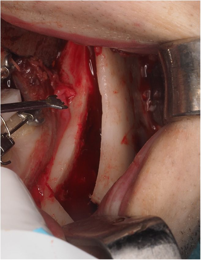
YIJOM-4335; No of Pages 9

                                                                            3D virtual prediction of bony interferences after BSSRO             3

                                                                                                      settlement of the PS were checked subjec-
                                                                                                      tively by the main surgeon (Fig. 5). In the
                                                                                                      event of any such interference, a green-
                                                                                                      stick osteotomy of the lingual cortical
                                                                                                      layer of the DS was performed, without
                                                                                                      stripping the soft tissues on the lingual
                                                                                                      surface in order to avoid IAN damage, and
                                                                                                      the osteotomized bone fragment was left
                                                                                                      in place (Figs 2, 6, and 7). This technique,
                                                                                                      referred to as a lingual osteotomy (LO),
                                                                                                      was first described by Ellis in 20079 – the
                                                                                                      only difference being that we performed
                                                                                                      the osteotomy with a piezoelectric saw
                                                                                                      device (Implant Center 2; Satelec-Acteon
                                                                                                      Group, Tuttlingen, Germany).
                                                                                                         This technique should enable smooth
                                                                                                      transition between the segments and prop-
Fig. 2. Sagittal split according to the Dolphin software design. The red dotted lines represent the   er 3D repositioning of the DS with passive
Hunsuck–Dal Pont–Obwegeser or so-called short lingual osteotomy (SLO). The black dotted               accommodation of the condyle at the gle-
line represents the lingual osteotomy (LO) (For interpretation of the references to colour in this    noid fossa, while increasing the contact
figure legend, the reader is referred to the web version of this article).
                                                                                                      surface between the two fragments. As a
                                                                                                      routine measure, proper seating of the
                                                                                                      condyles into the uppermost-anterior part
                                                                                                      of the fossa was ensured with a bidirec-
                                                                                                      tional manoeuvre. Then, rigid internal fix-
                                                                                                      ation with a hybrid technique (a miniplate
                                                                                                      fixed with four monocortical screws and a
                                                                                                      retromolar bicortical screw) was per-
                                                                                                      formed10, followed by removal of the
                                                                                                      intermaxillary fixation. Before removing
                                                                                                      the intermediate splint, proper condylar
                                                                                                      positioning and intermediate occlusion
                                                                                                      were checked again. Lastly, if necessary,
                                                                                                      the upper maxilla was repositioned
                                                                                                      according to the final splint.

Fig. 3. Virtual planning: distal segment repositioning according to the UI-STP protocol, and          Postoperative management
subsequent interference between the distal segment and left proximal segment.
                                                                                                      All patients wore a closed-circuit cold
                                                                                                      mask (17  C) during hospital admission
                                                                                                      and were discharged 24 hours after sur-
                                                                                                      gery. Standard antibiotic and anti-inflam-
Surgical procedure                                 split was performed using the Hunsuck–             matory medications for orthognathic
                                                   Dal Pont–Obwegeser technique or so-                surgery were prescribed. Functional train-
The patients were operated upon under              called short lingual osteotomy (SLO)4,8.           ing with light guiding elastics was fol-
general anaesthesia. In all cases, the man-        Next, interferences between the distal and         lowed for 1 month, with the observation
dible was operated on first, and the sagittal      proximal bony segments precluding gentle           of a soft diet for the same period of time.

Fig. 4. Three-dimensional settlement of the mandibular proximal segments in order to avoid mismatches between distal and proximal fragments is
shown in virtual orthognathic surgery planning.

 Please cite this article in press as: Valls-Ontañón A, et al. Relevance of 3D virtual planning in predicting bony interferences between
 distal and proximal fragments after sagittal split osteotomy, Int J Oral Maxillofac Surg (2020), https://doi.org/10.1016/j.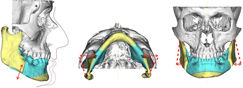
YIJOM-4335; No of Pages 9
4 Valls-Ontañón et al.
Evaluation
In order to evaluate the relevance of 3D VSP
in assessing 3D displacements of the PS
after mandibular distal fragment reposition-
ing in orthognathic surgery, the following
3D surgical movements were registered in
each virtually planned case: (1) B-point
movements (which are to be DS move-
ments) in all three axes: sagittal, vertical,
and transverse. (2) Mandibular occlusal
plane changes (which are to be DS move-
ments) on the right and left sides. (3) PS
angular movements while maintaining the
condyles in place (right and left sides) for
adaptation to the DS movement in all three
axes: pitch, roll, and yaw (Fig. 4).
Then, interferences between the distal
and proximal segments were compared
between the virtually predicted cases
and the real cases (those that required a
LO) in order to examine the true capacity
of 3D VSP to predict bony interferences.
Moreover, the LO technique described
above was subjectively tested by the main
surgeon (FHA) in terms of fragment inter-
ference and subsequent 3D gentle settlement
of the PS after performing the osteotomy.
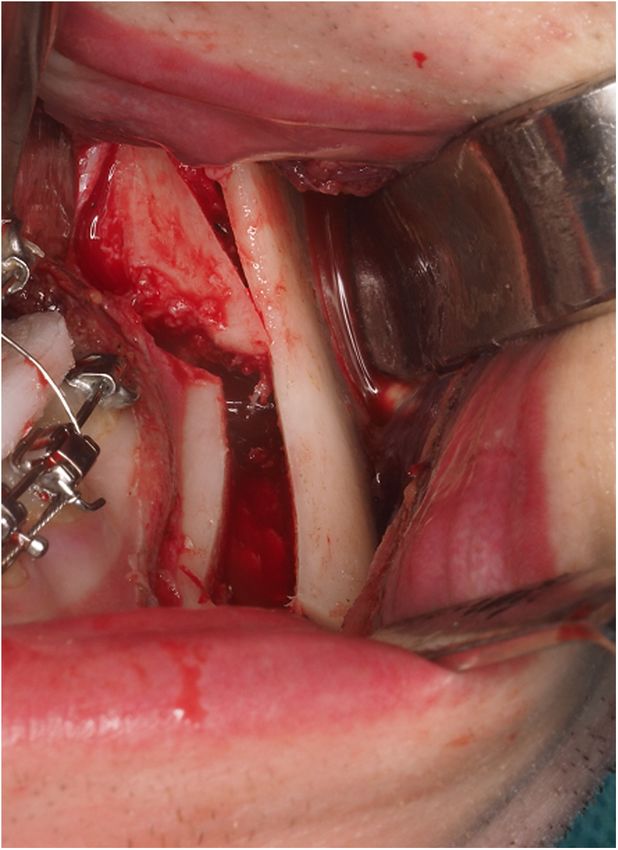
Fig. 5. Intraoperative view showing interference between the distal segment and left proximal Finally, in assessing the safety of surgery,
segment. the following conditions were considered
potential complications of the procedure:
IAN or lingual nerve damage, bone seques-
tration, intra- or postoperative malocclu-
sion secondary to condylar sag11,12, and
TMJ symptoms at 1 year of follow-up.
Statistical analysis
A descriptive analysis was made of the
study variables, with calculation of the
mean, standard deviation, minimum and
maximum values, and median for contin-
uous variables. Absolute and relative fre-
quencies (percentages) were used for
qualitative variables.
A one-sample t-test was used to deter-
mine whether the change in a certain ceph-
alometric parameter was relevant, and the
kappa concordance index was used to assess
agreement between planning and execution
of the osteotomy procedure. In addition,
simple binary logistic regression models
were used to evaluate the impact of cepha-
lometric changes upon the probability of
performing an osteotomy. The level of sig-
nificance was set at 5% (a = 0.05).
Results
Sample characterization
One hundred and twenty-seven patients
were scheduled for orthognathic surgery
during the study period; a total 100 were
Fig. 6. Intraoperative lingual osteotomy. enrolled based on the inclusion and exclu-
Please cite this article in press as: Valls-Ontañón A, et al. Relevance of 3D virtual planning in predicting bony interferences between
distal and proximal fragments after sagittal split osteotomy, Int J Oral Maxillofac Surg (2020), https://doi.org/10.1016/j.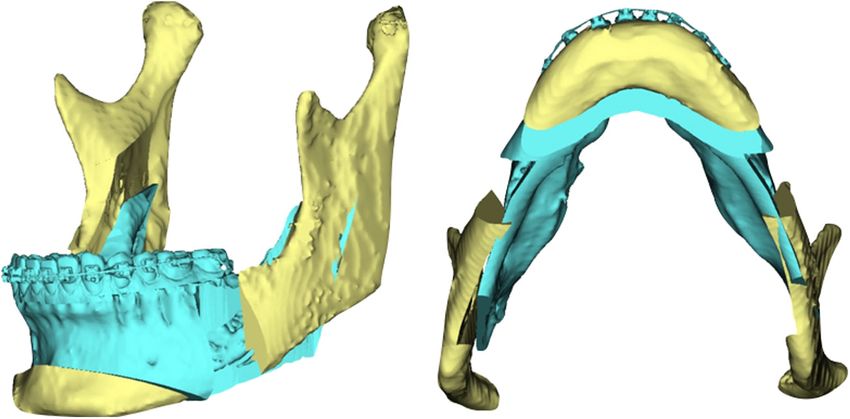
YIJOM-4335; No of Pages 9

                                                                         3D virtual prediction of bony interferences after BSSRO              5

                                                                                                      After DS repositioning and PS adapta-
                                                                                                   tion to it, the VSP predicted interferences
                                                                                                   between the proximal and distal segments
                                                                                                   in 51% of the patients (specifically accord-
                                                                                                   ing to side: right side 23%, left side 16%,
                                                                                                   and right and left side 12%).

                                                                                                   Surgical procedure
                                                                                                   All patients underwent bimaxillary
                                                                                                   orthognathic surgery, except for one pa-
                                                                                                   tient who only underwent mandibular sur-
                                                                                                   gery. Four setback surgeries were
                                                                                                   recorded versus 96 mandibular advance-
                                                                                                   ment surgeries. Of the total, five (5%)
                                                                                                   underwent LO (specifically according to
                                                                                                   side: right side 3% and left side 2%) – all
                                                                                                   of them in the context of forward mandib-
                                                                                                   ular movement. In addition, 26 (26%)
                                                                                                   genioplasties and 30 (30.3%) segmenta-
                                                                                                   tions of the upper maxilla were reported.
                                                                                                      Surgeon satisfaction with the procedure
                                                                                                   was high, since all true interferences were
                                                                                                   addressed with the LO technique.
                                                                                                      There were no complications related to
                                                                                                   the LO procedure during the perioperative
                                                                                                   period, such as IAN or lingual nerve dam-
                                                                                                   age, bone sequestration, or intra- or post-
                                                                                                   operative malocclusion secondary to
Fig. 7. Smooth transition between the left proximal and distal segments after lingual osteotomy.   condylar sag. Furthermore, no patients
Note that the osteotomized bone fragment is left in place.                                         reported postoperative TMJ symptoms at
                                                                                                   1 year of follow-up.

sion criteria. Five patients were excluded       Table 1. Although movements of the DS
                                                                                                   Statistical agreement between virtual
because of insufficient data, 15 because         were performed in all planes, only occlu-
                                                                                                   surgical planning and actual surgery
they had undergone isolated Le Fort I            sal plane in pitch changes on both sides
                                                                                                   regarding the need for lingual osteotomy
maxillary surgery, six because they were         and sagittal B-point movements proved
under-aged, and one patient because sur-         statistically significant (P < 0.001) when        Only five of the 51 virtually predicted
gery was in the context of a craniofacial        comparing the pre- and postoperative VSP          interferences ended with the application
syndrome.                                        (Figs 8 and 9).                                   of the LO technique. Thus, 46% of the
   The study sample comprised 61 women              In terms of PS accommodation dis-              previously predicted cases did not require
(61%) and 39 men (39%), with a mean age          placement, substantial results were               the application of this technique (Fig. 11).
of 27.6 years (range 18–56 years).               achieved for roll movements of both PS               Overall, there was agreement between
                                                 (P < 0.001), being positive on the right          VSP and actual surgery in 54% of the
                                                 side (+4.16 ) and negative on the left side      patients. It follows that virtual planning
Virtual surgical planning                        ( 4.88 ) (Fig. 10). Insignificant changes        is very sensitive (100%) but not specific
The magnitudes of the planned surgical           were reported for the pitch and yaw axes          (51.6%), because it predicts bony interfer-
mandibular movements are reported in             (Fig. 10).                                        ences that finally do not take place.
                                                                                                      Regarding the relationship between DS
Table 1. Planned surgical movements are reported (in millimetres): mean  standard deviation,      surgical movement and the need for LO, a
95% confidence interval, and single-sample t-test result.                                          correlation was found with the magnitude
                                        Mean  SD             95% CI                P-value
                                                                                                   of movement of B-point in the vertical
                                                                                                   axis (P = 0.081): for each additional unit
Right occlusal plane                      7.50  4.09           8.31; 6.70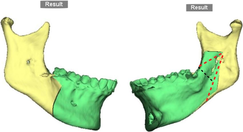
YIJOM-4335; No of Pages 9
6 Valls-Ontañón et al.
formities: prognathism, retrognathism,
and asymmetries. Despite its popularity,
one of the major drawbacks of BSSRO is
the eventual short- and long-term postsur-
gical relapse rate, which has been related
to the following contributing factors: the
method of fixation used; the amount and
direction of DS movement; proper seating
of the condyles into the fossa; 3D dis-
placement of the PS; idiopathic condylar
resorption; and interaction of the sur-
rounding para-mandibular tissues12–15.
Although the exact mechanism of relapse
is multifactorial in nature, one of the most
critical points is postsurgical instability
due to displacement of the PS from its
seated position in 3D space (sagittal, ver-
Fig. 8. Box plot illustrating that approximately 75% of the sample achieved an occlusal plane tical, and transverse) when the distal and
change of over 5 on both sides. proximal segments are not passively posi-
tioned to one another during the applica-
tion of fixation devices16–19.
Modifications of the conventional BSSRO
technique, rigid internal fixation (RIF)
methods, and condylar positioning tech-
niques have been suggested in order to
resolve this problem.
On the one hand, several conventional
BSSRO modifications have been pro-
posed: from the internal vertical ramus
osteotomy (IVRO)20, which can only be
used in cases of mandibular prognathism
because of the lack of area of bony con-
tact, to other procedures that reduce the
long length of the DS at the ascending
ramus, such as the technique used in our
study, i.e., SLO (traditionally known as
the Hunsuck–Dal Pont–Obwegeser tech-
nique)4,8. In this context, it has been dem-
Fig. 9. Box plot showing the significant advancement of B-point (P < 0.001), a strong tendency onstrated that SLO is more favourable
towards the left in the transverse axis (P = 0.086), and invariability in the vertical axis than conventional BSSRO or IVRO in
(P = 0.439). reducing flaring of the PS21. Subsequently,
additional methods have been described to
further reduce interferences between the
DS and PS, such as the abovementioned
LO9, the distal cutting technique22, and the
lingual short split technique14. In this
study, the LO technique was used because
we consider it easy to perform, complica-
tion-free, and safe.
Regarding the relationship between the
DS surgical movement and the need for
LO, a correlation was observed with the
amount of movement of B-point in the
vertical axis (P = 0.081) (Fig. 12), mean-
ing that clockwise rotation of the occlusal
plane is more prone to lingual interfer-
ences (26%, OR 1.26) than counterclock-
wise rotation. This can be explained by the
orientation of the sagittal osteotomy, be-
cause if the DS is rotated in a down
Fig. 10. Box plot showing that the roll dimension of the proximal segment underwent a position, the thick lingual bone segment
significant change (P < 0.001) to adapt to the movement of the jaw: positive (+4.16 ) on the will be near to the PS, with a greater
right side and negative ( 4.88 ) on the left side. The pitch and yaw dimensions remained chance of contact. Likewise, B-point
constant with surgery. backward movement in the sagittal plane
Please cite this article in press as: Valls-Ontañón A, et al. Relevance of 3D virtual planning in predicting bony interferences between
distal and proximal fragments after sagittal split osteotomy, Int J Oral Maxillofac Surg (2020), https://doi.org/10.1016/j.
YIJOM-4335; No of Pages 9

                                                                        3D virtual prediction of bony interferences after BSSRO              7

                                                                                                  tion, some flaring of the PS due to move-
                                                                                                  ment of the DS must be assumed, but this
                                                                                                  should be minimal in order not to increase
                                                                                                  the TMJ dysfunction rates.
                                                                                                     With regard to imaging techniques for
                                                                                                  PS positioning, sagittal and vertical dis-
                                                                                                  placement of the condyle has been widely
                                                                                                  studied using cephalometric and frontal
                                                                                                  radiographs, respectively18,19. However,
                                                                                                  it was not until the 3D virtual era when
                                                                                                  assessment of the changes in condylar
                                                                                                  position in the six degrees of freedom
                                                                                                  (sagittal, vertical, transverse, pitch, roll,
                                                                                                  and yaw) became feasible26.
Fig. 11. Agreement between the virtual planning and real surgery regarding the need for lingual      The benefit of VSP in orthognathic
osteotomy.                                                                                        surgery has been extensively documented
                                                                                                  over the last decades, because it allows
                                                                                                  more precise outcomes and reduces the
                                                                                                  surgery time and complications. For diag-
                                                                                                  nostic purposes it is especially relevant for
                                                                                                  the correction of facial asymmetries.
                                                                                                  Moreover, as seen in the present study,
                                                                                                  predicting interferences between proximal
                                                                                                  and distal segments may be helpful for
                                                                                                  planning specific DS and PS mandibular
                                                                                                  movements, and keeping in mind an even-
                                                                                                  tual additional LO in order to solve such
                                                                                                  problems. The results suggest that VSP is
                                                                                                  very sensitive (100%). This means that in
                                                                                                  all cases subjected to LO, bony interfer-
                                                                                                  ence had been predicted previously. Thus,
                                                                                                  if VSP shows no bone contact, the surgeon
                                                                                                  can go into the operating room being sure
                                                                                                  that LO will not be needed. In contrast,
                                                                                                  specificity was low, because far more
Fig. 12. The magnitude of variation at B-point in the vertical axis showed a tendency to be       interferences than those that actually oc-
associated with the need for lingual osteotomy.                                                   curred were predicted, finally implying an
                                                                                                  easier surgical procedure than expected.
                                                                                                  This low specificity is due to the software-
                                                                                                  based automated design of the osteotomy,
should give rise to interferences. Howev-        verse condylar displacement than the use         where the length of the DS is longer than
er, no interferences were reported in any of     of wire fixation. However, the latter fails      the real SLO, and the mandibular ramus is
the patients who received a backward             to provide enough stability between the          split sagittally just behind Spix’s spine
movement (a mean of 2 mm in a total of           fragments, and a longer maxillomandibu-          (Fig. 2).
four patients). No conclusions can be            lar fixation period is also required. With          Similarly, regarding the relevance of
drawn from this finding, since apart from        regard to RIF procedures, it is agreed that      virtual planning in assessing the 3D dis-
the small sample involved, this could be         miniplates with monocortical screws or           placement of the PS after DS positioning,
related to the minimum amounts of set-           position screws are to be preferred over         substantial results were achieved regard-
back movement that the authors imple-            compression or lag screws, because force-        ing the roll movements of both PS
ment in order to avoid airway narrowing          ful closure of a gap between the segments        (P < 0.001), and insignificant changes
and a double-chin appearance5.                   will cause the condyle to be displaced           were reported for the pitch and yaw axes
   On the other hand, there has been con-        medially or laterally, depending on where        (Fig. 10). These results cannot be trans-
siderable discussion as to which RIF meth-       the gap is located1.                             ferred to actual surgery for the same rea-
od produces the least condylar torque. In           Lastly, condylar positioning techniques       son as mentioned above (differences
this regard, some methodologies requiring        such as navigation systems or positioning        between software-designed and surgically
no fixation have been described in order to      devices are rarely used because they are         performed mandibular osteotomies). More
secure physiological positioning of the PS       too time-consuming, are difficult to use,        precise simulated osteotomies are there-
while avoiding condylar displacement,            and moreover cannot reproduce the origi-         fore needed to further validate the accura-
such as IVRO or SLO without fixation23.          nal condylar position intraoperatively be-       cy of virtual planning for PS settlement
However, the maxillomandibular fixation          cause of the supine position of the patient,     and the prediction of interferences be-
period is lengthened when these techni-          who is under general anaesthesia and mus-        tween mandibular segments.
ques are used. Similarly, several studies        cle relaxants24,25.                                 A manual osteotomy design together
have appeared to indicate that the use of           Despite the various methods seeking to        with cutting guides could be a potential
RIF after BSSRO results in greater trans-        maintain the condyle in its natural posi-        solution for transferring virtual planning

 Please cite this article in press as: Valls-Ontañón A, et al. Relevance of 3D virtual planning in predicting bony interferences between
 distal and proximal fragments after sagittal split osteotomy, Int J Oral Maxillofac Surg (2020), https://doi.org/10.1016/j.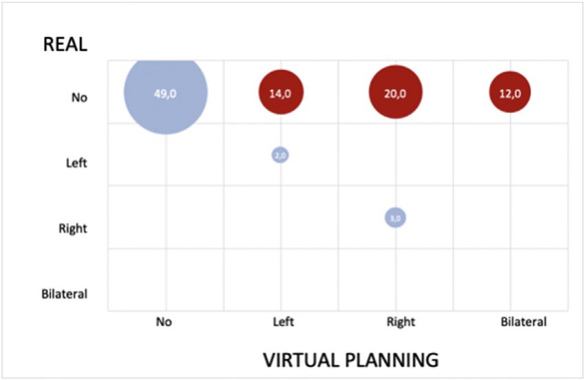
YIJOM-4335; No of Pages 9

8       Valls-Ontañón et al.

to the operating room, but neither manual         Acknowledgements. The authors would                   12. Arnett GW. A redefinition of bilateral sagit-
design nor cutting guides are sufficiently        like to thank all of the staff members at                 tal osteotomy (BSO) advancement relapse.
precise8,27. Furthermore, surgical guides         the Institute of Maxillofacial Surgery,                   Am J Orthod Dentofacial Orthop 1993;104:
are bulky, require invasive soft tissue de-       Teknon Medical Centre (Barcelona), for                    506–15.
tachment, and while they are useful for           their administrative and clinical support.            13. Yoshida K, Rivera RS, Kaneko M, Kurita K.
marking the osteotomy superficially, the                                                                    Minimizing displacement of the proximal
cutting direction is not totally transferred                                                                segment after bilateral sagittal split ramus
to the basal-most area of the mandible,                                                                     osteotomy in asymmetric cases. J Oral Max-
                                                                                                            illofac Surg 2001;59:15–8.
which is precisely where most bony inter-         References
                                                                                                        14. Sant’Ana E, Souza DPE, Temprano AB,
ferences arise.
                                                   1. Hernández-Alfaro F, Guijarro-Martı́nez R.            Shinohara EH, Faria PEP. Lingual short split:
   Thus, so far, VSP cannot replace the               New protocol for three-dimensional surgical           a bilateral sagittal split osteotomy technique
need for constant intraoperative monitor-             planning and CAD/CAM splint generation in             modification. J Craniofac Surg 2017;28:
ing of jaw movements and real-time com-               orthognathic surgery: an in vitro and in vivo         1852–4.
parisons between the planned and actual               study. Int J Oral Maxillofac Surg 2013;42:        15. Gassmann CJ, Van Sickels JE, Thrash WJ.
outcomes. It is therefore imperative to               1547–56.                                              Causes, location, and timing of relapse fol-
check proper condylar positioning and              2. Aboul-Hosn Centenero S, Hernández-Alfaro             lowing rigid fixation after mandibular ad-
occlusion before RIF as well as after                 F. 3D planning in orthognathic surgery:               vancement. J Oral Maxillofac Surg 1990;48:
maxillomandibular fixation, in order to               CAD/CAM surgical splints and prediction               450–4.
rule out condylar sag, asymmetries, and               of the soft and hard tissues results—our          16. Yang HJ, Hwang SJ. Contributing factors to
malocclusions2.                                       experience in 16 cases. J Craniomaxillofac            intraoperative clockwise rotation of the
   In conclusion, the reported results sug-           Surg 2012;40:162–8.                                   proximal segment as a relapse factor after
gest that clockwise rotation of the mandi-         3. Hernandez-Alfaro F. Upper incisor to soft             mandibular setback with sagittal split ramus
ble is the mandibular movement most                   tissue plane (UI–STP): a new reference for            osteotomy. J Craniomaxillofac Surg
prone to segment interferences. In daily              diagnosis and planning in dentofacial defor-          2014;42:e57–63.
practice, VSP may alert us to eventual                mities. Med Oral Patol Oral Cir Bucal             17. Schendel SA, Epker BN. Results after man-
interferences between mandibular frag-                2010;15:e779–81.                                      dibular advancement surgery: an analysis of
ments with a sensitivity of 100%, although         4. Hunsuck EE. A modified intraoral sagittal             87 cases. J Oral Surg 1980;38:265–82.
                                                      splitting technic for correction of mandibular    18. Becktor JP, Rebellato J, Becktor KB, Isaks-
intraoperatively most cases will not show
                                                      prognathism. J Oral Surg 1968;26:250–3.               son S, Vickers PD, Keller EE. Transverse
real interferences. When these arise
                                                   5. McCormick SU, Drew SJ. Virtual model                  displacement of the proximal segment after
intraoperatively, LO is a safe and compli-
                                                      surgery for efficient planning and surgical           bilateral sagittal osteotomy. J Oral Maxillo-
cation-free technique that enables passive            performance. J Oral Maxillofac Surg                   fac Surg 2002;60:395–403.
accommodation of the DS.                              2011;69:638–44.                                   19. Yoo JY, Kwon YD, Suh H, Ko SJ, Lee B, Lee
   VSP is an essential tool in the planning        6. Haas Jr OL, Becker OE, de Oliveira RB.                JW, Kim EC, Girod S. Transverse stability of
of orthognathic surgery, but is currently             Computer-aided planning in orthognathic               the proximal segment after bilateral sagittal
unable to reproduce either the settlement             surgery—systematic review. Int J Oral Max-            split ramus osteotomy for mandibular set-
of the PS according to DS positioning or              illofac Surg 2015;44:329–42.                          back surgery. Int J Oral Maxillofac Surg
interferences between mandibular seg-              7. Xia JJ, Shevchenko L, Gateno J, Teichgrae-            2013;42:994–1000.
ments, mainly because of the differences              ber JF, Taylor TD, Lasky RE, English JD,          20. Chen CM, Lai SS, Wang CH, Wu JH, Lee
between software-designed and surgically              Kau CH, McGrory KR. Outcome study of                  KT, Lee HE. The stability of intraoral verti-
performed mandibular osteotomies. Thus,               computer-aided surgical simulation in the             cal ramus osteotomy and factors related to
more precise simulated osteotomies are                treatment of patients with craniomaxillofa-           skeletal relapse. Aesthetic Plast Surg
needed to further validate the accuracy               cial deformities. J Oral Maxillofac Surg              2011;35:192–7.
of VSP for this purpose.                              2011;69:2014–24.                                  21. Yang HJ, Lee WJ, Yi WJ, Hwang SJ.
                                                   8. Wolford LM, Bennett MA, Rafferty CG.                  Interferences between mandibular proxi-
                                                      Modification of the mandibular ramus sagit-           mal and distal segments in orthognathic
Funding                                               tal split osteotomy. Oral Surg Oral Med Oral          surgery for patients with asymmetric man-
                                                      Pathol 1987;64:146–55.                                dibular prognathism depending on different
None.                                              9. Ellis E. A method to passively align the              osteotomy techniques. Oral Surg Oral Med
                                                      sagittal ramus osteotomy segments. J Oral             Oral Pathol Oral Radiol Endod 2010;110:
                                                      Maxillofac Surg 2007;65:2125–30.                      18–24.
Competing interests
                                                  10. Hernández-Alfaro F, Raffaini M, Paredes-         22. Kim MJ, Kim SG, Park YW. Positional
None.                                                 de-Sousa-Gil A, Magri AS, Guijarro-Martı́-            stability following intentional posterior
                                                      nez R, Valls-Ontañón A. Three-dimensional           ostectomy of the distal segment in bilateral
                                                      analysis of long-term stability after bilateral       sagittal split ramus osteotomy for correction
Ethical approval                                      sagittal split ramus osteotomy fixed with a           of mandibular prognathism. J Craniomaxil-
                                                      single miniplate with 4 monocortical screws           lofac Surg 2002;30:35–40.
The study was approved by the Ethics                  and 1 bicortical screw: a retrospective 2-        23. Ohba S, Yoshida M, Kohara H, Kawasaki T,
Committee at Teknon Medical Centre un-                center study. J Oral Maxillofac Surg                  Minamizato T, Koga T, Nakatani Y, Wana-
der number 3DIDPS.                                    2017;75:1036–45.                                      tabe E, Nakao N, Yoshida N, Asahina I.
                                                  11. Reyneke JP, Ferretti C. Intraoperative diag-          Short lingual osteotomy without fixation: a
                                                      nosis of condylar sag after bilateral sagittal        new strategy for mandibular osteotomy
Patient consent
                                                      split ramus osteotomy. Br J Oral Maxillofac           known as ‘‘physiological positioning’’. Br
Not required.                                         Surg 2002;40:285–92.                                  J Oral Maxillofac Surg 2014;52:e9–13.

    Please cite this article in press as: Valls-Ontañón A, et al. Relevance of 3D virtual planning in predicting bony interferences between
    distal and proximal fragments after sagittal split osteotomy, Int J Oral Maxillofac Surg (2020), https://doi.org/10.1016/j.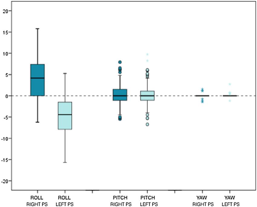
YIJOM-4335; No of Pages 9

                                                                              3D virtual prediction of bony interferences after BSSRO                9

24. Ellis E. Bimaxillary surgery using an inter-     26. Lee YC, Sohn HB, Kim SK, Bae OY, Lee JH.         Address:
    mediate splint to position the maxilla. J Oral       A novel method for the management of proxi-      Adaia Valls-Ontañón
    Maxillofac Surg 1999;57:53–6.                        mal segment using computer assisted simula-      Maxillofacial Institute
25. Berger M, Nova I, Kallus S, Ristow O,                tion surgery: correct condyle head positioning   Teknon Medical Centre
    Freudlsperger C, Eisenmann U, Dickhaus               and better proximal segment placement. Max-      Carrer de Vilana
    H, Engel M, Hoffmann J, Seeberger R. Can             illofac Plast Reconstr Surg 2015;37:21.          12 (desp 185)
    electromagnetic-navigated maxillary posi-        27. Suojanen J, Leikola J, Stoor P. The use of       08022 Barcelona
    tioning replace occlusional splints in               patient-specific implants in orthognathic sur-
                                                                                                          Spain
                                                                                                          Tel: +34 93 393 31 85
    orthognathic surgery? A clinical pilot               gery: a series of 30 mandible sagittal split
                                                                                                          E-mail: avalls@institutomaxilofacial.com
    study. J Craniomaxillofac Surg 2017;45:              osteotomy patients. J Craniomaxillofac Surg
    1593–9.                                              2017;45:990–4.

  Please cite this article in press as: Valls-Ontañón A, et al. Relevance of 3D virtual planning in predicting bony interferences between
  distal and proximal fragments after sagittal split osteotomy, Int J Oral Maxillofac Surg (2020), https://doi.org/10.1016/j.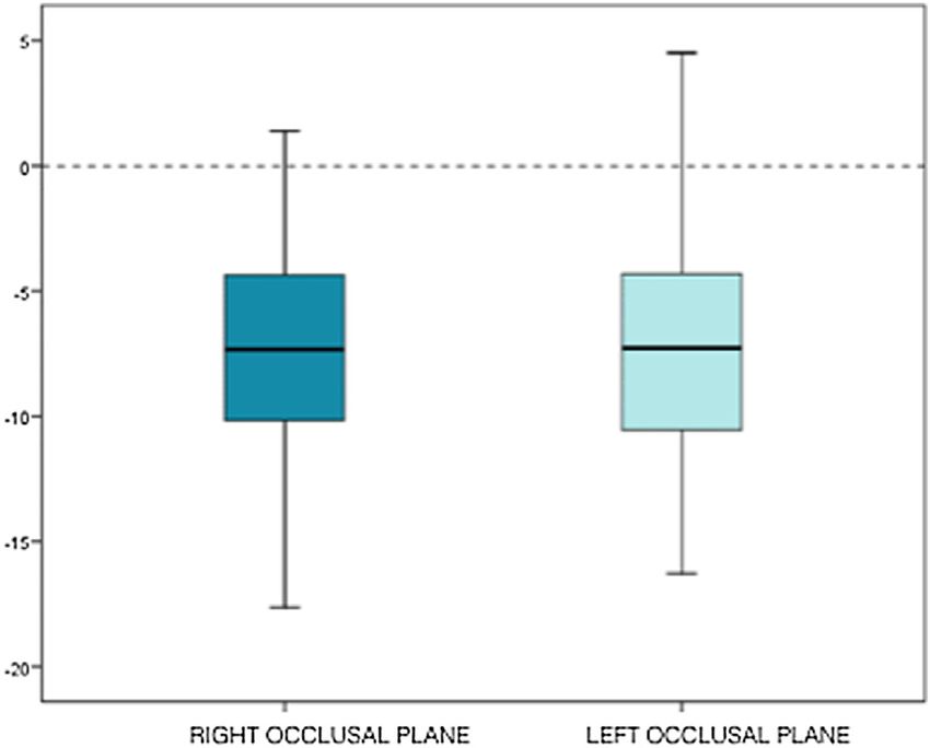
You can also read