RT QUIC DETECTION OF CWD PRION SEEDING ACTIVITY IN WHITE TAILED DEER MUSCLE TISSUES - NATURE
←
→
Page content transcription
If your browser does not render page correctly, please read the page content below
www.nature.com/scientificreports
OPEN RT‑QuIC detection of CWD prion
seeding activity in white‑tailed
deer muscle tissues
Manci Li1,2, Marc D. Schwabenlander1,2, Gage R. Rowden1,2, Jeremy M. Schefers2,3,
Christopher S. Jennelle4, Michelle Carstensen4, Davis Seelig2,5 & Peter A. Larsen1,2*
Chronic wasting disease (CWD) is a prion disease circulating in wild and farmed cervid populations
throughout North America (United States and Canada), Europe (Finland, Norway, Sweden), and South
Korea. CWD is a long-term threat to all cervid populations and to cervid hunting heritage, with the
potential to cause substantial economic losses across multiple sectors. In North America, hunting and
farming industries focused on the processing and consumption of white-tailed deer (WTD) venison
are particularly vulnerable to CWD prion contamination, as millions of WTD are consumed annually.
Real-time quaking-induced conversion (RT-QuIC) is a highly sensitive assay amplifying misfolded CWD
prions in vitro and has facilitated CWD prion detection in a variety of tissues and excreta. To date, no
study has comprehensively examined CWD prion content across bulk skeletal muscle tissues harvested
from individual CWD infected WTD. Here, we use RT-QuIC to characterize prion-seeding activity in a
variety of skeletal muscles from both wild and farmed CWD-positive WTD. We successfully detected
CWD prions in muscles commonly used for consumption (e.g., backstrap, tenderloin, etc.) as well as
within tongue and neck samples of WTD. Our results suggest that CWD prions are distributed across
the skeletal muscles of infected WTD. We posit that RT-QuIC will be a useful tool for monitoring CWD
prions in venison and that the method (with additional protocol optimization and high-throughput
functionality) could be used to reduce and/or prevent CWD prions from entering animal and human
food chains.
Chronic wasting disease (CWD) is an infectious and fatal prion disease transmitted among cervids, including
white-tailed deer (WTD; Odocoileus virginianus), mule deer, elk, red deer, caribou, reindeer, and moose. The
disease is a direct threat to a number of cervid-related multibillion-dollar economic sectors, including both
agricultural and hunting industries, and it is now prevalent in the USA and Canada with additional cases in
Korea, and Scandinavian regions1. As with other transmissible spongiform encephalopathies2,3, CWD prion seeds
(PrPCWD) consist of misfolded cellular prion protein ( PrPC) which form β-sheet-rich amyloid fibrils through
inducing conformational change and polymerization of native P rPC. The central nervous system (CNS) typically
contains the highest load of prions in a terminally diseased animal in comparison to peripheral tissues and body
excreta due to the abundance of P rPC in nervous t issues4.
Recent studies have shown that there are compelling reasons to suggest that CWD poses a non-zero risk to a
variety of mammals, including h umans1,5. Challenge experiments using CWD prions have shown that CWD can
cause neurodegenerative disease in numerous species, including ferrets, mink, domestic cats, sheep, goats, cows,
onkeys6. In vitro experiments showed that CWD prions can convert human prion proteins
pigs, and squirrel m
into a misfolded and potentially disease-causing f orm5. For these reasons, as of 2020, both the Food and Drug
Administration (FDA) and Food Safety and Inspection Service, United States Department of Agriculture (FSIS,
USDA) consider venison from CWD-positive animals as adulterated and unsuitable for c onsumption7,8. While
there is no evidence of CWD transmission to h umans1, the National Institutes of Health and Centers for Disease
Control and Prevention suggests that people should not consume known CWD-infected venison.
1
Department of Veterinary and Biomedical Sciences, University of Minnesota, 1971 Commonwealth Ave, Saint
Paul, MN 55108, USA. 2Minnesota Center for Prion Research and Outreach, College of Veterinary Medicine,
University of Minnesota, Saint Paul, MN 55108, USA. 3Veterinary Diagnostic Laboratory, Veterinary Population
Medicine Department, University of Minnesota, Saint Paul, MN 55108, USA. 4Minnesota Department of Natural
Resources, 5463 West Broadway, Forest Lake, MN 55025, USA. 5Department of Veterinary Clinical Sciences,
University of Minnesota, Saint Paul, MN 55108, USA. *email: plarsen@umn.edu
Scientific Reports | (2021) 11:16759 | https://doi.org/10.1038/s41598-021-96127-8 1
Vol.:(0123456789)
www.nature.com/scientificreports/
Currently, CWD diagnosis relies on the identification of Proteinase K (PK)-resistant P rPCWD by enzyme-
linked immunosorbent assay (ELISA) and immunohistochemistry (IHC)9. These standardized methods for
detecting CWD are designed to have consistent protocols with quantified estimates of test accuracy that are
scalable to meet the needs of agencies conducting surveillance and monitoring to manage the disease. However,
there are limitations to the existing antibody-based diagnostic approaches, namely relatively poor sensitivity
as well as the inability to screen biofluids and environmental samples. In the past two decades, the detection of
prion seeding activity has been greatly enhanced by highly sensitive methods involving amplification of protein
misfolding in vitro, such as protein misfolding cyclic amplification (PMCA) and real-time quaking-induced
conversion (RT-QuIC)9,10. PMCA uses rodent brain homogenates as the substrate to amplify misfolded prions
and Western Blotting as the o utput9,11,12. RT-QuIC utilizes recombinant P rPC, commonly from rodent sources,
as the substrate for prion amyloid formation, the real-time reporting of which is enabled by thioflavin T (ThT)
binding and d etection13–15. Although RT-QuIC demonstrates unparalleled detection sensitivity and specificity
for brain and lymphoid tissues, it has lower sensitivity for other sample types; this has in-part been ascribed to
lower prion density and RT-QuIC reaction i nhibitors16. Importantly, the inhibitory effect of certain tissues—likely
due to their biochemical compositions16,17, such as b lood18–20 and saliva, seemed to be specific for RT-QuIC
but not P MCA16. Dilutions (diluting out inhibitory effects) and phosphotungstic acid (PTA) precipitation are
commonly used to increase RT-QuIC sensitivity by enriching for prions and overcoming effects of RT-QuIC
inhibitors15–17,20, although other methods exist18,21.
CWD prions have previously been identified in a variety of tissue types and excreta using RT-QuIC, such as
the CNS, third eyelids, and feces22–25. Prior studies focused on the detection of CWD prions in skeletal muscle
using immunodetection methods have produced mixed results26–29. PMCA was used to amplify CWD prions in
hindlimb muscles from two W TD30. Skeletal muscle tissues from CWD-infected deer contain infectious prions
as determined in transgenic mice bioassay29. Despite clear advantages of RT-QuIC as a screening method9,31,32,
no comprehensive reports are available for detecting CWD prions using RT-QuIC in skeletal muscle.
Cervid skeletal muscles are consumed by a growing population of hunters and restaurant clientele and have
become a common ingredient in pet food (e.g., commercial cat and dog food). At the time of this publication,
there are no guidelines regarding venison-based detection of CWD and associated food-product surveillance.
This observation, combined with the limitations of existing CWD diagnostic tools (e.g., ELISA and IHC), has
resulted in a situation whereby venison processing can occur without the knowledge of an animal’s CWD status,
and it is estimated that at least 15,000 CWD positive cervids are consumed in the USA annually1. Underscoring
this statistic was a well-documented 2005 exposure event in which over 200 participants at a Sportsmen’s feast
consumed CWD-positive v enison33. Current estimates indicate a 20–50% CWD prevalence rate in harvested
WTD from focal areas of southern Wisconsin, however, only 1 out of 3 are tested for the d isease33. Collectively,
these observations highlight the need for post-harvest production-level monitoring of cervid products used for
human and animal consumption.
Here, we examine the utility of RT-QuIC for the detection of CWD prions within a broad set of WTD skel-
etal muscle tissues, including those frequently used for both human and animal consumption. We report the
RT-QuIC results for muscles sampled from the neck (brachiocephalicus/sternocephalicus) of wild WTD with
known CWD status. Further, we investigated whether CWD prion deposition is limited to certain groups of
muscles or if it is more generalized by using multiple WTD skeletal muscle groups across the body, including
muscles from the tongue, forelimb (suprascapularis), backstrap (longissimus dorsi), tenderloin (psoas major), and
hindlimb (semimembranosus/semitendinosus) from both wild and farmed CWD positive animals independently
determined by ELISA and/or IHC.
Results
RT‑QuIC detection of CWD prions in unilateral skeletal muscles from the neck of wild WTD. We
first developed a PrPCWD enrichment protocol for muscles—based on previous work20,25—herein referred to as
the freeze–thaw method as it consists of several rapid freeze–thaw cycles prior to PTA precipitation. To test the
performance of the freeze–thaw method, we processed unilateral muscles collected from the neck (brachioce-
phalicus/sternocephalicus) of 10 CWD-positive and 10 CWD-negative wild WTD (Table 1) and analyzed the
resultant homogenates using RT-QuIC. We found that we could detect significant prion seeding activity in 8 out
of 10 (80%) samples from 10 different CWD-positive animals (Table 1; Fig. 1a) with relatively consistent fluo-
rescent readings (Fig. 1b). In contrast to animals with official CWD-positive test results (i.e., ELISA and IHC),
none of the muscle samples from CWD-negative animals showed statistically significant prion seeding activity
in RT-QuIC (Fig. 1b), despite one of eight wells crossing the threshold from a single animal (Fig. 1d).
We then compared the rate of amyloid formation (RAF) among muscles, blood, and lymphoid tissues, all of
which were processed using mechanical extraction methods; with methods and results of blood and lymphoid
tissues reported by Schwabenlander et al.32. We note that although muscles appeared to have a lower RAF, it is
possible for an animal to have a statistically positive RT-QuIC result for muscles and lymphoid tissues but not
blood (e.g., animal 166; Fig. 2a; see Schwabenlander et al. In press). 1:10 dilution of the enriched homogenates
(after NaPTA precipitation) was chosen because of its consistency in producing results in different animals
(Fig. 2b). The optimal dilutions (a log10-based dilution that would produce the highest RAF for a given sample)
of each animal may differ, with dilution factors ranging from 0 to 3 (Fig. 2b). Because the initial concentration
of prion seeds added into RT-QuIC reaction is known to affect detectability and RAF15, it is then expected that
the method presented here—using suboptimal dilutions for some samples—would underestimate the RAF for
consistent detection purposes. Indeed, by comparing the RAF between 1:10 dilution of the enriched homogen-
ates and lymphoid tissues, which exhibit RAF 10 times lower than brain homogenates17, we found that indirectly
calculated brain/muscle ratio was 100–1000 times lower than previously reported in muscles of mice inoculated
Scientific Reports | (2021) 11:16759 | https://doi.org/10.1038/s41598-021-96127-8 2
Vol:.(1234567890)
www.nature.com/scientificreports/

                                               ELISA/IHC                      RT-QuIC wells
                                   MNPRO ID    CWD test result RT-QuIC result positive
                                   166         +              ***             7/8
                                   250         +              **              6/8
                                   333         +              **              6/8
                                   353         +              *               5/8
                                   360         +              ****            8/8
                                   363         +              *               5/8
                                   376         +              ***             7/8
                                   384         +              ***             7/8
                                   508         +              NS              2/8
                                   735         +              NS              1/8
                                   443         –              –               0/8
                                   239         –              –               0/8
                                   515         –              –               0/8
                                   536         –              –               0/8
                                   693         –              NS              1/8
                                   708         –              –               0/8
                                   723         –              –               0/8
                                   727         –              –               0/8
                                   734         –              –               0/8
                                   762         –              –               0/8

                                  Table 1.  RT-QuIC results of WTD neck muscles. All animals were collected through the Minnesota
                                  Department of Natural Resources 2019 agency culling operations. All animals’ medial retropharyngeal lymph
                                  nodes were tested for CWD through official regulatory means by ELISA, with IHC confirmation on ELISA
                                  positives. Mann–Whitney U test: NS, rate of amyloid formation is not 0 but not statistically significant from
                                  the corresponding negative controls; –, rate of amyloid formation is 0 in the given time period; ****p < 0.0001;
                                  ***p < 0.001; **p < 0.01; *p < 0.05. The freeze–thaw method was used for sample processing and RT-QuIC was
                                  performed at 45 °C.

                                  Figure 1.  Detection of prion seeding activity in unilateral neck muscles from white-tailed deer. (a) Rate of
                                  amyloid formation (1/h) was plotted using data collected from CWD-positive animals. Statistical significance
                                  was obtained through comparing with rate of amyloid formation with the respective negative controls on the
                                  same plate (****p < 0.0001; ***p < 0.001; **p < 0.01; *p < 0.05). (b) Examples of real-time fluorescence readings
                                  from positive animals (sample IDs 166 and 360). (c) Rate of amyloid formation (1/h) from CWD-negative
                                  animals. (d) Examples of real-time fluorescence readings from negative animals (443 and 693); plotted as
                                  described in (b) and showing one of eight wells for sample 693 having amyloid seeding activity (not significant).

Scientific Reports |   (2021) 11:16759 |              https://doi.org/10.1038/s41598-021-96127-8                                                     3

                                                                                                                                             Vol.:(0123456789)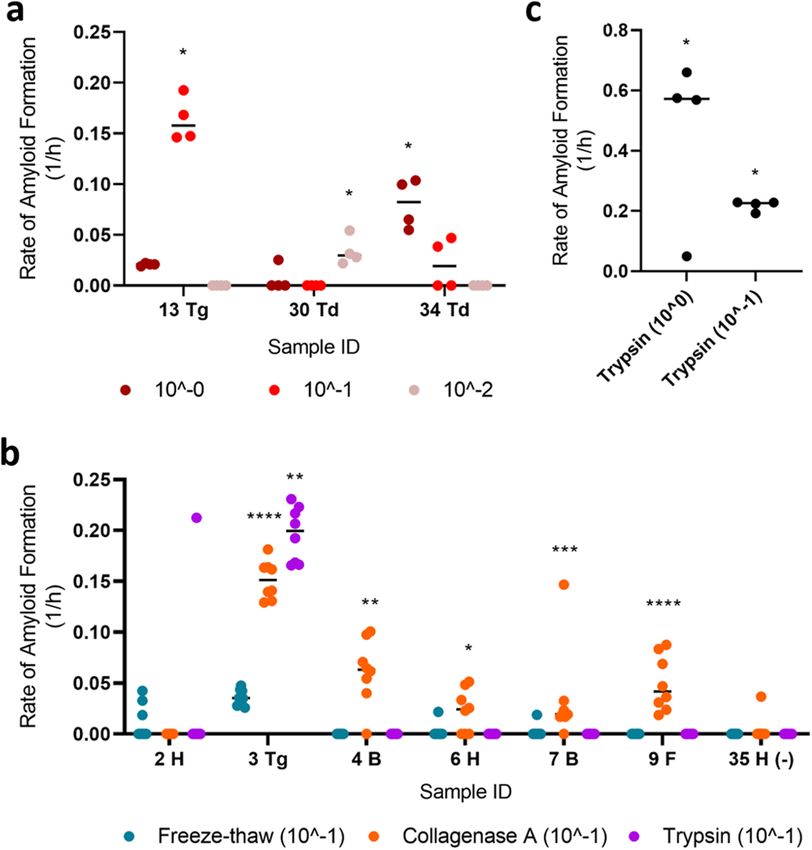
www.nature.com/scientificreports/
Figure 2. Comparison of prion-seeding activity in RT-QuIC. (a) Rate of amyloid formation was compared
among neck muscle, blood, and lymphoid tissues of three CWD-positive animals. (b) Rate of amyloid formation
compared between each sample and the negative control on the same plate. Statistical result for each sample
compared with its respective negative control was indicated (****p < 0.0001; ***p < 0.001; **p < 0.01; *p < 0.05).
RPLN, medial retropharyngeal lymph nodes.
with different strains of prions (Supplementary Fig. 1)34. Because the tenfold dilution for each sample was
likely not optimal, the difference could be attributed to a combination of the presence of RT-QuIC inhibitors in
muscle tissues and incomplete extraction (Bosque et al. pulverized muscles under liquid nitrogen34) in addition
to experimental design and species differences. It is also possible that particular CWD prion seeds within our
samples were partially degraded during autolysis (discussed further below).
CWD prions found in muscles from the tongue, neck, mid‑trunk, forelimb, and hindlimb of
WTD. To investigate if the CWD prions are found in WTD skeletal muscles and whether the freeze–thaw
method described above can be used to detect prions deposition in other skeletal muscles other than those from
the neck, we used a set of muscle tissues from another 10 WTD, including the tongue, forelimb (suprascapula-
ris), backstrap (longissimus dorsi), tenderloin (psoas major), and hindlimb (semimembranosus/semitendinosus).
In the blinded run, we were able to detect at least one significantly RT-QuIC positive sample in all the muscle
groups tested (Table 2; Fig. 3). We observed poor sensitivity of the freeze–thaw method with these particular
samples (Table 2; when compared to fresh samples), a result that is likely due to the deteriorated condition of
the muscle tissues upon receipt. Nevertheless, we recovered statistically significant RT-QuIC results for a variety
of muscle groups and we therefore conclude that PrPCWD occurs broadly throughout the skeletal muscles of
infected WTD (Fig. 3b) and are not limited to specific muscle groups, as previously reported in mouse models34.
Notably, the samples used for this experiment were undergoing various degrees of autolysis. Hypothesizing
that this may influence RT-QuIC’s ability to detect prion-seeding activity by changing the optimal dilutions of
the processed homogenate, we again looked at prion-seeding activities using serial dilutions of a selected number
of samples. As expected, the dilution with adequate positive wells for samples no longer consistently converged
at 10–1 (Fig. 4a), suggesting that the freeze–thaw method is not suitable for muscle tissue samples of sub-optimal
quality. To investigate whether other tissue processing methods would improve the detection of CWD prion-
seeding activity in RT-QuIC, given the sub-optimal tissue preservation described above, we examined a subset of
samples using enzymatic digestions (collagenase A and trypsin) instead of the freeze–thaw method. We hypoth-
esized that collagenase A and/or trypsin would sufficiently digest potential inhibitors and/or further “release”
CWD prions to a degree where extensive dilution of the processed homogenates was unnecessary. Surprisingly,
collagenase A digestion still required a tenfold dilution similar to the freeze–thaw method (Supplementary
Scientific Reports | (2021) 11:16759 | https://doi.org/10.1038/s41598-021-96127-8 4
Vol:.(1234567890)www.nature.com/scientificreports/
ELISA and/ RT-QuIC
or IHC CWD RT-QuIC wells
ID Region MNPRO ID test result result positive
1 F 287 + – 0/8
2 H 287 + NS 3/8
3 Tg 287 + *** 8/8
4 B 287 + – 0/8
5 F 288 + NS 1/8
6 H 288 + NS 1/8
7 B 288 + NS 1/8
8 F 289 + NS 1/8
9 H 289 + – 0/8
10 B 289 + – 0/8
11 F 290 + – 0/8
12 H 290 + NS 2/8
13 Tg 290 + – 0/8
14 Td 290 + – 0/8
15 H 295 + NS 1/8
16 F 295 + – 0/8
17 B 295 + NS 1/8
18 H 296 + – 0/8
19 F 296 + – 0/8
20 B 296 + – 0/8
21 H 297 + NS 1/8
22 F 297 + NS 1/8
23 B 297 + – 0/8
24 H 298 + NS 1/8
25 F 298 + – 0/8
26 B 298 + – 0/8
27 H 307 + *** 8/8
28 F 307 + **** 8/8
29 B 307 + * 4/8
30 Td 307 + NS 1/8
31 H 311 + – 0/8
32 F 311 + – 0/8
33 B 311 + – 0/8
34 Td 311 + * 5/8
Table 2. RT-QuIC results of various WTD muscle groups. All animals’ medial retropharyngeal lymph
nodes were tested for CWD through official regulatory means by ELISA, with IHC confirmation on ELISA
positives, except MNPRO ID 307, which was tested by IHC only on obex and medial retropharyngeal lymph
nodes. Mann–Whitney U test: NS, rate of amyloid formation is not 0 but not statistically significant from the
corresponding negative controls; –, rate of amyloid formation is 0 in the given time period; ****p < 0.0001;
***p < 0.001; **p < 0.01; *p < 0.05. RT-QuIC analyses of the forelimb (F), hindlimb (H), backstrap (B),
tenderloin (Td), and tongue (Tg) muscles were performed with the researcher blinded to official CWD testing
results (see methods). The freeze–thaw method was used for sample processing and RT-QuIC was performed
at 45 °C.
Fig. 2) although it appeared to be more sensitive [i.e., identified more muscle samples as RT-QuIC positive from
CWD positive animals (Fig. 4b)]; however, this was not observed when we re-tested a subset of neck muscle
samples. In addition, we confirmed that the collagenase method did not appear to produce false-positive RT-
QuIC signals (Supplementary Fig. 3). Alternatively, trypsin digestion produced an extremely high RAF without
requiring the tenfold dilution even though its sensitivity did not improve upon the freeze–thaw method in the
given sample set (Fig. 4c).
We note that all methods used in this study did result in positive prion-seeding activities using RT-QuIC
on muscle tissue (Fig. 4b). The results reported here indicate that the freeze–thaw method may not be enough
to facilitate RT-QuIC detection of CWD prions in aged or decomposing muscles but has utility for freshly col-
lected samples.
Scientific Reports | (2021) 11:16759 | https://doi.org/10.1038/s41598-021-96127-8 5
Vol.:(0123456789)www.nature.com/scientificreports/
Figure 3. Presence of CWD prions in the muscle tissues of tongue, neck, hindlimb, forelimb, backstrap, and
tenderloin. (a) Examples of the rate of amyloid formation (RAF) from RT-QuIC positive samples were plotted.
Statistical results as compared to the respective negative controls were indicated (****p < 0.0001; ***p < 0.001;
**p < 0.01; *p < 0.05). Tg Tongue, F forelimb, B backstrap, Td tenderloin, H hindlimb. Numbers on X-axis are
animal IDs listed in Table 2. (b) Prion-seeding activity was detected by RT-QuIC in muscles from the tongue,
forelimb, backstrap, tenderloin, and hindlimb. Deer image was created using BioRender.com.
Figure 4. Comparison of different methods used to extract CWD prions from skeletal muscles of WTD. (a)
Rate of amyloid formation (RAF) of samples diluted to different concentrations was visualized. (b) RAF from
RT-QuIC was plotted for a subset of samples treated by different extraction methods, including freeze–thaw,
collagenase A, and trypsin. The final suspension was diluted to 1 0−1. (c) RAF of trypsin-digested sample number
3 (tongue; undiluted and diluted to 1 0−1) was diagramed. Statistical result for each sample when compared with
its respective negative control was indicated (****p < 0.0001; ***p < 0.001; **p < 0.01; *p < 0.05). B backstrap, F
forelimb, H hindlimb, Td tenderloin, Tg tongue.
Scientific Reports | (2021) 11:16759 | https://doi.org/10.1038/s41598-021-96127-8 6
Vol:.(1234567890)www.nature.com/scientificreports/
Discussion
CWD is an emerging infectious prion disease currently affecting cervid populations across three continents
and negatively influencing all cervid-related industries within impacted regions. Infected animals can remain
asymptomatic for months while shedding CWD prions through e xcreta22,25, thus making the identification of
early-stage CWD-infected animals based on external diseased phenotypes impossible. Antibody-based ELISA
and IHC tests are the current diagnostic standards for CWD. Despite their reliability, such immunodetection
methods have limited sensitivity and application across various tissues and body excreta in comparison to in vitro
amplification methods for prion detection, such as PMCA and RT-QuIC9. Of the available in vitro amplifica-
tion methods, RT-QuIC is well-suited as a CWD screening tool because it can be easily scaled up as required by
industrial applications. Given the continued spread of CWD, and uncertainty surrounding potential health risks
to both animals and humans due to the consumption of CWD-positive venison1, it is clear that a highly sensitive
and reliable diagnostic method to detect CWD prions in skeletal muscles of cervids is needed.
In this study, we tested methods aimed at extracting and enriching P rPCWD from WTD skeletal muscles for
prion detection by RT-QuIC. We first found that CWD prions were present in bulk sampled neck muscles (bra
chiocephalicus/sternocephalicus) of CWD positive animals (Fig. 1a)32. This result prompted us to investigate the
general distribution of prions in skeletal muscles from the tongue, forelimb, mid-truck, and hindlimb of CWD
positive WTD tissue-sets available in our biorepository. We found that in addition to the neck, PrPCWD is also
present in a variety of skeletal muscle tissues (described above; Table 2; Fig. 3). Our results, based on a sampling
of various muscle groups, suggest that CWD prions are distributed across CWD infected WTD skeletal muscles.
Additional research is needed to determine the full extent to which CWD prions occur within particular muscle
tissue types of infected animals, including intra- and inter-individual variation of CWD prion accumulation in
WTD muscles.
It remains to be determined whether CWD prions are detectable in skeletal muscles that were not sampled
herein or in similar studies using amplification-based methods, such as PMCA and RT-QuIC. Although we were
unable to detect PrPCWD across all muscle types within a given CWD positive animal, this result is expected
because the successful detection of PrPCWD within an infected individual, and particular tissue type, can be
impacted by multiple factors. With respect to the neck samples screened here, we only had access to unilaterally
sampled muscles harvested from individual WTD heads and it has been shown recently that PrPCWD may not be
bilaterally present in select t issues32,35. The 10 positive animals (originating from wild WTD herds) selected for
testing of neck muscles were strongly positive across multiple tissues and likely were in relatively advanced, yet
pre-clinical stages of the disease (i.e., no clinical signs were observed at the time of euthanasia)32. The stability of
prions may vary depending on s trains36 and, to date, no RT-QuIC method is available to detect particular CWD
strain differences. Further, the progression of CWD affects the deposition of prions in peripheral tissues and it
is unclear at what time in the disease progression that prions accumulate in WTD muscles. We note the neck
muscles used herein were frozen less than 12 h after collection; however, the other muscle tissues were at various
stages of decomposition and underwent multiple freeze–thaw cycles prior to our possession. This difference in
tissue preservation and quality potentially accounts for the reduced sensitivity of RT-QuIC upon application, an
observation that suggests an altered balance of RT-QuIC inhibitors and active prion seeds and/or degradation
of particular CWD prion strains in decaying tissue. Thus, we recommend muscles for RT-QuIC-based analyses
of CWD prions be frozen (at either –20 °C or –80 °C) as soon as possible after collection, ideally less than 24 h.
Based on the results from well-preserved neck muscles, we posit that the freeze–thaw method has the most
potential for large-scale diagnostic screening of venison, as it is cheaper and easier to perform. For samples
with heavy prion loads, such as tongue, all methods used in this study agreed on the positivity of prion-seeding
activity. For poorly preserved sample types, collagenase A outperformed the freeze–thaw method and trypsin
digestion in terms of identifying more RT-QuIC positive muscle samples from CWD positive animals. Surpris-
ingly, trypsin digestion yielded a high RAF and did not require additional dilutions of the final resuspension as
needed by other methods. This could be due to the digestion of protein inhibitors by trypsin and/or superior
ability of trypsin to free prions from examined tissues. Additional optimization of the methods presented here is
needed for protocols focused on suboptimal sample types. It is possible that the prion seeding activity we detected
in the collected muscle tissues is from non-muscle cell types as reported by Daus et al.28. However, the cellular
origin of P rPCWD in skeletal muscle, whether in myocytes, erythrocytes, neurons, epithelial cells, or any other cell
type, is inconsequential to the recommendations of not consuming venison from CWD-positive animals or the
potential for RT-QuIC-based venison screening as venison products are a matrix of multiple tissues and cell types.
Our findings suggest that CWD prions occur throughout an array of WTD muscles and further investigation,
from an anatomical perspective, is needed to understand the extent of this distribution. Future studies focusing
on larger sample sizes with systematic, bilateral samplings of well-preserved muscle samples throughout the
body are needed to assess, validate, and improve the presented method for its application, as well as quantify
the load of CWD prions present. Longitudinal characterization of prion deposition (i.e., using cervid challenge
experiments) in a variety of high-quality muscle samples, such as those conducted for saliva, lymphoid tissues,
and feces is needed to better understand the pathophysiology of CWD in deer and other cervids. Our study pro-
vides the foundation for the development of RT-QuIC-based screening of venison and venison-related products
associated with food processing pipelines for CWD-prions.
Methods
Experimental design. The RT-QuIC muscle protocols (freeze–thaw and enzymatic digestion, detailed
below) were initially used on a small subset of CWD positive and not-detected neck muscle tissue samples. After
refining our methods, we then tested the protocol on a larger set of neck muscles from ten CWD positive and
ten CWD not detected deer, with CWD status determined by ELISA, IHC, and RT-QuIC analyses on lymphoid
Scientific Reports | (2021) 11:16759 | https://doi.org/10.1038/s41598-021-96127-8 7
Vol.:(0123456789)www.nature.com/scientificreports/
tissues from the animals reported by Schwabenlander et al.32. To blind investigators, researcher “A” subsampled
approximately 300 mg of each sample, placed them individually in 1.5 ml tubes, and re-labeled them in a ran-
domized numerical order. Researcher “B” carried out the muscle processing and RT-QuIC and was blinded to
the original identity of the samples. ELISA and IHC results for animals examined herein are presented in Sup-
plementary Table 1. Researcher “B” was unblinded after the first pass of all samples; investigation of different
extraction methods was done after unblinding.
Sample collection. RT-QuIC protocol development was initially performed utilizing neck muscle (brachio
cephalicus/sternocephalicus) tissue samples collected from wild WTD through 2019 agency culling operations in
southeast Minnesota conducted by the Minnesota Department of Natural Resources in conjunction with USDA
APHIS Wildlife Services as described by Schwabenlander et al.32 (Table 1). Muscle tissue samples for the quanti-
tative comparison study were obtained through disposal or necropsy of farmed and wild WTD at the University
of Minnesota Veterinary Diagnostic Laboratory and were stored at − 20 °C. All farmed and wild WTD exam-
ined in our study have been independently tested through official regulatory means by the National Veterinary
Services Laboratories or Colorado State University, respectively, for CWD infection based on immunodetection
analysis (ELISA and/or IHC) of the brain and/or lymphatic tissue for the presence of P rPCWD (Table 2).
Muscle preparation. Freeze–thaw method. This method was inspired by a combination of existing RT-
QuIC protocols20,25,28. Muscles were stored at − 20 °C within 12 h after collection then transferred to − 80 °C
until tested. 10% (weight/volume) muscle homogenates in 1× PBS were prepared in tubes with 1.5 mm diameter
zirconium oxide beads using a Beadbug homogenizer at top speed for 180 s. The homogenates underwent three
cycles of flash freeze–thaw consisting of 3 min in dry ice and 3 min at 37 °C. The homogenates were subjected
to additional homogenization at top speed for 180 s using the Beadbug homogenizer. The mixtures were then
centrifuged at 5000 rpm for 3 min. 500 µl of supernatants were used for centrifugation at 15,000 rpm, 4 °C for
40 min. The resultant pellets were resuspended in 100 µl of 1× PBS then incubated with 7 μl of 4% (w/v) phos-
photungstic acid (Sigma-Aldrich) in 0.2 M magnesium chloride. The mixtures were then incubated at 37 °C
and 1500 rpm for 1 h in a ThermoMixer (Eppendorf) before being subjected to centrifugation for 30 min at
15,000 rpm, 4 °C. Pellets were resuspended in 10 µl of 0.1% (v/v) SDS/PBS/N2. 2 μl of 10-1 diluted resuspension
was used for optimal result.
Collagenase A and trypsin digestion method. This method for RT-QuIC was modified from the PMCA method
developed by Daus et al.28. 10% (weight/volume, w/v) muscle homogenates and 180-s homogenization were
carried out as described above. 350 μl homogenates were mixed with equal volume of 2× collagenase A [4 mM
CaCl2 and 0.5% (w/v), Roche] or trypsin (Gibco) solutions. The mixture was incubated at 37 °C, 700 rpm for
four hours. After being homogenized again for 90 s, the mixtures were centrifuged at 5000 rpm for 3 min at 4 °C.
The supernatant was then transferred to another tube and mixed with an equal volume of 2× protease inhibitor
cocktail (Sigma-Aldrich). This was followed by steps including centrifugation at 15,000 rpm for 40 min as the
freeze–thaw method. 2 μl of the final suspensions were diluted tenfold and added to the RT-QuIC reaction. The
additional dilution was not necessary for trypsin digestion.
RT‑QuIC substrate preparation and reaction conditions. Recombinant hamster PrP (HaPrP90-231;
provided by NIH Rocky Mountain Laboratory) production and filtration followed the methods of Schwaben-
lander et al.32. All ingredients of RT-QuIC master mix (1× PBS, 500 µM EDTA, 50 µM ThT, 300 mM NaCl, and
0.1 mg/ml HaPrPrP) were filter-sterilized through 0.22 µm PVDF filters. 98 µL of the master mix was pipetted
into wells of 96-well black clear bottoms plates. The plate was sealed with clear tape after 2 µL samples were
added. Plates were then shaken on BMG FLUOstar Omega microplate readers (BMG LABTECH Inc., Cary,
North Carolina, USA) at 700 rpm (57 s double orbital shaking followed by 83 s resting). Fluorescence was
recorded after 21 shake/rest cycles using a 450 nm excitation filter and 480 nm emission filter. The gain was set
to 1600. The machine performed 21 flashes/well and no well-scan was conducted. 45 °C, 50 °C, and 55 °C were
used. 55 °C was only used for investigating whether decomposing tissues would have converging dilutions. 50 °C
was used for enzymatic digestions.
Data analysis. Statistical analysis and plotting of fluorescence data from RT-QuIC were conducted using
GraphPad Prism version 9.0 for Windows, GraphPad Software, San Diego, California USA, www.graphpad.com.
RT-QuIC data from four or eight replicates were used for calculating the rate of amyloid formation (RAF) for
muscles, which is defined by the inverse of the time to reach the fluorescent threshold15. The threshold was calcu-
lated as ten standard deviations above the average baseline fluorescence unless otherwise specified. We observed
variable RAF values across the four microplate readers used for our analyses (i.e. plate reader one consistently
exhibited earlier amyloid seeding rates vs. plate readers two, three, and four). In no instance did this impact our
positive or negative controls. However, due to RAF differences among plate readers, this threshold could not
be applied to all plates. In these rare circumstances, the threshold was calculated as two times the background
fluorescence in each well. The differences in RAF calculated by these two methods for a true RT-QuIC positive
sample is usually less than 0.01, therefore not influencing general comparisons of RAF among plates. The one-
tailed Mann–Whitney unpaired u-test was used to test the average difference between samples and correspond-
ing negative controls on the same plates. Quantitative analysis of CWD prion load in muscles was conducted as
described by Henderson et al.15.
Scientific Reports | (2021) 11:16759 | https://doi.org/10.1038/s41598-021-96127-8 8
Vol:.(1234567890)www.nature.com/scientificreports/
Animal research statement. No white-tailed deer were euthanized specifically for the research con-
ducted herein and all tissues were secured from dead animals or loaned for RT-QuIC analyses. For these reasons,
the research activities conducted herein are exempt from review and approval by the University of Minnesota
Institutional Animal Care and Use Committee (as specified https://research.umn.edu/units/iacuc/submit-maint
ain-protocols/overview). All CWD positive deer were submitted to the University of Minnesota College of Vet-
erinary Medicine for disposal of infectious prions and were sampled prior to their disposal. White-tailed deer
were euthanized by the Minnesota Department of Natural Resources (MN DNR) for annual culling efforts to
control the spread of CWD in Minnesota following MN DNR state regulations and euthanasia guidelines estab-
lished by the Animal Care and Use Committee of the American Society of M ammalogists37. All methods and all
experimental procedures carried out during the course of this research followed University of Minnesota guide-
lines and regulations as approved by the Institutional Biosafety Committee under protocol #1912-37662H. This
study was also carried out in compliance with the ARRIVE guidelines (https://arriveguidelines.org). We confirm
that no human tissues were used for the research performed herein.
Field research. CWD negative animals were euthanized by the Minnesota Department of Natural Resources
for routine annual culling efforts to control the spread of CWD in Minnesota and were not sampled specifically
for the current study. Tissue samples were provided by the MN DNR.
Received: 17 May 2021; Accepted: 29 July 2021
References
1. Osterholm, M. T. et al. Chronic wasting disease in cervids: Implications for prion transmission to humans and other animal spe-
cies. MBio 10, e01091-e1119 (2019).
2. Prusiner, S. B. Novel proteinaceous infectious particles cause scrapie. Science 216, 136–144 (1982).
3. Prusiner, S. B. Creutzfeldt-Jakob disease and scrapie prions. Alzheimer Dis. Assoc. Disord. 3, 52–78 (1989).
4. Ford, M. J., Burton, L. J., Morris, R. J. & Hall, S. M. Selective expression of prion protein in peripheral tissues of the adult mouse.
Neuroscience 113, 177–192 (2002).
5. Barria, M., Libori, A., Mitchell, G. & Head, M. Susceptibility of human prion protein to conversion by chronic wasting disease
prions. Emerg. Infect. Dis. J. 24, 1482 (2018).
6. Hannaoui, S., Schatzl, H. M. & Gilch, S. Chronic wasting disease: Emerging prions and their potential risk. PLoS Pathog. 13,
e1006619 (2017).
7. FDA. Use of Material from Deer and Elk in Animal Feed. http://www.fda.gov/media/69936. Accessed 05 Feb 2021.
8. FSIS, USDA. Notice 16-20. Voluntary inspection of cervid animals tested for chronic wasting disease. (2020).
9. Haley, N. J. & Richt, J. A. Evolution of diagnostic tests for chronic wasting disease, a naturally occurring prion disease of cervids.
Pathogen (Basel, Switzerland) 6, 35 (2017).
10. Haley, N. J. et al. Management of chronic wasting disease in ranched elk: Conclusions from a longitudinal three-year study. Prion
14, 76–87 (2020).
11. Kramm, C., Soto, P., Nichols, T. A. & Morales, R. Chronic wasting disease (CWD) prion detection in blood from pre-symptomatic
white-tailed deer harboring PRNP polymorphic variants. Sci. Rep. 10, 1–8 (2020).
12. Weber, P. et al. Cell-free formation of misfolded prion protein with authentic prion infectivity. Proc. Natl. Acad. Sci. 103, 15818–
15823 (2006).
13. Wilham, J. M. et al. Rapid end-point quantitation of prion seeding activity with sensitivity comparable to bioassays. PLoS Pathog.
6, e1001217 (2010).
14. Atarashi, R., Sano, K., Satoh, K. & Nishida, N. Real-time quaking-induced conversion: A highly sensitive assay for prion detection.
Prion 5, 150–153 (2011).
15. Henderson, D. M. et al. Quantitative assessment of prion infectivity in tissues and body fluids by real-time quaking-induced
conversion. J. Gen. Virol. 96, 210–219 (2015).
16. Davenport, K. A., Hoover, C. E., Denkers, N. D., Mathiason, C. K. & Hoover, E. A. Modified protein misfolding cyclic amplification
overcomes real-time quaking-induced conversion assay inhibitors in deer saliva to detect chronic wasting disease prions. J. Clin.
Microbiol. 56, e00947-18 (2018).
17. Clare, E. H., Kristen, A. D., Davin, M. H. & Mark, D. Z. Endogenous brain lipids inhibit prion amyloid formation in vitro. J. Virol.
91, 1–12 (2017).
18. Orrù, C. D., Wilham, J. M., Vascellari, S., Hughson, A. G. & Caughey, B. New generation QuIC assays for prion seeding activity.
Prion 6, 147–152 (2012).
19. Abdel-Haq, H. Factors intrinsic and extrinsic to blood hamper the development of a routine blood test for human prion diseases.
J. Gen. Virol. 96, 479–493 (2015).
20. Elder, A. M. et al. Immediate and ongoing detection of prions in the blood of hamsters and deer following oral, nasal, or blood
inoculations. J. Virol. 89, 7421–7424 (2015).
21. Denkers, N. D., Henderson, D. M., Mathiason, C. K. & Hoover, E. A. Enhanced prion detection in biological samples by magnetic
particle extraction and real-time quaking-induced conversion. J. Gen. Virol. 97, 2023–2029 (2016).
22. Henderson, D. M. et al. Longitudinal detection of prion shedding in saliva and urine by chronic wasting disease-infected deer by
real-time quaking-induced conversion. J. Virol. 89, 9338–9347 (2015).
23. Hoover, C. E. et al. Pathways of prion spread during early chronic wasting disease in deer. J. Virol. 91, e00077-17 (2017).
24. Cooper, S. K. et al. Detection of CWD in cervids by RT-QuIC assay of third eyelids. PLoS One 14, e0221654 (2019).
25. Tennant, J. M. et al. Shedding and stability of CWD prion seeding activity in cervid feces. PLoS One 15, e0227094 (2020).
26. Otero, A. et al. Prion protein polymorphisms associated with reduced CWD susceptibility limit peripheral PrP(CWD) deposition
in orally infected white-tailed deer. BMC Vet. Res. 15, 50 (2019).
27. Jewell, J. E., Brown, J., Kreeger, T. & Williams, E. S. Prion protein in cardiac muscle of elk (Cervus elaphus nelsoni) and white-tailed
deer (Odocoileus virginianus) infected with chronic wasting disease. J. Gen. Virol. 87, 3443–3450 (2006).
28. Daus, M. L. et al. Presence and seeding activity of pathological prion protein (PrPTSE) in skeletal muscles of white-tailed deer
infected with chronic wasting disease. PLoS One 6, 1–7 (2011).
29. Angers, R. C. et al. Prions in skeletal muscles of deer with chronic wasting disease. Science 311, 1117 (2006).
Scientific Reports | (2021) 11:16759 | https://doi.org/10.1038/s41598-021-96127-8 9
Vol.:(0123456789)www.nature.com/scientificreports/
30. Olszowy, K. M. et al. Six-year follow-up of a point-source exposure to CWD contaminated venison in an Upstate New York com-
munity: Risk behaviours and health outcomes 2005–2011. Public Health 128, 860–868 (2014).
31. Haley, N. J. et al. Antemortem detection of chronic wasting disease prions in nasal brush collections and rectal biopsy specimens
from white-tailed deer by real-time quaking-induced conversion. J. Clin. Microbiol. 54, 1108–1116 (2016).
32. Schwabenlander, M. D. et al. Comparison of chronic wasting disease detection methods and procedures: Implications for free-
ranging white-tailed deer (Odocoileus Virginianus) surveillance and management. J. Wildl. Dis. https://doi.org/10.1101/2021.03.
03.433751 (2021) (in press).
33. Wisconsin Department of Natural Resources. Wisconsin Department of Natural Resources. https://dnr.wisconsin.gov/. Accessed
05 Feb 2021.
34. Bosque, P. J. et al. Prions in skeletal muscle. Proc. Natl. Acad. Sci. 99, 3812–3817 (2002).
35. Bloodgood, J., Kiupel, M., Melotti, J. & Straka, K. Chronic wasting disease diagnostic discrepancies: The importance of testing
both medial retropharyngeal lymph nodes. J. Wildl. Dis. 57, 194–198 (2021).
36. Angers, R. C. et al. Prion strain mutation determined by prion protein conformational compatibility and primary structure. Science
328, 1154–1158 (2010).
37. Sikes, R. S. & The Animal Care and Use Committee of the American Society of Mammalogists. 2016 Guidelines of the American
Society of Mammalogists for the use of wild mammals in research and education. J. Mammal. 97(3), 663–688 (2016).
Acknowledgements
We thank NIH Rocky Mountain Labs, especially Byron Caughey, Andrew Hughson, and Christina Orru for
training and assistance with the implementation of RT-QuIC. We thank Fred Schendel, Tom Douville, and
the staff of the University of Minnesota Biotechnology Resource Center for critical support with respect to the
large-scale production of recombinant proteins. Evan Kipp and Suzanne Stone provided key assistance with
laboratory techniques and logistics. We thank Lon Hebl for graciously providing access to animals housed at
the Oxbow Park & Zollman Zoo. We are grateful to the following persons for their assistance with the collection
of biological samples used herein: MNDNR Wildlife staff (especially Erik Hildebrand, Patrick Hagen, Kelsie
LaSharr, and Margaret Dexter), Roxanne J. Larsen, Negin Goodarzi, Devender Kumar, the Minnesota Board of
Animal Health, USDA APHIS Wildlife Services staff, and University of Minnesota Veterinary Diagnostic Lab
Necropsy staff, especially Melissa Wolfe. The Minnesota Supercomputing Institute provided secure data storage
of computational products stemming from our work. Funding was provided by the Minnesota State Legislature
through the Minnesota Legislative-Citizen Commission on Minnesota Resources (LCCMR), Minnesota Agri-
cultural Experiment Station Rapid Agricultural Response Fund, University of Minnesota Office of Vice President
for Research, and start-up funds awarded to PAL through the Minnesota Agricultural, Research, Education,
Extension and Technology Transfer (AGREETT) program. The Minnesota Department of Natural Resources
provided funding for the staff needed to collect biological samples from wild deer and the costs for official CWD
testing. Figure 3 was organized and partially created using BioRender (biorender.com).
Author contributions
M.L., M.D.S., D.S., and P.A.L. conceived of the study. M.L. designed the RT-QuIC experiments, collected data,
and performed data analysis. M.L. and G.R.R. interpreted RT-QuIC results and prepared figures. M.L., M.D.S.,
D.S., and P.A.L. wrote the main manuscript. G.R.R., J.M.S., C.S.J., and M.C. helped to interpret results and write
the manuscript. All authors reviewed the manuscript.
Funding
Funding was provided by the Minnesota State Legislature through the Minnesota Legislative-Citizen Commis-
sion on Minnesota Resources (LCCMR; awarded to PAL and DS), Minnesota Agricultural Experiment Station
Rapid Agricultural Response Fund (awarded to PAL and DS), University of Minnesota Office of Vice President for
Research (awarded to PAL), and start-up funds awarded to PAL through the Minnesota Agricultural, Research,
Education, Extension and Technology Transfer (AGREETT) program. The funders had no role in study design,
data collection and analysis, decision to publish, or preparation of the manuscript.
Competing interests
The authors declare no competing interests.
Additional information
Supplementary Information The online version contains supplementary material available at https://doi.org/
10.1038/s41598-021-96127-8.
Correspondence and requests for materials should be addressed to P.A.L.
Reprints and permissions information is available at www.nature.com/reprints.
Publisher’s note Springer Nature remains neutral with regard to jurisdictional claims in published maps and
institutional affiliations.
Scientific Reports | (2021) 11:16759 | https://doi.org/10.1038/s41598-021-96127-8 10
Vol:.(1234567890)www.nature.com/scientificreports/
Open Access This article is licensed under a Creative Commons Attribution 4.0 International
License, which permits use, sharing, adaptation, distribution and reproduction in any medium or
format, as long as you give appropriate credit to the original author(s) and the source, provide a link to the
Creative Commons licence, and indicate if changes were made. The images or other third party material in this
article are included in the article’s Creative Commons licence, unless indicated otherwise in a credit line to the
material. If material is not included in the article’s Creative Commons licence and your intended use is not
permitted by statutory regulation or exceeds the permitted use, you will need to obtain permission directly from
the copyright holder. To view a copy of this licence, visit http://creativecommons.org/licenses/by/4.0/.
© The Author(s) 2021
Scientific Reports | (2021) 11:16759 | https://doi.org/10.1038/s41598-021-96127-8 11
Vol.:(0123456789)You can also read