Scanner Spectral Double Energie et Comptage Photonique Principes et Premiers Résultats - Philippe Douek, Loic Boussel, Monica Sigovan, Salim ...
←
→
Page content transcription
If your browser does not render page correctly, please read the page content below
Scanner Spectral

Double Energie et Comptage Photonique

 Principes et Premiers Résultats
 Philippe Douek, Loic Boussel, Monica Sigovan,
 Salim Si-Mohamed Daniel Bar ness
Plan

1. Rappel Techniques: Attenuation des RX
2. Double Energie:
 1. Principes
 2. Applications CV
3. Perspectives: SPCCT Scanner à Comptage Photonique:
•
 Computed Tomography
 CT: Imaging technology widely
 used in the world

• CT: Majors improvements the
 last 10 years
 – Large detectors:
 • Improved workflow with faster
 acquisitions
 • Improved diagnosis (PE, Stroke,
 Emergency Polytrauma etc..)
 • Cardio-vascular and coronary
 applications but still some limitations
 • Reduced dose of contrast agent
 – Iterative reconstructions
 • Reduced dose with improved S/N

3
X-Ray: Photons of High Energy
X-Ray Tube Spectrum
X Ray and matter Interactions

 • Probability of the photoelectric interaction is
 proportional to the atomic number Z ( ℎ ∝ 3 ).

 • Probability of a Compton interaction is almost
 independent of atomic number Z and is directly
 proportional to the number of electrons per gram
 (electron density) of the material.
X-Ray Attenuation • X-ray attenuation depends on the incident X-ray energy and on the effective atomic number of the traversed tissue • Different tissues exhibit different combinations of photoelectric absorption and Compton scattering ( E ) P ( E ) c ( E ) P f P ( E ) c f c ( E ) Calcium or Iodine ? Calcium or Iodine ? • Single X-ray acquisition cannot always help in the tissue characterization (finding out the contribution of each effect: photoelectric and Compton) and may lead to similar HU for different tissues
Double énergie
• Objectif : apporter une information spectrale supplémentaire

• Séparation hautes et basses énergies :
Double énergie
• Objectif : apporter une information spectrale supplémentaire

• Séparation hautes et basses énergies :
Double énergie
• Objectif : apporter une information spectrale supplémentaire

• Séparation hautes et basses énergies :
Double énergie
 Technology Paths to Dual-Energy Acquisition

 Dual Source kV Switch Dual Spin Detection Based
 No spectral mode:
Spectral mode: Spectral mode: Spectral mode: SPECTRAL ALWAYS
needs to be pre-selected needs to be pre-selected needs to be pre-selected @ 120 kVp & 140 kVp
2 tubes (80 0r 100/140 kVp) Fast kV switching: 80/140kVp 1st spin @ 80kVp Tube mA modulation
 Image Space Projection Space 2nd spin @ 140kVp Dose Neutral
 (interpolations) Image Space Projection Space
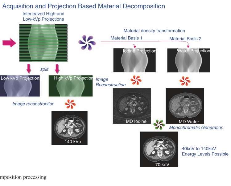
Double énergie

• Objectif : apporter une information spectrale supplémentaire

• Séparation hautes et basses énergies :
 •Emission du tube polychromatique
 •Séparation des matériaux : Iode, calcium…
 •Algorithmes de prédiction probabiliste (likelihood)
 •Cartographie : VNC, Energies (40->200 kV), iode, calcium
 •Correction du beam hardening
Photoelectric - Compton Decomposition
 Material pairs

 Material Specific Images

CT Image Calcium Image Iodine Image

 Calcium-Iodine pair
Photoelectric - Compton Decomposition
 Material pairs

 Material Specific Images

CT Image Iodine image Water image

 Water-Iodine pair
Photoelectric - Compton Decomposition Virtual Mono Energetic Imaging ( E ) P ( E ) c ( E ) P f P ( E ) c f c ( E ) Beam Hardening Correction De-noising MCI Prep PhotoE Image Low keV FBP PhotoE P Linear Raw (LE) Combination With fp and fc Raw (HE) Prep Compton Image FBP Compton c High keV Beam Hardening Correction De-noising
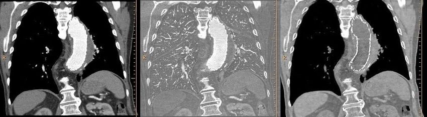
Virtual Monochromatic Spectral Imaging with Fast Kilovoltage Switching: Improved Image
 Quality as Compared with That Obtained with Conventional 120-kVp *
 62-keV 67-keV

 72-keV VMS imaging conventional 120-kVp CT
*Radiology 2012 Matzumoto et al
Photoelectric - Compton Decomposition
 Virtual Mono Energetic Imaging

 200 keV

 keV

40 keV
Photoelectric - Compton Decomposition
 Virtual Mono Energetic Imaging

 Iodine Boost @ low keV
 Conventional 120 kV CT Image 55 keV Mono-Energy CT Image

 Conventional 120 kV CT Image 55 keV Mono-Energy CT Image

ROI: 227.9 ± 25.9
Photoelectric - Compton Decomposition
 Virtual Mono Energetic Imaging

 Artefact reduction @ high keV
Conventional 120 kV CT Image 55 keV Mono-Energy CT Image
 120kVp 200keV
Impact of monochromatic coronary computed tomography angiography from single-source dual-energy CT on
coronary stenosis quantification

 J. Stehli et al. / Journal of Cardiovascular Computed Tomography 10 (2016)
Impact of monochromatic coronary computed tomography angiography from single-source dual-energy CT on
coronary stenosis quantification

 J Stehli et al. / Journal of Cardiovascular Computed Tomography 10 (2016)
Impact of monochromatic coronary computed tomography angiography from single-source dual-energy CT on
coronary stenosis quantification

 J. Stehli et al. / Journal of Cardiovascular Computed Tomography 10 (2016)
Diagnostic Accuracy of Rapid Kilovolt Peak–Switching Dual-Energy CT Coronary Angiography in Patients
With a High Calcium Score

 JA C C : C A R D I OV A S C U L A R IMA G I N G , 2 0 1 5
Perfusion

 • Réalisation d'une double acquisition* :
 • Repos (coronaires)
 • Stress pharmacologique ou effort(basse dose)

• Reconstruction itérative : dose et amélioration du contraste/détection iode
* Radiology. 2014 Jan;270(1):25-46
Perfusion
Coro-scanner
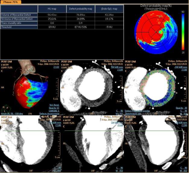
Analyse cardiaque Evaluation d’un défect de perfusion.
—50-year-old man with recurrent chest pain after prior myocardial infarction in left anterior descending
 artery territory and surgical revascularization.A, Short-axis images of SPECT (A) and MRI (B) examinations
 at rest show subendocardial perfusion defect (arrows).

 Vliegenthart R et al. AJR 2012;199:S54-S63

©2012 by American Roentgen Ray Society
—50-year-old man with recurrent chest pain after prior myocardial infarction in left anterior descending
 artery territory and surgical revascularization., Corresponding short-axis cross-section of contrast-
 enhanced dual-energy CT study at rest,

reconstructed as merged gray-scale image with superimposed iodine distribution
 color map.
 Vliegenthart R et al. AJR 2012;199:S54-S63
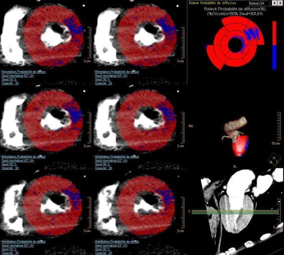
Perfusion – Double énergie
 • Amélioration de la
 quantification
 • Correction du beam hardening
 (paroi postérieure +++)

* AJR 2010; 195:639-646
Dual-energy CT might be a better way for optimising myocardial and coronary artery imaging

Pan et al, International Journal of Cardiology March 2016
Spectral Photon Counting CT
 SPCCT
 Imaging all the Photons…...

Common Scintillating Detector Photon counting detector
Dual Energy CT Photon counting CT
 Dual Layer Detector (PHILIPS) Direct Conversion Detector
Tube Spectrum
 X-Ray

 Low density/High Light Photo
 Diode Direct Conversion Detector
 Detector

 Output Scintillator

 High Output Scintillator h h
 h e e
 Photo
 Diode

 e

 Integrating ASIC Counting ASIC
Schematic diagram of energy
 discrimination
Potential Benefits

= High Spatial Resolution
= K-edge Imaging
= Multiple material characterization
= Precise energy separation
= Low-contrast resolution
= Low Dose
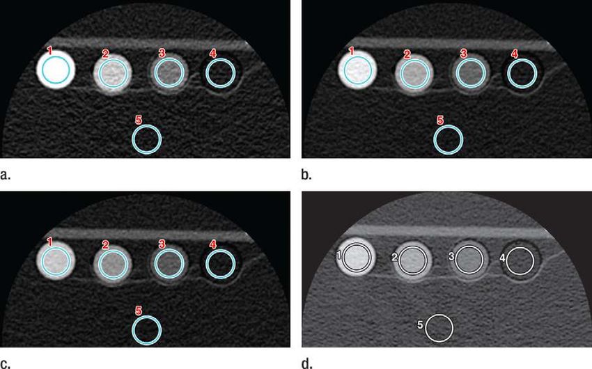
Methods and Materials
Biological material:
 – 8 calcified atherosclerotic plaques
 – 10 lipid-rich atherosclerotic plaques
 – Filled and immerged into an Iodine solution

Photon Counting Multi-Energy CT:
 – 70 keV, 20 mAs/Slice
 – FOV: 60 mm
 – Resolution: 0.1x0.1x0.2 mm3
 – Scan time: 200 sec/slice

 Photon counting CT, Philips, Germany

36
Results

 Significant differences between all elements for Photoelectric absorption and Iodine concentration (p
Results
Iodine map and calcifications

 CT-like image Iodine map

38 L Boussel Ph Douek , BJR 2014
Molecular imaging

 39
Molecular imaging
 •Gold high-density lipoprotein nanoparticle
 contrast agent (Au-HDL)*
 For characterization of macrophage
 burden, calcification and stenosis of
 atherosclerotic plaques
 In Apolipoprotein E knockout mice

Comode et al Radiology 2013 40
Molecular imaging

• Fibrin using bismuth loaded
 nanoparticles

 D Pan et al Computed Tomography in Color: NanoK-Enhanced Spectral CT
 Angew Chem Int Ed Engl. 2010 December 10; 49(50): 9635–9639.

 41
Installation of 1st iCT-based preclinical SPCCT –
prototype @ CERMEP Lyon
Parameter Specification
Platform Philips iCT
Tube voltages [kVp] 80, 100, 120
Tube currents [mA] 10-100
Focal spot [mmx 0.6 x 0.7
mm]
Gantry rotation [s] 1.0
Spatial Resolution > 20
[lp/cm]
FOV [mm] 168
# energy bins >2
Sensor Material CZT
Sensor Thickness 2 mm
Spatial Resolution

 Scan of anatomical leg phantom
Line-Pairs plastic phantom
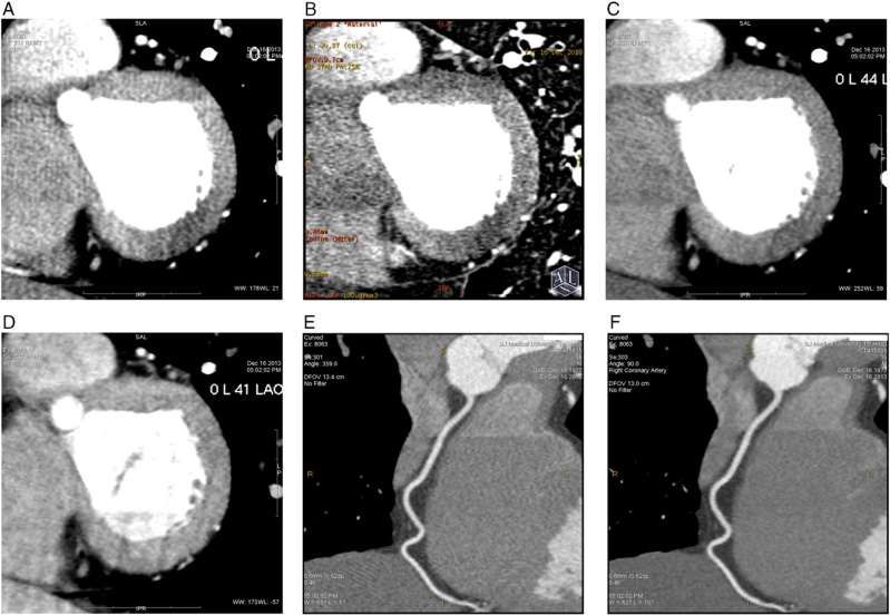
Spatial Resolution Stenosis Phantom
 W2000L400 Z =5.25 Z =11
= Scan Parameter : 0.2x0.25

 - Scan Type: Axial (stack)
 - 120kVp 50mA
 - Rotation 1sec R=1.5mm

= Reconstruction:
 - HU Image
 R=3.4mm W2000L400
 - Filter: Standard 0.2x0.25

 - Voxel Size: 0.2 x 0.2 x 0.25 [mm]

 Mean:1850

 Z=5.25 Z=11
 W2000L400 W2000L400
 0.2x0.2x0.25 0.2x0.2x0.25
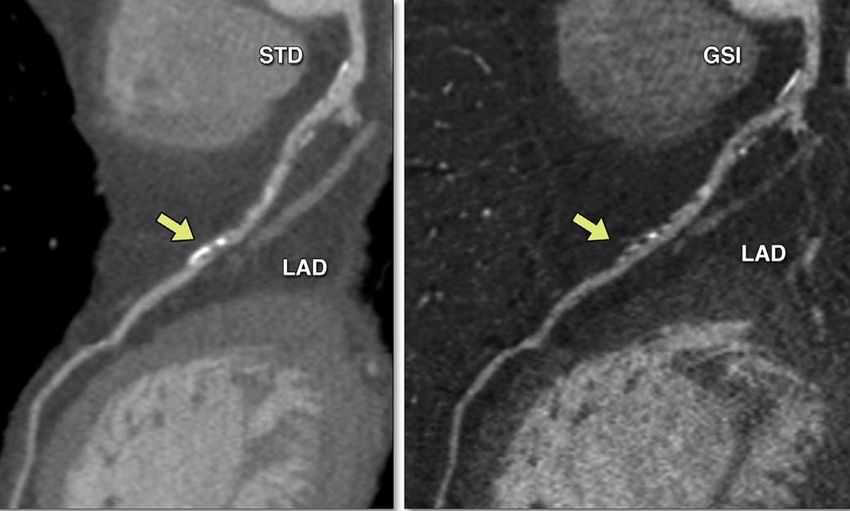
Spatial Resolution Stent
 Standard CT SPCCT

ww1500 wl470 ww2000 wl800 ww1500 wl470 ww2000 wl800
Voxel Size: 0.1 x 0.1 x 0.1 Voxel Size: 0.1 x 0.1 x 0.1 Voxel Size: 0.1 x 0.1 x 0.1 Voxel Size: 0.1 x 0.1 x 0.1
X-RAY

X-Ray Rabbit
Acquisition In-Vivo SPCCT : Contrast
• Acquisition : Axial, Z coverage = 2 mm, 120 kVp, 100 mA
• Reconstruction : FOV 80, Matrix size 1024, pixel size= 0,08, slice thickness
 0,1 mm, Detail filter
Acquisition In-Vivo SPCCT : Contrast
• Figure 3: Volume rendering of 2mm coverage
 with media contrast agent
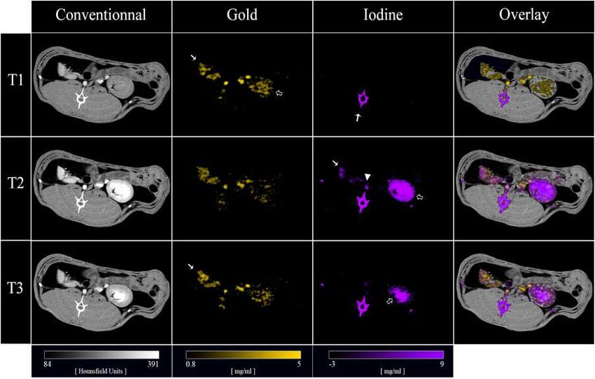
Material Decomposition Contrast Specificity Images

 HU Water [mg/cc]
Axial : 120kVp 100mA
 WL0 WW1000 : SW 2mm : Standard WL790 WW2764 : SW 2mm : Standard

Gadolinium [mg/cc] Iodine [mg/cc] Gold [mg/cc]
WL3.64 WW4.38 : SW 2mm : Smooth WL18.28 WW35.26 : SW 2mm : Standard WL4.76 WW6.02 : SW 2mm : Smooth

 Partnership with: University of Pennsylvania
Rabbit Gold Contrast

HU Gold
WL60 WW600 : SW 2mm : Standard WL1.5 WW3.2 : SW 2mm

11.2cc Gold (peg 65mg/cc ) injected 120 kVp 100 mA
Rabbit Gadolinium Contrast

HU Gadolinium [mg/cc]
WL60 WW360w : SW2mm WL580 WW180 : SW2mm

50 seconds after Gadolinium
Injection.
 = Cavity : 2.92 [mg/cc]
 = Parenchyma: 1.63 [mg/cc]
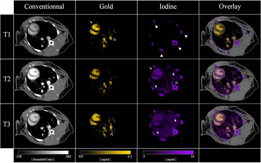
Rabbit 2

 CARDIAC ANGIOGRAPHY
 Parameters : Parameters :
 - Thickness 2 mm - Thickness 2 mm
 - No Filter - Gaussian filter 2 mm
 - L1172; W311
 Fig 3 : Reconstruction of the Kedge of Gold. Cardiac angiography after
 Fig 1 : Cardiac angiography after dynamic injection of Gold (12 mL at 1 cc/s) (gif) : 30 dynamic injection of Gold (12 mL at 1 cc/s) (gif) : 30 secondes of
 secondes of acquisition starting 5 s after injection, 15 cycles of 2 secondes acquisition, 15 cycles of 2 secondes

 Fig 2 : Cardiac angiography after dynamic injection of Iodine (3 mL at 1 cc/s) and Fig 4 : Reconstruction of the Kedge of Gold. Cardiac angiography after
 past injection of gold (gif) : 30 secondes of acquisition, 15 cycles of 2 secondes dynamic injection of Iodine (3 mL at 1 cc/s) and past injection of gold (gif) :
 30 secondes of acquisition, 15 cycles of 2 secondes
Rabbit 2

 DISCRIMINATION GOLD/IODINE
 Fig 1 : Dynamic angiography during 30 s with reconstruction of Fig 2 : Dynamic angiography during 30 s with reconstruction of
 Kedge of Gold after injection of iodine (3 mL at 1 cc/s) and past material decomposition of Iodine after injection of iodine (3 mL at
 injection of gold (12 mL) (gif) 1 cc/s) and past injection of gold (12 mL) (gif)
Material Decomposition Contrast Specificity Images
 Applications of Dual Contrast Agents
 HU

 I Gd I

 Gadolinium Iodine

 iodine

 phantom

 Axial : 120kVp 50mA
 HU: WL-350 WW1400
unenhanced Gd : WL545 WW45
polyp I : WL690 WW255

 gadolinium
 enhanced polyp
 Partnership with: Technical University Munich
Rabbit Gold & Iodine Contrast

3cc Iodine (400 mg/cc) injected 120 kVp 100 mA
Rabbit Gold & Iodine Contrast
HORIZON 2020
 HORIZON 2020

 SPCCT
H2020 SPCCT :Lyon university coordinator
To develop and validate a widely accessible, new quantitative and analytical imaging technology combining:

 Spectral Photon Counting Computed Tomography (SPCCT) AND
 Dedicated Contrast Agents

To accurately detect, characterize and monitor neurovascular and cardiovascular disease

 • Ultra-low dose imaging
 • CA dose reduction (reduction of entire scans)
 • Quantitative imaging (follow-up)
 • Functional imaging (K-edge)
 • Higher spatial resolution

 Clinical (sub-seconds, 1000mA)

 In-vivo pre-clinical (seconds, 100 mA)

 Ex-vivo, pre-clinical (hours, 100 µA)
Acknowledgements & Collaborators
Loic Boussel
Daniel Bar-Ness
Monica Sigovan
Salim Si Mohamed
Franck Lavenne
Marlene Wiart
Yves berthezene
Caroline Bouillot
Jean-Baptiste Langlois
Peter B. Noël
David Cormode
Frédéric LEROUGE
Simon Rit
Jean-Michel Létang
Gloria Vilches Freixas
Philippe Coulon
Bracco research
Philips Hamburg
Philips Haifa
Thank you

60
You can also read