SCWDS BRIEFS - University of Georgia College of Veterinary ...
←
→
Page content transcription
If your browser does not render page correctly, please read the page content below
SCWDS BRIEFS              A Quarterly Newsletter from the
                                  Southeastern Cooperative Wildlife Disease Study
                                          College of Veterinary Medicine
                                                          The University of Georgia
                              Phone (706) 542-1741         Athens, Georgia 30602                     FAX (706) 542-5865

                             Volume 36                        January 2021               Number 4

           Figure 1. Map showing the distribution of EHDV and BTV detections by SCWDS during 2020.
           Some symbols may represent more than a single detection.

2020 Hemorrhagic Disease Summary                             viruses does not provide the complete picture of
                                                             EHDV and BTV activity in wild ruminants in the US,
As was highlighted in the October 2020 issue of the          the data do provide situational awareness on
SCWDS BRIEFS, SCWDS has provided annual                      ongoing outbreaks for wildlife agencies, a means to
diagnostic testing for epizootic hemorrhagic                 validate parallel annual data provided by our Annual
disease virus (EHDV) and bluetongue virus (BTV)              National Hemorrhagic Disease (HD) Survey, and
for the last 30 years. Annually, we receive 200-400          the isolates represent valuable resources for future
submissions from state wildlife management                   research.
agencies, as well as some agriculture agencies and
veterinary      diagnostic    laboratories.    Most          Emerging viruses (i.e., SARS-CoV-2, rabbit
submissions consist of tissue samples (typically             hemorrhagic disease virus 2) were unfortunately a
lung and/or spleen) from wild white-tailed deer,             common theme in 2020. Although HD is certainly
although a minority come from other wild and                 not a new disease in North America, we have
captive ruminants. Samples are screened for EHDV             clearly observed changing dynamics over the last
and BTV using real-time reverse-transcription                two decades, such as the detection of new
polymerase chain reaction (rRT-PCR) assays, and              EHDV/BTV serotypes and changing epidemiologic
virus isolation is attempted on positive samples.            patterns (e.g., increasing expansion in the northern
Virus isolates are further identified to serotype.           US). In this sense, HD is an emerging disease in
While the identification and isolation of these              some regions of North America. Amid restrictions
                                                                                                            Continued…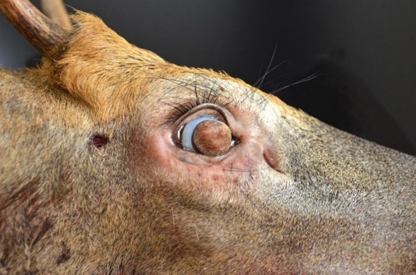
SCWDS BRIEFS, January 2021, Vol. 36, No. 4

associated with the COVID-19 pandemic, many                    and New York). A further example of this
agency staff were limited in their ability to                  phenomenon is West Virginia, a state with a 5-6
investigate deer mortalities. Despite these                    year HD outbreak cycle historically but that has now
limitations, biologists were still able to submit a            had annual outbreaks for 5 consecutive years
large number of samples to SCWDS, and                          (2016-2020). Work is needed to better understand
diagnostic results from 2020 clearly show the                  the potential underlying mechanisms that may
continuation of some of the changing patterns                  explain these observed changes. Finally, moving
mentioned above.                                               deeper into the Southeast, we isolated additional
                                                               BTV serotypes, including BTV-2 and BTV-3 in
During 2020, SCWDS received 274 submissions for                Florida and BTV-18 in Louisiana. Both BTV-3 and
EHDV and BTV diagnostic testing from 25 different              BTV-18 represent historically exotic BTV serotypes.
states. The vast majority of submissions (239/274)             This high diversity of viruses observed in the
were from white-tailed deer, although we also                  southernmost states like Louisiana and Florida is
received tissue samples from 16 pronghorn, 13                  expected and is likely associated with their unique
mule deer, three elk, two bighorn sheep, and two               habitats and climate, which in turn drive vector
moose. Overall, we had 162 virus detections (128               population dynamics. For example, in addition to
EHDV and 34 BTV) from 22 states, including 86                  the historically endemic EHDV and BTV serotypes
virus isolates representing numerous serotypes                 known to occur in the United States, over 10 exotic
(EHDV-2, -6, and BTV-1, -2, -3, -5, -17, and -18)              viruses have been confirmed in Florida over the last
(see Figure). As is the case every year, most                  20 years. This region of high virus diversity may
detections were from white-tailed deer and EHDV-               serve as a potential source of virus for other parts
2 was the most common virus identified (42                     of the country via movement of infected hosts or
isolations from 10 states). However, other                     vectors across the landscape.
ruminants were involved in some regions. In
particular, EHDV-2 was isolated from white-tailed              We thank the many wildlife professionals who
deer, mule deer, pronghorn, and elk in Montana                 submitted tissue samples for diagnostic testing this
and/or North Dakota. Additionally, BTV-17 was                  past season and are grateful that you continued to
isolated from bighorn sheep (Nebraska) and mule                contribute samples in spite of interferences related
deer (Kansas), and BTV-1 was isolated from a                   to the COVID-19 pandemic. The data obtained
pronghorn (Kansas). Aside from the diversity of                through such effort are critical to documenting the
hosts represented, the diversity of viruses                    viruses associated with HD outbreaks in wild
recovered from Kansas and Nebraska was notable.                ruminant populations throughout much of the US
In these two states we detected two common and                 and help to better document and understand the
historically endemic viruses (EHDV-2 and BTV-17),              changing patterns of HD. (Prepared by Mark Ruder,
as well as two historically exotic viruses (BTV-1 and          Natalie Stilwell and Dave Stallknecht)
BTV-5) that had not been previously documented in
this part of the country. Further, a white-tailed deer         COVID-19 and Wildlife
in Nebraska was co-infected with EHDV-2 and BTV-
5. The outbreaks in the Great Plains during 2020               It has now been a year since the first human cases
highlight the complexity of hemorrhagic disease in             of a novel coronavirus infection known as SARS-
North America – a system that involves multiple                coronavirus-2 (SARS-CoV-2) surfaced in Asia and
viruses, multiple ruminant hosts, and likely multiple          spread rapidly in human populations around the
Culicoides vector species.                                     globe. The pandemic has resulted in more than 105
                                                               million human cases worldwide and infection rates
Moving into the eastern United States, all EHDV                are still rising in many areas. During this pandemic,
and BTV detections were in white-tailed deer. Much             several questions have emerged regarding the
of the HD activity in the eastern US was driven by             potential role of wildlife. These questions have
EHDV-6 (27 virus isolates from 11 states),                     primarily related to the origin of this virus and the
especially in the mid-Atlantic and northeastern                susceptibility of wildlife species to SARS-CoV-2.
states. This included the first reports of EHDV-6 in
New York, Delaware, and Georgia. Another trend                 Early genetic analyses suggest SARS-CoV-2 likely
that is apparent when examining the 2020 map is                originated after spilling over into human populations
the continued expansion of HD into the upper                   from an animal reservoir, but the exact source and
Midwest and Northeast (e.g., Wisconsin, Michigan,              species remain unknown. The virus appears

                                                         -2-                                            Continued…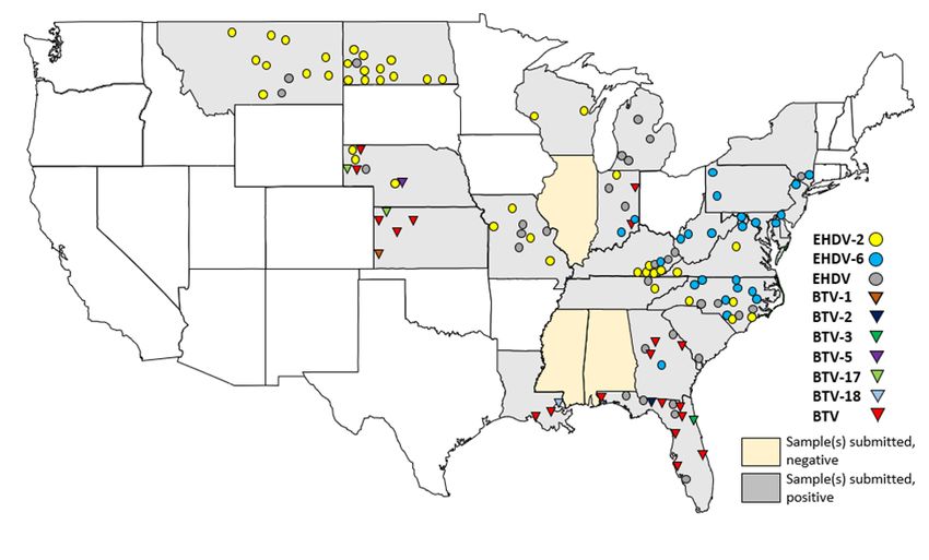
SCWDS BRIEFS, January 2021, Vol. 36, No. 4

genetically and structurally similar to bat and                could act as a potential animal reservoir for SARS-
pangolin coronaviruses, suggesting that one or both            CoV-2. Results from controlled laboratory studies
species played a role in evolution of the virus prior          are also insufficient to predict population-level
to transmission into humans. While wildlife may be             disease risks in the natural environment. Therefore,
the initial source of SARS-CoV-2 exposure to                   to put these susceptibility studies into perspective
humans, zoonotic transmission was likely facilitated           it’s important also to focus on natural infection data.
by actions related to the commercialization and                To date, SARS-CoV-2 has been detected in a
utilization of wildlife. Similar transmission patterns         limited range of non-domestic species, including
have occurred in the past with the highly pathogenic           felids (i.e., lion, tiger, snow leopard, puma),
SARS- and MERS-coronavirus outbreaks, which                    mustelids (i.e., mink, ferret), and non-human
arose from human contact with horseshoe bats and               primates (i.e., gorilla). The most extensive effects
camels, respectively. To further elucidate                     have been observed in farmed mink. According to
information on the source of SARS-CoV-2, the                   recent CDC figures, SARS-CoV-2 infection has
World Health Organization recently deployed a                  been confirmed at more than 400 mink farms
team of scientists to Wuhan, China, where the first            worldwide; most cases have occurred in Denmark
human infections were identified in connection with            and the Netherlands where the fur industry is
a live animal market.                                          prevalent and individual facilities house thousands
                                                               of animals. Many facilities were depopulated due to
Meanwhile, researchers around the world are                    the observed high infection rate and subsequent
focusing on SARS-CoV-2 susceptibility in different             risk of zoonotic spillover to farm personnel, along
hosts through two main methods: 1) examining the               with the detection of novel, mutated variants of the
angiotensin-converting enzyme 2 (ACE2) receptor,               virus in some cases. Similar outbreaks have
and 2) performing live animal experimental infection           occurred on mink farms in the United States.
studies. The ACE2 receptor, which is the host cell             Although these cases were dramatic and widely
binding site for the coronavirus spike protein, varies         publicized, it’s important to remember that
significantly among animal species and its structure           outbreaks in high-density, industrial facilities such
implies whether SARS-CoV-2 can attach and enter                as those used for mink farming do not directly
host cells. Experimental infections go one step                correlate with potential risks to wild populations, as
further to evaluate in vivo susceptibility, along with         captive animals are typically held in artificial
pathology, virus transmission and shedding                     conditions where they have high contact rates with
patterns, which can provide insight into how SARS-             humans and other animals. Farmed animals may
CoV-2 behaves in individual animals.                           also undergo generations of selective breeding for
                                                               desired characteristics, such as fur color and quality
So far, published studies show that certain bat,               in mink, which can result in decreased genetic
rodent, felid, rabbit, mustelid, non-human primate,            diversity and impaired immune function compared
skunk, bovid, canid, and deer species demonstrate              to their wild counterparts.
a degree of susceptibility to SARS-CoV-2 infection,
whereas other rodents and all examined bird, pig,              Aside from farmed mink, natural infections in other
insect vector, raccoon, and North American bat                 animal hosts have rarely been reported, which
species were not susceptible. Regarding wildlife, a            suggests the vast majority of animal species are
recent study conducted by the USDA Agricultural                poorly susceptible to SARS-CoV-2. For example, a
Research Service examined the potential for                    very low number of SARS-CoV-2 cases have been
experimental infection in white-tailed deer. In this           reported in domestic dogs (n=41) and cats (n=53)
study, fawns were found to be susceptible to                   in the United States. These numbers are particularly
intranasal inoculation with a high dose of SARS-               striking considering there are more than 150 million
CoV-2. Although infected fawns remained                        dogs and cats in US households. In non-domestic
asymptomatic, viral transmission to naïve,                     animals, infections (while still rare) have largely
cohabitating fawns was documented. A follow-up                 been restricted to animals in captivity. Notable
study will further examine transmission and                    exceptions in the United States have been free-
shedding patterns in white-tailed deer.                        ranging, wild mink or escaped farmed mink
                                                               captured on or near affected mink farms in Utah and
While susceptibility studies give some idea of the             Oregon, respectively. In general, the risk of viral
relative risk of infection in various species, these           spread is inherently higher when animals are
data only provide partial evidence that a species              housed in high densities (e.g., farm facilities) or

                                                         -3-                                               Continued…
SCWDS BRIEFS, January 2021, Vol. 36, No. 4
have increased contact with humans (e.g., in bred and raised for meat. All rabbits died or were
zoological or rehabilitation settings), compared to in culled over a two-week period. The outbreak
natural environments. In contrast, there is no represents the first confirmed detection of RHDV2
evidence yet to suggest SARS-CoV-2 infection can in rabbits (domestic or wild) in Florida. Clinical signs
be sustained in wild animal populations. included bleeding from the nose and sudden death.
The location has since been disinfected and placed
Infection rates over the past year show that COVID- under quarantine and a fallow (rabbit free) period
19 is largely a disease affecting humans, not for 90 days. The backyard rabbit operation was
animals. Efficient human-to-human transmission closed, meaning no rabbits were recently imported
has been the single most important driver of the or exported and the route of RHDV2 introduction is
global spread of SARS-CoV-2. Furthermore, not currently known. However, the Florida
SARS-CoV-2 introduction into animal populations Department of Agriculture & Consumer Services
has occurred only under specific conditions (FDACS) and United States Department of
involving direct contact with infected humans. Still, Agriculture (USDA) are conducting an ongoing
appropriate measures need to be taken to minimize epidemiologic investigation into the outbreak. The
viral transmission between humans and animals, Florida Fish and Wildlife Conservation Commission
not only for the consideration of the current SARS- and FDACS are continuing outreach efforts to
CoV-2 outbreak but to prevent similar pandemics educate citizens of the risk and encourage prompt
from occurring in the future. Regarding SARS-CoV- reporting of sick or dead wild and domestic rabbits.
2, those working closely with captive or free-ranging To date, no additional cases of RHDV2 have been
wildlife should follow the same measures used to reported in Florida.
prevent human-to-human virus transmission, which
include wearing appropriate PPE, disinfecting This recent RHD outbreak in Florida represents the
spaces frequently, and undergoing testing and self- most southeastern detection of RHDV2 in the US,
isolation if COVID-19 exposure is suspected. With with the next closest report being in domestic
several SARS-CoV-2 vaccines now being rabbits in central Texas. However, isolated RHDV2
deployed, widespread vaccination of humans will outbreaks in domestic rabbits in the eastern US
also play a crucial role in obtaining herd immunity occurred previously in Ohio (fall 2018) and New
and reducing viral spread in human and animal York (spring 2020) and genetic analysis of these
populations. (Prepared by Natalie Stilwell, Mark viruses by USDA suggests these viruses were
Ruder, and David Stallknecht) distinct from those currently circulating in the
western US and likely represent different
Rabbit Hemorrhagic Disease Virus 2 introductions. The USDA is performing a similar
Continues Lurking genetic analysis of the RHDV2 detection from Lake
County, Florida. This information will help
In the April 2020 issue of the SCWDS BRIEFS, we determine the genetic relatedness of the Florida
discussed the ongoing outbreak of rabbit RHDV2 to other viruses detected in North America
hemorrhagic disease (RHD) virus 2 (RHDV2) in the from 2018-2020, which will help understand if the
southwestern United States and Mexico. At the Florida outbreak represents significant spread of
time, RHDV2 had been reported in wild rabbits and the ongoing outbreak in the Southwest, or yet
hares in New Mexico, Arizona, Colorado, and another separate introduction.
Texas, and in domestic rabbits (Oryctolagus
cuniculus) in New Mexico, Arizona, and Texas. As RHDV2 is a highly infectious and lethal virus that
feared, the RHD outbreak continued to expand and may persist in the environment for extended
by mid-December 2020, RHDV2 had been periods. Clinical signs may include fever, lethargy,
confirmed in wild and domestic or feral rabbits or ocular and nasal bleeding. However, sudden
throughout a vast and largely contiguous region of death is often the only sign of infection. Virus is
the western US (Arizona, California, Colorado, New shed in most bodily secretions and transmission is
Mexico, Texas, Utah, and Wyoming). Unfortunately, through direct or indirect contact and the virus can
in late December 2020, RHDV2 was confirmed in remain stable in carcasses/tissues and the
domestic rabbits in Lake County, Florida. environment for months. With such efficient
transmission and the multitude of susceptible
The RHD outbreak in Florida occurred in a small, domestic and wild lagomorph hosts, the risk of
non-commercial, backyard population of rabbits RHDV2 spread is high. Biosecurity (e.g., sanitation,
-4- Continued…SCWDS BRIEFS, January 2021, Vol. 36, No. 4
limiting movement) remains key to preventing the pathogen, and the environment. Influenza is no
spread of RHDV2 among domestic and wild exception. We have known for more than 50 years
populations. Movement of live or dead rabbits, that waterfowl represent an important reservoir for
rabbit parts, equipment, bedding, or any other a genetically diverse population of influenza A
materials rabbits have contacted are significant risk viruses (IAV). However, our understanding of the
factors for the spread of RHDV2 and preventive interactions between host, IAV, and the
measures should target minimizing the risk of long- environment that provide a means for IAV
distance movement of the virus. transmission and maintenance in wild birds is
incomplete. It is widely accepted that IAV
There is potential for RHDV2 to significantly impact transmission in waterfowl populations primarily
wild rabbit and hare populations, as well as pet occurs via an indirect fecal/oral route that involves
rabbits and production rabbits. The effects of contaminated water. Infectious IAV have been
potential wild lagomorph population declines on isolated directly from water samples collected from
predator populations remains unknown but also waterfowl habitats, and it has been demonstrated
raises concern. To date, RHDV2 has been experimentally that these viruses can remain
confirmed in the black-tailed jackrabbit (Lepus infective in water for extremely long durations
californicus), antelope jackrabbit (Lepus alleni), depending on water temperature and other physical
desert cottontail rabbit (Sylvilagus audubonii), and chemical properties such as pH and salinity. In
mountain cottontail rabbit (Sylvilagus nuttallii), and a distilled water laboratory model, for example, IAV
eastern cottontail rabbit (Sylvilagus floridanus), can remain infective for more than one year in water
although the wild lagomorph host range in North at 4ºC. Most of what we know about the stability of
America remains unclear and all lagomorphs IAV in water is derived from controlled experimental
should be considered susceptible until further studies; validating these results in the field has
information is gathered. Prompt detection of the proven challenging. This challenge relates to the
virus in new areas, followed by robust disinfection physical, chemical, and biological complexity of
and containment measures are the best chance to natural water bodies, direct testing limitations
control outbreaks. However, once RHDV2 is related to the large volumes of water associated
circulating in wild populations, management options with waterfowl habitats, and biosafety concerns
become limited. Increased vigilance with prompt associated with potential field release of laboratory
reporting and investigation of wild or domestic rabbit propagated IAV.
mortality events are critically important, especially
in unaffected areas. Robust communication and In a collaboration with the United States Geological
cooperation among state and federal wildlife and Survey (USGS) Alaska Science Center, we recently
agricultural agencies, as well as citizen stakeholder developed a system to safely evaluate the infectivity
groups, is paramount to the successful prevention of IAV in water under monitored field conditions.
and management of RHDV2 in the US. This system relies on the periodic testing of
cloacal/oropharyngeal (CL/OP) swab material from
Currently, RHDV2 is classified by USDA-APHIS- ducks that are diluted in filter-sterilized water
Veterinary Services as a foreign animal disease and collected from the corresponding waterfowl
is reportable to the World Organization for Animal habitats. The swab-inoculated water samples are
Health (OIE). Therefore, close cooperation between contained in individual tubes within a larger barrel-
state and federal wildlife and agricultural agencies type container and submerged in a natural water
is important. Please contact SCWDS if assistance body for subsequent retrieval and testing. This
is needed with outreach, prevention, or response system was originally described by Reeves et al.
activities. To submit a wild lagomorph carcass to (2020). By retrieving and testing contained
SCWDS for RHDV2 testing, please work with our swab/water samples over time, this model system
Diagnostic Service to coordinate shipment. was shown to provide a safe method to assess the
(Prepared by Katie Vivirito, University of Illinois, and long-term viability of IAVs that are naturally shed by
Mark Ruder) infected waterfowl into water under the physical and
chemical conditions that are present in the specific
Influenza Persistence in Waterfowl Habitats habitats that ducks are utilizing.
The epidemiology of any infectious wildlife disease In a more recent study, we applied this technique to
is dependent on interactions between host, waterfowl habitats in Alaska, Louisiana, and
-5- Continued…SCWDS BRIEFS, January 2021, Vol. 36, No. 4
Minnesota. The goal was to better understand the isolated from 51 (7.4%) of the replicate #1 samples.
potential for IAV to overwinter in the environment in These included 40 samples from Minnesota and 11
these habitats. This study was done not only with from Alaska. IAV was not isolated from samples
our colleagues at the Alaska Science Center but collected from Louisiana. Replicate #2 samples
also involved collaborators from the California from Alaska and Minnesota were retrieved during
Water Science Center (USGS), Western Ecological April (approximately 6 – 7 months later). Of the 51
Research Center (USGS), Memorial University of swab/water samples that originally yielded an IAV
Newfoundland, US National Poultry Research isolate, 10 (20%) remained infective when
Center (USDA), Louisiana Department of Wildlife inoculated into eggs. These included an assortment
and Fisheries, and Patuxent Wildlife Research of IAV subtypes including H2N9, H3N6, H3N8,
Center (USGS). In this study, two field sites were H4N6, H4N8, and low pathogenicity (LP) H7N3.
selected from wetlands at Izembek National Wildlife These samples had remained infective for 209 -229
Refuge (Alaska), Agassiz National Wildlife Refuge days in filtered lake water under natural water
(Minnesota), and Cameron Parish (Louisiana). temperature conditions. To confirm that the positive
replicate #2 water samples still contained virus
capable of infecting waterfowl, we experimentally
challenged 10 groups of three mallards with
approximately 0.5 mL of these water samples.
Infection of mallards, as measured by three criteria:
detection of viral RNA, virus isolation, and
seroconversion, was observed with two of ten
samples corresponding to an H3N8 and LP H7N3
IAV. The field study was further supported by a
laboratory trial using the residual water in the 40
positive replicate #1 samples from Minnesota.
Following initial testing, these were maintained at
4ºC and tested monthly from September to April. Of
these 40 samples, five (13%) remained infective for
at least seven months (April represented the last
month of testing for this experiment).
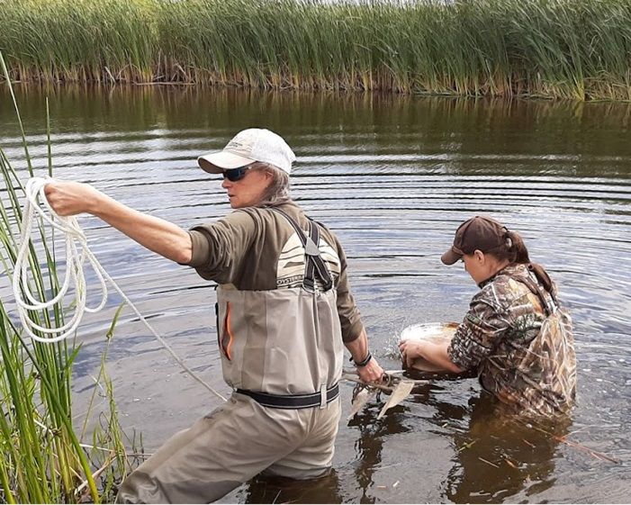
Figure 2. SCWDS biologists, Deb Carter and Alinde Fojtik,
deployed a submersible chamber containing CL/OP swabs Results from this work confirm that IAV can persist
at Agassiz National Wildlife Refuge (Minnesota) where they in the natural environment during the time when
remained overwinter.
waterfowl are not present, or are in greatly reduced
Surface water from all sites was collected, filter numbers, on northern waterfowl habitats. This
sterilized, chemically characterized, and divided potentially provides a means for infectious IAV to be
among tubes that were filled with 40 mL of the maintained from one IAV season to the next.
filtered water. Individual tubes were then inoculated Although the model system employed in these
with a combined CL/OP swab collected from a studies provides an improved and more relevant
single wild duck utilizing these wetlands and each method to access environmental stability of IAV in
inoculated water sample was evenly divided (13 mL waterfowl habitats, it does not capture all of the
each) into two replicate samples. Replicate #1 was biological, chemical, physical, and hydrologic
submitted to the SCWDS laboratory for immediate factors and interactions that can exist in a natural
IAV testing. Replicate #2 samples were sealed and water body. A great deal of work remains to be done
placed in a larger barrel-like container and to better understand and confirm the role and
submerged for 6-7 months in the waterfowl habitat importance of environmental transmission in the
for subsequent retrieval (see photo). Sampling in maintenance of these viruses. With current
Alaska and Minnesota was done in September detections and wild bird mortality associated with
(corresponding to early fall migration); ducks were highly pathogenic H5 viruses of the
sampled in Louisiana during November as birds A/Goose/Guangdong/1/1996 lineage in waterfowl in
arrived on wintering areas. Asia, Europe, and Africa, this understanding may
prove valuable if we are faced with a similar
Of the 686 surface water samples that were problem in North America.
inoculated with a CL/OP swab, IAV was initially
-6- Continued…SCWDS BRIEFS, January 2021, Vol. 36, No. 4
For additional details related to this work see: consistent with “corneal dermoids.” The internal
structures of the eye were intact and no microscopic
Ramey AM, Reeves AB, Drexler JZ, Ackerman JT, De
La Cruz S, Lang AS, Leyson C, Link P, Prosser DJ, abnormalities were noted in the eyes or brain.
Robertson GJ, Wight J, Youk S, Spackman E, Pantin- Additionally, laboratory tests for hemorrhagic
Jackwood M, Poulson RL, Stallknecht DE. Influenza A disease (HD) and chronic wasting disease (CWD)
viruses remain infectious for more than seven months were conducted on the submitted tissues. In
in northern wetlands of North America. Proc Biol Sci. addition to the corneal dermoids, epizootic
2020 Sep 9;287(1934):20201680. hemorrhagic disease virus (EHDV) serotype 2 was
doi: 10.1098/rspb.2020.1680. isolated from the lung. Thus, hemorrhagic disease
was the presumed cause of the bleeding observed
Reeves AB, Ramey AM, Koch JC, Poulson RL,
by the private citizen. It is not clear whether this
Stallknecht DE. Field-based method for assessing
buck’s abnormal behavior was attributed primarily
duration of infectivity for influenza A viruses in the
environment. J Virol Methods. 2020 Mar;277:113818. to HD, corneal dermoids, or a combination. The
doi: 10.1016/j.jviromet.2020.113818 prion that causes CWD was not detected in the
retropharyngeal lymph nodes.
(Prepared by Dave Stallknecht and Rebecca
Poulson) Dermoids are a type of choristoma, which is defined
as normal tissue in an abnormal location.
Accordingly, dermoids are characterized by skin-
Corneal Dermoids in a White-tailed Deer
like tissue occurring on the body in a location other
than the dermis (skin). Corneal dermoids, as in the
During late August 2020, a private citizen in case of this deer, often contain elements of normal
Knoxville, Tennessee reported a white-tailed deer skin, including hair follicles, sweat glands, collagen,
buck that was circling, bleeding, and lacked an and fat. The masses generally are benign (non-
appropriate fear response to humans. The yearling invasive) and are congenital, likely resulting from an
buck subsequently was dispatched and Tennessee embryonal developmental defect. Dermoids over
Wildlife Resources Agency (TWRA) personnel the eyes likely obscure vision and disrupt an
performed a field necropsy. Strikingly, TWRA staff animal’s ability to forage, engage in normal social
noted that hair appeared to be growing from the interactions, evade predation, and avoid hazards. In
surface of both eyes and submitted the head and the present case, the age of the buck suggests that
selected fresh tissues to SCWDS for diagnostic it was able to adapt and survive with this condition
examination. but ultimately succumbed to hemorrhagic disease.
Frankly, it is impressive the young buck was able to
survive as long as he did.
Dermoids have been reported in numerous
domestic animal species, most commonly the dog
and cow, but have rarely been reported in deer. In
some domestic animal species and breeds,
dermoids are presumed to be an inherited trait.
They can affect just one eye, but often affect both
eyes in cows. In contrast to the present case
involving both eyes, the only previous diagnosis by
SCWDS of dermoids in a white-tailed deer affected
only one eye (LaDouceur et al. 2012. Journal of
Wildlife Diseases, 48(3);826-828). Aside from
Figure 3. Corneal dermoid covering the surface of the eye of a
obscured vision, these masses do not pose a health
white-tailed deer. threat, either at the individual or population level.
The growths were densely haired and completely SCWDS would like to thank Sterling Daniels and
covered the surface (cornea) of both eyes, others at the TWRA for submission of this case
presumably drastically reducing visual capacity showing a rare and interesting condition. (Prepared
(Figure). Microscopic examination of the haired by Michelle Willis and Nicole Nemeth)
masses revealed they consisted of haired skin,
-7-SCWDS BRIEFS
SCWDS BRIEFS, January 2021, Vol. 36, No. 4 Nonprofit Organization
U.S. Postage
PAID
Southeastern Cooperative Wildlife Disease Study Athens, Georgia
Permit No. 11
College of Veterinary Medicine
The University of Georgia
Athens, Georgia 30602-4393
RETURN SERVICE REQUESTED
Information presented in this newsletter is not intended for citation as scientific literature. Please contact the
Southeastern Cooperative Wildlife Disease Study if citable information is needed.
Information on SCWDS and recent back issues of the SCWDS BRIEFS can be accessed on the internet at
https://vet.uga.edu/scwds. If you prefer to read the BRIEFS online, just send an email to Jeanenne Brewton
(brewton@uga.edu) or Michael Yabsley (myabsley@uga.edu) and you will be informed each quarter when
the latest issue is available.You can also read