STRANGER THAN A SCORPION: A REASSESSMENT OF PARIOSCORPIO VENATOR, A PROBLEMATIC ARTHROPOD FROM THE LLANDOVERIAN WAUKESHA LAGERST €ATTE
←
→
Page content transcription
If your browser does not render page correctly, please read the page content below
[Palaeontology, Vol. 64, Part 3, 2021, pp. 429–474]

STRANGER THAN A SCORPION: A REASSESSMENT
OF PARIOSCORPIO VENATOR, A PROBLEMATIC
ARTHROPOD FROM THE LLANDOVERIAN
                  €
WAUKESHA LAGERST ATTE
by EVAN P. ANDERSON 1 , JAMES D. SCHIFFBAUER 1 , 2 ,
SARAH M. JACQUET 1 , JAMES C. LAMSDELL 3 , JOANNE KLUESSENDORF 4 , †
and DONALD G. MIKULIC 4
1
 Department of Geological Sciences, University of Missouri, 101 Geology Building, Columbia, MO 65211, USA; andersonep@missouri.edu
2
 X-ray Microanalysis Core Facility, University of Missouri, 101 Geology Building, Columbia, MO 65211, USA
3
 Department of Geology & Geography, West Virginia University, 98 Beechurst Avenue, Brooks Hall, Morgantown, WV 26505, USA
4
 Weis Earth Science Museum, University of Wisconsin–Oshkosh, Fox Valley Campus, 1478 Midway Road, Menasha, WI 54952, USA

Typescript received 17 December 2019; accepted in revised form 21 December 2020

Abstract: A relatively uncommon arthropod of the Wauke-                           filamentous elements organized into stiff bundles. The preser-
sha lagerst€atte, Parioscorpio venator, is redescribed as an                      vation habits of P. venator are characterized and compared to
arthropod bearing a combination of characters that defy ready                     previous assessments of Waukesha lagerst€atte taxa. Four
classification. Diagnostic features include sub-chelate ‘great                    preservation habits are observed: a phosphatized habit show-
appendages’, a lack of antennae, multiramous anterior trunk                       ing flattened to partly three-dimensional mineralization in
appendages, filamentous fan-like rear trunk appendages, and                       francolite; a mouldic habit largely left behind by removed fran-
apparently thin and poorly preserved pleural fields. Phylo-                       colite that shows no carbon enrichment despite a darkened
genetic analysis resolves this organism as basal to crown-group                   colour; sheet-like or speckled carbonaceous compressions; and
Mandibulata and Chelicerata, but its exact placement is incon-                    scattered pyrite crystals. This redescription highlights both the
clusive. Thus, we compare its morphology to several stem                          palaeobiological value of ‘small’ lagerst€atten typical of the
groups of arthropods in a discussion of its plausible taxonomic                   middle Palaeozoic and the caution that must be taken when
affinities. The examined specimens are probably carcasses and                     interpreting their more enigmatic constituents.
preserve a variety of soft-tissue details, including muscle
blocks in the head, eyes and eye facets, likely ventral nerve                     Key words: stem-group Arthropoda, taphonomy, phospha-
cords, a central gut tract and trunk legs with multiple                           tization, nerve cord, appendage morphology, tagma.

A R T H R O P O D S represent a particularly diverse group of                     besides these. Unfortunately, the fate of many of these
living organisms, and have been integral components of                            arthropod taxa has proven difficult to track, given the
animal ecosystems since the early Cambrian. Understand-                           paucity of post-Cambrian marine lagerst€atten, particularly
ing how extant arthropods came to occupy their modern                             in the middle Palaeozoic (Muscente et al. 2017).
niches requires accurate accounts of past taxonomic                                  Discoveries of novel non-biomineralized arthropod taxa
diversity, morphological disparity, and the succession and                        from the middle Palaeozoic highlight the diversity of the
evolution from early to modern forms. Soft-bodied faunas                          arthropod bauplan (e.g. Orr et al. 2000; Rudkin et al.
from the Cambrian contain a great diversity of arthro-                            2013; Siveter et al. 2014), help determine age ranges for
pods, particularly those from the celebrated Burgess (e.g.                        clades (e.g. Rudkin et al. 2008; K€
                                                                                                                    uhl et al. 2009; Lamsdell
Briggs et al. 1994; Briggs & Collins 1999; Garcıa-Bellido                        et al. 2015a) and provide crucial links for phylogenetic
& Collins 2007; Haug et al. 2012a, b) and Maotianshan                             analyses connecting Cambrian taxa to their relatives or
(Hou & Bergstr€       om 1997; Hou 1999; Hou et al. 2004)                         descendants (e.g. Briggs et al. 2012; Rak et al. 2013;
shales, although there are many noteworthy deposits                               Lamsdell et al. 2015b). Many such studies have attempted
                                                                                  to relate early arthropods over the past several decades,
                                                                                  some considering only fossil characters and taxa (Budd
†
    Deceased.

© 2021 The Palaeontological Association                                               doi: 10.1111/pala.12534                                  429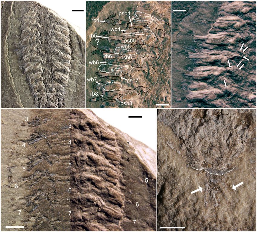
430   PALAEONTOLOGY, VOLUME 64

2002; Hendricks & Lieberman 2008; Lamsdell et al. 2013;          Unlike surrounding sediments, which are composed
Lerosey-Aubril et al. 2017), others also incorporating        primarily of wavy and crinkly laminated calcilutites
modern taxa and characters to relate extinct and              signifying intertidal zone deposition, the Waukesha
modern groups (Vaccari et al. 2004; Scholtz & Edge-           lagerst€atte beds are planar laminated and dark in colour,
combe 2006; Legg et al. 2013). In either case, these phylo-   suggesting a relatively high organic content. The lithology
genies are constantly evolving and the steady stream of       of the laminae varies in varve-like fashion (Kluessendorf
discovery of new or better-preserved taxa has the poten-      1990) between smooth, shaly and dolomitized calcilu-
tial to clarify (e.g. Dunlop 2002; Waloszek & Dunlop          tites and coarser, lighter-coloured dolosiltstones, the so-
2002; Yang et al. 2013; Lerosey-Aubril et al. 2017), or       called f€aulen and flinzen, respectively, of Wendruff et al.
occasionally upend (M€   uller & Walossek 1987; Ma et al.     (2020b). The interlaminations between the two lithologies
2012; Lamsdell et al. 2013), our understanding of arthro-     may be at a sub-millimetric scale, although thicker and
pod relationships.                                            slightly coarser dolosiltstone beds are common. Thicker
   Here, we rediagnose and redescribe an unusual arthro-      beds of the calcilutite may also be found, and some calci-
pod from the Silurian Waukesha lagerst€atte of Wisconsin,     lutite finely interlaminates with very thin organic-rich
USA, which bears a character combination that has simi-       laminae.
larities with various arthropod groups. This taxon, Par-         Kluessendorf (1990) interpreted the dark beds forming
ioscorpio venator Wendruff et al., 2020a, was originally      the lagerst€atte to be a hydrodynamic trap, where moults
figured by Mikulic et al. (1985a), called a ‘branchiopod      and carcasses carried along on tidal currents were
or remipede crustacean’ by Mikulic et al. (1985b), and        dropped out of suspension when the currents washed
recently described as the earliest known scorpion by Wen-     onto a locally developed, subaerially exposed palaeoscarp
druff et al. (2020a). Our observations refute a placement     adjacent to the trap. The confined nature and high
in Scorpiones, and instead initially suggested a placement    organic input of the trapped materials caused anoxic con-
of the species in the ‘short great appendage’ Megacheira      ditions to develop, facilitating preservation, probably in
due to the lack of antennae and the possession of great       conjunction with the permeability-sealing effects of
appendages. However, incorporating P. venator into the        microbial mats, which have been found in association
character matrix of Aria & Caron (2017a) produced phy-        with the exceptionally preserved fossils (Wendruff et al.
logenies that do not consistently place this species within   2020b).
a well-established arthropod group. The purpose of this          Despite the limited extent of the deposit, the Waukesha
report is to redescribe the morphologies of this species in   lagerst€atte represents one of the most diverse Silurian
greater detail, based on additional specimens, and con-       soft-bodied fossil deposits of Laurentia (Kluessendorf
sider how its characters compare to other basal arthro-       1994). Many of the taxa, including an abundant, appar-
pods, with particular attention focused on short-great        ently highly specialized dalmanitid trilobite found
appendage Megacheira, Fuxianhuiida and Mandibulata.           nowhere else, remain undescribed. In addition to Par-
Regardless of its ultimate taxonomic placement, the           ioscorpio venator, other fossils that have been formally
revised diagnosis presented herein highlights intriguing      described include: a synziphosuran chelicerate, Venustulus
soft-bodied morphological details of this species, and        waukeshaensis Moore et al., 2005; a thylacocephalan
serves to underscore the preservation potential of fossils    arthropod, Thylacares brandonensis Haug et al., 2014; a
at this undercharacterized lagerst€atte.                      dasycladalean alga, Heterocladus waukeshaensis LoDuca
                                                              et al., 2003; and three species of phyllocarid crustacean
                                                              within the genus Ceratiocaris (Jones et al. 2015).
GEOLOGICAL SETTING

The Waukesha lagerst€atte is a soft-bodied fossil deposit     MATERIAL AND METHOD
in the Brandon Bridge Formation of south-eastern Wis-
consin, laid down during the Telychian Age (late Llan-        Studied material
doverian) of the Silurian (Kluessendorf 1990; Mikulic &
Kluessendorf 1999). The deposit is of limited area and        The material referred to in this paper was loaned from
stratigraphic extent, the main interval being a 12 cm         the University of Wisconsin Geology Museum (UWGM),
thick exposure at Waukesha Lime and Stone Quarry in           located in the Department of Geology and Geophysics in
Waukesha County, WI. The specimens considered in this         Madison, Wisconsin. All types and material are perma-
study were sourced from this exposure, although finer         nently reposited at this location. A total of 15 specimens
stratigraphic control is no longer possible as samples were   were analysed, and the reanalysis and redescription efforts
collected quickly from areas of active quarrying (Klues-      were based primarily on the following specimens:
sendorf 1990).                                                UWGM2793 (Fig. 1A), 2785 (Fig. 1B), 2764 (Fig. 1C and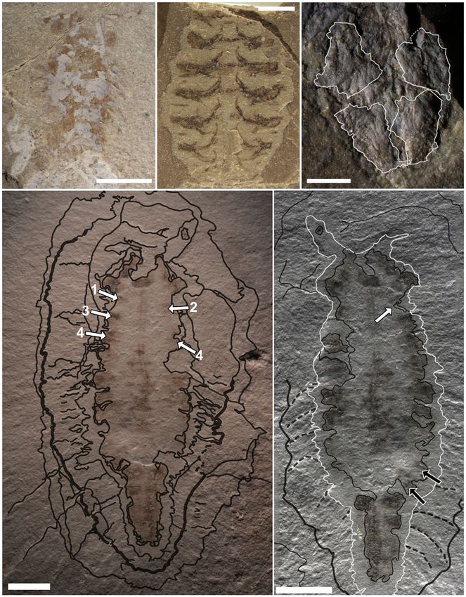
ANDERSON ET AL.: COMPLEX LIMBS ON A SILURIAN ARTHROPOD                          431

counterpart to the designated paratype UWGM2163),             et al. 2021, appendix S1). This dataset was chosen because:
2857 (Fig. 1D, E), 2854 (Fig. 1F, G), 2798 (Fig. 1H) and      (1) it is a recently published dataset; (2) it was assembled
2885 (Fig. 1I, J). These specimens had multiple measure-      from multiple sources; (3) its characters have been opti-
ments taken of their morphology (Anderson et al. 2021,        mized for the inclusion of fossil taxa; and (4) it is compre-
tables S1, S2). Since the pleural regions of the head and     hensive in the breadth of represented extinct and extant
trunk and anterior region of the head shield are usually      arthropod clades. This final point figured particularly heav-
poorly preserved, if they are preserved at all, lengths and   ily in our selection of Aria & Caron (2017a), as we were
widths only incorporate those regions that can plausibly      unsure of where to place P. venator, and phylogenetic anal-
be inferred to belong to the axial portion of the body.       yses which cover all the major clades of Panarthropoda in
Further, rather than separately measuring features on each    more than cursory detail are unusual. While we were aware
individual trunk segment, segments 2, 7 and 11 were           of some of the limitations of the finer scale resolutions of
measured as representatives of the anterior, medial and       the dataset (e.g. Phosphatocopina and Ostracoda resolving
posterior trunk, respectively. These specific segments were   as sister groups in the cladograms of Aria & Caron 2017a),
selected as most were preserved well enough to be confi-      the inclusion of primarily fossil-based characters that
dently measured. Additional figured specimens that aided      would not leave phalanxes of uncertain and inapplicable
in the rediagnosis and redescription include UWGM2778         character states in the character row for Parioscorpio was
and 2787 (Fig. 2A, B), 2803 (Fig. 2C), 2779 (Fig. 2D,         appealing. Our purpose was to determine, at least generally,
counterpart to UWGM2785, although it was not available        where in the arthropod family tree Parioscorpio fits in
during initial study of the species), 4558 (Fig. 2E) and      accordance with parsimony.
2796 (Fig. 2F). These specimens were examined to pro-            In our analysis, character gaps were coded as ‘-’, miss-
vide details on specific morphologies and preservation        ing or ambiguous states as ?, and inapplicable states
habits. Photographs of UWGM 2436, 2437 and 2575 from          were treated as missing data (Aria et al. 2015). There were
Wendruff (2016) and Wendruff et al. (2018) were con-          some characters for which multiple interpretations were plau-
sulted for their insights on the pleural regions of the       sible. For example, character 19 codes for the presence of
exoskeleton and the terminus of the animal, although          median eyes, of which convincing evidence was never
they were not available for physical examination.             found in the studied specimens of P. venator. This would
                                                              suggest entering a state of 0 for the absence of this char-
                                                              acter. Yet, the portion of the exoskeleton in which the
Imaging methods and analysis                                  median eyes are likely to be found was usually poorly
                                                              preserved on specimens of P. venator, such that it could
Standard photographs were taken using a Nikon D3300           also reasonably be coded as missing, or ?.
and processed with open-source software digiCamControl           In these circumstances, we ultimately coded the charac-
v. 2.1 (Istvan 2018). Photomicrographs were taken using      ter with what we felt was the most likely state for the pri-
a Nikon D600 camera attached to a Nikon SMZ1500               mary analysis, but retained note of alternative potential
binocular microscope. Select specimens were imaged for        states. We then ran further analyses where: (1) alternative
compositional analysis with scanning electron microscopy      states were coded ? for characters where ? was an optional
(SEM) and energy dispersive x-ray spectroscopy (EDS)          alternative state; (2) alternative states were coded 1 (or
using a Zeiss Sigma 500 VP field emission SEM with dual       whatever character state represented a higher number for
coplanar Bruker X Flash 6|30 spectrometers. SEM imaging       a character, such as podomere number) when this was an
was conducted with a signal mix between a five-segment        optional alternative state; and (3) alternative states were
high definition backscattered electron detector and a cas-    coded as 0 (or whatever character state represented a
cade current low vacuum secondary electron detector.          lower number for a character) when this was an optional
Photos and images were edited using Affinity Photo            alternative state. Additionally, these three sets of analyses
(v. 1.6.4.104) and Affinity Designer (v. 1.6.4.104) on a      were run for two interpretations of the anteriormost (as it
Wacom Cintiq 27QHD Creative Pen Display tablet. Mea-          is preserved in the fossils) ramus of the trunk limbs: one
surements were taken on photographs and photomicro-           with it coded as an exopod (the default interpretation, see
graphs using ImageJ (v. 1.49; Schneider et al. 2012).         the Redescription, below), the other with it coded as
                                                              an epipod (the alternative interpretation, see Features of
                                                              the Trunk, below). These alternative arrangement analy-
Phylogenetic analysis                                         ses were done in order to observe how relatively
                                                              minor changes in the interpretation of the morphology of
Phylogenetic analysis was based on the dataset of Aria &      P. venator could affect its taxonomic placement.
Caron (2017a), where Parioscorpio venator Wendruff et al.,       Analysis follows the standard of Aria & Caron (2017a)
2020a was added as a new, ninety-second taxon (Anderson       using PAUP* v. 4.0a167 (X86) (Swofford 2002). In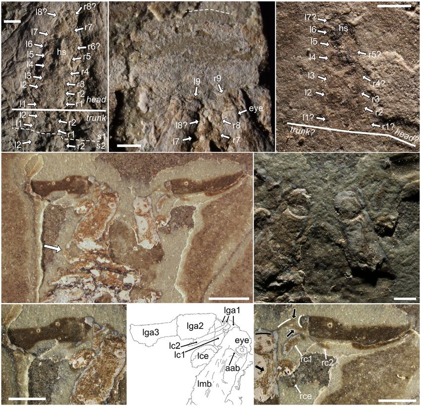
432   PALAEONTOLOGY, VOLUME 64

summary, the dataset was processed with parsimony anal-    and branch support was evaluated using Bremer, boot-
ysis using an heuristic search with tree bisections and    strap and jackknife support values.
reconnection using 1000 replicates and a maximum of 10        Bremer support was calculated by re-running the analy-
trees with a score above 1 for each replicate. The back-   sis with 10 locally optimal trees retained for each of the
bone constraints of Aria & Caron (2017a) were retained.    1000 replicates, whether or not their score was optimal
Strict consensus trees were constructed and compared,      for all replicates. This allowed for the retention of
ANDERSON ET AL.: COMPLEX LIMBS ON A SILURIAN ARTHROPOD 433
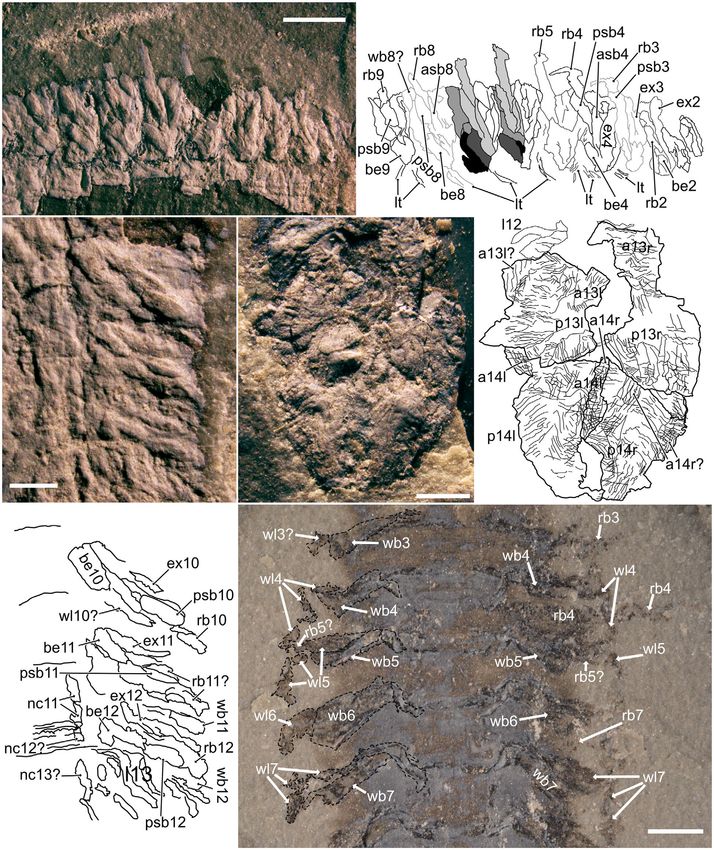
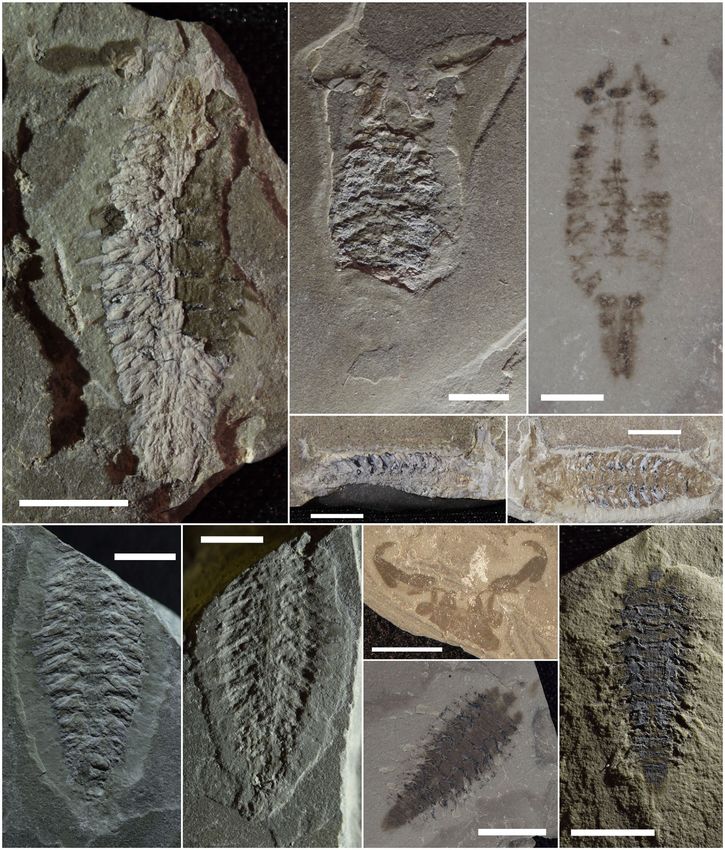
FIG. 1. Specimens upon which the rediagnosis and redescription of Parioscorpio venator Wendruff et al., 2020a are primarily based.
A, UWGM2793, a nearly complete specimen with an entire left great appendage. B, UWGM2785, a specimen with all cephalic appen-
dages intact, including both great appendages, which are nearly complete; note that the posterior portion of the body is still buried
beneath the matrix. C, UWGM2764, paratype and counterpart to UWGM2163, preserved as a thin film with the right great appendage
barely visible on the upper right; no trunk appendages are preserved, which makes the posterior constriction of the axial trunk easy to
see compared to other specimens. D–E, part and counterpart of UWGM2857, a nearly complete specimen with numerous head and
trunk details. F–G, part and counterpart of UWGM2854, which preserves many three-dimensional limbs, but whose head is cut off by
the border of the matrix. H, UWGM2798, a largely mouldic specimen showing excellent preservation of the cephalic appendages,
including two complete great appendages. I–J, part and counterpart of UWGM2885, a nearly complete specimen which shows limited
three-dimensional preservation, but preserves many walking legs as dark compressions. All scale bars represent 5 mm.
suboptimal scores, and consensus trees were computed SYSTEMATIC PALAEONTOLOGY
for the most parsimonious trees and trees one step
longer, then for the most parsimonious trees and trees Phylum ARTHROPODA von Siebold, 1848
one and two steps longer, and so on (Bremer 1988) to Subphylum INCERTAE SEDIS
suboptimal trees five steps longer than the most parsimo-
nious tree. Bremer scores were assigned to nodes based Genus PARIOSCORPIO Wendruff et al., 2020a
on how many additional steps were needed to collapse it,
with nodes surviving in consensus trees with five addi- Type species. Parioscorpio venator Wendruff et al., 2020a.
tional steps allowed assigned a score of ‘>5’.
Bootstrap and jackknife support analyses were con- Rediagnosis. As for species.
ducted using their respective commands in PAUP*
v. 4.0a167 (Swofford 2002). For both jackknife and boot-
strap analyses, 500 replications of a ‘fast’ stepwise-addition Parioscorpio venator Wendruff et al., 2020a
search were run with a random number seed of 1, groups Figures 1–10, 13A
compatible with the 50% majority-rule consensus trees
were retained for display, and for the jackknife analysis 1985a ?branchiopod crustacean; Mikulic et al., p. 716,
10% of characters were randomly deleted. In the primary fig. 2d.
analysis, the results of Bremer, bootstrap, and jackknife 1985b branchiopod or remipede crustacean; Mikulic et al.,
support analyses appeared broadly similar, so only Bremer p. 79, pl. 2 fig. 16.
analyses were run for the alternative interpretations. 2016 Latromirus tridens Wendruff, pp 150–153 (pars),
After the analysis, we compared the characters of P. ve- figs 5.1. 4–7, 5.1.9–11 (non figs 5.1.1–3, 5.1.8,
nator to those of several stem-group taxa featured in the 5.1.12–13).
character table of Aria & Caron (2017a) to analyse poten- 2018 Xus yus Wendruff et al., pp 7–10 (pars), fig. 1e–l
tial synapomorphies. The selected taxa include: (1) Suru- (non fig. 1a–d).
2020a Parioscorpio venator Wendruff et al., figs 1a, c, 2a.
sicaris elegans Aria & Caron, 2015 representing Isoxyidae;
2020b scorpion; Wendruff et al. p. 1, 7, fig. 5a.
(2) Leanchoiliidae Raymond, 1935 and Yohoia tenuis Wal-
2020b cheloniellid arthropod; Wendruff et al. fig. 7b (non
cott, 1912 representing Megacheira, along with Oelando-
fig. 7c).
caris oelandica M€ uller, 1983, which is more likely to be a
member of Crustacea sensu lato (e.g. Stein et al. 2008;
Haug et al. 2010) but resolved with Megacheira in Aria & Holotype. UWGM2162 from the Waukesha Lime and
Caron (2017a); (3) Offacolus kingi Orr et al., 2000 repre- Stone Quarry, Waukesha, Wisconsin, USA.
senting stem-group Euchelicerata; (4) Sidneyia inexpectans
Walcott, 1911 representing Artiopoda; (5) Fuxianhuia Paratypes. UWGM2163 and UWGM2764 (Figs 1C, 3E,
Hou, 1987, representing Fuxianhuiida and its relatives; 6A–D, 10D), part and counterpart; from the same locality
(6) Tokummia katalepsis Aria & Caron, 2017a represent- as the holotype.
ing Hymenocarina and stem-group Mandibulata; (7)
Marrella splendens Walcott, 1912; and (8) Agnostus pisi- Additional material. UWGM2436; UWGM2437; UWG-
formis Wahlenberg, 1818. These last two species were cho- M2575; UWGM2778 (Figs 2A, 5E) and UWGM2787
sen not because of a particularly close morphological (Figs 2B, 5F), part and counterpart; UWGM2779
resemblance to P. venator, but because they have also (Fig. 2D) and UWGM2785 (Figs 1B, 4D–F, H), part and
proven to be difficult to place phylogenetically (e.g. counterpart; UWGM2793 (Figs 1A, 3A, B, 5A, H, 7A, F,
Walossek & M€ uller 1990; Rak et al. 2013). G, 8A, C, 13A); UWGM2796 (Figs 2F, 9F); UWGM 2798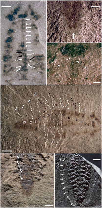
434   PALAEONTOLOGY, VOLUME 64

                                                                                      FIG. 2.    Additional available speci-
 A                                                                        B           mens of Parioscorpio venator that
                                                                                      aided in the rediagnosis and
                                                                                      redescription. A–B, UWGM2778
                                                                                      and UWGM2787, part and counter-
                                                                                      part of a nearly complete but largely
                                                                                      effaced specimen associated with a
                                                                                      conulariid. C, UWGM2803, a nearly
                                                                                      completely flattened specimen that
 C                                                                        D           preserves the axial body in stark
                                                                                      contrast with the matrix; the pleural
                                                                                      field may be partly preserved as a
                                                                                      halo of darker speckled material on
                                                                                      the specimen’s left side.
                                                                                      D, UWGM2779, the counterpart to
                                                                                      UWGM2785, which was not initially
                                                                                      available for study; its great appen-
                                                                                      dages are preserved by three-dimen-
                                                                                      sional phosphate. E, UWGM4558, a
                                                                                      poorly preserved specimen which
                                                                                      nevertheless shows some traces of
                                                                                      segmentation and limbs.
                                                                                      F, UWGM2796, an extensively
                                                                                      deformed specimen; the posterior
                                                                                      on the left side of the sample is lar-
                                                                                      gely articulated (white arrow with
                                                                                      black outline), but the anterior is
                                                                                      discombobulated on the right.
                                                                                      Abbreviations: ga, great appendage
                                                                                      article; mb, muscle block. All scale
                                                                                      bars represent 5 mm.
 E

                                                                          F

(Figs 1H, 5C, G); UWGM2801; UWGM2803 (Fig. 2C);             second article roughly trapezoidal, third article is the lar-
UWGM 2827, part and counterpart (Fig. 10A); UWGM            gest and longest, fourth article is smaller, pointed, and
2854, part and counterpart (Figs 1F, G, 6F, 7I, 9A–C, E);   projects at a nearly 90° angle to the long axis of the third
UWGM2857, part and counterpart (Figs 1D, E, 3C, 4A,         article. Second cephalic appendage is biramous, and both
B, 7C, E, 8E, 10C); UWGM2858, part and counterpart          exopod and endopod are considerably smaller than the
(Fig. 10B); UWGM2885, part and counterpart (Figs 1I–J,      great appendage; exopod may be distally setose. Axial
6E, 7H, 8G, 9D); UWGM4558 (Figs 2E, 4C); and                region of the head is trapezoidal in dorsoventral view
UWGM4718.                                                   with compound eyes preserved in anterolateral corners of
                                                            the trapezoid. A larger, faint, semicircular(?) head shield
Rediagnosis. A great appendage-bearing arthropod with a     with anterolaterally directed posterior margins covers the
transversely wide ovoid body outline and two cephalic       axial region. Trunk consists of 14 somites. Axially, two
appendages. First cephalic appendage, the great appen-      pseudotagmata (sensu Lamsdell 2013) are evident: the
dage, is uniramous with four articles. First article has    first 10 somites form a broad pear-shaped preabdomen,
highly reduced, broadly y-shaped sclerotized portion,       while the last 4 somites form a posteriorly tapering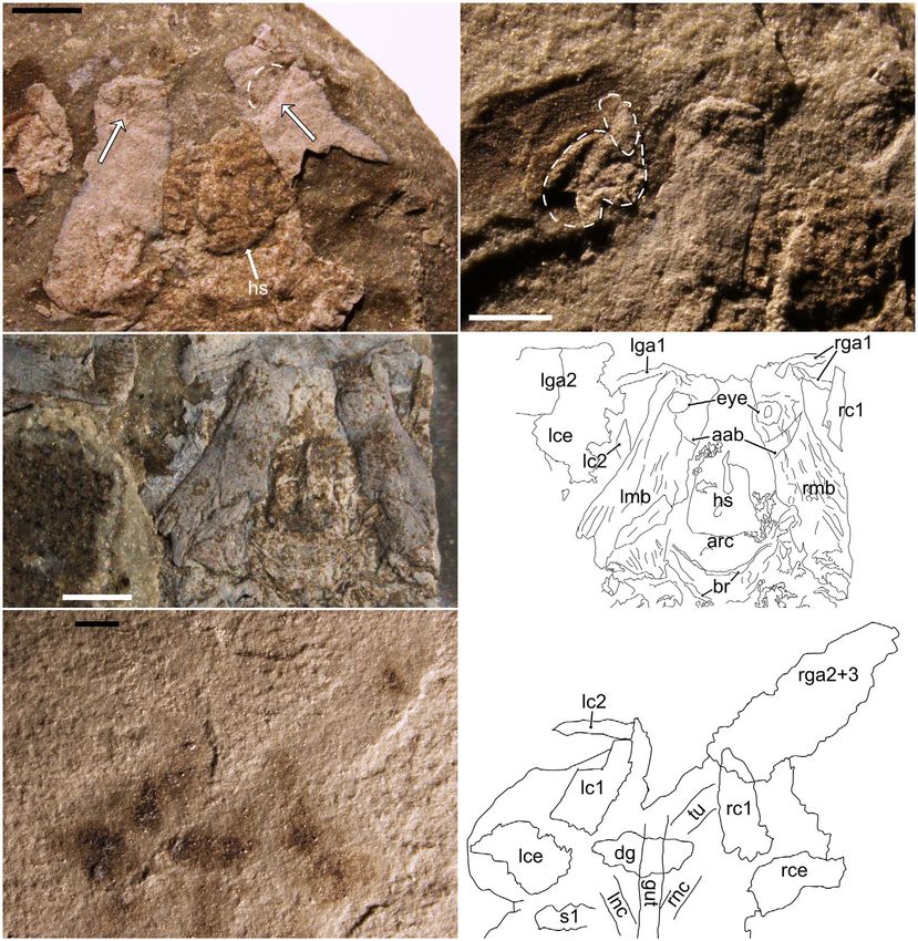
ANDERSON ET AL.: COMPLEX LIMBS ON A SILURIAN ARTHROPOD                                    435

                                                                  A                                                                   B

  C                                                                    D

  E                                                                    F

FIG. 3.    Features of the head of Parioscorpio venator. A, head of UWGM2793; white arrows with black outlines point to the posterior
of the depressions interpreted as the great appendage insertion points; dashed white arc highlights the anteromedial half of the eye,
which appears as an oval with a dotted outline (the dark areas may represent facets). B, low angle light photograph of the upper left
corner of the head and cephalic appendages of UWGM2793; the dashed white lines trace the second cephalic appendage rami, includ-
ing the first segment of the endopod (smaller, anteriorward trace) and the exopod (larger, posteriorward trace). C–D, photograph and
interpretive drawing of the head of paratype UWGM2857a; the striations in the trapezoidal muscle blocks are particularly well pre-
served in this specimen; these are interpreted as muscle fibres and suggest a complex arrangement of multiple muscles within the
blocks. E–F, photograph and interpretive drawing of the head of paratype UWGM2764; preserved as a film, different from most other
available specimens. Abbreviations: prefix r or l, indicates right or left of some elements; aab, appendage articulation boundary, i.e. of
the great appendage; arc, arcuate structure; br, brace-like structures; c, cephalic appendage endopod podomere (1 or 2; r/l);
ce, cephalic appendage exopod (r/l); dg, early digestive structure associated with the anterior of the gut; ga, great appendage element
(1–3; r/l); hs, hypostome; mb, muscle block (r/l); nc, nerve cord (r/l); s1, cuticle of segment 1; tu, indeterminate tube. All scale bars
represent 1 mm.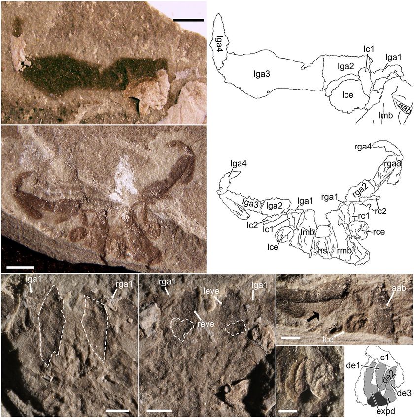
436   PALAEONTOLOGY, VOLUME 64

subrectangular postabdomen. Pleural regions of the trunk        a transverse, rounded suture; a small telson dorsal to
typically faint. The anterior two (or more) tergopleurae        the lateral spines extends posteriorly to about their
are anterolaterally directed, with subsequent tergopleurae      length.
directed first laterally, then increasingly posterolaterally.
The first 12 trunk somites have a complex biramous              Redescription. The axial portion of Parioscorpio venator
limb with multiple filamentous elements that can be             measures between 16.43 and 28.03 mm in length and
lobose, sub-lanceolate, to lanceolate in shape. The final       between 5.41 and 11.34 mm in width (Anderson et al.
two trunk somites with fan-shaped, filamentous primary          2021, tables S1, S2). Specimens UWGM2857 (Fig. 1D, E)
rami and an indeterminate number of smaller, filamen-           and 2885 (Fig. 1I, J) are the smallest of those measured
tous, fan-shaped elements; filaments are elongate and           while UWGM2785 (Fig. 1B) and 2854 (Fig. 1F, G) are
originate from indistinct rami. Terminal segment bears          the widest (the length of both is incomplete and cannot
an anus and two lateral spines separated from tergite by        be accurately assessed) and UWGM2764 (Fig. 1C) the

  A                               B                                             C

  D                                                                        E

  F                                     G                                  H
ANDERSON ET AL.: COMPLEX LIMBS ON A SILURIAN ARTHROPOD                                    437

F I G . 4 . Further features of the head and cephalic appendages of Parioscorpio venator. A–C, paired circles or rings of unknown func-
tion seen in the head and trunk of multiple specimens, indicated by arrows and sequentially labelled from posterior to anterior. A–
B, rings of UWGM2857a: A, rings of the posterior head (1–9) and anterior trunk (1 or 2); those by the brace structures (see Fig. 3C–
D) are the most discernible, but anteriorward they are partially obscured by the hypostome; posteriorward, there appear to be two
pairs of circular structures per trunk somite; B, the seventh to ninth pairs of rings, anterior to the hypostome and highlighted due to
a different lighting angle; the dashed line denotes the discernible anterior boundary of a broad, flat, lightly mineralized surface inter-
preted as the (possibly partially displaced) head shield. C, circles or rings of UWGM4558, most strongly developed just posterior to
the hypostome and with three pairs apparently overprinting the hypostome, as in UWGM2857. D–H, the head and cephalic appen-
dages of UWGM2785: D, overview of the head and anterior trunk segments; the arrow indicates the single clawed terminus of the
walking portion of the first trunk leg; E, low angle light photography of the head, revealing the depressions marking the insertion
zones for the great appendages; F–G, photograph and interpretive drawing of the left muscle block and cephalic appendages; for the
sake of clarity, only the outline of the many overlapping elements of the great appendage and second cephalic appendage rami are
traced; note that the second cephalic appendage’s second endopod podomere (lc2) may end flush with the right edge of the first podo-
mere (lc1) or just medial to it; H, right muscle block and cephalic appendages; black arrows with white outlines point to the distal
branches of the y-shaped first great appendage article; the white arc to their right indicates the angled anteroproximal corner of the
second great appendage article, which would have been rotated counterclockwise in life to lie flush with the distal branches of the first
great appendage article; the left white arc and black arc show the estimated placement of the first great appendage article and its inser-
tion zone under the head, respectively; the solid black arrow indicates fibres within the muscle block. Abbreviations: prefix r or l, indi-
cates right or left of some elements; aab, appendage articulation boundary, i.e. of the great appendage; c, cephalic appendage endopod
podomere (1 or 2); ce, cephalic appendage exopod; hs, hypostome; lga, left great appendage element (1–3); lmb, left muscle block;
1–9, ring structures in the head (1–9; r/l) or trunk (1 or 2; r/l); ?, dubious ring structures. Scale bars represent: 0.5 mm (A); 1 mm
(B–C, E); 3 mm (D); 2 mm (F, H).

longest. Differences in major morphological details, like               the trapezoidal muscle blocks which expand anteriorly
segment number, are not evident between smaller and                     and terminate near or just anterior to the eyes (eyes not
larger specimens. Proportion differences in finer details,              readily evident in Fig. 5E). The centre of the kite-shaped
such as the length:width ratios of the great appendage                  depressions corresponds in location to a pair of pits pos-
articles, are minor and more likely to be natural within-               terior to the eyes on UWGM2787 (white dashed outlines
species variation or taphonomic than a reflection of onto-              in Fig. 5F). The kite-shaped depressions of UWGM2778
geny or taxonomy.                                                       are interpreted as units of muscle that originally passed
                                                                        through the ventral pits on UWGM2787 and articulated
Morphology of the head. The axial portion of the head                   with the now-displaced great appendages (the first articles
containing soft tissue is roughly trapezoidal in outline with           of which are labelled in Fig. 5E, F). Depressions in
rounded corners, with a length between 3.12 and 4.84 mm                 UWGM2787 are not directly beneath the eye as they
and a width between 4.55 and 7.24 mm (Anderson et al.                   appear to be in other specimens (Figs 3A, B, D, 4D–H;
2021, table S1). The lateral portions of the axial head are             5G); it may be a taphonomic effect.
dominated by two trapezoidal blocks (Fig. 3A, C, D). They                  In UWGM2793 and 2857 (Fig. 3A, C, D) a pear-
may show extensive striations (Figs 3C, D, 4D–H). Depres-               shaped, topographically elevated structure is located on
sions are visible under raking light at the anterolateral cor-          the sagittal midline of the head centred a little over half-
ner of each trapezoidal block (Figs 3A, B, D, 4E–H, 5A, B,              way along the length of the muscle blocks. This struc-
G). On UWGM2793 a partial, oval-shaped, dotted outline                  ture’s posterior is marked by a convex arc (arrow labelled
probably represents the eye (Fig. 3A, B); on other speci-               ‘hs’ in Fig. 3A; ‘arc’ in Fig. 3D). Based on the shape of
mens a simple circular to sub-circular ring denotes the eye             the head, this arc lies just anterior to a transversely elon-
(Figs 3C, D, 4B, D, F, G, 5F).                                          gated oval in UWGM2764 which overprints an elongate,
   The depressions were probably the insertion points for               parallel-sided structure that terminates anterior to the
the great appendages (cf. Liu et al. 2007, fig. 3b; Haug                oval (Fig. 3E, F) and runs posteriorly along much of the
et al. 2012b, fig. 3d) which in all observed specimens have             length of the body (Fig. 1C). The parallel-sided structure
been displaced laterally from the head to some extent                   is interpreted as a simple gut, with the dark oval inter-
(e.g. Fig. 4F, H). The striated trapezoidal structures are              preted as an initial digestive structure (Fig. 3F). The pear-
here interpreted as muscle blocks (see Features of the                  shaped structure is thus the hypostome.
Head, below). UWGM2778 and 2787 (Fig. 5E, F), a part/                      Paired circular features along the axis of the head are
counterpart pair, preserve different aspects of the muscle              seen on several specimens, usually only under low-angle
blocks’ anatomy. On UWGM2778, two kite-shaped                           light. They are usually most strongly expressed postero-
depressions (white dashed outlines on Fig. 5E) are seen in              medially in the head and appear to overprint features like
438    PALAEONTOLOGY, VOLUME 64

                                                                A                                                                  B

  C                                                                 D

  E                                           F                                            G

                                                                                           H                     I

FIG. 5.    Features of the cephalic appendages of Parioscorpio venator. A–B, photograph and interpretive drawing of the great appen-
dage and second cephalic appendage of UWGM2793. C–D, photograph and interpretive drawing of UWGM2798, which preserves the
cephalic appendages exquisitely; note the split in the cuticle along the length of the third great appendage articles. E–F, UWGM2778
and UWGM2787, part and counterpart, which provide evidence of the mechanical operation of the great appendages by the muscle
blocks in the head; white dashed outlines indicate the mouldic outline of a muscle block pair in E and the insertions of the great
appendages in F. G, left second cephalic appendage of UWGM2798; black arrow points to the distal edge of the endopod, the arrow-
head’s width reflecting the breadth of its potentially setose terminus. H–I, photograph and interpretive drawing of the left second
cephalic appendage exopod of UWGM2793, also seen in Figure 3B; I, for ease of interpretation, pertinent exopod units are lightly
shaded and pertinent phosphate removed by erosion is darkly shaded. Abbreviations: prefix r or l, indicates right or left of some ele-
ments; aab, appendage articulation boundary, i.e. of the great appendage; c, cephalic appendage endopod podomere (1 or 2; r/l);
ce, cephalic appendage exopod (r/l); de, distal element (1–3); expd, exopod podomere; eye, eye (r/l); ga, great appendage element
(1–4; r/l); hs, hypostome; mb, muscle block (r/l). Scale bars represent: 1 mm (A, E–G); 2 mm (C); 0.5 mm (H).
ANDERSON ET AL.: COMPLEX LIMBS ON A SILURIAN ARTHROPOD 439 the hypostome (Fig. 4A, C). There may be at least nine podomere that arcs from an anteromedial to posterome- (Fig. 4A, B) or as few as five (Fig. 4C) and they appear to dial position and has two or three processes anterior to extend into the trunk with up to two pairs per segment its distal tip. The length is simply measured as the long (Fig. 4A). Other structures are preserved in the head, but axis of the overall oval shape, and its width the short axis. the interpretation of most is dubious; such as a pair of Its length ranges between 1.07 and 1.60 mm and its width curious brace-like structures in UWGM2857a (labelled between 0.65 and 1.25 mm. ‘br’ in Fig. 3D). These are axially oriented posteriorly, in Whilst not apparent in all specimens, the second contrast to ventral features such as the legs, which are cephalic appendage is inserted ventrally below the trape- oriented anteriorly, and may represent the preservation of zoidal muscle blocks, much like the great appendage, a dorsal feature, like head segmentation. approximately halfway or posterior to halfway along the The great appendage consists of only four articles muscle blocks’ length. In UWGM2785, bundles of stria- (Fig. 5A–D). The first segment preserves only a small tions in the muscle blocks, here interpreted as relict mus- amount of material and when complete is roughly y- cular fibres, lead to the second cephalic appendage shaped (Figs 3C, D, 4F, G), with the open end of the ‘Y’ exopod, which is just lateral to the head approximately pointing toward the second article (Fig. 4H). The second halfway along its length (Fig. 4F–H). A pair of dark article may appear either rectangular (Fig. 5A, B) or trape- stains, of roughly the same shape as the first podomere of zoidal, with rounded anteromedial corners (Figs 4F–H, the endopod and the exopod, are visible in the anterior 5C, D). If length is considered to be the axis perpendicular and posterior halves of the head, respectively, in to the main body’s length, the article’s length (range 1.51– UWGM2764 (Fig. 3E, F). These carbonaceous stains are 2.17 mm) is slightly greater than the width (range 0.94– interpreted as the compressional remnants of the endo- 1.40 mm; Anderson et al. 2021, table S1). This article was pod and exopod of the second cephalic appendage. apparently well-sclerotized, as it sometimes shows cracking Pleural and dorsal regions of the head shield are poorly patterns consistent with brittle fracture (e.g. Fig. 3B). The preserved in the observed specimens. UWGM2857a third article is the largest, between 2.21 and 3.86 mm in (Fig. 4B) exhibits a thin, lightly mineralized sheet accom- length and between 0.82 and 1.63 mm in width, and panied by a distinctly flat region anterior to the axial fea- resembles a kitchen knife in outline. Basally is a rectangu- tures of the head. It is not clear if it is compressed or was lar ‘handle,’ which distally expands posteriorly into a displaced from the body in this instance. The posterior ‘blade.’ Distal to this expansion, the posterior side of the margin of the head shield appears to cover the an- article curves anteriorly towards its termination (Figs 4D, teriormost trunk, either to the first (e.g. UWGM2436 in H, 5A–D). This termination is concave, and the convex Wendruff et al. 2018, fig. 1e–f) or second (e.g. base of article 4 articulates with it (Fig. 5B, D). Article 4 is UWGM2764, Fig. 6D; there are no clear tergopleural small, between 1.48 and 1.87 mm long and 0.57–0.69 mm demarcations anterior to the posterior boundary of seg- wide, with a conical outline and a mesial bulge more pro- ment 2) trunk segment. nounced on its inner side than its outer (Fig. 5A–D). The conical tip appears straight in UWGM2793 (Fig. 5A, B), Morphologies found on both head and trunk. Two major but bends inward toward the body axis in UWGM2798 features cross the boundary between the head and trunk. (Fig. 5C, D). As mentioned above, in UWGM2764 a dark oval inter- The second cephalic appendage is much smaller than preted as digestive glands overprints on a simple, elongate the first (Figs 3B, 4D, F–H, 5A–D, G) and is biramous, gut (Fig. 3E, F). This bends off to the right and becomes although the endopod is often displaced anteriorly to the ambiguous posterior to segment 6 (Fig. 6A). It is faintly exopod (Figs 4F–H, 5C, D, G). The endopod is composed seen again under raking light in the terminal segments of at least two segments, the first preserved somewhat (Fig. 6B), still a straight, simple tube. The digestive tract three-dimensionally and considerably longer than wide is not evident in any of the other available specimens. (Anderson et al. 2021, table S1) and may have gentle lon- Along the length of the gut tract, small paired patches gitudinal striations visible (Fig. 4F). The second podo- may be found, one or two for each segment, and with mere is as long as, or longer than, the first, but is one pair behind the main digestive glands in the head narrower (Figs 4F–H, 5C, D, G) and often does not pre- (white arrows in Fig. 6A). These could either be small serve well (Fig. 3C–F), if at all (Fig. 5A, B). There is lim- diverticulae or the compressional version of the paired ited evidence of setae extending along and beyond this circles or rings seen in Figure 4A–C. podomere on UWGM2798 (Fig. 5G). The exopod is The second feature is a pair of parallel, tube-like struc- curled in on itself and under higher-angle incident light tures oriented on the midline of most of the available assumes an oval outline (Figs 4F–H, 5A–D). Under low- specimens. These structures are slightly nodulose in the angle raking light, a more complex structure becomes vis- anterior segments (black arrow in Fig. 7E), but are simple ible (Figs 3B, 5G–I) which consists of a banana-shaped cords in the posterior segments (Fig. 7F). They are best
440   PALAEONTOLOGY, VOLUME 64

                A                B

                                 C

                D

                                 F

                E
ANDERSON ET AL.: COMPLEX LIMBS ON A SILURIAN ARTHROPOD 441
F I G . 6 . Features that cross the head and trunk of Parioscorpio venator and of axial and tergopleural segmentation. A–D, features of the
nearly flattened UWGM2764 with dark film preservation: A, head and anterior trunk showing the continuation of the gut beyond the
head (Fig. 3E–F); white arrows in the trunk and a white arrow with a black outline in the head point to pairs of darkened patches that
may be equivalent to the circles or rings in Figure 4A–C, or may represent digestive diverticulae; they become increasingly dubious pos-
terior to the bend in the gut; black arrows point to this divergence to the right, probably severed during decay, posterior to the sixth seg-
ment; B, termination of the gut at the posterior of the specimen, visible as a faint mouldic impression under very low angle incident
light; arrow points up the gut from its terminus; C, photograph taken under low angle light, showing the divergence of the putative nerve
cords at the posterior head and anterior trunk; white arrows with black outlines point to the right and left boundaries of the right and
left nerve cords; D, photograph taken under low angle light to demonstrate the tergopleurae of the trunk and how their width and struc-
ture relate to changes in the axial trunk’s morphology; numbers indicate segment number, with those in the preabdomen black and those
in the postabdomen white; plausible segment boundaries continuing into the tergopleurae are traced on the left side of the specimen by
dashed lines (black lines indicate the posterior boundary of even-numbered segments while white lines indicate the posterior boundary of
odd-numbered segments); arrows indicate putative pleural spines; because the specimen was not compressed perfectly perpendicular to
bedding, there may appear to be multiple transverse divisions within a segment; these are traced in solid white on the boundaries between
segments 6–9; this is also likely to account for the ambiguity of many tergopleural boundaries. E, posterior of UWGM2885a, demonstrat-
ing the relative length of the terminal axial segments; arrows demarcate the anterior boundaries of segments 11–14. F, low angle light
photograph of UWGM2854a, showing the segmentation of the tergopleurae beyond the three-dimensionally preserved axial body; tergo-
pleural boundaries on the left flank are indicated with arrows, and those that are less clear (three anteriorly, two posteriorly) are marked
with ‘?’. Scale bars represent: 2 mm (A–B), 0.5 mm (C), 3 mm (D, F) and 1 mm (E).
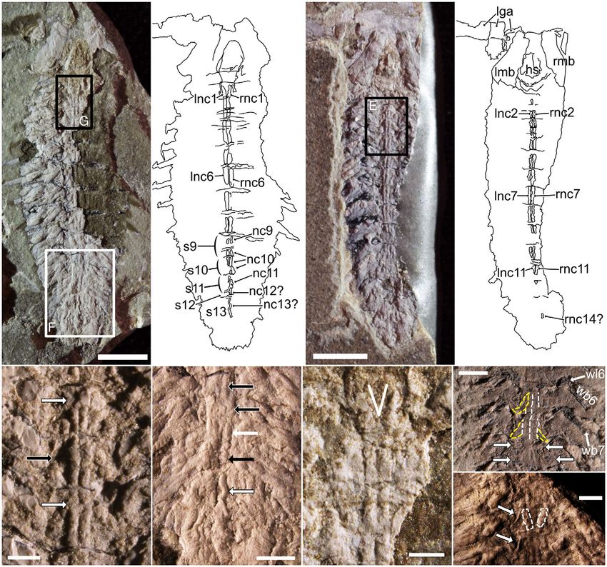
seen in UWGM2793 (Fig. 7A, B), the part of than that of somites 1–10 (segment 11 is 5.34 mm wide).
UWGM2857 (Fig. 7C, D) and are visible in the flattened Segment 14 appears to be the longest on both
UWGM2764 (Fig. 6C). The width of the tubes matches UWGM2764 (Fig. 6B, D) and UWGM2885 (Fig. 6E),
trends in the width of the trunk somites: narrower an- both of which are unobscured by legs on this final somite.
teriorly and posteriorly and widest medially (Anderson Other specimens with legs show similar axial segment
et al. 2021, table S2). These structures diverge anteriorly length and width trends, but the transverse contraction
in the vicinity of the first trunk segment and continue after somite 8 appears more gradual (e.g. Fig. 1A, B, E–
into the posterior of the head (Figs 6C, 7G). Given their G).
paired, ventral nature, we cautiously interpret these struc- The dorsoventral shape of the axial body appears ovoid
tures as nerve cords. based on ring-shaped structures, probably representing
unevenly compressed segment boundaries, seen in the
Morphology of the trunk. The axial region of the trunk of middle trunk of UWGM2764 (Fig. 6D). Differentiating
Parioscorpio venator consists of 14 somites, the shape of the tergites from the sternites, however, is difficult. On
which is partly obscured in most of the specimens by the some specimens, spindle-shaped units with crescentic lat-
legs, which are usually three-dimensionally mineralized. eral boundaries may represent the sternites (Fig. 7H–I;
Morphological features of the trunk itself are best pre- Wendruff et al. 2018, fig. 1i; Wendruff et al. 2020a,
served in one specimen, UWGM2764 (Figs 1C, 3E–F, 6A– fig. 2a). If so, they would be considerably narrower than
D). In this specimen, no legs are preserved and the struc- the overlying tergites.
ture of the axial segment divisions are thus visible The pleural field is visible in several of the specimens,
(Figs 1C, 6A, D). It is clear that trunk segment 1 is the although it is usually subtle and without clear lateral mar-
shortest and transversely its axial portion is narrower than gins. The tergopleural divisions between segments are best
the head. Successive axial segments are wider and longer seen in UWGM2764 (Fig. 6D) and UWGM2854a (Fig. 6F)
(segment 2 is 1.16 mm long and 6.66 mm wide), under low angle light, and also in UWGM2436 (Wendruff
although the increase in size is subtle after segment 3, et al. 2018, fig. 1e–f), UWGM2437 (Wendruff et al. 2018,
and maximum width for both the axial trunk and the fig. 1l) and UWGM2575 (Wendruff et al. 2018, fig. 1i).
entire axial body is achieved in the vicinity of somites 6 They generally show increasing posterolateral deflection on
and 7 (segment 7 is 2.55 mm long and 10.08 mm wide; successive segments. Unlike the sharp contraction in axial
Anderson et al. 2021, table S2). The axial lengths of seg- segment width seen between somites 8–11 in UWGM2764,
ments 8–13 are roughly equal, though shorter than seg- the contraction in width of the pleural fields appears more
ment 7 (segment 11 is 1.67 mm long). Transversely, the gradual (Fig. 6D; Wendruff et al. 2018, fig. 1e–f) There is
axial portion of segment 8 is narrower than 7, and the some suggestion of spines projecting posterolaterally off
axial portions of segments 9 and 10 have lateral margins the tergopleural margins of posterior segments in
that are directed posteromedially (Figs 1C, 6D). The axial UWGM2764 (Fig. 6D) and UWGM2436 (Wendruff et al.
portion of the final four somites is considerably narrower 2018, fig. 1f).442 PALAEONTOLOGY, VOLUME 64
A B C D
E G
H
I
F
F I G . 7 . Features of the nervous system and medial axis of Parioscorpio venator. A–B, photograph and interpretive drawing of
UWGM2793, positions of enlargements F and G indicated on A. C–D, photograph and interpretive drawing of UWGM2857a; position
of enlargement E indicated on C. E, paired nerve cords of segments 2–4; black arrow with white outline indicates slight anterior bulge
interpreted as a pair of ganglia; white arrows with black outlines indicate where the ganglia should be but have been effaced, perhaps
because of their slightly higher relief. F, nerve cords in the posterior of UWGM2793; the right cord appears better preserved and is
traced out by arrows on segments 10 (black arrows with white outlines), 11 (white arrow), 12 (black arrow) and 13 (white arrow with
black outline), becoming highly dubious posterior to this. G, anterior end of the nerve cords in UWGM2793; a white ‘V’ shows their
divergence in the first segment, indicating the posterior of a possible oesophageal foramen. H, central trunk of UWGM2885b, demon-
strating multiple types of impression on a single axial trunk segment, with the walking portions of some of the legs labelled for refer-
ence; on segments 6 and 7, the discernible impressions of the nerve cords (white dashed outlines) and crescentic-shaped structures
(yellow dashed outlines) are highlighted, but they are visible on segments anterior and posterior to these, too; on segment 8, two pairs
of circular or ring-like structures also observed in the head and anterior trunk are indicated (white arrows with black outlines; see
Fig. 4A–C). I, anterior trunk of UWGM2854a with subparallel strap-like structures unlikely to be nerve cords highlighted (white
dashed outline) on segment 3; crescentic structures that may indicate the borders of the sternites are indicated (white arrows with
black outlines) and may be uncompressed homologues of the structures highlighted in H. Abbreviations: prefix r or l, indicates right or
left of some elements; hs, hypostome; lga, left great appendage elements; mb, muscle block (r/l); nc, nerve cord of segment (1–14; ?
indicates less confident assignment to segment; r/l); s, segment (9–13); wb, walking leg bundle (6, 7); s, segment (9–13); wl6, walking
leg 6. Scale bars represent: 3 mm (A, C), 0.5 mm (E, G), 2 mm (F) and 1 mm (H–I).ANDERSON ET AL.: COMPLEX LIMBS ON A SILURIAN ARTHROPOD 443 The trunk legs are visible and preserved to varying margins of the trunk, particularly in legs 5–8 (Figs 8A, B, degrees in most specimens (Figs 1, 2, 8, 9). They are usu- 9A–C, E). These filamentous bundles are racemose in ally three-dimensionally mineralized, often robustly so, to shape and the apical tip is occasionally broken off (legs the point that their structure can be difficult to determine 3–7 in Fig. 8A, B). Alternatively, some of these could be and smaller or less-mineralized components of the legs displaced walking leg tips (e.g. legs 4–6 in Fig. 9B, C; are obscured. The legs of somites 1–12 have the same perhaps legs 6–7 in Fig. 9E). The racemose bundles are basic components. Basally the leg has a basipod, which both distinct in shape and often preserved, so their trans- may consist of a single solid unit (Fig. 8A, B), or it may verse width makes for a good proxy of total leg size for have one to several lobes along its length (legs 6–8 in somites 1–12 (e.g. compare the ‘racemose bundle width’ Fig. 9B, C, legs 3, 5–7 in Fig. 9E). Towards its distal end, values for UWGM2793 and 2854 in Anderson et al. the basipod has an endite bearing a series of relatively (2021, table S2) to the trends in leg size seen in Fig. 8A– short filaments, forming a bundle with a lobose shape D for UWGM2793 and Fig. 9A for UWGM2854a). Like (legs 2–9 in Fig. 8A, B; legs 10–12 in Fig. 8C, D; legs 4–8 the legs themselves, the racemose bundles increase in size in Fig. 9B, C; legs 3–6 in Fig. 9E). quickly to leg 3, then increase in size slowly to a maxi- Distal to the basipod, the leg then splits into an exopod mum width between somites 6–8, then decrease in width and an endopod. The number of podomeres on the exo- to somite 12. pod is difficult to determine (there may be at least 10, The two apical bundles are sublanceolate to lanceolate based on leg 4 in Fig. 9E), and in many cases the exopod in shape and ‘sheath’ around the racemose bundle. The appears as a simple rod (leg 2 in Fig. 8A, B; leg 12 in first, the anterior sheathing bundle, is quite small, often Fig. 8C, D; legs 5–7 in Fig. 9B, C). In a few legs, distally poorly preserved and directed anterolaterally (legs 4, 6–8 the exopod bears anterolaterally to posterolaterally ori- in Fig. 8A, B). The second, larger and more prominent ented filaments in one or multiple bundles (legs 6 and 7 posterior sheathing bundle is directed posterolaterally in Fig. 8A, B; probably legs 4 and 8 in Fig. 9B, C). More (legs 3–4, 6–9 in Fig. 8A, B; perhaps legs 10–12 in typically, the exopods may bear grooves along their length Fig. 8C, D; legs 4–8 in Fig. 9B, C; legs 3–7 in Fig. 9E). (legs 10 and 11 in Fig. 8C, D; legs 4–6 in Fig. 9E). The structure of legs 13 and 14 (Fig. 8E) appear to Whether these grooves are elongate filaments or a preser- consist of a primary ramus of two ranks of parallel, pos- vational artefact is difficult to determine. teriorly directed filaments (Fig. 8F, p#). Towards the base The endopod is complex and consists of a series of of the legs are at least two accessory rami with two ranks podomeres, presumably used for walking, and a basal of smaller filaments directed perpendicular or subperpen- exite that forms the most distinctive component of the dicular to the ramus axis (Fig. 8F, a#). leg. The walking portion of the endopod is usually largely Another feature of the legs worth mentioning is the pres- hidden by other components of the legs (legs 6 and 7 in ence of small straps of material that cross posterolaterally Fig. 8A, B) or is simply poorly preserved (e.g. legs 10–12 from the body axis adjacent to the nerve cords towards the in Fig. 8C, D). On UWGM2885, however, the walking trunk legs. Sometimes, a secondary, anterolaterally trending portions of the endopods are clearly visible (legs 3–7 in set is also present. These are seen on multiple specimens, Fig. 8G; legs 3–7 in Fig. 9D). There appear to be around but are best developed on UWGM2793 (Fig. 8A, B, labelled six or seven podomeres, but the number is not clear. The ‘lt’ on Fig. 8B). Whether they originate on the body axis trend of the walking endopods is roughly perpendicular and extend into the legs, or vice versa, is unclear, although to the body axis, with a sharp posterior bend in the ter- when they are preserved they can be highly distinctive. minal one or two podomeres. The walking leg tip is gen- The caudal termination of Parioscorpio venator may be erally not well preserved, but when it is (Fig. 4D), it buried, obscured by legs, or simply poorly preserved terminates in a single, stout claw. On many of the walk- (Figs 1A, B, D–G; 2A, B, E; 7A–D; 8E, F). When evident, ing legs, a filamentous bundle seems to originate on the the terminus appears as either a simple semicircle (Figs 1C, second or third podomere and expands posterolaterally. I, J; 6D, E) or as a distinct, three-pronged apparatus Preserved in UWGM2885 as a black film with thin bluish (Figs 2F, 9F; Wendruff et al. 2018, fig. 1e–f, i, k–l). The coats (Figs 8G, 9D) these bundles are preserved in three- three processes are of approximately equal length, with the dimensions in other specimens, even if the walking legs central process separate from and dorsal to the two lateral to which they correspond are not. This is especially evi- processes (Fig. 9F). The central process is probably the true dent in UWGM2854 (legs 4–7 in Fig. 9B, C; legs 3–6 in telson, while the lateral processes are separated from the Fig. 9E), where the filamentous bundle can be seen largely fourteenth segment by a curving suture (Fig. 9F). Since tucked behind components of the endopod exite. segment 14 lacks pleural spines (Fig. 6D), these lateral pro- The endopod exite has three filamentous bundles, cesses may reflect the posteriorly directed tergopleurae of although its segmentation or annulation is dubious. The the terminal somite, rather than furcae or caudal rami basal bundle is longest and projects beyond the axial (sensu Aria & Caron 2017a).
444   PALAEONTOLOGY, VOLUME 64

Remarks. Though Parioscorpio venator is not a rare com-      to elemental analysis of the fossils’ composition and
ponent of the Waukesha biota, its morphology is suffi-       phylogenetic analysis of the species’ affinities, we first
ciently chimerical (Fig. 11) that, even with evidence from   present an overview of the preservational habits of P.
multiple specimens, its characters defy ready homologiza-    venator, then consider some of the more unusual mor-
tion with established arthropod groups. Before proceeding    phologies of the organism in greater detail. We examine

  A                                                          B

                                     C                                     E                                       F

                                     D                                                                             G
ANDERSON ET AL.: COMPLEX LIMBS ON A SILURIAN ARTHROPOD 445
F I G . 8 . Features of the legs of Parioscorpio venator. A–B, photograph and interpretive drawing of the first nine left trunk legs of
UWGM2793; B, key units are labelled in legs 2–5, 8 and 9, with successive legs outlined in different shades of black and grey to aid in
differentiation; legs 6 and 7 are shaded according to leg unit: black shading denotes the basipod, dark grey the basipod endite, medium
grey the walking portion of the endopod, light grey the endopod exite, and white the exopod. C–D, photograph and interpretive draw-
ing of right legs 10–12 of UWGM2793; some structure of leg 13 appears visible, but is ambiguous. E–F, photograph and interpretive
drawing of hind legs 13 and 14 of UWGM2857a; heavy outline denotes the boundary of the legs and their respective filaments; the
thinner lines denote filaments of primary and accessory rami. G, photograph of legs 3–7 of UWGM2885a, which preferentially pre-
serves the walking portions of the endopods; the left flank is traced with major units labelled, while the more poorly preserved right
flank only has easily discernible major units labelled. Abbreviations r or l, as suffix, indicates right or left of some elements; a, accessory
filamentous ramus of posterior leg 13 or 14 (r/l); asb, anterior sheathing bundle of leg (4 or 8); be, basipod endite of leg (2–12); ex,
exopod of leg (2–12); l, leg elements of somite (12 or 13); lt, potential tendon or muscle of legs; nc, nerve cord of segment (11–13);
p, primary filamentous ramus of posterior leg (13 or 14; r/l); psb, posterior sheathing bundle of leg (3–12); rb, racemose bundle of leg
(2–12); wb, walking leg bundle (3–12); wl, walking leg (3–10); ?, uncertain assignment of leg or nerve cord unit. Scale bars represent:
2 mm (A), 0.75 mm (C, E) and 1 mm (G).
alternative interpretations of these morphologies where appendages of P. venator may certainly be thought of as
appropriate in an attempt to resolve where in the arthro- great appendages, although they are unusual in bearing
pod family tree this species may fit and where it cannot. only four articles, a single sub-chelate termination and a
Consequently, we also evaluate how the morphology of P. reduced, y-shaped first article (Figs 4D–H, 5A–D, 11B,
venator is incompatible with its initial placement in Scor- 12A).
piones by Wendruff et al. (2020a). The reason for this shape is a consequence of its func-
The preservational pathways expressed in the specimens tion: rather than being optimized for articulation against
vary somewhat, even within individuals. Many specimens succeeding elements within an appendage, as in most
preserve considerable quantities of soft tissue as a partially great appendages (Haug et al. 2012a), the great appen-
three-dimensional, white to blue material (e.g. Fig. 1A) dages of P. venator articulated toward one another, prob-
previously interpreted as calcium phosphate (e.g. Jones ably to hold prey in a vice (Fig. 12B). The entire
et al. 2015). On companion pieces, or when removed by structure of the head and great appendage has been mod-
erosion, these leave dark coloured mouldic depressions ified to facilitate this movement. If the great appendage is
(compare Fig. 1D, E). A separate preservation habit of reconstructed such that the reduced first article is just an-
limited patches of shiny, black, compressional material terior to the oval depressions underneath the trapezoidal
(best seen in Fig. 1A, D, J) has been interpreted as being muscle blocks (Figs 4H, 11A), then this article can serve
carbonaceous (e.g. Wendruff 2016). Specimens with a as an articulation point for the muscles held in the head
substantial phosphatic composition may be somewhat (Fig. 5E, F). In fact, the observed y-shaped first article is
three-dimensional (Figs 1A, B, D–H, 2A, B, D, F) or probably just the sclerotized portion of a more extensive
slightly three-dimensional (Figs 1I, J, 2C, E). On the and membranous first element that escaped preservation
other hand, phosphate-poor specimens, such as in the available specimens (Fig. 11B). The membranous
UWGM2764 (Fig. 1C), which is preserved as a dark film portion would have stretched from the posterior end of
with virtually no phosphate, are nearly completely flat- the oval depressions and loosely enveloped the sclerotized
tened. y-shaped structure. A membranous cuticle would be nec-
essary to facilitate the broad range of movement needed
Features of the head. The first morphology to consider is to bring the great appendages together (Fig. 12B); when
probably the most noticeable aspect of the animal: the great the muscles contracted against the medial portion of the
appendages projecting forward from the body. The notion first article, the entire great appendage would swing
of what constitutes a ‘great appendage’ has shifted with inward. When the muscles were contracted against the
time (see the introduction of Aria & Caron 2015) and distal portion of the first article, the appendages would
despite attempts to create a scenario of homology for the swing back out. A range of movement of at least 90°
spectrum of great appendages (Haug et al. 2012a), it is would be easily possible, and when working with the sub-
becoming increasingly evident that at least some ‘great chelate motion of the terminal and penultimate articles
appendages’ are analogous to one another (e.g. Fu et al. (Fig. 12A), the great appendages would be capable of
2011; Cong et al. 2014; Aria & Caron 2015). They can even quickly and effectively seizing prey items.
evolve de novo in lineages, as in the artiopod Kodymirus The closest analogues to these raptorial appendages are
vagans Chlupac & Havlıcek, 1965 (Lamsdell et al. 2013). If not found in the great appendage arthropods of the Cam-
‘frontalmost pairs of appendages’ that are ‘more spinose brian, but in the true bugs of Nepomorpha (Borror &
and prehensile . . . [are] broadly referred to as “great appen- White 1970; Carver et al. 1991), specifically the giant
dages”’ (Aria & Caron 2015, p. 2), then the first water bugs (Belostomatidae), water scorpions (Nepidae),You can also read