Subthreshold Nano-Second Laser Treatment and Age-Related Macular Degeneration - MDPI
←
→
Page content transcription
If your browser does not render page correctly, please read the page content below
Journal of
              Clinical Medicine

Review
Subthreshold Nano-Second Laser Treatment and Age-Related
Macular Degeneration
Amy C. Cohn 1, * , Zhichao Wu 1,2 , Andrew I. Jobling 3 , Erica L. Fletcher 3 and Robyn H. Guymer 1,2

                                          1   Centre for Eye Research Australia, The Royal Victorian Eye and Ear Hospital, Melbourne 3002, Australia;
                                              wu.z@unimelb.edu.au (Z.W.); rh.guymer@unimelb.edu.au (R.H.G.)
                                          2   Department of Ophthalmology, Department of Surgery, The University of Melbourne, Parkville 3052,
                                              Australia
                                          3   Department of Anatomy and Neuroscience, The University of Melbourne, Parkville 3052, Australia;
                                              aij@unimelb.edu.au (A.I.J.); e.fletcher@unimelb.edu.au (E.L.F.)
                                          *   Correspondence: amycohn1@gmail.com

                                          Abstract: The presence of drusen is an important hallmark of age-related macular degeneration
                                          (AMD). Laser-induced regression of drusen, first observed over four decades ago, has led to much
                                          interest in the potential role of lasers in slowing the progression of the disease. In this article, we
                                          summarise the key insights from pre-clinical studies into the possible mechanisms of action of
                                          various laser interventions that result in beneficial changes in the retinal pigment epithelium/Bruch’s
                                          membrane/choriocapillaris interface. Key learnings from clinical trials of laser treatment in AMD
                                          are also summarised, concentrating on the evolution of laser technology towards short pulse, non-
                                          thermal delivery such as the nanosecond laser. The evolution in our understanding of AMD, through
                                          advances in multimodal imaging and functional testing, as well as ongoing investigation of key
                                          pathological mechanisms, have all helped to set the scene for further well-conducted randomised

                                   trials to further explore potential utility of the nanosecond and other subthreshold short pulse lasers
Citation: Cohn, A.C; Wu, Z.; Jobling,
                                          in AMD.
A.I; Fletcher, E.L; Guymer, R.H
Subthreshold Nano-Second Laser            Keywords: drusen; age-related macular degeneration; laser
Treatment and Age-Related Macular
Degeneration. J. Clin. Med. 2021, 10,
484. https://doi.org/10.3390/
jcm10030484                               1. Introduction
                                                Tremendous advances have been made in the treatment of neovascular age-related
Academic Editor: Gianrico Spagnuolo
                                          macular degeneration (AMD) with the introduction of anti-vascular endothelial growth
Received: 14 December 2020
                                          factor (anti-VEGF) intravitreal injections [1–3]. However, there has been very little advance
Accepted: 17 January 2021
                                          in our ability to intervene in the early or intermediate stages of the disease, in order to
Published: 28 January 2021
                                          prevent or slow disease progression. There remains an urgent unmet need for proven,
                                          efficacious intervention strategies at earlier stages of AMD to prevent progression to vision-
Publisher’s Note: MDPI stays neutral
                                          threatening, late stages of this common and devastating disease [4].
with regard to jurisdictional claims in
                                                Drusen are extracellular, lipid-rich deposits that accumulate over time in between
published maps and institutional affil-
                                          the retinal pigment epithelium (RPE) and Bruch’s membrane (BM) and are one of the
iations.
                                          earliest clinical hallmarks of AMD, representing an important biomarker for risk of disease
                                          progression to vision threatening late complications of AMD [5]. Drusen composition using
                                          histological markers has been well documented. Although drusen are known to contain
                                          carbohydrates [6], zinc [7], proteins [8–10] and constituents of the complement system [11],
Copyright: © 2021 by the authors.
                                          the largest component is lipids [12–15]. Another well-known hallmark of AMD—albeit
Licensee MDPI, Basel, Switzerland.
                                          one not readily imaged in the clinic, but seen histopathologically—is thickening of the
This article is an open access article
                                          BM, where an accumulation of lipid-rich debris reduces essential transport across the
distributed under the terms and
                                          membrane [16–19]. With advances in multi-modal imaging, in particular optical coherence
conditions of the Creative Commons
Attribution (CC BY) license (https://
                                          tomography (OCT), other biomarkers have been identified that confer an increased risk of
creativecommons.org/licenses/by/
                                          AMD disease progression, including reticular pseudodrusen (RPD) [20–24], hyper-reflective
4.0/).                                    foci [25], drusen with heterogeneous internal reflectivity [26] and nascent geographic

J. Clin. Med. 2021, 10, 484. https://doi.org/10.3390/jcm10030484                                                 https://www.mdpi.com/journal/jcm
J. Clin. Med. 2021, 10, 484                                                                                            2 of 14

                              atrophy [27]. These features have enhanced our understanding of the disease stage, the
                              risk of progression, and the appreciation of various clinical phenotypes within AMD. This
                              is especially evident with the increasing appreciation of RPD—both in its prevalence and
                              potential underlying pathophysiology [23,24]. This new, more granular ability to phenotype
                              the disease, will likely need to be considered as we work towards targeted intervention
                              strategies to prevent progression to late atrophic or neovascular AMD complications.
                                    The time of progression from the development of drusen to vision-threatening late
                              stage complications is often many decades, providing a large window of time in which
                              to intervene to slow progression. Laser, in particular its non-thermal application through
                              subthreshold, very short pulses, offers a potential therapeutic option to explore. In this
                              review, we discuss the evolution of laser use in AMD from the early observations using
                              continuous wave (CW) thermal lasers through to the newer short pulse, subthreshold laser
                              treatment trials and histological findings. We also present a body of preclinical work that
                              explores the potential mechanism of action of a nanosecond laser that provides a rational
                              for its possible efficacy in slowing AMD disease progression.

                              2. Historical Use of Ophthalmic Lasers in Age-Related Macular Degeneration
                                   As early as the 1970s, Gass made the incidental observation that when thermal,
                              continuous wave (CW) ruby or argon lasers were applied to the retina for diabetes, drusen
                              were noted to regress [28]. As a result of these observations, several trials in the 1990s
                              sought to explore whether thermal CW laser treatment could be used to slow AMD disease
                              progression, with the hypothesis being that clearance of drusen could alter the underlying
                              pathology of AMD, thereby slowing the progression to end-stage disease [29]. Some
                              studies indicated a beneficial effect of lasers in preventing visual loss [30,31], while others
                              suggested a possible increase in neovascular AMD (nAMD) [32,33]. However, a Cochrane
                              systemic review of 11 clinical laser trials found that whilst drusen did indeed regress with
                              laser therapy (with an odds ratio of >9), there was neither a beneficial effect in slowing
                              AMD progression, nor evidence of an increased risk of nAMD, geographic atrophy (GA),
                              or vision loss [34].
                                   Hypotheses were put forward at the time to explain a potential therapeutic benefit of
                              laser treatment in AMD. One histological report suggested an increase in, or activation of,
                              normal choroidal endothelial protrusions which increased their surface area within Bruch’s
                              membrane and may have resulted in greater clearance of debris [35]. Others suggested a
                              possible release of immune mediators from the retinal pigment epithelium (RPE) [36] or
                              induction of phagocytic cell activity [37]. However, it was also evident from natural history
                              studies that drusen regression also occurred as part of the characteristic progression to
                              atrophy [27,38,39], even in the absence of laser therapy. As such, witnessing the reduction
                              in drusen alone is not sufficient to determine whether the progression of the disease has
                              been slowed.
                                   The application of thermal CW laser treatment may result in biological processes
                              that influence AMD progression, but whilst standard CW laser energy is absorbed by
                              melanosomes within the RPE, it is also converted to thermal energy within the RPE and
                              choroid, resulting in collateral or bystander damage in the adjacent neuro-retina [40,41].
                              This destruction is thought to give conventional thermal CW laser its therapeutic effect
                              (known as “laser induced retinal damage”, or LIRD) for a wide variety of diseases such
                              as retinal ischaemia [42–44], diabetic macula oedema [45], polyps in polypoidal choroidal
                              neovascular membranes [46], and retinal artery macro-aneurysms [47], where the aim is
                              often to destroy tissue. However, this type of collateral damage is disadvantageous when
                              the aim of treatment is to maintain the structure and function of surrounding cells, such
                              as the photoreceptors, BM, and other neural cells. Thermal CW laser photocoagulation
                              also results in an overt inflammatory response within the retina, resulting in exacerbation
                              of retinal damage [48]. In particular, thermal injury has been reported to upregulate
                              inflammatory mediators such as cytokines [49,50] and growth factors, [36,51] while retinal
J. Clin. Med. 2021, 10, 484 3 of 14
glia exhibit an increase in gliotic markers, including the intermediate filament and glial
fibrillary associated protein (GFAP) [51,52].
Given the lack of any evidence for a beneficial effect of thermal CW lasers in reducing
progression to late-stage AMD and the collateral retinal damage that ensues, potentially
resulting in scotomas and increased risk of choroidal neovascularization (CNV) [40,53],
these lasers were not pursued as a treatment option for early and intermediate AMD. The
damaging effects of thermal CW lasers restricts the majority of their use to the peripheral
retina, where secondary off-target cell damage is of less significant clinical consequence.
3. Development of Newer Retinal Laser Technology to Allow Shorter Duration Pulses
and a More Targeted Effect
In order to treat diseases of the macula, researchers have sought a means whereby they
could harness the potential positive effects of thermal CW lasers in a way that avoided the
bystander thermal damage to the neurosensory retina and choroid. The ability to restrict
laser-induced effects to just the RPE was introduced by Anderson and Parrish in 1983
in a method termed “selective photothermolysis” [54]. They proposed the application
of extremely brief laser pulses to the RPE to limit heat dissipation into the surrounding
tissues. This led to the development of lasers with pulse durations in the microsecond
range, such as the retinal laser described by Pankratov [55] that delivered laser energy in
short pulses (“micro-pulse”) rather than as a continuous wave. The technology allowed
for greater control over laser treatments due to the innate concept of alternating an active
“on” cycle with an “off” cycle, where the duty cycle refers to the “pulsing” and is defined
as the length of time the power is “on” divided by the total time the laser is used. Using
this definition, a CW laser has a duty cycle of 100%, whereas a 5% duty cycle laser refers
to a laser that is pulsed “on” for 100 milliseconds (ms), with a 1900 ms “off” time. The
advantage of pulsed lasers is that the temperature rise within the tissue during the “on”
time is dissipated during the “off” cycle [56]. Modelling has shown that the ideal duty
cycle is less than 5% to maximise efficacy and safety [56,57].
The diode micro-pulse (SDM) lasers, developed in the 1990s, employed the rapid
application of a burst of laser pulses with a pulse duration of 100–300 microseconds over
a 100–500 ms time window. More recently, selective retinal therapy (SRT) is an approach
that utilises the application of a burst of very short laser pulses of 1.4 ms in duration, and
a duration between pulses of about 10 ms. Although the laser pulses in both SDM and
SRT systems induce a temperature rise within the RPE (i.e., cause thermal effects), the
time between each pulse is sufficient for the temperature to theoretically return to baseline,
thereby reducing the potential for diffusion of heat into surrounding tissues, such as the
neural retina. The thermal relaxation time, a measure of ability of thermal energy to diffuse
through the cell, is calculated to be approximately 10 ms for the RPE. This suggests that
intervals between pulses that are >10 ms would result in very little, if any, thermal energy
diffusion into photoreceptors [58]. Thus, the length of the interval between laser pulses,
together with the pulse duration, determines whether thermal damage extends beyond the
RPE [59].
The mechanism(s) of cell destruction induced by short-pulsed lasers are distinct to
those induced by thermal CW lasers [60]. Laser energy is absorbed by melanosomes within
the RPE, and when laser pulse durations are >4 ms, there is liberation of heat within the
cell that can extend into the surrounding neural retina [60]. When RPE cells are irradiated
with pulse durations that are less than 4 ms, mechanical disruption of the cell is thought to
occur, because heating of melanosomes is below the temperature to cause thermal effects
within the cell, such as the coagulation of proteins. Rather, small bubbles of steam develop
around the melanosomes within the RPE which lead to the transient expansion of the
cell and ultimately mechanical disruption [60]. Based on this information, it is likely that
even some short pulse lasers could induce thermal damage to surrounding tissue, whereas
nanosecond and microsecond lasers could potentially deliver more selective loss of the
RPE. Indeed, evidence to suggest thermal changes can be seen when using micro-pulse
lasers comes from an evaluation of the heat shock proteins in the RPE, especially HSP70, anJ. Clin. Med. 2021, 10, 484 4 of 14
indicator of thermal changes, in response to the SDM laser [61]. More research is needed
to determine the extent of any more widespread thermal effects when using pulses in
the microsecond range, and what the effect of repetitive laser bursts could be if there is a
gradual increase in cell temperature over time. These would be an important consideration
in the application of these lasers for the treatment of macula diseases.
A laser in the range of nanoseconds has been developed (2RT® , Ellex Pty Ltd. Adelaide,
Australia), which uses a Q-switched frequency doubled laser to deliver 3 nanosecond (ns)
pulses to the posterior eye [62]. The energy absorbed by the RPE in response to these short
pulses is 1/500th of that delivered by thermal CW lasers, and it employs a speckled beam,
resulting in sporadic and selective loss of RPE cells [62,63]. In view of the extremely short
pulse duration, the nanosecond laser provides a mechanism for inducing selective changes
in the RPE in the absence of thermal cellular changes with a wide safety margin.
The precise mechanism by which lasers induce protective effects on the posterior eye
remain to be definitively elucidated, but one possibility is via the release of various protec-
tive factors from the RPE. In this section, we provide an overview of the cellular effects of
nanosecond laser application (2RT® , Ellex Pty Ltd. Adelaide, Australia) to the posterior eye.
The positive effects of this laser provide the foundation for understanding how nanosecond
lasers might be efficacious when used to treat macular diseases, including AMD.
4. Mechanisms of Action of Nanosecond Laser Treatment and Evidence of Safety in
Animal Models
Although there are no naturally occurring animal models of AMD, the effects of
nanosecond laser irradiation on the posterior eye and its selectivity for the RPE have been
demonstrated in in vitro porcine explants, in vivo rodents, as well as in vitro in human RPE
cell cultures and also in two exenterated human eyes [62–66]. Using porcine cultures, the
level of laser energy that can be delivered before damage to the overlying photoreceptors
occurs (called the therapeutic range ratio) is almost three time higher for the nanosecond
laser (e.g., 3.6:1) than a thermal CW laser (1.3:1), suggesting that there is a greater safety
margin with this class of laser than for thermal CW lasers. This is consistent with the notion
that the nanosecond laser delivers high levels of laser energy to the RPE, but that cellular
effects on neighbouring tissues is minimal in contradistinction to thermal CW lasers [65].
In all model systems evaluated to date, the nanosecond laser has been shown to
selectively ablate small areas of the RPE, leaving the adjacent neuroretina intact. In the
mouse posterior eye, small regions of the RPE showed restricted cell death within 5 h of
laser irradiation and healing over a 1–7-day period that was characterised by an increase in
individual RPE cell size within laser treated areas, as well as labelling of RPE nuclei with
the proliferation marker, cyclin D1 [63]. Similarly, five days following nanosecond laser
treatment, exenterated human eyes showed enlargement and migration of RPE cells into
the treated area, while in vitro human RPE cells show increased labelling of the S-phase
marker, bromodeoxyuridine, suggesting cell proliferation [63]. However, it should be
noted that despite the indication of RPE proliferation, the formation of daughter cells in the
human RPE is yet to be demonstrated. Indeed, RPE cells in laser-treated regions often show
high numbers of nuclei within individual cells, suggesting that there may be a modification
of nuclei numbers, but not the generation of daughter cells.
As noted above, a unique feature of the nanosecond laser is the selectivity of its
effect to the RPE. This has been confirmed in in vivo rat and mouse experiments, where
retinal integrity was assessed at low and high energy doses of nanosecond lasers. Using
a “low” or clinically relevant nanosecond laser energy dose (0.21 mJ in rat and 0.065 mJ
in mouse), minimal cell death within the outer nuclear layer was observed [64,66,67] and
only a small increase in gliosis markers was observed, including increased expression of
glial fibrillary acidic protein in Müller cells and increased expression of the intermediate
filament, nestin [64,65]. In addition, repeat laser application in the same region of the
posterior eye after three weeks did not exacerbate these retinal changes [68].
The effect of clinically relevant and suprathreshold energy doses of nanosecond
laser have also been assessed in the human eye five days after laser application [63].J. Clin. Med. 2021, 10, 484 5 of 14
Nanosecond laser pulses with a clinically relevant dose of 0.3 mJ showed no disruption
of the outer retina, nor localised gliosis, cell death, or activation of resident immune cells,
microglia. In contrast, the application of a thermal CW laser was associated with significant
disruption of the outer retina, combined with activation of innate immune cells within
the subretinal space [63]. This is shown in Figure 1, where cross sections of retinas are
shown corresponding to regions away from laser-treated areas, and areas treated with
either a nanosecond laser or continuous wave laser. Importantly, photoreceptor disruption,
which is evident in areas of CW laser treatment, is not seen in areas receiving nanosecond
laser treatment.
Figure 1. Human retinas treated with continuous wave (CW) or nanosecond laser irradiation. Cross sections of human
retinas are shown immunolabelled for the neuronal marker calretinin (green), and the nuclear maker bisenzimide (blue).
Sections corresponding to an area well away are shown in Figure 2. RT® (2RT) or CW treatment (CW). Significant disruption
of photoreceptors is evident in the region treated with the continuous wave laser. Abbreviations: ONL—outer nuclear layer;
INL—inner nuclear layer; GCL—ganglion cell layer. Figure adapted from Jobling et al. (2005).
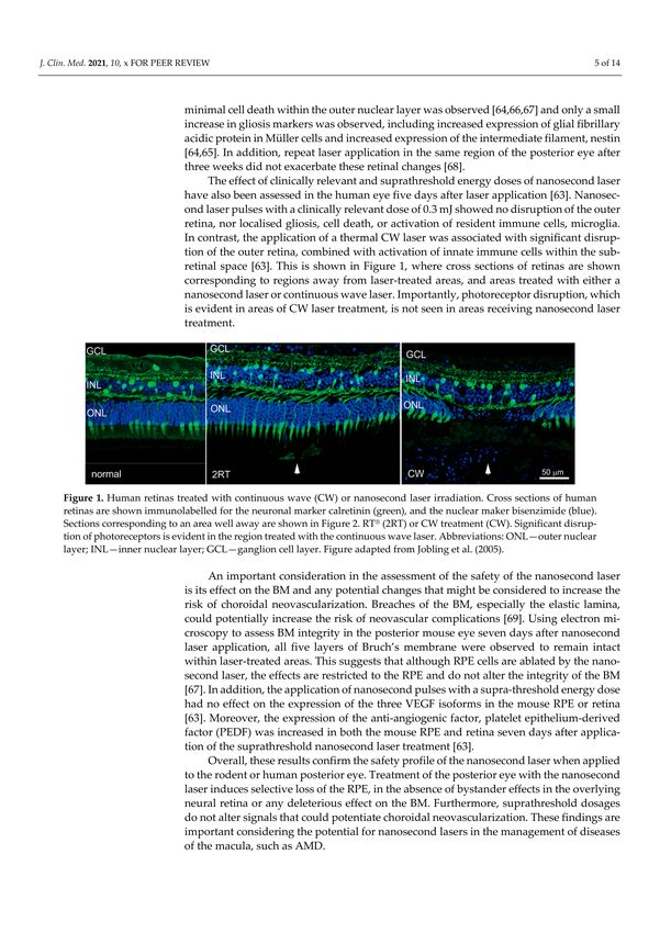
Figure 2. Nanosecond laser thins Bruch’s membrane in a mouse model with features of early AMD. (A,B) Electron
micrographs of the Bruch’s membrane (BM) of a non-laser treated 12-month-old ApoEnull mouse and an ApoEnull mouse
that had received nanosecond laser treatment 3 months prior to fixation. Abbreviations: BM-Bruch’s membrane; RPE-retinal
pigmental epithelium. (C) Graph showing percentage change in Bruch’s membrane thickness in control, lasered, and
unlasered fellow eyes of ApoENull mice that had received laser treatment 3 months prior. Application of the nanosecond
laser induced significant thinning of Bruch’s membrane compared to control or fellow unlasered eyes (one-way ANOVA,
Tukey’s post-hoc test; ** p < 0.01). Figure adapted from Jobling et al. (2005).
An important consideration in the assessment of the safety of the nanosecond laser is
its effect on the BM and any potential changes that might be considered to increase the risk
of choroidal neovascularization. Breaches of the BM, especially the elastic lamina, could
potentially increase the risk of neovascular complications [69]. Using electron microscopy
to assess BM integrity in the posterior mouse eye seven days after nanosecond laser
application, all five layers of Bruch’s membrane were observed to remain intact within
laser-treated areas. This suggests that although RPE cells are ablated by the nanosecond
laser, the effects are restricted to the RPE and do not alter the integrity of the BM [67]. In
addition, the application of nanosecond pulses with a supra-threshold energy dose hadJ. Clin. Med. 2021, 10, 484 6 of 14
no effect on the expression of the three VEGF isoforms in the mouse RPE or retina [63].
Moreover, the expression of the anti-angiogenic factor, platelet epithelium-derived factor
(PEDF) was increased in both the mouse RPE and retina seven days after application of the
suprathreshold nanosecond laser treatment [63].
Overall, these results confirm the safety profile of the nanosecond laser when applied
to the rodent or human posterior eye. Treatment of the posterior eye with the nanosecond
laser induces selective loss of the RPE, in the absence of bystander effects in the overlying
neural retina or any deleterious effect on the BM. Furthermore, suprathreshold dosages
do not alter signals that could potentiate choroidal neovascularization. These findings are
important considering the potential for nanosecond lasers in the management of diseases
of the macula, such as AMD.
5. Nanosecond Laser Treatment Abrogates Changes in the Posterior Eye Important in
the Development of AMD
Having established that the nanosecond laser selectively ablates the RPE in the absence
of damage to neighbouring structures, it is important to address its effect on the posterior
eye that has the potential to reduce the progression of AMD. The formation of drusen
and a thickening in the BM are critical in the development of early AMD. Investigation of
BM thickness in an animal model with features of early AMD demonstrated a thinning
of the BM in response to nanosecond laser application [63]. ApoEnull mice, which have
a thickened BM, were treated with the 2RT® laser. Ten spots were delivered in each eye
at nine months of age, and eyes were then evaluated three months later. In contrast to
ApoEnull mice eyes that were sham-treated and showed a substantially thickened BM
(~900 nm thick), animals that had received nanosecond laser treatment to one eye showed
a significant reduction in thickness (~700 nm thick) in the treated eye (Figure 2) [63].
In order to investigate the mechanism of this apparent nanosecond laser effect, it is
important to realise that the BM is a dynamic structure consisting of extracellular matrix,
including alternating layers of collagen and elastin. Its turnover is controlled by signalling
pathways within the RPE, including the expression of matrix metalloproteinases (MMP)
and tissue inhibitors of matrix metalloproteinases (TIMPs), which are important for the for-
mation and degradation of constituents of the BM. In vitro studies on cultured human RPE
cells have revealed that treatment with the nanosecond laser showed induced expression
of MMP2 and MMP9, with these enzymes being released within two days of subthreshold
nanosecond laser (SNL) treatment [66]. Expressional analysis of genes associated with the
formation and degradation of the extracellular matrix has also been carefully examined in
a mouse model with features of AMD. Changes in gene expression of 84 genes associated
with extracellular matrix turnover have been examined in 12-month-old C57Bl6 (control)
and ApoEnull (AMD-like model) mice three months after nanosecond laser treatment.
A total of nine genes were significantly dysregulated by more than two-fold, including
Mmp2 and Mmp3, a finding that was also confirmed by quantitative RT-PCR [63]. These
data suggest that treatment of the RPE of aged ApoEnull mice with a nanosecond laser
alters the turnover of extracellular matrix components of Bruch’s membrane by altering
the expression of MMPs within the RPE [63].
One of the more intriguing findings in animals treated with the nanosecond laser
was the observation that changes in gene expression in the RPE occurred in both the laser
treated and the untreated fellow eye. Indeed, both Mmp2 and Mmp3 were upregulated by
similar amounts in both eyes, alongside seven other genes associated with extracellular
matrix turnover [63]. Although the BM was not significantly thinned in untreated con-
tralateral eyes, these results suggest that the nanosecond laser could have distant effects,
the mechanisms and significance of which require further study.
Overall, these findings suggest that nanosecond laser application selectively ablates
RPE cells without inducing overt visible damage in adjacent structures. Moreover, ab-
sorption of nanosecond laser energy by the RPE induces gene expressional changes that
are associated with thinning of the BM, particularly involving the MMPs. These findings
support the evaluation of this laser in macular conditions, including AMD.J. Clin. Med. 2021, 10, 484 7 of 14
6. Human Proof of Concept Study of Nanosecond Laser Treatment in AMD
The 2RT® nanosecond laser was used in a human pilot study in 2012–2013 [70]. The
study recruited 50 people with bilateral large drusen (>125 µm; meeting the definition of
intermediate AMD) and with best corrected visual acuity (BCVA) >20/63 in both eyes. The
treatment protocol was a single session in one eye of 12 laser spots, 400 µm in diameter
(at the retina) and placed >500 µm from the fovea. The 12 spots were chosen because this
was a similar approach taken in one of the original thermal CW laser studies [34]. The
energy was titrated for each individual by establishing a “threshold”—the energy at which
a visible burn was seen. The treatment energy was then turned down from threshold
so as to deliver sub-threshold laser to the macula (range 0.15–0.45 mJ; average 0.24 mJ).
A natural history cohort was included for comparison and consisted of 58 untreated
participants with intermediate AMD (bilateral large drusen). Both groups were followed
up at six-month intervals for two years [70]. Drusen load was assessed using multimodal
imaging, including colour fundus photographs, OCT, and fundus autofluorescence (FAF).
Eyes that reached late disease (nAMD or GA) were excluded from the analysis of drusen
load grading; the development of late disease invalidates any grading on drusen load
because drusen disappear when late AMD occurs. After 12 months, 40% of eyes receiving
the 2RT® laser had a reduction in drusen area compared with baseline [70]. This was
statistically significant when comparing the treatment cohort to the natural history group,
where a reduction in drusen area only occurred in 5% of eyes (p < 0.001) [70]. This effect
was maintained over two years, with 35% of the treated eyes demonstrating reduction
in drusen area compared to 11% in the natural history group (p < 0.01). An interesting
observation was that the untreated fellow eyes in participants receiving the 2RT® laser also
demonstrated a reduction in drusen area at 12 months compared to the natural history
cohort (p = 0.05), although this effect was not maintained into the second year of follow-
up [70]. Importantly, this pilot study demonstrated that laser-induced drusen resolution
did not result in progression to atrophy to the two-year follow-up time point. Moreover,
FAF imaging of specific regions where drusen regressed were not hypoautofluorescent, a
potential indicator of progression towards GA [70]. This is important because spontaneous
resolution of drusen often heralds the development of atrophy [39]. This pilot study
concluded that at two years, there were resolutions of drusen in 2RT® -treated eyes without
evidence of progression to atrophy.
7. Nanosecond Laser Treatment in Early Age-Related Macular Degeneration: The
Laser Intervention in the Early Stages of Age-Related Macular Degeneration Study
Following the pilot study, a larger randomised clinical trial was conducted: The Laser
Intervention in the Early Stages of Age-Related Macular Degeneration (LEAD) study [71,72].
The LEAD study was a 36-month, multicentre, randomised, sham-controlled trial, designed
to evaluate the effect of the 2RT® in individuals with bilateral large drusen. The treatment
was referred to as “subthreshold nanosecond laser” (SNL). A total of 292 participants
with BCVA >20/40 in both eyes were randomised to receive either SNL therapy or sham
treatment to a study eye every six months and were followed over a three-year study
period. The main outcome measure was the time to the development of late AMD in
the study eye, as defined by MMI, which for this study was defined as colour fundus
photography, OCT, FAF, and fluorescein or indocyanine green angiography (as clinically
indicated) [71,72]. The LEAD trial was the first trial to use a combined atrophic endpoint
of atrophy, as defined either on OCT as nascent geographic atrophy (nGA) through to GA
as defined on colour fundus photography [71]. SNL treatment was applied using the 2RT®
laser with 12 spots, 400 µm in diameter applied to the macula area—six spots just inside the
superior arcade and six spots inside the inferior arcade. Test spots were used to determine
threshold energy for each participant, and then reduced to perform the treatment to ensure
subthreshold delivery of the laser [71]. Sham laser was performed in the same way, but
short bursts of light from the 2RT® illumination system were used to simulate the laser.J. Clin. Med. 2021, 10, 484 8 of 14
Analysis of the LEAD study showed that in participants with bilateral large drusen,
there was no significant difference in the overall progression to late AMD when comparing
the group randomised to SNL to the group receiving sham treatment. At 36 months of
follow-up, 45 patients (15.4%) developed late AMD in the study eye, with this occurring in
20 (13.6%) participants in the SNL group and 25 (17.2%) participants in the sham group.
However, a proportion of individuals with bilateral large drusen also exhibited the high-
risk RPD phenotype. Given that these individuals might have a more dysfunctional RPE
and may not respond as well to laser treatment compared to those with conventional
drusen, a post-hoc analysis was performed. The post-hoc analyses revealed evidence that
the effect of the SNL treatment was modified based on the coexistence of RPD at baseline
(interaction p = 0.002). Specifically, for the 222 (76.0%) participants without coexisting RPD
at baseline, the rate of progression to late AMD showed a more than four-fold reduction
in the SNL group compared to the sham group (p = 0.002). Conversely, in the 70 (24.0%)
participants with coexisting RPD at baseline, there was potentially an increased rate of
progression to late AMD in the SNL-treated arm compared to the sham arm (p = 0.112) [72].
This was the first study to suggest a potential differential result of an intervention based
upon the AMD phenotype with regards to the drusen subtype. Whilst these findings from
the post-hoc analysis should be interpreted with caution, they are biologically plausible and
provide an hypothesis that this form of laser treatment may be effective at slowing disease
progression in those with intermediate AMD without RPD. However, this hypothesis
requires validation in future, larger randomised trials [72].
8. Secondary Outcomes and Other Research from the LEAD Study
The secondary and exploratory outcomes of the LEAD study looked at the time to
develop late AMD in the non-study eye based on MMI, change in visual function (BCVA,
LLVA and microperimetric sensitivity) and drusen volume in the study and non-study
eyes, and participant-reported outcomes from the Night Vision Questionnaire (NVQ-10)
and Impact of Vision Impairment (IVI) questionnaire [73]. Overall, SNL treatment did not
significantly delay overall progression to late AMD in the fellow non-study (untreated) eye.
Although not significant, there was a trend of effect modification based on the coexistence
of RPD in the non-study eye (interaction p = 0.065), with similar trends as seen in the study
eye (i.e., reduced rate of progression for those without coexistent RPD with SNL treatment,
while a potentially increased rate of progression in those with RPD). These findings are
consistent with the preclinical animal studies that revealed a thinning of the BM in the
lasered eye and changes in gene expression in both the lasered eye and non-lasered fellow
eye, suggesting a possible systemic effect.
There was no significant difference in the change in the visual function measures
and drusen volume in both the study and non-study eyes, and no significant difference in
the participant-reported outcomes, between those in the SNL and sham treatment groups.
The only exception was a slightly greater drop in BCVA in the SNL group compared
with the sham for study eyes, but no consistent reasons for this finding were found
based on a clinical review of the cases showing a ≥10-letter drop. Furthermore, this
observation did not correlate with other visual function measures, but indeed warrants
further evaluation [73]. The absence of evidence for a reduction in drusen load for study
eyes in the SNL treatment group was unexpected [73] as this had been observed in our
pilot study [70]. However, these findings suggest that the potential positive effects of
SNL treatment on delaying progression to late AMD may not necessarily be reflected by
changes in drusen load. This adds complementary knowledge to the previous findings that,
despite inducing drusen regression, CW thermal lasers did not lead to beneficial effects for
slowing late AMD development [34], highlighting the importance of evaluating late AMD
development as the main outcome in such trials.
Further examination of the potential impact of SNL treatment parameters on the
progression to late AMD was performed using the data from the LEAD study. The lack
of real-time visual feedback of a sub-threshold laser application can make it difficult toJ. Clin. Med. 2021, 10, 484 9 of 14
determine if the treatment has been delivered adequately to the RPE to bring about the
desired effect. Whilst the laser spots were not clinically visible at the time of the intervention,
they were often readily visible on FAF imaging obtained at subsequent visits in the LEAD
study. We therefore sought to examine if there was a “dose–response” relationship between
the number of visible spots on FAF imaging, as well as the treatment laser energy used
from the first two LEAD treatments on late AMD progression over the three-year study
period. Multivariable analyses revealed that there were no significant associations between
the time to develop late AMD and number of FAF-visible laser spots, nor the laser energy
used during the SNL treatments, delivered early in the trial. Thus, there was no evidence
to suggest that a dose–response relationship existed for the effect of laser treatment using
the LEAD study treatment parameters on the progression of AMD (unpublished data).
9. Future Applications and Directions of Subthreshold Laser Treatments for Treating
Macula Disease
Extensive research into short duration lasers have heralded the development of se-
lective retinal therapy (SRT) and subthreshold diode micro-pulse (SDM) and nanosecond
lasers. Although the use of many short duration lasers has been explored for use in retinal
disease, to the best of our knowledge, the 2RT® laser developed by Ellex (now Nova Eye
Medical, Pty Ltd. Fremont, CA, USA) represents the only laser functioning in the nanosec-
ond range for ophthalmic use. As such, the results of the LEAD study are only applicable
for use with such a laser and cannot be extrapolated for other short pulse lasers. In addition,
the LEAD study is, to the best of our knowledge, the only large randomised-controlled trial
to examine the potential efficacy of a subthreshold, nanosecond laser in slowing progres-
sion of intermediate AMD to advanced disease. Nova Eye Pty Ltd. (Fremont, CA, USA)
plans to continue its research using the 2RT® laser in management of iAMD.
Another development in short duration laser use for ophthalmic conditions is the
release of the R:GEN laser by Lutronic Vision (South Korea). The R:GEN is an SRT laser
with a 527 nm wavelength and 1.7 µs pulse duration designed to selectively target the
RPE, with its effect delivered through microbubble formation in the RPE. As discussed
previously, lasers delivered at subthreshold levels have no visual feedback at the time
of application, which can make the titration of laser power for adequate tissue effect
extremely difficult. The R:GEN laser utilises Dual Dosimetry technology to measure
reflectometry (back-scattered light) and opto-acoustic signalling (thermo-elastic pressure
waves) to offer real-time titration of laser energy delivery to the RPE. Opto-acoustic (OA)
imaging technology (also known as photo-acoustic imaging) is a non-invasive way to
determine the temperature rise in the RPE cell at the time of laser treatment, utilising
both light and sound wave principles. When short duration laser light is absorbed by
chromophores within a tissue (such as melanosomes within the RPE), the cell undergoes
thermoelastic expansion and acoustic waves are generated. These optoacoustic signals
can be measured by an ultrasonic transducer. During irradiation of the RPE, the baseline
temperature of the cell increases, resulting in a change to the pressure signal and acoustic
waves emitted and microbubble formation can be detected within the RPE cells using
OA techniques [74,75]. These methods can then indicate when sufficient energy has been
generated by the laser within the RPE cell, and at this point the laser automatically switches
off. It is possible that this will result in a more accurate, individualised titration of laser
energy delivery. The R:GEN laser has already been studied in macular disease central
serous chorioretinopathy with promising results [76], and further studies are planned in
other diseases.
Another difficulty in conducting interventional trials for the early stages of AMD is the
natural history of the disease itself. The disease progresses slowly over years, which renders
reaching clinically meaningful results within a reasonable time frame difficult. Significant
advances have been made to address this, through describing potential early disease
endpoints. Nascent geographic atrophy (nGA) is one such early biomarker, signifying
early atrophic changes as seen on OCT imaging. These changes were incorporated into
a combined atrophic endpoint in the LEAD study (the first trial to do so), and in soJ. Clin. Med. 2021, 10, 484 10 of 14
doing, enabled a more time- and cost-efficient study to be conducted [25]. Similarly, the
Classification of Atrophy Meeting (CAM) international consensus group have proposed
a classification of atrophy defined on OCT features of both incomplete and complete
retinal pigment epithelium and outer retinal atrophy (iRORA and oRORA, respectively) in
AMD [77,78]. Having consensus on nomenclature around early atrophic changes in AMD
will help facilitate early intervention studies, making it more feasible to assess the efficacy
of novel early interventions.
10. Conclusions
Apart from lifestyle modifications and dietary supplements, there are no specifically
targeted treatments to slow the progression from the early stages of AMD to advanced
disease. Whilst nanosecond laser is not yet a recognised intervention for AMD, we have
reviewed a body of work to demonstrate that it may offer an intervention where there
currently is none. Preclinical models show that a nanosecond laser (2RT® ) can be safely
delivered to the retina where it is selectively taken up by RPE cells, and this treatment
shows biological plausibility given our current understanding of AMD pathogenesis. The
LEAD study provided clinical results supporting continued research into the potential
of subthreshold delivery of nanosecond laser to provide a possible early intervention to
slow AMD progression. Further well-conducted, randomised clinical trials are required to
determine the efficacy and safety of the 2RT® , as well as all other lasers aiming to target
this indication.
Author Contributions: Each of the contributing authors to this paper undertook the following:
conceptualization, R.H.G., Z.W., E.L.F.; methodology, R.H.G., Z.W., E.L.F.; validation, A.C.C., Z.W.,
A.I.J., E.L.F., R.H.G.; formal analysis, Z.W., E.L.F., R.H.G., A.C.C.; investigation, A.C.C., Z.W., A.I.J.,
E.L.F., R.H.G.; data curation, Z.W., A.C.C., R.H.G.; writing—original draft preparation, A.C.C., E.L.F.,
R.H.G.; writing—review and editing, A.C.C., Z.W., A.I.J., E.L.F., R.H.G. All authors have read and
agreed to the published version of the manuscript.
Funding: This study was supported by the National Health & Medical Research Council of Australia
(project grant no.: APP1027624 [RHG and CDL], and fellowship grant no.: GNT1103013 (RHG),
APP1104985 [ZW], APP1054712 [FKC], APP1142962 [FKC] and GNT1128343 [SSW]), and the BUPA
Health Foundation (Australia) (RHG and CDL). The Centre for Eye Research Australia (CERA)
receives operational infrastructure support from the Victorian Government. Ellex R&D Pty Ltd.
(Adelaide, Australia) provided partial funding of the central coordinating centre and the in-kind
provision of Ellex 2RTTM laser systems, ongoing support of those systems, and the Macular Integrity
Assessment micro-perimeters for the duration of the study. The web-based Research Electronic Data
Capture (REDCap) application and open-source platform OpenClinica allowed secure electronic
data capture. The study is sponsored by CERA, an independent medical research institute and a
not-for-profit company.
Institutional Review Board Statement: The LEAD study was a multicentre, randomised, sham-
controlled clinical trial conducted at six sites between 2012 to 2018, with five in Australia and one in
Northern Ireland. The coordinating centre and sponsor of this trial was the Centre for Eye Research
Australia (CERA). The study was registered with the Australian New Zealand Clinical Trials registry
(ACTRN12612000704897) and clinicaltrials.gov (NCT01790802). The LEAD study was undertaken
according to the International Conference on Harmonization Guidelines for Good Clinical Practice
and the Declaration of Helsinki. The protocol was approved by the relevant review boards at all
involved institutions. All study participants gave written informed consent.
Informed Consent Statement: Informed consent was obtained from all subjects involved in the study.
Data Availability Statement: Data sharing not applicable. No new data were created or analyzed in
this study. Data sharing is not applicable to this article.
Conflicts of Interest: The authors declare no conflict of interest.J. Clin. Med. 2021, 10, 484 11 of 14
References
1. Brown, D.M.; Kaiser, P.K.; Michels, M.; Soubrane, G.; Heier, J.S.; Kim, R.Y.; Sy, J.S.; Schneider, S. ANCHOR Study Group.
Ranibizumab versus verteporfin for neovascular age-related macular degeneration. N. Engl. J. Med. 2006, 355, 1432–1444.
[CrossRef]
2. Rosenfeld, P.J.; Brown, D.M.; Heier, J.S.; Boyer, D.S.; Kaiser, P.K.; Chung, C.Y.; Kim, R.Y.; MARINA Study Group. Ranibizumab
for neovascular age-related macular degeneration. N. Engl. J. Med. 2006, 355, 1419–1431. [CrossRef]
3. Brown, D.; Michels, M.; Kaiser, P.K.; Heier, J.S.; Sy, J.P.; Ianchulev, T. ANCHOR Study Group. Ranibizumab versus Verteporfin Pho-
todynamic Therapy for Neovascular Age-Related Macular Degeneration: Two-Year Results of the ANCHOR Study. Ophthalmology
2008, 116, 57–65. [CrossRef]
4. The Age-Related Eye Disease Study Research Group. The Age-Related Eye Disease Study (AREDS): Design Implications AREDS
Report No. 1. Control Clin. Trials 1999, 20, 573–600. [CrossRef]
5. Ferris, F., III; Wilkinson, C.P.; Bird, A.; Chakravarthy, U.; Chew, E.; Csaky, K.; Sadda, S.R. Beckman Initiative for Macular
Research Classification Committee. Clinical Classification of Age-related Macular Degeneration. Ophthalmology 2013, 129,
844–851. [CrossRef]
6. Mullins, R.F.; Hageman, G.S. Human ocular drusen possess novel core domains with a distinct carbohydrate composition.
J. Histochem. Cytochem. 1999, 47, 1533–1539. [CrossRef]
7. Lengyel, I.; Flinn, J.M.; Peto, T.; Linkud, D.H.; Bird, A.C.; Lanzirotti, A.; Frederickson, C.J.; van Kuijk, F.J. High concentration of
zinc in sub-retinal pigment epithelial deposits. Exp. Eye Res. 2007, 84, 772–780. [CrossRef]
8. Pikuleva, I.; Curcio, C.A. Cholesterol in the retina: The best is yet to come. Prog. Retin. Eye Res. 2014, 41, 64–89. [CrossRef]
9. Mullins, R.F.; Russell, S.R.; Anderson, D.H.; Hageman, G.S. Drusen associated with aging and age-related macular degeneration
contain proteins common to extracellular deposits associated with atherosclerosis, elastosis, amyloidosis, and dense deposit
disease. FASEB J. 2000, 14, 835–846. [CrossRef]
10. Malek, G.; Li, C.M.; Guidry, C.; Medeiros, N.E.; Curcio, C.A. Apolipoprotein B in cholesterol-containing drusen and basal deposits
in eyes with age-related maculopathy. Am. J. Pathol. 2003, 162, 413–425. [CrossRef]
11. Johnson, L.; Ozak, S.; Staples, M.K.; Erikson, P.A.; Anderson, D.H. A potential role for immune complex pathogenesis in drusen
formation. Exp. Eye Res. 2000, 70, 441–449. [CrossRef]
12. Anderson, D.H.; Ozaki, S.; Nealon, M.; Neitz, J.; Mullins, R.F.; Hageman, G.S.; Johnson, L.V. Local cellular sources of apolipopro-
tein E in the human retina and retinal pigmented epithelium: Implications for the process of drusen formation. Am. J. Ophthalmol.
2001, 131, 767–781. [CrossRef]
13. Pauleikhoff, D.; Zuels, S.; Sheraidah, G.S.; Marshall, J.; Wessing, A.; Bird, A.C. Correlation between biochemical composition and
fluorescein binding of deposits in Bruch’s membrane. Ophthalmology 1992, 99, 1548–1553. [CrossRef]
14. Curcio, C.; Presley, J.B.; Malek, G.; Medeiros, N.E.; Avery, D.V.; Kruth, H.S. Esterified and unesterified cholesterol in drusen and
basal deposits of eyes with age-related maculopathy. Exp. Eye Res. 2005, 81, 731–741. [CrossRef]
15. Wang, L.; Clark, M.E.; Crossman, D.K.; Kojima, K.; Messinger, J.D.; Mobley, J.A.; Curcio, C.A. Abundant lipid and protein
components of drusen. PLoS ONE 2010, 5, e10329. [CrossRef]
16. Booji, J.; Baas, D.C.; Beisekeeva, J.; Gorgels, T.G.M.F.; Bergen, A.A.B. The dynamic nature of Bruch’s membrane.
Prog. Retin. Eye Res. 2010, 29, 1–18. [CrossRef]
17. Karampelas, M.; Sim, D.A.; Keane, P.A.; Papastefanou, V.P.; Sadda, S.R.; Tufail, A.; Dowler, J. Evaluation of retinal pigment
epithelium-Bruch’s membrane complex thickness in dry age-related macular degeneration using optical coherence tomography.
Br. J. Ophthalmol. 2013, 97, 1256–1261. [CrossRef]
18. Starita, C.; Hussain, A.A.; Pagliarini, S.; Marshall, J. Hydrodynamics of ageing Bruch’s membrane: Implications for macular
disease. Exp. Eye Res. 1996, 62, 565–572. [CrossRef]
19. Hussain, A.; Starita, C.; Hodgetts, A.; Marshall, J. Macromolecular diffusion characteristics of ageing human Bruch’s membrane:
Implications for age-related macular degeneration (AMD). Exp. Eye Res. 2010, 90, 703–710. [CrossRef]
20. Finger, R.P.; Wu, Z.; Luu, C.D.; Kearney, F.; Ayton, L.N.; Lucci, L.M.; Hubbard, W.C.; Hageman, J.L.; Hageman, G.S.; Guymer,
R.H. Reticular pseudodrusen: A risk factor for geographic atrophy in fellow eyes of individuals with unilateral choroidal
neovascularization. Ophthalmology 2014, 121, 1252–1256. [CrossRef]
21. Zhou, Q.; Daniel, E.; Maguire, M.G.; Grunwald, J.E.; Martin, E.R.; Martin, D.F.; Ying, G.S. Pseudodrusen and incidence of late
age-related macular degeneration in fellow eyes in the Comparison of Age-related Macular Degeneration Treatment Trials.
Ophthalmology 2016, 123, 1530–1540. [CrossRef] [PubMed]
22. Domalpally, A.; Agron, E.; Pak, J.W.; Keena, T.D.; Ferrris, F.L., III; Clemon, T.E.; Chew, E.Y. Prevalence, Risk, and Genetic
Association of Reticular Pseudodrusen in Age-related Macular Degeneration. Age-Related Eye Disease Study 2 Report 21.
Ophthalmology 2019, 126, 1659–1666. [CrossRef] [PubMed]
23. Curcio, C.; Messinger, J.D.; Sloan, K.R.; McGwin, G.; Medeiros, N.E.; Spaide, R.F. Subretinal Drusenoid Deposits in Non-
Neovascular Age-related Macular Degeneration: Morphology, Prevalence, Topography and Biogenesis. Retina 2013, 33, 265–276.
[CrossRef] [PubMed]
24. Spaide, R.F.; Ooto, S.; Curcio, C.A. Subretinal drusenoid deposits AKA pseudodrusen. Surv. Ophthalmol. 2018, 63, 782–815.
[CrossRef] [PubMed]J. Clin. Med. 2021, 10, 484 12 of 14
25. Christenbury, J.G.; Folgar, F.A.; O’Connell, R.V.; Chiu, S.J.; Farsiu, S.; Toth, C.A. Progression of intermediate age-related macular
degeneration with proliferation and inner reitnal migration of hyperreflective foci. Ophthalmology 2013, 120, 1038–1045. [CrossRef]
26. Tan, A.C.S.; Pilgrim, M.G.; Fearn, S.; Bertazzo, S.; Tsolaki, E.; Morell, A.P.; Li, M.; Messinger, J.D.; Dolz-Marco, R.; Lei, J.; et al.
Calcified nodules in retinal drusen are associated with disease progression in age-related macular degeneration. Sci. Transl. Med.
2018, 10, eaat4544. [CrossRef] [PubMed]
27. Wu, Z.; Luu, C.D.; Ayton, L.N.; Goh, J.K.; Lucci, L.M.; Hubbard, W.C.; Hageman, J.L.; Hageman, G.S.; Guymer, R.H. Optical
Coherence tomography defined changes preceding the development of drusen-associated atrophy in age-related macular
degeneration. Ophthalmology 2014, 121, 2415–2422. [CrossRef]
28. Gass, J. Photocoagulation of macula lesions. Trans. Am. Acad. Ophthalmol. Otolaryngol. 1971, 75, 580–608.
29. Olk, R.; Friberg, T.R.; Stickney, K.L.; Akduman, L.; Wong, K.L.; Chen, M.C.; Levy, M.H.; Garcia, C.A.; Morse, L.S. Therapeutic
benefits of infrared (810-nm) diode laser macular grid photocoagulation in prophylactic treatment of nonexudative age-related
macular degeneration: Two-year results of a randomized pilot study. Ophthalmology 1999, 106, 2082–2090. [CrossRef]
30. Wetzig, P. Treatment of drusen-related aging macular degeneration by photocoagulation. Trans. Am. Ophthmol. Soc. 1988, 86,
276–290.
31. Frennesson, C.; Nilsson, S.E. Prophylactic laser treatment in early age-related maculopathy reduced the incidence of exudative
complications. Br. J. Ophthalmol. 1998, 82, 1169–1174. [CrossRef]
32. Kaiser, R.S.; Berger, J.W.; Maguire, M.G.; Ho, A.C.; Javornik, M.S. CNVPT Study Group. Laser burn intensity and the risk for
choroidal neovascularisation in the CNVPT Fellow Eye Study. Arch Ophthmol. 2001, 119, 826–832. [CrossRef]
33. Owens, S.; Bunce, C.; Brannon, A.J.; Wormald, R.; Bird, A.C. Prophylactic laser treatment appears to promote choroidal
neovascularisation in high-risk ARM: Results of an interim analysis. Eye (Lond.) 2003, 15, 623–627. [CrossRef] [PubMed]
34. Virgili, G.; Michelessai, M.; Parodi, M.B.; Bacherini, D.; Evans, J.R. Laser treatment of drusen to prevent progression to advanced
age-related macular degeneration. Cochrane Libr. 2015, 10, CD006537. [CrossRef] [PubMed]
35. Guymer, R.; Hageman, G.; Bird, A. Influence of laser photocoagulation on choroidal capillary cytoarchitecture. Br. J. Ophthalmol.
2001, 85, 40–46. [CrossRef] [PubMed]
36. Yamamoto, C.; Ogata, N.; Yi, X. Immunolocalization of basic fibroblast growth factor during wound repair in rat retina after laser
photocoagulation. Graefes Arch. Clin. Exp. Ophthalmol. 1996, 234, 695–702. [CrossRef] [PubMed]
37. Duvall, J.; Tso, M.O.M. Cellular Mechanisms of Resolution of Drusen after Laser Coagulation. An Experimental Study. Arch.
Ophthalmol. 1985, 103, 695–703. [CrossRef] [PubMed]
38. Toy, B.C.; Krishnadev, N.; Indaram, M.; Cunnigham, D.; Cukras, C.A.; Chew, E.Y.; Wong, W.T. Drusen regression is associated
with local changes in fundus autofluorescence in intermediate age-related macular degeneration. Am. J. Ophthalmol. 2013, 156,
532–542. [CrossRef]
39. Klein, M.L.; Ferris, F.L., III; Armstrong, J.; Hwang, T.S.; Chew, E.Y.; Bressler, S.B.; Chandra, S.R. AREDS Research Group. Retinal
precursors and the development of geographic atrophy in age-related macular degeneration. Ophthalmology 2008, 115, 1026–1031.
[CrossRef]
40. Varley, M.; Frank, E.; Purnell, E.W. Subretinal neovascularization after focal argon laser for diabetic macular edema. Ophthalmology
1988, 95, 567–573. [CrossRef]
41. Marshall, J.; Hamilton, A.M.; Bird, A.C. Histopathology of ruby and argon laser lesions in the monkey and human retina.
Br. J. Ophthalmol. 1975, 59, 610–630. [CrossRef] [PubMed]
42. Paulus, Y.; Jain, A.; Gariano, R.F. Healing of retinal photocoagulation lesions. Investig. Ophthalmol. Vis. Sci. 2008, 49, 5540–5545.
[CrossRef]
43. The Diabetic Retinopathy Study Research Group. Photocoagulation treatment of proliferative diabetic retinopathy. Clinical
application of Diabetic Retinopathy Study (DRS) findings, DRS report number 8. Ophthalmology 1981, 88, 583–600.
44. The Branch Vein Occlusion Study Group. Argon laser scatter photocoagulation for prevention of neovascularization and vitreous
hemorrhage in branch vein occlusion. A randomized clinical trial. Arch. Ophthalmol. 1986, 104, 34–41. [CrossRef] [PubMed]
45. The Early Treatment Diaebtic Retinopathy Study Group. Early photocoagulation for diabetic retinoapthy. ETDRS report number
9. Ophthalmology 1991, 98, 766–785. [CrossRef]
46. Lee, M.; Yeo, I.; Wong, D.; Ang, C.L. Argon laser photocoagulation for the treatment of polypoidal choroidal vasculopathy. Eye
2009, 23, 145–148. [CrossRef] [PubMed]
47. Rabb, M.; Gagliano, D.A.; Teske, M.P. Retinal arterial macroaneurysms. Surv. Ophthalmol. 1988, 33, 73–96. [CrossRef]
48. Marc, R.E.; Jones, B.W.; Watt, C.B.; Strettoi, E. Neural remodeling in retinal degeneration. Prog. Retin. Eye Res. 2003, 22, 607–655.
[CrossRef]
49. Richardson, P.; Boulton, M.E.; Duvall-Young, J.; McLeod, D. Immunocytochemical study of retinal diode laser photocoagulation
in the rat. Br. J. Ophthalmol. 1996, 80, 1092–1098. [CrossRef]
50. Musashi, K.; Kiryu, J.; Miyamoto, K. Thrombin inhibitor reduces leukocyte-endothelial cell interactions and vascular leakage
after scatter laser photocoagulation. Investig. Ophthalmol. Vis. Sci. 2005, 46, 2561–2566. [CrossRef]
51. Humphrey, M.; Chu, Y.; Mann, K.; Rakoczy, P. Retinal GFAP and bFGF expression after multiple argon laser photocoagulation
injuries assessed by both immunoreactivity and mRNA levels. Exp. Eye Res. 1997, 64, 361–369. [CrossRef] [PubMed]
52. Tackenberg, M.; Tucker, B.A.; Swift, J.S. Muller cell activation, proliferation and migration following laser injury. Mol. Vis. 2009,
15, 1886–1896. [PubMed]J. Clin. Med. 2021, 10, 484 13 of 14
53. Matsunaga, H.; Nangoh, K.; Uyama, M.; Nanbu, H.; Fujiseki, Y.; Takahashi, K. Occurrence of choroidal neovascularization
following photocoagulation treatment for central serous retinopathy. Nippon Ganka Gakkai Zasshi 1995, 99, 460–468. [PubMed]
54. Anderson, R.R.; Parrish, J.A. Selective photothermolysis: Precise microsurgery by selective absorption of pulsed radiation. Science
1983, 220, 524–527. [CrossRef] [PubMed]
55. Pankratov, M.M. Pulse delivery of laser energy in experimental thermal retinal photocoagulation. Proc. Soc. Photo Opt. Instrum.
Eng. 1990, 1202, 205–213.
56. Framme, C.; Schuele, G.; Roider, J.; Birnbruger, R.; Brinkman, R. Influence of pulse duration and pule number in selective RPE
laser treatment. Lasers Surg. Med. 2004, 34, 206–215. [CrossRef]
57. Dorin, G. Subthreshold and mircopulse diode laser photocoagulation. Semin. Ophthalmol. 2003, 18, 147–153. [CrossRef]
58. Brinkmann, R.; Roider, J.; Birngruber, R. Selective retina therapy (SRT): A review on methods, techniques, preclinical and first
clinical results. Bull. Soc. Belge Ophtalmol. 2006, 302, 51–69.
59. Tode, J.; Richert, E.; Koinzer, S.; Klettner, A.; vonder Burchard, C.; Brinkman, R.; Lucius, R.; Roider, J. Selective Retina Therapy
Reduces Bruch’s Membrane Thickness and Retinal Pigment Epithelium Pathology in Age-Related Macular Degeneration Mouse
Models. Transl. Vis. Sci. Technol. 2019, 8, 11. [CrossRef]
60. Brinkmann, R.; Huttmann, G.; Rogener, J.; Roider, J.; Birngruger, R.; Lin, C.P. Origin of retinal pigment epithelium cell damage by
pulsed laser irradiance in the nanosecond to microsecond time regimen. Lasers Surg. Med. 2000, 27, 451–464. [CrossRef]
61. Wang, J.; Quan, Y.; Dalal, R.; Palanker, D. Comparison of Continuous-Wave and Micropulse Modulation in Retinal Laser Therapy.
Investig. Ophthalmol. Vis. Sci. 2017, 58, 4722–4732. [CrossRef] [PubMed]
62. Wood, J.P.; Plunkett, M.; Previn, V.; Chidlow, G.; Casson, R.J. Nanosecond pulse lasers for retinal applications. Lasers Surg. Med.
2011, 43, 499–510. [CrossRef]
63. Jobling, A.I.; Guymer, R.H.; Vessey, K.A.; Greferath, U.; Mills, S.A.; Brassington, K.H.; Luu, C.D.; Aung, K.Z.; Trogrlic, L.; Plunkett,
M.; et al. Nanosecond laser therapy reverses pathologic and molecular changes in age-related macular degeneration without
retinal damage. FASEB J. 2015, 29, 696–710. [CrossRef] [PubMed]
64. Chidlow, G.; Shibeeb, O.; Plunkett, M.; Casson, R.J.; Wood, J.P.M. Glial cell and inflammatory responses to retinal laser treatment:
Comparison of a conventional photocoagulator and a novel, 3-nanosecond pulse laser. Investig. Ophthalmol. Vis. Sci. 2013, 54,
2319–2332. [CrossRef] [PubMed]
65. Wood, J.P.; Shibeeb, O.; Plunkett, M.; Casson, R.J.; Chidlow, G. Retinal damage profiles and neuronal effects of laser treatment:
Comparison of a conventional photocoagulator and a novel 3-nanosecond pulse laser. Investig. Ophthalmol. Vis. Sci. 2013, 54,
2305–2318. [CrossRef] [PubMed]
66. Zhang, J.J.; Sun, Y.; Hussain, A.A.; Marshall, J. Laser-mediated activation of human retinal pigment epithelial cells and concomitant
release of matrix metalloproteinases. Investig. Ophthalmol. Vis. Sci. 2012, 53, 2928–2937. [CrossRef] [PubMed]
67. Vessey, K.A.; Ho, T.; Jobling, A.I.; Mills, S.A.; Tran, M.X.; Brandli, A.; Lam, J.; Guymer, R.G.; Fletcher, E.L. Nanosecond Laser
Treatment for Age-Related Macular Degeneration Does Not Induce Focal Vision Loss or New Vessel Growth in the Retina. Investig.
Ophthalmol. Vis. Sci. 2018, 59, 731–745. [CrossRef]
68. Chidlow, G.; Plunkett, M.; Casson, R.J.; Wood, J.P.M. Investigations into localized re-treatment of the retina with a 3-nanosecond
laser. Lasers Surg. Med. 2016, 48, 602–615. [CrossRef]
69. Chong, N.H.; Keonin, J.; Luthert, P.J.; Frennesson, C.I.; Weingeist, D.M.; Wolf, R.L.; Mullins, R.F.; Hageman, G.S. Decreased
thickness and integrity of the macular elastic layer of Bruch’s membrane correspond to the distribution of lesions associated with
age-related macular degeneration. Am. J. Pathol. 2005, 166, 241–251. [CrossRef]
70. Guymer, R.H.; Brassington, K.H.; Dimitrov, P.; Makeyeva, G.; Plunkett, M.; Xia, W.; Chauhan, D.; Vingrys, A.; Luu, C.D.
Nano-second laser application in intermediate AMD: 12 month results of fundus appearance and macular function. Clin. Exp.
Ophthalmol. 2014, 42, 466–479. [CrossRef]
71. Lek, J.; Brassington, K.H.; Luu, C.D.; Chen, F.K.; Arnold, J.J.; Heriot, W.J.; Durkin, S.R.; Chakravarthy, U.; Guymer, R.H. Laser
in Early Stages of Age-related Macular Degeneration Study Writing Committee Subthreshold nanosecond laser intervention
in intermediate age-related macular degeneration: Study design and baseline characteristics of the Laser in Early Stages of
Age-Related Macular Degeneration Study (report number 1). Ophthalmol. Retina 2017, 1, 227–239. [PubMed]
72. Guymer, R.H.; Wu, Z.; Hodgson, L.A.; Caruso, E.; Brassington, K.H.; Tindill, N.; Aung, K.Z.; McGuinness, M.B.; Fletcher, E.L.;
Chen, F.K.; et al. Subthreshold Nanosecond Laser Intervention in Age-Related Macular Degeneration: The LEAD Randomized
Controlled Clinical Trial. Ophthalmology 2019, 126, 829–838. [CrossRef] [PubMed]
73. Wu, Z.; Luu, C.D.; Hodgson, L.A.B.; Sharangan, P.; Guymer, R.G. For the LEAD Study Group. Secondary and Exploratory
Outcomes of the Subthreshold Nanosecond Laser Intervention Randomized Trial in Age-Related Macular Degeneration: A LEAD
Study Report. Ophthalmol. Retina 2019, 3, 1026–1034. [CrossRef] [PubMed]
74. Wang, L.; Yao, J. A practical guide to photoacoustic tomography in the life sciences. Nat. Methods 2016, 13, 11. [CrossRef]
75. Schüle, G.; Huettman, G.; Roider, J.; Wirbelauer, C.; Birngruger, R.; Brinkman, R. Optoacoustic measurements during µs-irradiation
of the retinal pigment epithelium. Proc. SPIE 2000.
76. Park, Y.; Kang, S.; Kim, M.; Yoo, N.; Roh, Y.J. Selective retina therapy with automatic real-time feedback-controlled dosimetry for
chronic central serous chorioretinopathy in Korean patients. Graefes Arch. Clin. Exp. Ophthalmol. 2017, 255, 1375–1383. [CrossRef]You can also read