Successful treatment of early cutaneous squamous cell carcinoma with hypofractionated radiation therapy in an African lion (Panthera leo)
←
→
Page content transcription
If your browser does not render page correctly, please read the page content below
Journal of the South African Veterinary Association
ISSN: (Online) 2224-9435, (Print) 1019-9128
                                                                   Page 1 of 6            Case Report

         Successful treatment of early cutaneous squamous
           cell carcinoma with hypofractionated radiation
               therapy in an African lion (Panthera leo)

 Authors:                                Cutaneous squamous cell carcinoma (SCC) is a slow growing but locally invasive neoplasm,
 Louise van der Weyden1                  most commonly caused by prolonged exposure to ultraviolet (UV) radiation. Whilst SCC
 Nicolize O’Dell2,3
 Alida Avenant2                          accounts for 15% of skin tumours in domesticated cats, cutaneous SCC in non-domesticated
 Paolo Pazzi4                            felids (apart from captive snow leopards) appears to be uncommon, with only three reports
 Katja N. Koeppel3,5                     in the literature to date. In this report, a captive African lion (Panthera leo) presented with
                                         two ulcerative lesions on the nasal planum. Histopathology of the lesions revealed epidermal
 Affiliations:
 1
  Wellcome Genome Institute,             keratinocyte dysplasia and neoplastic basal- and supra-basal epithelial cells with dyskeratosis
 Wellcome Sanger Campus,                 and evidence of basement membrane breaching and dermal invasion, consistent with a
 Cambridge, United Kingdom               diagnosis of SCC. There was also evidence of laminar fibrosis and inflammation of the
                                         subjacent dermis suggesting that the SCC most likely resulted from UV-induced neoplastic
 2
  Department of Paraclinical             transformation of the epidermal squamous epithelium following actinic keratosis. The lion
 Sciences, Faculty of Veterinary
 Science, University of Pretoria,        was treated with hypofractionated radiation therapy and remained in remission until his
 Onderstepoort, South Africa             death (euthanised 17 months later because of age-related chronic renal failure). This is the
                                         first report of cutaneous SCC in a lion with evidence of actinic damage and resolution after
 3
  Centre for Veterinary Wildlife         radiation therapy.
 Studies, Faculty of Veterinary
 Science, University of Pretoria,        Keywords: lion; skin; cancer; radiation therapy; actinic damage; laminar fibrosis; UV.
 Onderstepoort, South Africa

  Department of Companion
                                      Introduction
 4

 Animal Studies, Faculty of
 Veterinary Science, University       Squamous cell carcinoma (SCC) is a malignant neoplasm arising from squamous epithelium.
 of Pretoria, Onderstepoort,
                                      Squamous cell carcinomas account for 15% of skin tumours in domestic cats, with most feline
 South Africa
                                      cutaneous SCCs occurring on the head, often involving the pinna, eyelid and nasal planum (Miller
 5
  Production Animal Science,          et al. 1991). In contrast, SCC in non-domestic felids is rarely reported, except in captive snow
 Faculty of Veterinary Science,       leopards, where one survey of 424 animals found SCC accounted for 9% of the mortalities (Joslin
 University of Pretoria,              et al. 2000). In this species, SCC is usually found on the ventral surface of the tongue, face and
 Onderstepoort, South Africa
                                      forelimbs and is mostly associated with papillomavirus-induced malignant transformation (Terio,
 Corresponding author:                McAloose & Mitchell 2018). Histopathological changes that support a viral aetiology consist of
 Katja Koeppel,                       koilocytes (keratinocytes with shrunken basophilic nuclei surrounded by a clear halo) and/or
 katja.koeppel@up.ac.za               keratinocytes with increased greyish-blue granular cytoplasm and occasional eosinophilic
                                      intranuclear inclusions. This is referred to as viral cytopathology and is associated with a
 Dates:
 Received: 03 Dec. 2020               hyperplastic epithelium (Munday 2014).
 Accepted: 08 Apr. 2021
 Published: 24 June 2021              Excluding snow leopards, reports of SCC in non-domesticated felids have mostly been in the oral
                                      cavity (in Lynx species [Altamura et al. 2011; Gunson, Klein & Reid 1978; Sladakovic et al. 2016], a
                                      North American Amur leopard (Panthera pardus orientalis) (Napier et al. 2018) and a Siberian tiger
                                      (Panthera tigris altaica) [De Oliveira et al. 2018]), with only three reports of cutaneous SCC. Firstly,
                                      a 16-year-old female captive tiger (Panthera tigris) who had SCC on the left rear limb underwent
                                      surgery to remove the mass; however, recurrence was documented 2 years later (Owston, Ramsay
                                      & Rotstein 2008). Secondly, a 3-year-old female wild African lion (Panthera leo) had severe swelling
                                      and draining sinus tracts on the front left paw and the lesions were unresponsive to 7 months of
                                      treatment with various antibiotic regimes such that her deteriorating condition resulted in
                                      humane euthanasia. At postmortem, SCC was confirmed, presenting as discharging sinuses lined
                                      by neoplastic squamous cells (Mwase et al. 2013). Thirdly, a 15-year-old female captive clouded
                                      leopard (Neofelis nebulosa) had a swelling and an abscess on the right hind paw and biopsy
     Read online:
                    Scan this QR
                                      revealed a well-differentiated SCC. Attempts at surgical excision and cryosurgery proved
                    code with your
                    smart phone or    How to cite this article: Van der Weyden, L., O’Dell, N., Avenant, A., Pazzi, P. & Koeppel, K.N., 2021, ‘Successful treatment of early
                    mobile device     cutaneous squamous cell carcinoma with hypofractionated radiation therapy in an African lion (Panthera leo)’, Journal of the South African
                    to read online.   Veterinary Association 92(0), a2134. https://doi.org/10.4102/jsava.v92i0.2134
                                      Copyright: © 2021. The Authors. Licensee: AOSIS. This work is licensed under the Creative Commons Attribution License.

                                                     http://www.jsava.co.za               Open Access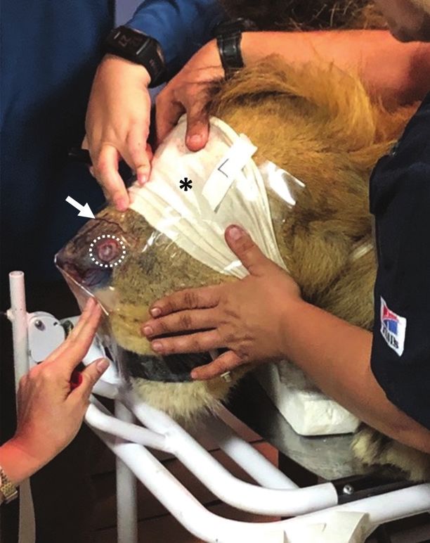
Page 2 of 6 Case Report
unsuccessful and subsequent recurrence of the mass led to a Health and Hygiene Ltd., Florida Hills, South Africa) to
mid-femoral amputation (Kesdangsakonwut et al. 2014). reduce inflammation and irritation. However, the lesions
continued to expand.
Neither actinic damage as the underlying cause of SCC, nor
use of hypofractionated radiation therapy as the sole means One month later the 260 kg lion was darted with
of successful therapy have been previously reported in any medetomidine (7 mg, Wildlife Pharmaceuticals, South
non-domestic felid. The present report describes a SCC on Africa) and Zoletil (160 mg, Virbac, South Africa) to allow
the nasal planum of an African lion that showed concomitant samples to be taken for further analysis. Blood was
evidence of actinic damage in the surrounding area and collected from the femoral vein for routine haematology
successful treatment with radiation therapy. and clinical biochemistry analysis. The blood sample
revealed all parameters to be within normal ranges
Case presentation (Appendix Table 1-A1). Four punch biopsies from the
ulcerative lesion on the nasal planum were taken with a 6
A 16-year-old intact male African lion from the Lory Park
mm biopsy punch: one from the centre and one from the
Zoo and Owl Sanctuary in Midrand, South Africa, who had
periphery of each of the two large lesions. Biopsies were
lived at the property since he was a few days old, was
fixed in 10% buffered formalin, embedded in paraffin wax
presented to the Wildlife Clinic at Onderstepoort Veterinary
and sectioned for histopathological evaluation. All the
Academic Hospital with a 2-month history of two round,
sections were stained with haematoxylin and eosin (H&E)
well demarcated, laterally positioned, ulcerative lesions
and examined by light microscopy by veterinary
approximately 5 cm and 3 cm diameter and a midline
elliptical lesion approximately 0.5 cm × 0.2 cm, on the nasal pathologists (N.O’D. and L.A.). All four of the
planum (Figure 1). No other abnormalities were reported examined sections revealed similar changes that
and all vital parameters were within normal limits. consisted of moderate acanthosis of the epidermis with
Differential diagnoses considered included trauma, fungal mild to moderate parakeratotic and/or orthokeratotic
infection, bacterial infection or hypersensitivity to black hyperkeratosis. There was evidence of epidermal rete peg
flies (Simulium spp), a common presentation in large accentuation, keratinocyte dysplasia and dyskeratosis and
captive carnivores in South Africa (Myburgh & Nevill 2003). occasional brightly eosinophilic apoptotic cells. The basal-
The lesions were treated daily with oral corticosteroids and supra-basal epithelial cells displayed loss of polarity,
(Lenisolone, Pharmacare Ltd., Woodmead, South Africa) moderate pleomorphism, hyperchromatic nuclei with 1–2
initially 120 mg once daily for 3 days then tapered prominent nucleoli and a mitotic count of 25 in 10 high-
(over 8 days) and topical ointment (F10 barrier ointment, powered fields (Figure 2a). In one section, evidence of
dermal invasion and breaching of the basement membrane
by an island of neoplastic keratinocytes could
be observed (Figure 2b). All sections revealed some degree
of laminar fibrosis of the subjacent superficial dermis
(Figure 2c) associated with mild perivascular inflammation
consisting of predominantly plasma cells and lymphocytes
accompanied by fewer neutrophils, macrophages and
occasional eosinophils. Ulceration of the epithelial surface,
with associated pleocellular inflammation and serocellular
crusting were present in areas. The diagnosis was SCC
with actinic keratosis.
The decision was made to use radiotherapy to treat the SCC
lesion, as surgical resection was not possible because of its
location and cutaneous SCC in domestic cats has been
reported to respond well to radiotherapy (Cunha et al. 2010;
Gasymova et al. 2017; Theon et al. 1995). In addition,
radiotherapy has been previously used in conjunction with
immunotherapy and surgical excision in a lion to successfully
treat a melanoma of the lip (4 weekly treatments of 8 gray
(Gy) external-beam hypofractionated radiation and
4 bimonthly immunotherapy treatments were used to reduce
the tumour size by 50%, after which surgical excision was
performed) (Steeil et al. 2013). In contrast, a single fraction of
22 Gy using stereotactic radiotherapy was unsuccessful in
treatment of a facial leiomyosarcoma in a tiger (Panthera
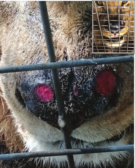
FIGURE 1: Photograph of the nasal planum of the lion showing two well-
demarcated ulcerative lesions. Inset: Nasal planum 2 months after diagnosis and
tigris), which succumbed to severe metastatic disease
receiving radiotherapy. 4 months later (Boudreaux et al. 2019). The radiotherapy was
http://www.jsava.co.za Open Access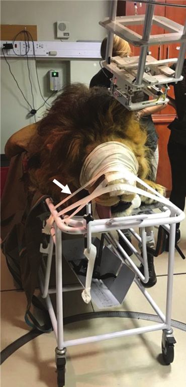
Page 3 of 6           Case Report

                                                   a                                                       b                                                       c

FIGURE 2: Histological features of the lesions. (a) Epidermal rete peg accentuation (arrows) and subepidermal perivascular inflammation (asterisk) (HE stain, x100
magnification). (b) Dyskeratosis (arrowhead), mitoses (arrows) and island of neoplastic epithelial cells below the basement membrane (dashed circle) (HE
stain, x200 magnification). (c) Acanthosis (asterisk) and pale band of subepidermal laminar fibrosis (area between parallel dashed lines) (HE stain, x40
magnification).

                                                           a                                                                                                       b

FIGURE 3: Photograph of the lion being prepared for radiotherapy treatment. (a) Lion strapped onto a patient trolley in the prone position, with the head secured with
tape (arrow) to prevent movement. (b) Blindfold applied for protection of the eyes (asterisk) and Elasto-Gel applied and marked for correct positioning (arrow) to ensure
the correct dose of radiation was uniformly delivered to the affected area of the nasal planum (dashed circle).

performed at the Mediclinic Muelmed Hospital in Pretoria,                                affected area of the nasal planum (Benson et al. 1991)
where the lion was sedated as described here (topped up                                  (Figure 3b). Using an Elekta Synergy machine (Elekta
with intravenously administered 0.5 mg/kg ketamine as                                    Instrument AB, Stockholm), the lion was administered 8 Gy
needed). The lion was strapped onto a patient trolley in the                             radiation directly to the lesions (~5 min duration). Upon
prone position, with the head secured with tape to prevent                               recovery, the lion was transferred back to Lory Park Zoo and
movement (Figure 3a) and an Elasto-Gel used to ensure the                                Owl Sanctuary. This procedure was repeated on three
correct dose of radiation was uniformly delivered to the                                 further occasions, each 8 days apart. In total, the lion received

                                                       http://www.jsava.co.za            Open Access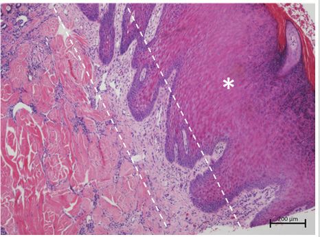
Page 4 of 6   Case Report

32 Gy radiation. During the month of treatment, the lion was         SCC (Röwert-Hubert et al. 2007). Here we have described the
kept in the shade in his enclosure (with his female                  first report of a non-domesticated felid showing signs of SCC
companion). At the end of radiation therapy, a blood sample          associated with actinic dermatosis, which suggest UV-
was taken and all values were again within the normal range          induced injury resulting in neoplastic transformation. No
(Appendix Table 1-A1).                                               viral cytopathology was present in this case, therefore
                                                                     making papillomavirus infection, as seen in snow leopards, a
Follow-up punch biopsies of the nasal planum 14 months               less likely aetiology.
later, because of the presence of ulcerating lesions at the site
of the original SCC lesion, showed no evidence of neoplastic         Although a range of different treatments have been used in
changes. The new lesions were associated with mild                   domestic cats with nasal planum SCC such as strontium
eosinophilic dermatitis, indicative of an allergic reaction of       (Sr90) plesiotherapy (Berlato et al. 2019; Hammond et al.
unknown aetiology (possibly an insect bite hypersensitivity          2007), boron neutron capture therapy (Trivillin et al. 2008)
reaction). Three months later, his condition deteriorated such       and intralesional carboplatin and superficial radiotherapy
that euthanasia was required. Histopathological findings at          (De Vos, Burm & Focker 2004), radiation therapy remains the
autopsy suggested chronic renal failure, which was most              most common therapy. Several radiation therapy protocols
likely age-related. There were no neoplastic lesions observed        are in use including a Monday–Wednesday–Friday schedule
in any of the organs examined (kidney, stomach, spleen,              (Theon et al. 1995), hypofractionated (Cunha et al. 2010) and
heart, liver, pancreas, lymph node, lung, oesophagus and             accelerated protocols (Gasymova et al. 2017). In terms of
diaphragm).                                                          efficacy, the results from different studies reflect the different
                                                                     levels of radiation used; however, in general a tumour
Discussion                                                           response (and manageable toxicity) is observed in most cats,
                                                                     with long tumour control durations achieved in some cats,
Older domestic cats are at greater risk of developing SCC,
                                                                     although complete remission is rare. The cutaneous SCC
with the average age at presentation being 10–12 years
(Hauck 2012; Miller et al. 1991). Factors contributing to the        diagnosed in the lion in this report was successfully treated
development of cutaneous SCC are prolonged exposure to               with hypofractionated radiation therapy and remained
ultraviolet (UV) radiation, lack of skin pigment and a sparse        disease-free until it was euthanised for age-related chronic
hair coat (Goldschmidt & Goldschmidt 2017) and infection             renal failure (17 months later).
by papilloma virus. UV radiation can produce
deoxyribonucleic acid (DNA) damage either directly or                In conclusion, cutaneous SCC, although rarely reported
through the production of reactive oxygen species, leading to        in non-domesticated felids, should be considered as a
the activation of oncogenes or inactivation of tumour                differential diagnosis for a long-term, non-healing skin
suppressor genes, which results in the survival and                  wound in these animals and can be successfully treated with
proliferation of the damaged keratinocytes (D’Orazio et al.          radiation therapy. It is hoped that further investigations
2013). Initiation and progression of skin carcinogenesis             of African lions, both in wildlife reserves and zoological
mediated by UV radiation involve many complex pathways,              facilities, will add to a better knowledge and understanding
including proliferation, apoptosis, autophagy, DNA repair,           of cutaneous tumours in this species.
checkpoint signalling, metabolism and inflammation (Kim &
He 2014). Feline skin chronically exposed to the sun presents        Acknowledgements
clinically as mild erythema in the affected areas (particularly
                                                                     The authors would like to thank Professor Gerhard
the ear margins, preauricular areas, periocular areas, nose
                                                                     Steenkamp at the University of Pretoria for his assistance
and lips [Almeida et al. 2008]), which may histologically
                                                                     with treatment of the lion, the radiation and oncology staff of
show preneoplastic and early neoplastic changes. As the
                                                                     the Mediclinic Muelmed Hospital for allowing use of their
lesions worsen, scaling and marginal crusting may form,
                                                                     radiation facilities and the staff of the Lory Park Zoo and Owl
with extensive ulceration and destruction of the affected
                                                                     Sanctuary for their care of the lion.
areas seen in the later stages (Sherding 1994). Domestic
shorthair cats showed a high level of ear-skin hypersensitivity
to solar radiation, with a positive correlation also observed        Competing interests
between age, degree of oedema and sclerosis in the upper             The authors declare they have no financial or personal
dermis, telangiectasia, squamatisation of basal keratinocytes        relationships that may have inappropriately influenced them
and epidermal thickness and the degree of photodamage                in writing this article.
(Almeida et al. 2008). UV exposure can lead to actinic damage
known as solar or actinic keratosis, which is a pre-neoplastic
lesion composed of dysplastic cells that do not breach the           Authors’ contributions
epithelial basement membrane. With a range of presentations          L.v.d.W. conceptualised and wrote the initial manuscript.
from an erythematous, scaly thickening of the skin to shallow,       L.A. and N.O’D. performed the histopathological analyses.
crusting lesions, it typically occurs on lightly haired, non-        P.P. conceptualised the manuscript. K.K. did all treatments,
pigmented skin (Murphy et al. 2013). With prolonged UV               anaesthesia and radiation therapy on the lion and all authors
radiation, most actinic keratosis lesions progress to invasive       edited and formatted the manuscript.

                                            http://www.jsava.co.za   Open Access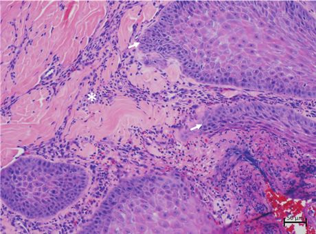
Page 5 of 6 Case Report
Ethical considerations Goldschmidt, M.H. & Goldschmidt, K.H., 2017, ‘Epithelial and melanocytic tumors of
the skin’, in D.J. Meuten (ed.), Tumors in domestic animals, 5th edn., pp. 88–141,
Blackwell Publishing, Ames, IA.
The owner of Lory Park Zoo and Owl Sanctuary gave written
Gunson, D.E., Klein, L.V. & Reid, C.F., 1978, ‘Gingival squamous cell carcinoma in a
consent for the surgery to be performed and the case to be Canadian lynx’, Journal of the American Veterinary Medical Association 173(9),
1228–1230.
written up. The Research Ethics Committee of the Faculty of
Hammond, G.M., Gordon, I.K., Theon, A.P. & Kent, M.S., 2007, ‘Evaluation of strontium
Veterinary Science at the University of Pretoria approved this Sr 90 for the treatment of superficial squamous cell carcinoma of the nasal
case report (REC089-20). planum in cats: 49 cases (1990–2006)’, Journal of the American Veterinary Medical
Association 231(5), 736–741. https://doi.org/10.2460/javma.231.5.736
Hauck, M.L., 2012, ‘Tumors of the skin and subcutaneous tissues’, in S. Withrow, D.
Vail & R. Page (eds.), Withrow and MacEwen’s small animal clinical oncology, 5th
Funding information edn., pp. 305–320, Elsevier/Saunders, St. Louis, MO.
L.v.d.W. was supported by the Wellcome Trust grant 20619. Joslin, J.O., Garner, M., Collins, D., Kamaka, E., Sinabaldi, K., Meleo, K. et al., 2000,
‘Viral papilloma and squamous cell carcinomas in snow leopards (Uncia uncia)’, in
Proceedings of the American association of zoo veterinarians (AAZV) and
International association for aquatic animal medicine (IAAAM) joint conference,
Data availability pp. 155–157, New Orleans, LA.
Kesdangsakonwut, S., Sanannu, S., Rungsipipat, A. & Banlunara, W., 2014, ‘Well-
Samples are stored at the University of Pretoria Pathology differentiated squamous cell carcinoma in a captive clouded leopard (Neofelis
nebulosa)’, Thai Journal of Veterinary Medicine 44(1), 153–157.
Department.
Kim, I.Y. & He, Y.-Y., 2014, ‘Ultraviolet radiation-induced non-melanoma skin cancer:
Regulation of DNA damage repair and inflammation’, Genes and Diseases 1(2),
188–198. https://doi.org/10.1016/j.gendis.2014.08.005
Disclaimer Miller, M.A., Nelson, S.L., Turk, J.R., Pace, L.W., Brown, T.P., Shaw, D.P. et al., 1991,
‘Cutaneous neoplasia in 340 cats’, Veterinary Pathology 28(5), 389–395. https://
The views and opinions expressed in this article are those of doi.org/10.1177/030098589102800506
the authors and do not necessarily reflect the official policy or Munday, J.S., 2014, ‘Papillomaviruses in felids’, The Veterinary Journal 199(3),
340–347. https://doi.org/10.1016/j.tvjl.2013.11.025
position of any affiliated agency of the authors.
Murphy, S., 2013, ‘Cutaneous squamous cell carcinoma in the cat: Current
understanding and treatment approaches’, Journal of Feline Medicine and Surgery
References 15(5), 401–407. https://doi.org/10.1177/1098612X13483238
Mwase, M., Mumba, C., Square D., Kawari S. & Madarme, H., 2013, ‘Cutaneous
squamous cell carcinoma presenting as a wound with discharging sinus tracts in
Almeida, E.M.P., Caraça, R.A., Adam, R.L., Souza, E.M., Metze, K. & Cintra, M.L., 2008,
‘Photodamage in feline skin: Clinical and histomorphometric analysis’, Veterinary a wild African lion (Panthera leo)’, Journal of Comparative Pathology 149,
Pathology 45(3), 327–335. https://doi.org/10.1354/vp.45-3-327 520–523.
Altamura, G., Eleni, C., Meoli, R., Cardeti, G., Friedrich, K.G. & Borzacchiello, G., 2011, Myburgh, E. & Nevill, E.M., 2003, ‘Review of blackfly (Diptera: Simuliidae) control in
‘Tongue squamous cell carcinoma in a European lynx (Lynx lynx): Papillomavirus South Africa’, Onderstepoort Journal of Veterinary Research 70(4), a295. https://
infection and histologic analysis’, Veterinary Sciences 5(1), 1–6. https://doi. doi.org/10.4102/ojvr.v70i4.295
org/10.3390/vetsci5010001 Napier, J.E., Lund, M.S., Armstrong, D.L. & McAloose, D., 2018, ‘A retrospective
Benson, K.E.N., Share, F., Shore, N. & Miami, N.W.S.T., 1991, ‘Study of elasto-gel pads study of morbidity and mortality in the North American Amur leopard (Panthera
used as surface bolus material high energy photon and electron therapy’, pardus orientalis) population in zoologic institution from 1992 to 2014’, Journal
International Journal of Radiation Oncology and Biology 22, 191–193. https://doi. of Zoo and Wildlife Medicine 49(1), 70–78. https://doi.org/10.1638/2017-
org/10.1016/0360-3016(92)90999-X 0019R2.1
Berlato, D., Murphy, S., Laberke, S. & Verganti, S., 2019, ‘Response, disease-free Owston, M.A., Ramsay, E.C. & Rotstein, D.S., 2008, ‘Neoplasia in felids at the
interval and overall survival of cats with nasal planum squamous cell carcinoma Knoxville Zoological gardens, 1979–2003’, Journal of Zoo and Wildlife Medicine
treated with a fractionated vs. a single-dose protocol of strontium plesiotherapy’, 39(4), 608–613. https://doi.org/10.1638/2008-068.1
Journal of Feline Medicine and Surgery 21(4), 306–313. https://doi.org/ Röwert-Huber, J., Patel, M.J., Forschner, T., Ulrich, C., Eberle, J., Kerl, H. et al., 2007,
10.1177/1098612X18773913 ‘Actinic keratosis is an early in situ squamous cell carcinoma: A proposal for
Boudreaux, B.B., LaRue, S.M., Rademacher, N., Neck, D., Grasperge, B., Wood, C. et al., reclassification’, British Journal of Dermatology 156(S3), 8–12. https://doi.
2019, ‘Treatment of leiomyosarcoma in a tiger (Panthera tigris) with stereotactic org/10.1111/j.1365-2133.2007.07860.x
radiotherapy’, Veterinary Radiology & Ultrasound 60(3), E33–E37. https://doi.
org/10.1111/vru.12720 Sherding, R.G., 1994, ‘Diseases of the skin’, in R.G. Sherding (ed.), The cat diseases and
clinical management, 2nd edn., pp. 1994–2046, Churchill Livingstone, London.
Cunha, S.C., Carvalho, L.A., Canary, P.C., Reisner, M., Corgozinho, K.B., Souza, H.A.
et al., 2010, ‘Radiation therapy for feline cutaneous squamous cell carcinoma Sladakovic, I., Burnum, A., Blas-Machado, U., Kelly, L.S., Garner, B.C., Holmes, S.P.
using a hypofractionated protocol’, Journal of Feline Medicine and Surgery 12(4), et al., 2016, ‘Mandibular squamous cell carcinoma in a Bobcat (Lynx rufus)’,
306–313. https://doi.org/10.1016/j.jfms.2009.10.005 Journal of Zoo and Wildlife Medicine 47(1), 370–373. https://doi.
org/10.1638/2015-0197.1
De Oliveira, A.R., Carvalho, T., Arenales, A. & De Lima Santos, R., 2018, ‘Mandibular
squamous cell carcinoma in a captive Siberian tiger (Panthera tigris altaica)’, Steeil, J.C., Schumacher, J., Baine, K., Ramsay, E.C., Sura, P., Hodshon, R. et al., 2013,
Brazilian Journal of Veterinary Pathology 11(3), 97–101. https://doi.org/10.24070/ ‘Diagnosis and treatment of a dermal malignant melanoma in an African lion
bjvp.1983-0246.v11i3p97-101 (Panthera leo)’, Journal of Zoo and Wildlife Medicine 44(3), 721–727. https://doi.
org/10.1638/2013-0023R2.1
D’Orazio, J., Jarrett, S., Amaro-Ortiz, A. & Scott, T., 2013, ‘UV radiation and the skin’,
International Journal of Molecular Sciences 14(6), 12222–12248. https://doi. Terio, K.A., McAloose, D. & Mitchell, E., 2018, ‘Chapter 10: Felidae’, in K.A. Terio, D.
org/10.3390/ijms140612222 McAloose & J.S. Leger (eds.), Pathology of wildlife and zoo animals, pp. 269–272,
Academic Press, London.
De Vos, J.P., Burm, A.G. & Focker, B.P., 2004, ‘Results from the treatment of advanced
stage squamous cell carcinoma of the nasal planum in cats, using a combination Theon, A.P., Madewell, B.R., Shearn, V.I. & Moulton, J.E., 1995, ‘Prognostic factors
of intralesional carboplatin and superficial radiotherapy: A pilot study’, Veterinary associated with radiotherapy of squamous cell carcinoma of the nasal plane in
and Comparative Oncology 2(2), 75–81. https://doi.org/10.1111/j.1476- cats’, Journal of the American Veterinary Medical Association 206(7), 991–996.
5810.2004.00040.x
Trivillin, V.A., Heber, E.M., Rao, M., De los Angeles Cantarelli, M., Itoiz, M.E., Nigg, D.W.
Gasymova, E., Meier, V., Guscetti, F., Cancedda, S., Roos, M. & Rohrer Bley, C., 2017, et al., 2008, ‘Boron neutron capture therapy (BNCT) for the treatment of
‘Retrospective clinical study on outcome in cats with nasal planum squamous cell spontaneous nasal planum squamous cell carcinoma in felines’, Radiation and
carcinoma treated with an accelerated radiation protocol’, BMC Veterinary Environmental Biophysics 47, 147–155. https://doi.org/10.1007/s00411-007-
Research 13, 86. https://doi.org/10.1186/s12917-017-1018-3 0138-8
Appendix start on the next page →
http://www.jsava.co.za Open Access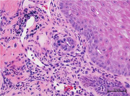
Page 6 of 6            Case Report

Appendix 1
TABLE 1-A1: Diagnostic laboratory report for the lion, showing haematology, biochemistry and blood gas electrolyte values at the two times blood was taken for analysis.
Test                                                         Units                       Reference range            Result (pre-therapy)     Result (post-therapy)
Haemoglobin (Hb)                                              g/L                              85–170                       150                      139
Red cell count                                               ×1012/L                          5.3–10.39                    9.40                      9.53
Haematocrit                                                    L/L                           0.250–0.528                   0.41                      0.43
Mean corpuscular volume                                        fL                             41.2–58.0                    43.8                      44.9
Mean corpuscular Hb                                            Pg                             14.1–18.4                    16.0                      14.6
Mean corpuscular Hb concentration                             g/dL                            28.8–39.0                    36.6                      32.5
Red cell distribution width                                    %                              15.1–25.6                    16.9                      16.3
White cell count                                             ×109/L                           5.4–18.5                     12.27                     14.16
Segmented neutrophil                                         ×109/L                          0.01–15.92                    9.82                      11.47
Band neutrophil                                              ×109/L                           0.00–1.80                    0.49                      0.00
Lymphocyte                                                   ×109/L                           0.00–4.72                    0.49                      1.98
Monocyte                                                     ×109/L                          0.074–0.944                   0.61                      0.28
Eosinophil                                                   ×109/L                          0.000–1.299                   0.86                      0.42
Basophil                                                     ×109/L                          0.000–0.070                   0.00                      0.00
Platelet count                                               ×109/L                           122–555                       312                      422
Parasite ID                                                     -                                 -                 None seen on smear        None seen on smear
Total serum protein                                           g/L                              53–83                       68.6                      64.7
Albumin                                                       g/L                               N/A                        33.9                      31.0
Globulin                                                      g/L                              21–57                       34.7                      33.7
A/G ratio                                                       -                              0.1–1.4                      1.0                       0.9
Alanine aminotransferase                                      U/L                              15–109                      72.0                      45.6
Alkaline phosphatase                                          U/L                               0–57                         6                         5
Glucose                                                      mmol/L                          0.39–12.90                     8.3                       NT
Serum inorganic phosphate                                    mmol/L                             N/A                        1.59                       NT
Cholesterol                                                  mmol/L                           2.12–6.64                     3.3                       NT
Urea nitrogen                                                mmol/L                           6.2–31.4                     17.6                      23.4
Creatinine                                                   umol/L                            41–326                       246                      184
Sodium                                                       mmol/L                           136–155                       147                      149
Potassium                                                    mmol/L                            3.1–6.0                     4.25                      4.74
Chloride                                                     mmol/L                            94–130                      116.0                      NT
Calcium                                                      mmol/L                            1.7–3.1                     2.42                       NT
Ionised calcium                                              mmol/L                           1.13–1.65                    1.19                       NT
Magnesium                                                    mmol/L                           0.37–1.19                    1.07                       NT
A/G, albumin/globulin; NT, not tested; ID, identification.

                                                                    http://www.jsava.co.za            Open Access
You can also read