The anti inflammation effect of strontium ranelate on rat chondrocytes with or without IL 1β in vitro
←
→
Page content transcription
If your browser does not render page correctly, please read the page content below
EXPERIMENTAL AND THERAPEUTIC MEDICINE 23: 208, 2022

                The anti‑inflammation effect of strontium ranelate
                on rat chondrocytes with or without IL‑1β in vitro
      HAO YU1,2, YAN LIU1,2, XIANGWEN YANG1,2, JIAJING HE1,2, QUN ZHONG1,2 and XIAOJING GUO1,2

               1
               Department of Prosthodontics, Shanghai Stomatological Hospital; 2Shanghai Key Laboratory of
          Craniomaxillofacial Development and Diseases, Fudan University, Huangpu, Shanghai 200001, P.R. China

                                    Received August 27, 2021; Accepted December 10, 2021

                                                   DOI: 10.3892/etm.2022.11131

Abstract. Temporomandibular joint osteoarthritis (TMJ‑OA)           Introduction
is a common disease with a high level of inflammation in
the joint micro‑environment and cartilage degradation.              Temporomandibular disease (TMD) is one of the most common
Anti‑inflammation and cartilage regeneration are the key            dental diseases and is characterized by temporomandibular
therapies for TMJ‑OA, but currently, there are no novel medi‑       joint (TMJ) pain, snapping and difficulty in opening the mouth,
cines or treatments that can control its pathogenic progression.    which seriously affect quality of life. There are several causes of
Strontium ranelate (SrR) is an anti‑osteoporosis drug and is        TMJ inflammation, including abnormal biomechanical stress,
now considered a promising anti‑OA drug, but the anti‑inflam‑       injury, or systematic disease, such as rheumatoid arthritis,
matory effect of SrR remains to be elucidated. In the present       which result in TMD (1). TM joint osteoarthritis (TMJ‑OA) is
study, the anti‑inflammatory effect of SrR in a normal or high      one of the most common cases of TMD and it is typically char‑
IL‑1β environment was observed. Cell viability under the treat‑     acterized by cartilage degradation and is very difficult to cure.
ment of SrR was tested using Cell Counting Kit‑8. Toluidine         At present, the main pathological changes of TMJ‑OA include
blue staining, immunofluorescence staining, hydroxyproline          an increase in inflammatory factors, chondrocyte degeneration
assay, PCR assay and western blotting were used to detect           and apoptosis, matrix collagen decomposition and abnormal
the expression of collagen (Col)II, proteoglycans (PG) and          remodeling of subchondral bone (2). Chondrocytes are the
aggrecan as a reflection of extracellular matrix synthesis          only cell component in cartilage tissue (3). When the balance
and MMP‑9,13 hydroxyproline was used as an inflamma‑                of the joint microenvironment is disturbed by factors such
tion indicator. IL‑1β of 10 ng/ml was added to the culture          as abnormal stress, ageing and genetic factors, inflammatory
medium as inflammation environment and the tests of those           factors such as IL‑1β, TNF‑α and nitric oxide (NO) increase (4),
biomarkers were done again. Then, the changes in β‑catenin          chondrocytes become irritated and secrete excessive MMPs,
were also studied by immunofluorescence staining, PCR assay         e.g. MMP‑13, aggrecanases (ADAMTS‑4, ‑5), nitric oxide
and western blotting to explore the possible involvement of         synthase (iNOS) and cyclooxygenase‑2 (COX‑2), which leads
the Wnt/β‑catenin pathway. The results showed a significant         to extracellular matrix (ECM) decomposition (5,6). Moreover,
inhibition of MMP‑9, MMP‑13, β‑catenin and promotion of             chondrocytes in an inflammatory state also secrete various
Col‑II, PG and aggrecan in normal chondrocytes. The pres‑           inflammatory factors, such as IL‑1β, TNF‑α, IL‑6 and IL‑8,
ence of IL‑1β markedly upregulated the expression of MMP‑9,         further leading to the aggravation of inflammation in the joint
MMP‑13 and β‑catenin while suppressing Col‑II and PG and            microenvironment (7). This vicious cycle of inflammation will
SrR partially reversed this trend. In conclusion, SrR decreased     eventually lead to cartilage destruction and the development
MMPs but promoted Col‑II, aggrecan and PG synthesis in              of OA (8).
rat chondrocytes with or without the presence of IL‑1β and              A previous study emphasizes that the altered joint
SrR attenuated the IL‑1β‑induced increase in β‑catenin, thus        mechanics that cause OA are addressed in first‑line therapy,
reducing the inflammatory reaction.                                 which stresses the rehabilitation of normal biomechanical
                                                                    and non‑inflammation microenvironments (9). Unfortunately,
                                                                    there are currently no pharmacological treatments or effec‑
                                                                    tive interventions that can alter the joint mechanics to halt
                                                                    or reverse the progression of OA in the long term. As OA
Correspondence to: Dr Xiaojing Guo, Department of Prosthodontics,
                                                                    involves a number of pathways and risk factors, personalized
Shanghai Stomatological Hospital, 356 East Beijing Road, Huangpu,
Shanghai 200001, P.R. China                                         therapy is the ultimate goal, which requires different targeted
E‑mail: guo_xiaojing@fudan.edu.cn                                   disease‑modifying OA drugs (DMOADs) for suitable thera‑
                                                                    peutic options (10). The pool of DMOADs is always in need
Key words: strontium ranelate, chondrocytes, anti‑inflammation,     of growth.
IL‑1β, β‑catenin                                                        Strontium ranelate (SrR) is an anti‑osteoporosis drug that
                                                                    has the dual effect of promoting bone formation and inhibiting
                                                                    bone resorption and has been recently considered a possible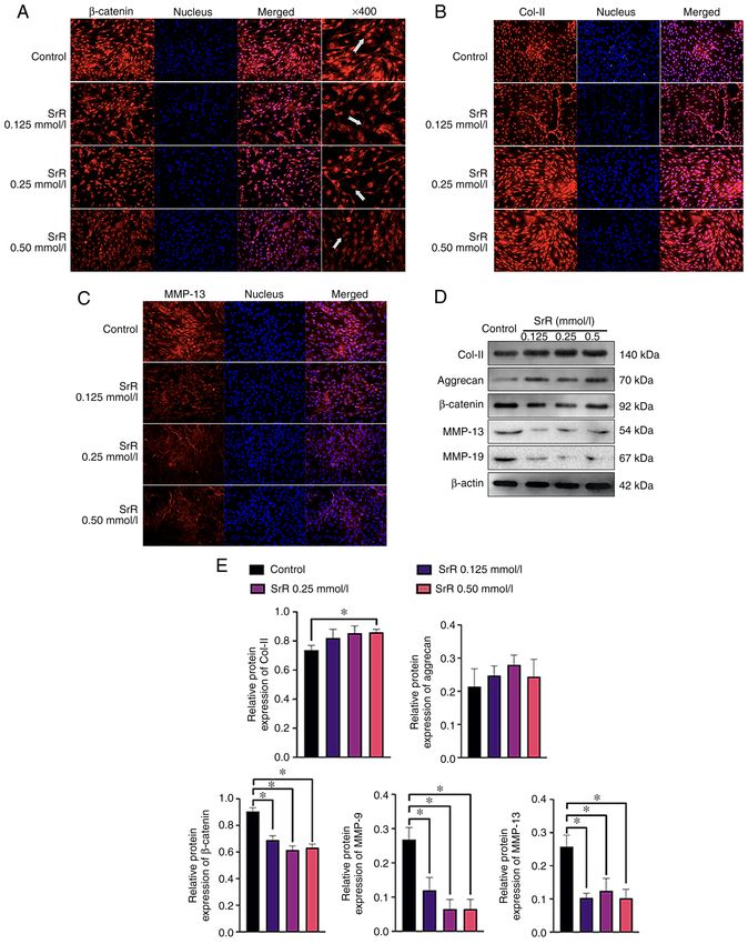
2                  YU et al: STRONTIUM RANELATE DECREASES THE INFLAMMATION OF RAT CHONDROCYTES

DMOAD (11). As a promising DMOAD, SrR has some special              pieces and incubated in HBSS (containing Ca 2+ and Mg2+)
advantages. First, SrR has a good tolerability and safety profile   with 200 U/ml type II collagenase (Gibco; Thermo Fisher
and is well tolerated by the majority of patients in long‑term      Scientific, Inc.; cat. no. 17101‑015) at 37˚C and 5% CO2 for
treatment (12). Second, SrR can modify subchondral bone             12 h. The cells were centrifuged at 300 x g for 5 min (room
turnover, thus indirectly modifying chondrocytes in cartilage       temperature), resuspended in high‑glucose DMEM (Gibco;
via factors released from bone (13,14). Third, in our previous      Thermo Fisher Scientific, Inc.) with 10% FBS (Gibco;
study, it was shown that SrR has a chondrogenic induction           Thermo Fisher Scientific, Inc.) and then subjected to routine
effect on bone mesenchymal stem cells (BMSCs) that promotes         cell culture procedures. Chondrocytes with 3‑5 passages
cartilage regeneration and suppresses cartilage degradation by      were used for the present study.
inhibiting the formation of MMPs in vitro and in vivo (15).             The present study was performed strictly in accordance
However, the anti‑inflammatory effect of SrR has not yet been       with the recommendations in the Guide for the Care and
fully elucidated.                                                   Use of Laboratory Animals of the National Institutes of
    Evidence of IL‑1β in progressing OA is well established         Health (20). All experiments were approved by the Animal
and is also an essential factor when simulating the OA              Research Committee of the Shanghai Stomatological Hospital
environment in vitro (6). Increased IL‑1β leads to abnormal         and Shanghai Research Center of Model Animal Organization
regulation of MMPs, induces chondrocytes to produce large           (IACUC no. 2020‑0010‑06).
amounts of NO, causes abnormal mitochondrial function and
leads to chondrocyte apoptosis and promotes chondrocytes to         Cell treatment with SrR and IL‑1β. Chondrocytes were
produce PEG‑2 and other inflammatory mediators, leading             treated with different concentrations of SrR and induced by
to degradation of PG and collagen (16). However, few studies        IL‑1β to simulate inflammation. As described in our previous
of the anti‑inflammation effect of SrR have been conducted.         study (15,21), 51.35 mg of SrR (MilliporeSigma) was dissolved
Alves et al (17) published research on antinociceptive effects      in 50 ml of culture medium to obtain a maximum soluble
of SrR; that orally taken SrR could reduce TNF‑ α levels in         concentration of 2.0 mmol/l. Then, the samples were diluted
periarticular tissues and trigeminal ganglion, but did not          to different concentrations of 1.0, 0.5, 0.25 and 0.125 mmol/l.
decrease IL‑1β expression, nor inhibit HO‑1 pathway. As the             IL‑1β‑treated chondrocytes or cartilage tissues have been
gastrointestinal barrier would block ranelate acid outside the      widely adopted as in vitro models to study OA (6). The recom‑
blood serum, it was not possible to predict the same result in an   binant rat IL‑1β protein was purchased from BioVision, Inc.
in vitro study. The in vitro study of Henrotin et al (18) showed    (cat. no. 4130‑10) and a final concentration of 10 ng/ml was
that SrR had a significant inhibitory effect on MMPs and simu‑      used for the present study.
lated PG synthesis even under an inflammatory environment,
but that study did not advance mechanistic investigations.          Chondrocytes cell proliferation assay. The proliferation
Above all, whether SrR could suppress the inflammation              of chondrocytes under different concentrations of SrR was
level of IL‑1β, MMPs and stabilize ECM proteins directly            detected by CCK‑8 assay. Briefly, the chondrocytes were inoc‑
on chondrocytes and the underlying mechanism remain to be           ulated in 96‑well plates (initial cell density of 3x10³ cells/well)
elucidated.                                                         and treated with SrR (0, 0.125, 0.25, 0.5, 1.0 and 2.0 mmol/l)
    The treatment of TMJ‑OA involves the regeneration of            for 1, 3, 5 and 7 days. CCK‑8 (10 µl) solution in 5% CO2 was
cartilage, which, using the method of tissue engineering,           added to each plate and incubated at 37˚C for 1 h under dark
requires plenty of cells. The chondrocytes from healthy             conditions. Optical density (OD) values reflecting cell viability
TMJ are limited and it is reasonable to obtain chondrocytes         were measured by a microplate reader (BioTek Instruments,
from other site of cartilage (19). The present study employed       Inc.; ELX800) at a wavelength of 450 nm.
the chondrocytes from rat femurs and aimed to investigate
whether SrR exerted a protective effect by reducing rat chon‑       Toluidine blue staining. The chondrocytes were seeded on
drocyte inflammation caused by IL‑1β. To explore its effect on      24‑well plates at an initial density of 1x10 4 cells/well with
chondrocyte cell viability, ECM matrix synthesis, the expres‑       DMEM added with SrR at 0.125, 0.25 and 0.5 mmol/l for
sion of cartilage‑forming or inflammation genes and proteins        14 days induction, while initial cell density of 1x10⁵ cells/well
and the involvement of the molecular mechanism of SrR in            for the cultural medium with IL‑1 and 0.25 mmol/l SrR, and
Wnt/β‑catenin signaling pathways was examined.                      for 3 days, at 37˚C. Then, the chondrocytes were fixed with 4%
                                                                    paraformaldehyde at 4˚C for 30 min and stained with a tolu‑
Materials and methods                                               idine blue solution at room temperature for 30 min (Beijing
                                                                    Solarbio Science & Technology Co., Ltd.) following 3 or
Isolation and culture of rat chondrocytes. Rat chondro‑             14 days of induction. Images were captured with an inverted
cytes cells were isolated from Sprague‑Dawley (SD) rats.            light microscope (Leica DMI 3000B; Leica Microsystems
Briefly, 12 male SD rats of 4‑6 weeks (weight 100‑120 g)            GmbH) at x200 magnification.
were purchased from Vital River Laboratories. Rats were
sacrificed on receipt by sodium pentobarbital at a dosage of        Hydroxyproline (Hyp) assay. An Hyp assay revealed the
150 mg/kg intraperitoneal injection and followed with strict        degradation condition of collagen. Hyp is the characteristic
disinfection, the cartilage tissue on the top of the metaphysis     amino acid that is composed of collagen and does not exist in
of the knee side was cut with a knife and washed twice              other human tissues (22). A hydroxyproline test kit (Abcam;
with PBS and twice with Hanks' Balanced Salt Solution               cat. no. ab222941) was used in accordance with the manu‑
(HBSS). After washing, cartilage tissues were cut into small        facturer's instructions. Cell culture supernatant, ddH 2O and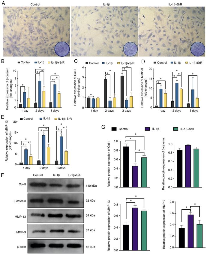
EXPERIMENTAL AND THERAPEUTIC MEDICINE 23: 208, 2022                                             3

Table I. Primer sequences used for the rat chondrocytes.

Gene                                              Forward                                                       Reverse

Col‑II                               ATCGCCACGGTCCTACAATG                                          GGCCCTAATTTTCGGGCATC
Aggrecan                             CAAGTCCCTGACAGACACCC                                          GTCCACCCCTCCTCACATTG
MMP‑9                                GATCCCCAGAGCGTTACTCG                                          GTTGTGGAAACTCACACGCC
MMP‑13                               TGCTGCATACGAGCATCCAT                                          TGTCCTCAAAGTGAACCGCA
β‑catenin                            ACTCCAGGAATGAAGGCGTG                                          GAACTGGTCAGCTCAACCGA
GAPDH                                AGTGCCAGCCTCGTCTCATA                                          GATGGTGATGGGTTTCCCGT

standard protein samples were prepared and an equal volume            10 µl 2X SuperReal PreMix Plus (cat. no. FP205; KR118),
of NaOH was added, evaporated, cooled and neutralized                 1.2 µl forward/reverse primer, 2 µl cDNA template and 6.8 µl
with an equal amount of HCl. Then, the supernatant was                RNase‑free ddH 2O. The PCR cycling conditions were as
centrifuged at 10,000 x g for 5 min (room temperature) and            follow: Initial denaturation at 95˚C for 15 min, followed by
collected into a new tube. An oxidation reagent was added             40 cycles of denaturation at 95˚C for 10 sec, annealing at 60˚C
and the solution was incubated at room temperature for                for 20 sec and extension at 72˚C for 20 sec. All experiments
20 min. A developer was added and the solution was incu‑              were repeated three times and the relative fold‑change of gene
bated at 37˚C for 5 min. DMAB concentrate was added and               expression (2‑ΔΔCq method) was calculated by the Ct value (23)
the solution was incubated at 65˚C for 45 min. The OD value           (LightCycler 96 PCR system; Roche Diagnostics GmbH). The
of each group was measured by microplate reader at 560 nm             primer sequences are in Table I. All tests were performed in
wavelength. The test was repeated for three times and the             triplicate.
hydroxyproline concentration was calculated by the standard
curve method.                                                         Western blotting assay. The chondrocytes were cultured in
                                                                      6‑well plates at a density of 5x10⁴ cells/well and treated with
Immunofluorescence staining assay. The chondrocytes were              or without SrR at 0.125, 0.25 and 0.5 mmol/l for 14 days, an
cultured in DMEM with or without IL‑1 and SrR at 0.125, 0.25          initial density of 5x10⁵ cells/well treated with or without IL‑1
and 0.5 mmol/l for 14 days at 37˚C. First, the cells were rinsed      and 0.25 mmol/l SrR for 3 days at 37˚C. After rinsing with PBS
with PBS and fixed at 4˚C with 4% paraformaldehyde for                several times, total proteins were collected by RIPA buffer
15 min. Second, the cells were treated with PBST for 10 min           (Beyotime Institute of Biotechnology) on ice and measured by
under room temperature for permeability and rinsed with PBS           a BCA protein assay kit (Beyotime Institute of Biotechnology).
several times (5 min; room temperature). Then, non‑specific           Equivalent amounts of protein (20 µg/lane) were transferred
interactions were blocked with donkey serum and incubated             to PCDF membranes on 12% SDS‑PAGE gels. Following
with primary antibodies against β ‑catenin (Abcam; cat.               blocking with 5% skimmed milk at room temperature for
no. ab16051), collagen (Col)‑II (ProteinTech Group, Inc.; cat.        1 h, the membrane was exposed to primary antibodies at
no. 15943‑1‑AP) and MMP‑13 (Novus Biologicals, LLC; cat.              4˚C overnight, including Col‑II (1:500; cat. no. 28459‑1‑AP;
no. NBP2‑17310). Cyanine 3‑conjugated donkey anti‑rabbit              ProteinTech Group, Inc.), aggrecan (1:500; cat. no. 13880‑1;
IgG (cat. no. GB21403; Wuhan Servicebio Technology Co.,               ProteinTech Group, Inc.), β‑catenin (1:1,000; cat. no. ab16051;
Ltd.) was used as a secondary antibody and nuclei were                Abcam), MMP‑9 (1:500; cat. no. 10375‑2; ProteinTech
stained with DAPI (MilliporeSigma; cat. no. D9642). Images            Group, Inc.), MMP‑13 (1:500; cat. no. Nbp2‑17310; Novus
were captured by a fluorescence microscope at x200 magni‑             Biologicals) and β‑actin (1:1,000; cat. no. 4970; Cell Signaling
fication.                                                             Technology, Inc.). Then, PBST was washed and incubated with
                                                                      HRP‑labelled goat anti‑rabbit IgG (1:1,000; cat. no. A0208;
Reverse transcription‑quantitative (RT‑q) PCR. The                    Beyotime Institute of Biotechnology) or HRP‑labelled goat
chondrocytes were cultured in 6‑well plates at a density of           anti‑mouse IgG (1:1,000; cat. no. A0216; Beyotime Institute
5x10⁴ cells/well with DMEM added with SrR at 0.125, 0.25 and          of Biotechnology) at room temperature for 2 h. The membrane
0.5 mmol/l for 14 days induction and total RNA extracted at           was thoroughly cleaned and visualized using ECL reagents
days 1, 7 and 14. With initial cell density of 5x10⁵ cells/well for   (Pierce; Thermo Fisher Scientific, Inc.). The bands were
the cultural medium with IL‑1 and 0.25 mmol/l SrR the total           imaged and measured by the Quantity One Analysis system
RNA was extracted at days 1, 2 and 3 using TRIzol® reagent            (version 4.6.6.; Bio‑Rad Laboratories, Inc.).
(Thermo Fisher Scientific, Inc.) according to the manufac‑
turer's protocol. RNA purity and quantification were tested           Statistical analysis. The results are shown as the mean ± stan‑
by NanoDrop One Microvolume Spectrophotometer (Thermo                 dard deviation of three repeated experiments and were analyzed
Fisher Scientific, Inc.). The RNA was used as a template              by SPSS 26.0 software (IBM Corp.). One‑way analysis of vari‑
for cDNA reverse by Tiangen FastKing cDNA Dispelling                  ance (ANOVA) with a subsequent post hoc Tukey's test was
RT SuperMix (cat. no. KR118; Tiangen Biotech Co., Ltd.)               used to determine the statistical significance of the differences
following the manufacturer's instructions. The resultant cDNA         among groups. P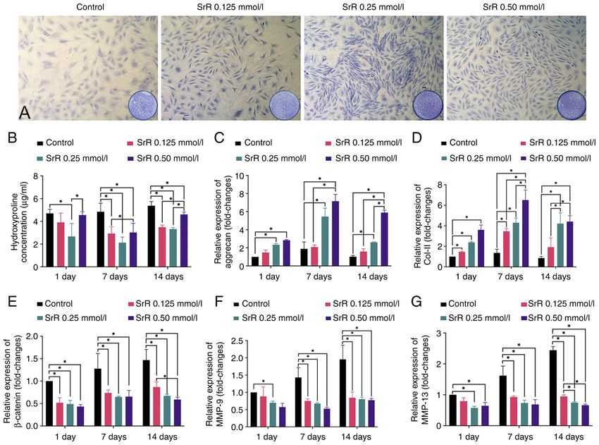
4                      YU et al: STRONTIUM RANELATE DECREASES THE INFLAMMATION OF RAT CHONDROCYTES

                                                                                14 days, which was followed by immunofluorescence staining
                                                                                to visualize the location and concentration of β‑catenin, Col‑II
                                                                                and MMP‑13 protein. The results showed a clear dose‑depen‑
                                                                                dent effect of SrR on promoting chondrocyte function.
                                                                                β‑catenin was highly expressed and located inside the nucleus
                                                                                in the control group, while SrR treatment resulted in lighter
                                                                                staining and a significantly lower expression of β‑catenin in the
                                                                                0.50 mmol/l treatment group (Fig. 3A) compared to that of the
                                                                                control. The same situation could be seen in Fig. 3C: MMP‑13
                                                                                was positively stained in cytoplasm around the nucleus and its
                                                                                expression was lower in the higher concentration group. Col‑II
Figure 1. Effect of SrR on rat chondrocytes viability. The OD values of every   located in cytoplasm and extracellular area and had the opposite
group at day 1,3,5 and 7 are presented and no significant differences were
                                                                                expression trend: the control group had the lowest expression
observed among the control, SrR 0.125, 0.25 and 0.5 mmol/l groups, while
the OD values of the SrR 2.0 mmol/l at day 3, 5 and 7 and SrR 1.0 mmol/l        and a higher concentration of SrR resulted in higher expression
at day 5 and 7 groups were significantly lower compared with the control        (Fig. 3B). Fig. 3D presents the WB results, which provided a
(*P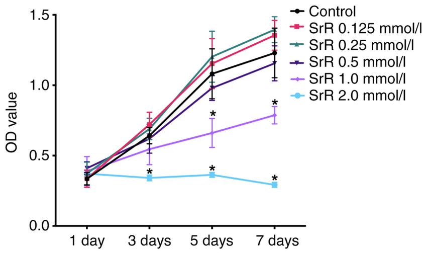
EXPERIMENTAL AND THERAPEUTIC MEDICINE 23: 208, 2022                                                                5

Figure 2. Toluidine blue staining. (A) Hyp concentration and (B) relative gene expression. Relative gene expression of (C) aggrecan, (D) Col‑II, (E) β‑catenin,
(F) MMP‑9 (G) and MMP‑13 of rat chondrocytes treated with different concentrations of SrR. The images of 2A were captured under x200 magnification.
*
 P
6                     YU et al: STRONTIUM RANELATE DECREASES THE INFLAMMATION OF RAT CHONDROCYTES

Figure 3. Immunofluorescence staining assay of (A) β‑catenin, (B) Col‑II and (C) MMP‑13. (D) Western blot assay of Col‑II, aggrecan, β‑catenin, MMP‑13,
MMP‑9 and β‑actin and (E) relative protein expression. The images were captured under x200 magnification, save for the right hand column of (A). Arrows
indicate the position of β‑catenin. *P
EXPERIMENTAL AND THERAPEUTIC MEDICINE 23: 208, 2022                                                             7

Figure 4. Chondrocytes respond to IL‑1β and SrR. (A) Toluidine blue staining and (B), relative gene expression of β ‑catenin. Relative gene expression of
(C) Col‑II, (D) MMP‑9 and (E) MMP‑13. (F) Western blotting analysis and (G) relative protein expression of chondrocytes treated with 10 ng/l IL‑1β and 0 or
0.50 mmol/l SrR for three days. The images were captured under x200 magnification. *P
8                      YU et al: STRONTIUM RANELATE DECREASES THE INFLAMMATION OF RAT CHONDROCYTES

Figure 5. Immunofluorescence staining assay of (A) β ‑catenin, (B) Col‑II and (C) MMP‑13 following three days of induction with IL‑1β and SrR. Arrow
showed the position of β‑catenin. The images were captured under x200 magnification, save for the right hand column of (A). Arrows indicate the position of
β‑catenin. Col, collagen; SrR, strontium ranelate.

canonical Wnt signaling by upregulating Wnt ligands (40).                      TCF/lEF induces the expression of MMP‑3 and MMP‑13,
Subsequently, the transcription complex of β ‑catenin with                     leading to cartilage destruction (41). On the other hand,
EXPERIMENTAL AND THERAPEUTIC MEDICINE 23: 208, 2022                                                  9

activation of the Wnt/ β ‑catenin pathway may lead to                XG performed the study design, statistical analysis and wrote
abnormal osteogenesis but depress chondrogenesis (42). The           the manuscript. XG and QZ confirm the authenticity of all the
reciprocal inhibitory effect between β ‑catenin and Sox‑9 was        raw data. All authors read and approved the final manuscript.
observed by Akiyama et al (43), in which Sox‑9 represses
β ‑catenin/Tcf/lef complex activities and shows an inhibitory        Ethics approval and consent to participant
effect on β ‑catenin when the cells were in the chondrogenic
differentiation trend but a controversial trend when osteo‑          All experiments involving animals were approved by the
genesis was dominant. The upregulation of β ‑catenin led to          Animal Research Committee of the Shanghai Stomatological
the inhibition of Col‑II and PG synthesis. Taken together,           Hospital and Shanghai Research Center of Model Animal
cartilage degradation is correlated to the promotion of              Organization (IACUC No. 2020‑0010‑06).
β ‑catenin activity. The present study showed that SrR could
inhibit β ‑catenin synthesis and accumulation. In normal             Patient consent for publication
chondrocytes, β ‑catenin and MMP synthesis decreased with
increasing SrR concentration (≤0.50 mmol/l). The addition            Not applicable.
of IL‑1β significantly activated β ‑catenin expression and
SrR attenuated the increase in β ‑catenin induced by IL‑1β.          Competing interests
However, further studies with more bio‑markers, including
in vivo experiments, are required to further determine               The authors declare that they have no competing interests.
the mechanism and confirm the role of β ‑catenin in the
anti‑inflammatory effect of SrR.                                     References
    SrR has specific characteristics, such as a regular effect
on the RANK/RANKL/OPG system that promotes bone                       1. Joseph R, Rahena A, Hassan N, Glen H, James W and Soichiro I:
formation and inhibits bone resorption (44), increases the               Epidemiology of temporomandibular disorder in the general
                                                                         population: A systematic review. Adv Dent Oral Health 10:
vascularization of new bone tissue (21), encourages chondro‑             555787, 2019.
genesis in cartilage tissue (35) and reduces the expression of        2. Wang XD, Zhang JN, Gan YH and Zhou YH: Current under‑
inflammatory factors. All these results made SrR a promising             standing of pathogenesis and treatment of TMJ osteoarthritis.
drug for DMOADs. However, more reliable evidence, espe‑                  J Dent Res 94: 666‑673, 2015.
                                                                      3. Charlier E, Deroyer C, Ciregia F, Malaise O, Neuville S, Plener Z,
cially well‑designed clinical trials, is needed to confirm the           Malaise M and de Seny D: Chondrocyte dedifferentiation and
anti‑OA effects.                                                         osteoarthritis (OA). Biochem Pharmacol 165: 49‑65, 2019.
    In conclusion, SrR decreased MMPs but promoted Col‑II,            4. Wojdasiewicz P, Poniatowski ŁA and Szukiewicz D: The role of
                                                                         inflammatory and anti‑inflammatory cytokines in the pathogen‑
aggrecan and PG synthesis in rat chondrocytes with or without            esis of osteoarthritis. Mediators Inflamm 2014: 561459, 2014.
the presence of IL‑1β and SrR attenuated the increase in β‑catenin    5. Loeser RF: Molecular mechanisms of cartilage destruction:
induced by IL‑1β, thus reducing the inflammatory reaction.               Mechanics, inflammatory mediators, and aging collide. Arthritis
                                                                         Rheum 54: 1357‑1360, 2006.
                                                                      6. Johnson CI, Argyle DJ and Clements DN: In vitro models for the
Acknowledgements                                                         study of osteoarthritis. Vet J 209: 40‑49, 2016.
                                                                      7. Samavedi S, Diaz‑Rodriguez P, Erndt‑Marino JD and Hahn MS:
                                                                         A three‑dimensional chondrocyte‑macrophage coculture system
The authors would like to thank Dr Xinxin Han and Dr                     to probe inflammation in experimental osteoarthritis. Tissue Eng
Shangfeng Liu (Shanghai Key Laboratory of Craniomaxillofacial            Part A 23: 101‑114, 2017.
Development and Diseases, Fudan University, Shanghai, China),         8. Gu YT, Chen J, Meng ZL, Ge WY, Bian YY, Cheng SW, Xing CK,
                                                                         Yao JL, Fu J and Peng L: Research progress on osteoarthritis
and Mr. Xiaolong Feng and Mr. Longyu Li (Shanghai Research               treatment mechanisms. Biomed Pharmacother 93: 1246‑1252,
Centre of Model Organisms, Shanghai, China) for the assistance           2017.
and execution of the experiments.                                     9. Xia B, Chen D, Zhang J, Hu S, Jin H and Tong P: Osteoarthritis
                                                                         pathogenesis: A review of molecular mechanisms. Calcif Tissue
                                                                         Int 95: 495‑505, 2014.
Funding                                                              10. Grässel S and Muschter D: Recent advances in the treatment of
                                                                         osteoarthritis. F1000Res 9: 325, 2020.
                                                                     11. Pelletier JP, Roubille C, Raynauld JP, Abram F, Dorais M,
The authors acknowledge the financial support from the                   Delorme P and Martel‑Pelletier J: Disease‑modifying effect of
Shanghai Commission of Science and Technology (grant                     strontium ranelate in a subset of patients from the phase III knee
no. 19YF1442400).                                                        osteoarthritis study SEKOIA using quantitative MRI: Reduction
                                                                         in bone marrow lesions protects against cartilage loss. Ann
                                                                         Rheum Dis 74: 422‑429, 2015.
Availability of data and materials                                   12. Tenti S, Cheleschi S, Guidelli GM, Galeazzi M, Fioravanti A:
                                                                         What about strontium ranelate in osteoarthritis? Doubts and
                                                                         securities. Mod Rheumatol 24: 881‑884, 2014.
The datasets used and/or analyzed during the current study are       13. Tat SK, Pelletier JP, Mineau F, Caron J and Martel‑Pelletier J:
available from the corresponding author on reasonable request.           Strontium ranelate inhibits key factors affecting bone remodeling
                                                                         in human osteoarthritic subchondral bone osteoblasts. Bone 49:
                                                                         559‑567, 2011.
Authors' contributions                                               14. Yu DG, Ding HF, Mao YQ, Liu M, Yu B, Zhao X, Wang XQ,
                                                                         Li Y, Liu GW, Nie SB, et al: Strontium ranelate reduces cartilage
HY performed most of the cell molecular biological tests. YL             degeneration and subchondral bone remodeling in rat osteoar‑
                                                                         thritis model. Acta Pharmacol Sin 34: 393‑402, 2013.
and XY helped HY with the primary chondrocytes cell isola‑           15. Yu H, Liu Y, Yang X, He J, Zhang F, Zhong Q and Guo X:
tion and culture. JH performed the statistical analysis. QZ was          Strontium ranelate promotes chondrogenesis through inhibition
responsible for the study design and revised the manuscript.             of the Wnt/β‑catenin pathway. Stem Cell Res Ther 12: 296, 2021.
10 YU et al: STRONTIUM RANELATE DECREASES THE INFLAMMATION OF RAT CHONDROCYTES
16. Sun Y, Zhou L, Lv D, Liu H, He T and Wang X: Poly(ADP‑ribose) 32. Pelletier JP, Kapoor M, Fahmi H, Lajeunesse D, Blesius A,
polymerase 1 inhibition prevents interleukin‑1β‑induced inflam‑ Maillet J and Martel‑Pelletier J: Strontium ranelate reduces the
mation in human osteoarthritic chondrocytes. Acta Biochim progression of experimental dog osteoarthritis by inhibiting
Biophys Sin (Shanghai) 47: 422‑430, 2015. the expression of key proteases in cartilage and of IL‑1β in the
17. Alves SM, Abreu SC, Lemos JC, Gomes FI, Alves SM, synovium. Ann Rheum Dis 72: 250‑257, 2013.
do Val DR, Freitas RS, Pereira KM, de Paulo Teixeira Pinto V, de 33. Lefebvre V, Behringer RR and de Crombrugghe B: L‑Sox5, Sox6
Castro Brito GA, et al: Anti‑inflammatory and anti‑nociceptive and Sox9 control essential steps of the chondrocyte differen‑
effects of strontium ranelate on the zymosan‑induced temporo‑ tiation pathway. Osteoarthritis Cartilage 9 (Suppl A): S69‑S75,
mandibular joint inflammatory hypernociception in rats depend 2001.
on TNF‑α inhibition. Pharmacol Rep 69: 764‑772, 2017. 34. Oh CD, Lu Y, Liang S, Mor i‑A k iyama Y, Chen D,
18. Henrotin Y, Labasse A, Zheng SX, Galais P, Tsouderos Y, de Crombrugghe B and Yasuda H: SOX9 regulates multiple
Crielaard JM and Reginster JY: Strontium ranelate increases genes in chondrocytes, including genes encoding ECM proteins,
cartilage matrix formation. Bone Miner Res 16: 299‑308, 2001. ECM modification enzymes, receptors and transporters. PLoS
19. Wang L, Lazebnik M and Detamore MS: Hyaline cartilage One 9: e107577, 2014.
cells outperform mandibular condylar cartilage cells in a TMJ 35. Kim HJ and Im GI: Electroporation‑mediated transfer of
fibrocartilage tissue engineering application. Osteoarthritis SOX trio genes (SOX‑5, SOX‑6, and SOX‑9) to enhance the
Cartilage 17: 346‑353, 2009. chondrogenesis of mesenchymal stem cells. Stem Cells Dev 20:
20. National Research Council. Guide for the care and use of labo‑ 2103‑2114, 2011.
ratory animals: Eighth edition. Washington, DC: The National 36. Rodrigues TA, Freire AO, Bonfim BF, Cartágenes MS and
Academies Press, 2011. https://doi.org/10.17226/12910. Garcia JB: Strontium ranelate as a possible disease‑modifying
21. Guo X, Wei S, Lu M, Shao Z, Lu J, Xia L, Lin K and Zou D: osteoarthritis drug: A systematic review. Braz J Med Biol Res 51:
Dose‑dependent effects of strontium ranelate on ovariectomy rat e7440, 2018.
bone marrow mesenchymal stem cells and human umbilical vein 37. Sassi N, Laadhar L, Allouche M, Achek A, Kallel‑Sellami M,
endothelial cells. Int J Biol Sci 12: 1511‑1522, 2016. Makni S and Sellami S: WNT signaling and chondrocytes:
22. da Silva CM, Spinelli E and Rodrigues SV: Fast and sensitive From cell fate determination to osteoarthritis physiopathology.
collagen quantification by alkaline hydrolysis/hydroxyproline J Recept Signal Transduct Res 34: 73‑80, 2014.
assay. Food Chem 173: 619‑623, 2015. 38. Xia H, Cao D, Yang F, Yang W, Li W, Liu P, Wang S and Yang F:
23. Livak KJ and Schmittgen TD: Analysis of relative gene expres‑ Jiawei Yanghe decoction ameliorates cartilage degradation
sion data using real‑time quantitative PCR and the 2(‑Delta Delta in vitro and vivo via Wnt/β‑catenin signaling pathway. Biomed
C(T)) method. Methods 25: 402‑408, 2001. Pharmacother 122: 109708, 2020.
39. Ma B, van Blitterswijk CA and Karperien M: A Wnt/β‑catenin
24. Aimaiti A, Maimaitiyiming A, Boyong X, Aji K, Li C and Cui L: negative feedback loop inhibits interleukin‑1‑induced matrix
Low‑dose strontium stimulates osteogenesis but high‑dose doses metalloproteinase expression in human articular chondrocytes.
cause apoptosis in human adipose‑derived stem cells via regula‑ Arthritis Rheum 64: 2589‑2600, 2012.
tion of the ERK1/2 signaling pathway. Stem Cell Res Ther 8: 282, 40. Yoshida Y, Yamasaki S, Oi K, Kuranobu T, Nojima T, Miyaki S,
2017. Ida H and Sugiyama E: IL‑1β enhances Wnt signal by inhibiting
25. Mao Z, Fang Z, Yang Y, Chen X, Wang Y, Kang J, Qu X, Yuan W DKK1. Inflammation 41: 1945‑1954, 2018.
and Dai K: Strontium ranelate‑loaded PLGA porous micro‑ 41. Yun K and Im SH: Transcriptional regulation of MMP13 by
spheres enhancing the osteogenesis of MC3T3‑E1 cells. RSC Lef1 in chondrocytes. Biochem Biophys Res Commun 364:
Adv 7: 24607‑24615, 2017. 1009‑1014, 2007.
26. Pilmane M, Salma‑Ancane K, Loca D, Locs J and Berzina‑ 42. Zhu M, Tang D, Wu Q, Hao S, Chen M, Xie C, Rosier RN,
Cimdina L: Strontium and strontium ranelate: Historical review O'Keefe RJ, Zuscik M and Chen D: Activation of beta‑catenin
of some of their functions. Mater Sci Eng C Mater Biol Appl 78: signaling in articular chondrocytes leads to osteoarthritis‑like
1222‑1230, 2017. phenotype in adult beta‑catenin conditional activation mice.
27. Deepthi S, Abdul Gafoor AA, Sivashanmugam A, Nair SV and J Bone Miner Res 24: 12‑21, 2009.
Jayakumar R: Nanostrontium ranelate incorporated injectable 43. Akiyama H, Lyons JP, Mori‑Akiyama Y, Yang X, Zhang R,
hydrogel enhanced matrix production supporting chondrogenesis Zhang Z, Deng JM, Taketo MM, Nakamura T, Behringer RR, et al:
in vitro. J Mater Chem B 4: 4092‑4103, 2016. Interactions between Sox9 and beta‑catenin control chondrocyte
28. Pan FY, Li ZM, Liu XW, Luo Y, Ma Z, Feng SX and Xu N: Effect differentiation. Genes Dev 18: 1072‑1087, 2004.
of strontium ranelate on rabbits with steroid‑induced osteone‑ 44. Han W, Fan S, Bai X and Ding C: Strontium ranelate, a promising
crosis of femoral head through TGF‑β1/BMP2 pathway. Eur Rev disease modifying osteoarthritis drug. Expert Opin Investig
Med Pharmacol Sci 24: 1000‑1006, 2020. Drugs 26: 375‑380, 2017.
29. Jackson A and Gu W: Transport properties of cartilaginous
tissues. Curr Rheumatol Rev 5: 40, 2009. This work is licensed under a Creative Commons
30. Troeberg L and Nagase H: Proteases involved in cartilage matrix Attribution-NonCommercial-NoDerivatives 4.0
degradation in osteoarthritis. Biochim Biophys Acta 1824: International (CC BY-NC-ND 4.0) License.
133‑145, 2012.
31. Liu‑Bryan R and Terkeltaub R: Emerging regulators of the
inflammatory process in osteoarthritis. Nat Rev Rheumatol 11:
35‑44, 2015.You can also read