The combined role of NT-proBNP and LV-GLS in the detection of early subtle chemotherapy-induced cardiotoxicity in breast cancer female patients
←
→
Page content transcription
If your browser does not render page correctly, please read the page content below
Sulaiman et al. The Egyptian Heart Journal
https://doi.org/10.1186/s43044-021-00142-z
                                                      (2021) 73:20
                                                                                                                                        The Egyptian Heart
                                                                                                                                                   Journal

 RESEARCH                                                                                                                                            Open Access

The combined role of NT-proBNP and
LV-GLS in the detection of early subtle
chemotherapy-induced cardiotoxicity in
breast cancer female patients
Laila Sulaiman1, Dina Hesham2, Magdy Abdel Hamid1 and Ghada Youssef1*

  Abstract
  Background: Chemotherapeutic agents have many side effects; among them is cardiotoxicity. Ejection fraction fails
  to detect the subtle alterations of left ventricular (LV) function; that is why there is a need for a more sensitive tool.
  The aim is to detect subclinical LV systolic dysfunction after chemotherapeutic treatment, using NT-BNP plasma
  level as well as speckle tracking echo-global longitudinal strain (STE-GLS). Seventy-four asymptomatic, non-
  metastasizing breast cancer female patients without risk factors were included. They were assessed before and 6
  weeks after taking their first chemotherapeutic session. Assessment included clinical characteristics, conventional
  two-dimensional (2D) and three-dimensional (3D) echocardiography, and 2D STE-GLS. Blood samples for NT-BNP
  plasma level were collected on both visits and were later analyzed using a Sandwich ELISA technique.
  Results: The median NT-proBNP almost doubled after 6 weeks of chemotherapy (73.50 vs 34.4 pg/L, p value <
  0.001). Only two patients showed significant reduction of LVEF >10% to less 15%) and
  all of them had a relatively higher NT-proBNP. A 2.2 relative elevation of the NT-proBNP was able to define a
  relative reduction of LV-GLS >15% by a 100% sensitivity and 81.8% specificity.
  Conclusion: The relative reduction of LV-GLS and the relative elevation of NT-proBNP were successful in defining
  subclinical, subtle chemotherapy-induced cardiotoxicity after 6 weeks of the first chemotherapeutic agent
  administration.
  Keywords: Cardiotoxicity, Chemotherapy, Echocardiography, Brain Natriuretic peptide

Background                                                                                In Egypt, the incidence of breast cancer among fe-
Breast cancer is a major public health and economical                                   males/100,000 population of individual cancer in Lower,
issue; that is why research on new therapies, and moni-                                 Middle, and Upper Egypt were 33.22%, 26.84%, and
toring their safety use, should be a priority. Globally,                                38.72% respectively [3]. The technical advances in strat-
breast cancer is the most commonly diagnosed cancer in                                  egies of early detection and therapies for cancer and can-
women, and it is the fifth leading cause of cancer death                                cer survival have been significantly improved and cancer
[1, 2].                                                                                 recurrence has been significantly reduced in the recent
                                                                                        years. Despite this improvement in cancer therapy, how-
                                                                                        ever, several treatment-related adverse effects have
* Correspondence: ghadayoussef@kasralainy.edu.eg                                        caused serious issues for cancer survivors [4]. It is
1
 Cardiology Department, Faculty of Medicine, Cairo University, Cairo, Egypt
Full list of author information is available at the end of the article

                                        © The Author(s). 2021 Open Access This article is licensed under a Creative Commons Attribution 4.0 International License,
                                        which permits use, sharing, adaptation, distribution and reproduction in any medium or format, as long as you give
                                        appropriate credit to the original author(s) and the source, provide a link to the Creative Commons licence, and indicate if
                                        changes were made. The images or other third party material in this article are included in the article's Creative Commons
                                        licence, unless indicated otherwise in a credit line to the material. If material is not included in the article's Creative Commons
                                        licence and your intended use is not permitted by statutory regulation or exceeds the permitted use, you will need to obtain
                                        permission directly from the copyright holder. To view a copy of this licence, visit http://creativecommons.org/licenses/by/4.0/.
Sulaiman et al. The Egyptian Heart Journal (2021) 73:20 Page 2 of 11
essential that specific oncologic therapies should be safe, as patients with abnormal baseline LVEF (less than 50%)
as the life expectancy of cancer survivors is increasing. were also excluded.
Numerous studies had shown the cardiotoxicity of This study complies with the Declaration of Helsinki
specific classes of chemotherapeutic agents. Congestive (2013), and the Faculty of Medicine, Cairo University
heart failure and left ventricular dysfunction are associ- ethics committee has approved the research protocol. A
ated with the use of anthracyclines, a cumulative-dose written informed consent has been obtained from the
reaction, which is more frequently seen in women with patients.
previous cardiac diseases and after mediastinal irradi-
ation [5]. Methods
Recommendations for the diagnosis of chemotherapy- The baseline visits
induced cardiotoxicity included functional and structural All patients were assessed clinically and this included
changes derived from conventional echocardiography, oncological history, type and site of cancer, a family his-
such as left ventricular diameters and volumes, fractional tory of breast cancer, body weight and height measure-
shortening (FS), and left ventricular EF (LVEF) [6, 7]. ments, body mass index (BMI) {BMI was calculated
However, these conventional measurements allow only according to the following formula; BMI = body weight
the late diagnosis of cardiac dysfunction, which by the (kg)/height (m2)}, and heart rate and blood pressure (BP)
time of diagnosis, might have been already irreversible. measurements (BP was measured using the mercury
Hence, it is essential to find accurate and reproducible sphygmomanometer).
measures that could detect an early subtle LV dysfunc- Some data were derived from the patients’ files and the
tion and, thus, be able to identify patients at risk of irre- pathology report like the TNM staging, the identified re-
versible cardiac damage, and who may benefit from early ceptors (progesterone, estrogen or HER receptors), the
cardioprotective measures. Global longitudinal strain chemotherapeutic agents used (cyclophosphamide, doxo-
(GLS) was found to be a strong predictor of cardiac dys- rubicin, epirubicin, and docetaxel), and the doses to be
function in several diseases and a reliable marker of car- given. The cardiotoxic effects have been reported with
diotoxicity [8]. doxorubicin doses >400 mg/m2, epirubicin doses >900
There are several cardiac biomarkers of myocardial in- mg/m2, and cyclophosphamide doses >140 mg/kg [11].
jury, such as troponin T (TnT), troponin I (TnI), B-type A twelve-lead ECG was done via the department ma-
natriuretic peptide (BNP), N-terminal pro-BNP (NT- chine SCHILLER, AT-101, looking out for changes in
ProBNP), and myeloperoxidase (MPO). These biomarkers voltage, PR interval, QRS duration, QTc interval using
could detect early cardiotoxic effect of chemotherapeutic Bazett’s formula [12] (normal corrected QT for females
drugs after and preferably during their administration [9, is 350–440 ms) [13], any arrhythmias, or any other
10]. Recent interest has focused on using NT proBNP pathological abnormalities.
plasma level to identify asymptomatic patients who are at Laboratory workup included complete blood count
risk of cardiovascular events. Integrated approach where (CBC) and kidney function tests (serum creatinine and
GLS be combined with cardiac biomarkers could be a blood urea nitrogen).
promising tool to define and follow up patients with
chemotherapy-induced cardiotoxicity. * NT-pro BNP level assessment
The aim of this study is the early detection of subtle It was measured using the enzyme-linked immunosorb-
chemotherapy-induced cardiotoxicity, using echocardi- ent assay (ELISA) technique (the kit catalogue number is
ography LV-GLS and NT-proBNP plasma level. E-EL-H0902) [14]. Three milliliters of blood was with-
drawn from an arm vein of the patient; samples were left
to clot for 2 h at room temperature or overnight at 4°C
Methods before centrifugation for 15 min at 1000×g at 2~8 °C.
Study population The supernatant was collected for the assay and stored
This is a non-randomized, observational cohort study at – 20 °C or – 80 °C till analysis [14].
that included 74 asymptomatic female patients who were
planned to receive chemotherapy for breast cancer in * Two-dimensional (2D) transthoracic echocardiography
the period between November 2017 and July 2018. Ex- (TTE)
clusion criteria included a history of coronary artery dis- Resting conventional 2D-TTE was done by a single ex-
ease, a hemodynamically significant valvular heart perienced operator using a commercially available ma-
disease, and a previous chemotherapy or chest radiother- chine (Philips IE-33), equipped with a 2.5 MHz
apy. Patients with known cardiovascular risk factors (old transducer, where a 2D, M-mode and Doppler images
age >65 years, hypertension, diabetes, dyslipidemia, were digitally recorded for subsequent analysis. Mea-
smoking, and obesity (BMI more than 30 kg/m2)) as well surements included LV dimensions and volumes, LVEFSulaiman et al. The Egyptian Heart Journal (2021) 73:20 Page 3 of 11
(apical biplane modified Simpson’s method), left atrial The software automatically detects the endocardial bor-
volume index, mitral inflow Doppler parameters, and ders in the three dimensions, throughout the cardiac
mitral lateral annulus tissue Doppler (TDI) values. The cycle and calculates LV volumes and EF [15–17].
Tei index was calculated by the following formula:
(IVCT+IVRT/ET), where IVCT is the isovolumetric con- The follow-up visits Patients were asked to follow up
traction time (the time from closure of the mitral valve after 6 weeks of the 1st chemotherapy session. Patients
till the opening of the aortic valve), IVRT is the isovolu- went through routine clinical assessment, searching for
metric relaxation time (the time from closure of the aor- symptoms and signs of heart failure.
tic valve till the opening of the mitral valve), and the ET Electrocardiography was done and analyzed for any
is the ejection time (the time from opening till closure changes (compared to the baseline ECG). Echocardio-
of the aortic valve). graphic assessment (conventional 2D, 2D-STE, and 3D
Loops of the different apical views (two (A2), four echocardiography) was repeated, and the same study pa-
(A4), and three (A3) chamber views) were stored for off- rameters were reported). Blood samples were collected
line analysis of STE and GLS using a (Q-LAB 10.0) pro- for NT-pro BNP plasma level using the same ELISA
gram. A good quality ECG was ensured during technique.
recording as a prerequisite for a proper speckle tracking
analysis. LV longitudinal strain parameters were mea- End points
sured from the apical views, and the myocardium was The primary end point for this study was the early de-
divided into 6 segments per view (basal, mid and apical tection of left ventricular systolic dysfunction, based on
segments for the 2 walls in each view). A2-GLS, A3- elevated NT-pro BNP plasma levels as well as reduction
GLS, and A4-GLS were reported. The overall GLS was of the LV-GLS in breast cancer patients receiving
automatically calculated as the average of the 18 seg- chemotherapy.
ments measured in the 3 apical views. Cardiotoxicity is defined as a decrease of LVEF from
baseline values by >5% to an EF 10% to an EF < 55% [18,
perienced operator using Philips IE-33 machine 19]. Abnormal LV-GLS is defined by an absolute value
equipped with an xMATRIX transducer. Within a single less than 20 (less negative values) and/or a relative re-
breath-hold, 4 wedge-shaped sub-volumes were acquired duction of GLS > 15% of the baseline values [19].
from an apical view to create full-volume data sets. Care
was taken to include the entire LV within the 3D scan Statistics
volume by decreasing the depth and sector width as Data was analyzed using Statistical Package of Social Sci-
much as possible to improve the temporal and spatial ence (SPSS) version 20. Categorical data are described as
resolution of the images. numbers and percentages and continuous data are de-
Quantitative analysis was done by using (Q-LAB 10.0) scribed as means and SD or median and range. A paired
software. A semi-automated border detection biplane LV sample Student’s t test (for data that was normally dis-
analysis was performed. For the quantification of LV vol- tributed) or a Mann–Whitney test (for data that was not
umes and function, the longitudinal axes were aligned in normally distributed) was used to compare data at the
the first frame of the loop which corresponds to LV baselines and follow-up visits. Spearman correlation test
end-diastole in both the apical four-chamber and two- was used to define the degree and direction of the rela-
chamber views. Care was taken for the proper definition tionship between the delta change of LV GLS and that
of both apical views and orthogonal views to avoid fore- of NT-proBNP plasma levels. The delta values of the
shortening. Then, tracing was performed by marking five NT-proBNP, 2D-LVEF, 3D LVEF, and 2D LV-GLS were
points: septal, lateral, anterior, and inferior points of the calculated as the follow-up value minus the baseline
mitral annulus and a fifth point on the LV apex. A semi- value. The relative change of the GLS and the NT-
automated blood–endocardial interface detection algo- proBNP were calculated as follows ({follow-up value −
rithm then automatically identified the endocardial baseline value}/baseline value). A multivariate linear re-
border and calculated the LV end-diastolic volume gression analysis was used and age, laboratory data, and
(LVEDV). Unsatisfactory delineation of the endocardial echocardiographic data were introduced into the regres-
border was manually adjusted. The end-systole was se- sion model to identify the independent predictors of the
lected by visually identifying the frame with the smallest delta change of NT-proBNP level as well as the delta
LV cavity size just before mitral valve opening where tra- change of the GLS. A receiver operator characteristic
cing was repeated in the same manner as for the end- (ROC) analysis was used to detect the relative change of
diastole to obtain the LV end-systolic volume (LVESV). NT-proBNP that was capable of defining a 15% relativeSulaiman et al. The Egyptian Heart Journal (2021) 73:20 Page 4 of 11 change of the GLS with a good sensitivity and specificity. maximum was 65 years, and 9 patients (12%) were older Two-tailed p value
Sulaiman et al. The Egyptian Heart Journal          (2021) 73:20                                                                   Page 5 of 11

Table 2 Clinical parameters and laboratory results in the baseline and follow-up visits
Variables                                                         Baseline (mean± SD)                           FU (mean± SD)          P value
SBP (mmHg)                                                        118.9 ± 8.5                                   115.8 ± 7.5            0.001
DBP (mmHg)                                                        71 ± 7.8                                      69.1 ± 8.1             0.084
HR (bpm)                                                          81.4 ± 12.3                                   85.2 ± 13.7            0.012
Weight (kg)                                                       72.4 ± 9.7                                    71.4 ± 9.4             0.117
BMI (kg/m2)                                                       26.4 ± 2.7                                    26.2 ± 2.61            0.256
Hb (g/dl)                                                         12.8 ±0.8                                     12.1 ± 1.1             200 pg/ml). Those
had elevated follow-up NT-proBNP (with relative values                            5 patients had IDC II, pre-chemotherapy surgery, and all
are 1.9 times and 1.7 times the baseline values respect-                          were positive in HER 2 receptors (Table 6). There were
ively). Using 3D images, only one patient fulfilled the                           no significant ECG or lab abnormalities in those pa-
criteria of reduction of LVEF (patient 57 had a follow-up                         tients. Patient number 19 was the only patient with a
3D LVEF that was 15% lower than the baseline with an                              high baseline NT-proBNP. There was no clinical or
absolute value of 54.4%).                                                         echocardiographic explanation for her elevated NT-pro
   In concordance with the 2D echocardiography results,                           BNP level at baseline, except for the abnormally low
3D LVESV was significantly higher and the EF was sig-                             baseline LV-GLS (− 16).
nificantly lower in the follow-up visit compared to base-                            ROC analysis of the 50 patients with elevated follow-up
line visit, yet remained within the normal range.                                 NT-proBNP showed that a 2.2 times elevation of NT-
                                                                                  proBNP from the baseline value was able to define a rela-
                                                                                  tive reduction of GLS (>15%) by a sensitivity of 100% and
Speckle tracking data (Table 4)                                                   a specificity of 81.8% (AUC = 0.929, p 15%, of whom, 8 patients (66.7%) had an abnormally                               in NT-proBNP level with an adjusted R square of 29.2%.
low GLS (less negative than − 20). Overall, 26 patients                              While a multivariate regression model with the delta
(42.6%) showed an abnormal GLS in the follow-up visit.                            GLS as the dependent variable showed that the Tei index
   The median NT-proBNP was higher in patients with                               (B = − 8.6, p = 0.001), 3D-LVEDV (B = 0.167, p = 0.02),
abnormally low (less negative) GLS (n=26) in the FU                               3D-LVESV (B = − 0.383, p = 0.028), and 3D-EF (B = −
visit compared to patients with normal GLS (92.75 vs                              0.30, p = 0.038) were able to predict the change in the
73.5 pg/ml, p=0.375). On the other hand, the median                               GLS with an adjusted R square of 21.1%.
NT-proBNP levels were significantly higher in patients                               None of the other tested clinical or echocardiographic
with bigger (>15%) relative reduction of GLS compared                             variables showed a predictive relation with either the
to patients with less relative reduction of GLS (137.2 vs                         delta NT-proBNP or the delta GLS.
66.0, p < 0.001). Figure 1 shows the strong positive cor-
relation between the relative change of the 2D-GLS and                            Discussion
the relative change of the NT-proBNP plasma level (r =                            The large number of cancer survivors increases the like-
0.833, p < 0.001).                                                                lihood of developing chemotherapy-induced LV dysfunc-
   A comparison was made between patients who had el-                             tion and overt heart failure. This is especially true in
evated follow-up NT-proBNP (n = 50) and those who                                 breast cancer female patients where female gender, per
did not (n = 23) (Table 5). The former patients showed                            se, is a well-established risk factor for cardiotoxicity. In
Sulaiman et al. The Egyptian Heart Journal (2021) 73:20 Page 6 of 11 Table 3 Conventional 2D and 3D echocardiographic data of the study patients in the baseline and follow-up visits Parameters Baseline (mean± SD) FU (mean± SD) P value M-Mode LVEF (%) 64.6 ± 5.60 62.5 ± 6.98 0.028 LVFS (%) 35.3 ± 4.83 34.12 ± 5.80 0.174 LVDD (cm) 4.5 ± 0.53 4.61 ± 0.60 0.103 LVSD (cm) 2.91 ± 0.40 3.54 ± 4.12 0.189 SWT (cm) 0.84 ± 0.11 0.83 ± 0.11 0.584 PWT (cm) 0.9 ± 0.10 0.9 ± 0.11 0.887 2D volumes LVEDV (ml) 62.51±17.10 64.7±17.81 0.373 LVESV (ml) 23.8±6.93 26.2±8.14 0.029 LVEF (%) 61.7±5.10 60.32±5.60 0.115 Mean delta 2D LVEF (%) − 1.4±7.3 LAVI (mL/m2) 20.24±5.44 19.21±4.50 0.052 Diastolic function E (cm/s) 0.8±0.17 1.71±7.9 0.323 A (cm/s) 0.65±0.21 0.7±0.17 0.365 E/A ratio 1.32±0.39 1.24±0.39 0.049 EDT (ms) 182.14±33.99 183.1±23.20 0.839 S TDI MV (cm/s) 11.7 ±9.15 10.9±1.90 0.435 E’ (cm/s) 11.65 ±3.13 11.7±3.05 0.924 A’ (cm/s) 9.31 ±3.10 10.12±2.62 0.028 E/E’ ratio 6.96±2.40 6.9±2.40 0.823 Tei index 0.31±0.10 0.34±0.10 0.014 RV function TAPSE (cm) 2.5±0.49 2.8±0.47
Sulaiman et al. The Egyptian Heart Journal          (2021) 73:20                                                                                     Page 7 of 11

Table 4 Comparison of the baseline and FU levels of GLS
Variable                                               Baseline (mean SD)                                 FU (mean SD)                                   P value
A4GLS (%)                                              − 22.0 ± 2.6                                       − 20.1 ± 2.6
Sulaiman et al. The Egyptian Heart Journal               (2021) 73:20                                                                              Page 8 of 11

Table 5 Comparing patients who had lower versus those who had higher NT-proBNP at the follow-up visit
Variables                                            Patients with lower FU NT-proBNP (n = 23)            Patients with higher FU NT-proBNP (n = 50)
                                                     Baseline              FU              p value        Baseline           FU                      p value
2D-LVEF                                              61.3 ± 5.8            62.5 ± 7.1      0.47           61.8 ± 4.7         59.3 ± 4.4              0.012
Mean delta 2D-LVEF                                                            1.2 ± 7.7                                       − 2.5 ± 6.9
3D-LVEF                                              60.6 ± 5.6            60.3 ± 6.7      0.87           62.4 ± 5.8         59.0 ± 6.4              0.01
Mean delta 3D-LVEF                                                          − 0.3 ± 8.3                                       − 2.7 ± 6.9
2D LV-GLS                                            − 21.0 ± 1.9          − 20.1 ± 1.7    0.055          − 21.7 ± 2.4       − 19.4 ± 2.2
Sulaiman et al. The Egyptian Heart Journal (2021) 73:20 Page 9 of 11
echocardiographic parameters of the study population,
the Tei index was found to be the only predictor of the
change in the NT-proBNP plasma level.
Kittiwarawut et al. collected data from 52 female breast
cancer patients receiving doxorubicin (mean cumulative
dose 237 mg/m2) and cyclophosphamide every 3 weeks
for four cycles. Cardiac function evaluations by echocardi-
ography and NT-pro BNP were done at baseline and at
the end of the fourth cycle of chemotherapy. No symp-
tomatic heart failure was detected during the study period.
However, there were significant asymptomatic reductions
of left ventricular ejection fraction (LVEF) from a mean of
70.7% at baseline to 67.0% at the follow-up. After chemo-
therapy, a significant rise of serum NT-proBNP occurred
in patients who subsequently developed an LVEF reduc-
tion compared with patients with normal LVEF. They
concluded that asymptomatic reductions in cardiac func-
tion are common in breast cancer patients treated with
doxorubicin and that NT-proBNP may serve as a conveni-
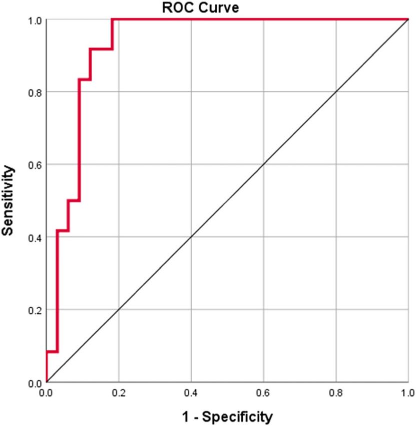
Fig. 2 ROC curve analysis of the relative elevation of the NT-proBNP ent serum biomarker for early detection of cardiotoxicity
in relation to the relative reduction of the GLS (>15%), AUC = 0.929 induced by anthracycline [28]. These findings were also
and p value15%. This is similar to what
came to follow-up, except for one patient who died of un- Calle et al. [30] had found, as they studied 66 breast can-
known etiology just before her scheduled FU visit. cer patients for early cardiotoxicity associated with
Fifty patients (68.5%) had their NT-pro BNP elevated anthracyclines-trastuzumab regimen. They found that 13
in the FU visit. They did not experience any heart failure patients (19.7%) developed early reduction in their LV-
symptoms or signs, yet there was a significant reduction GLS, despite normal 2D-LVEF values. In contrast to our
in the 2D and 3D LVEF, as compared to their baseline study, they followed up their patients for at least 1 year
levels, a finding that was not shown in patients who did to explore whether the early LV-GLS changes could pre-
not have elevated FU levels of NT-proBNP. In a multi- dict future reduction of LVEF. They concluded that ab-
variate analysis model that included all clinical and normal values of 2D-GLS in the presence of a normalSulaiman et al. The Egyptian Heart Journal (2021) 73:20 Page 10 of 11
EF can predict a future drop in LVEF. They also recom- include larger sample size because of the restricted fi-
mended that GLS should be used as a marker of stage B nancial resources.
heart failure in patients given a combined anthracycline-
trastuzumab regimen [30]. In our study, we were only Conclusion
concerned with the early changes of LV function and we Integration of relative elevation of NT-proBNP plasma
did not follow up the patients for the delayed level and relative reduction of the 2D LV-GLS may be
chemotherapy-induced cardiotoxic effects. helpful in the early detection (as early as 6 weeks after the
A relative reduction in the GLS >15% compared first chemotherapy dose) of chemotherapy-induced cardi-
with baseline values was found to be of clinical sig- otoxicity in asymptomatic breast cancer female patients.
nificance in chemotherapy induced cardiotoxicity [19]. The conventional 2D and 3D measurement of LVEF failed
In this study, the median NT-proBNP was signifi- to detect these early cardiac abnormalities. Patients who
cantly higher in patients with a high relative reduc- develop a higher relative elevation of NT-proBNP and a
tion of LV GLS (>15%) versus patients with a less higher relative reduction of the LV-GLS (>15%) should be
relative reduction. On the other hand, patients with candidates for more meticulous and frequent follow-ups.
low absolute LV GLS showed a higher, yet not statis- Early detection of subclinical LV dysfunction will prompt
tically significant, NT-proBNP median values when early cardioprotective measures and thus helps to improve
compared to patients with normal absolute LV GLS. the clinical outcomes. Future studies focusing on the role
So, the integration between the elevated NT-proBNP of cardioprotective drugs in patients with subclinical LV
and the relative reduction of the LV-GLS was more dysfunction are recommended.
successful in identifying patients with cardiac injury
than the integration between the NT-proBNP and the Abbreviations
absolute LV-GLS values. It was also apparent that 2D: Two-dimensional; 3D: Three-dimensional; BMI: Body mass index;
BP: Blood pressure; ECG: Electrocardiogram; EF: Ejection fraction;
there is a strong positive correlation between the ELISA: Enzyme-linked immunosorbent assay; ESC: European Society of
positive change in the NT-proBNP (the difference be- Cardiology; ET: Ejection time; FS: Fractional shortening; FU: Follow-up;
tween the follow-up and the baseline values) and the GLS: Global longitudinal strain; Hb: Hemoglobin; HER: Human epidermal
growth factor receptor; HF: Heart failure; IVCT: Isovolumetric contraction time;
relative change in the LV GLS. IVRT: Isovolumetric relaxation time; LV: Left ventricle; LVEDV: Left ventricular
A marked elevation of the follow-up NT-pro BNP end-diastolic volume; LVESV: Left ventricular end-systolic volume;
plasma levels (>200 pg/ml) was found in 5 patients MPO: Myeloperoxidase; NT-proBNP: N-terminal brain natriuretic enzyme;
PR: Progesterone receptor; SBP: Systolic blood pressure; SD: Standard
(6.8%). All of them had a lower LV-GLS in the FU visit deviation; STE: Speckle tracking echocardiography; TAPSE: Tricuspid annular
and 4 of them had a high relative reduction of the LV- plane systolic excursion; TDI: Tissue Doppler imaging; TnT: Troponin T;
GLS (>15%). Neither the 2D-LVEF nor the 3D-LVEF TTE: Transthoracic echocardiography; TV: Tricuspid valve
could identify any of these patients as being abnormal.
Acknowledgements
This study demonstrated the inability of the absolute Authors acknowledge all members of the Cardiology and Clinical Pathology
LVEF values (either 2D or 3D) to detect the early subtle Departments, Cairo University Hospitals, for their help throughout this work.
chemotherapy-induced myocardial injury, which was
Authors’ contributions
shown by the elevated NT-proBNP values. Even the ab- G.Y proposed the idea, did the echocardiography, did the statistical analysis,
solute GLS values were not as accurate as the relative and critically revised the manuscript. L.S wrote the first draft of the manuscript.
GLS reduction in defining cardiac dysfunction. On the D.H did the laboratory analysis. D.H and M.A critically revised the manuscript. All
authors have read and approved the manuscript.
other hand, the association between the relative eleva-
tion of the NT-proBNP and the relative reduction of the Funding
LV-GLS showed promising results in defining patients This study was self-funded.
with cardiac injury with an excellent sensitivity and a
good specificity. Identification of those patients is pertin- Availability of data and materials
The datasets used and/or analyzed during the current study are available
ent as management plans including cardioprotective from the corresponding author on reasonable request.
agents (renin angiotensin blockade and/or beta blockers)
could reverse the myocardial injury or at least prevent Ethics approval and consent to participate
the progression to cardiac dysfunction. This study complies with the Declaration of Helsinki (2013), and the Faculty
of Medicine, Cairo University ethics committee has approved the research
protocol (the reference number is not available). A written informed consent
has been obtained from all patients.
Limitations
This study focused only on the short-term outcomes. Consent for publication
Not applicable.
Future studies should extend the follow-up beyond 6
months for better evaluation of the long-term effects on Competing interests
patients with apparently normal LVEF. We could not The authors declare that they have no competing interests.Sulaiman et al. The Egyptian Heart Journal (2021) 73:20 Page 11 of 11
Author details Echocardiography and the European Association of Cardiovascular Imaging.
1
Cardiology Department, Faculty of Medicine, Cairo University, Cairo, Egypt. Eur Heart J Cardiovasc Imaging 15(10):1063–1093
2
Chemical Pathology Department, Faculty of Medicine, Cairo University, 20. Hooning MJ, Botma A, Aleman BM, Baaijens MH, Bartelink H, Klijn JG et al
Cairo, Egypt. (2007) Long-term risk of cardiovascular disease in 10-year survivors of breast
cancer. J Natl Cancer Inst 99(5):365–375
Received: 22 December 2020 Accepted: 8 February 2021 21. Felker GM, Thompson RE, Hare JM, Hruban RH, Clemetson DE, Howard DL
et al (2000) Underlying causes and long-term survival in patients with
initially unexplained cardiomyopathy. N Engl J Med 342(15):1077–1084
22. Jensen BV, Skovsgaard T, Nielsen SL (2002) Functional monitoring of
References anthracycline cardiotoxicity: a prospective, blinded, long-term observational
1. Ferlay J, Soerjomataram I, Dikshit R, Eser S, Mathers C, Rebelo M et al (2015) study of outcome in 120 patients. Ann Oncol 13(5):699–709
Cancer incidence and mortality worldwide: sources, methods and major 23. Smiseth OA, Torp H, Opdahl A, Haugaa KH, Urheim S (2016) Myocardial
patterns in GLOBOCAN 2012. Int J Cancer 136(5):E359–E386 strain imaging: how useful is it in clinical decision making? Eur Heart J
2. Bray F, Ferlay J, Soerjomataram I, Siegel RL, Torre LA, Jemal A (2018) Global 37(15):1196–1207
cancer statistics 2018: GLOBOCAN estimates of incidence and mortality 24. Ledwidge M, Gallagher J, Conlon C, Tallon E, O'Connell E, Dawkins I et al
worldwide for 36 cancers in 185 countries. CA Cancer J Clin 68(6):394–424 (2013) Natriuretic peptide-based screening and collaborative care for heart
3. Ibrahim AS, Khaled HM, Mikhail NN, Baraka H, Kamel H (2014) Cancer failure: the STOP-HF randomized trial. Jama. 310(1):66–74
incidence in Egypt: results of the national population-based cancer registry 25. Fernández AE. Chemotherapy-induced dysfunction. E-Journal of Cardiology
program. J Cancer Epidemiol 2014:437971 Practice. 2017;14 (N° 40 ). https://www.escardio.org/Journals/E-Journal-of-Ca
4. Minami M, Matsumoto S, Horiuchi H (2010) Cardiovascular side-effects of rdiology-Practice/Volume-14/Chemotherapy-induceddysfunction
modern cancer therapy. Circ J 74(9):1779–1786 26. Duncan AE, Alfirevic A, Sessler DI, Popovic ZB, Thomas JD (2014)
5. Yeh ET, Tong AT, Lenihan DJ, Yusuf SW, Swafford J, Champion C et al (2004) Perioperative assessment of myocardial deformation. Anesth Analg 118(3):
Cardiovascular complications of cancer therapy: diagnosis, pathogenesis, 525–544
and management. Circulation. 109(25):3122–3131 27. Mornos C, Petrescu L (2013) Early detection of anthracycline-mediated
6. Ewer MS, Ali MK, Mackay B, Wallace S, Valdivieso M, Legha SS et al (1984) A cardiotoxicity: the value of considering both global longitudinal left
comparison of cardiac biopsy grades and ejection fraction estimations in ventricular strain and twist. Can J Physiol Pharmacol 91(8):601–607
patients receiving Adriamycin. J Clin Oncol 2(2):112–117 28. Kittiwarawut A, Vorasettakarnkij Y, Tanasanvimon S, Manasnayakorn S,
7. Sawaya H, Sebag IA, Plana JC, Januzzi JL, Ky B, Cohen V et al (2011) Early Sriuranpong V (2013) Serum NT-proBNP in the early detection of
detection and prediction of cardiotoxicity in chemotherapy-treated patients. doxorubicin-induced cardiac dysfunction. Asia Pac J Clin Oncol 9(2):155–161
Am J Cardiol 107(9):1375–1380 29. Cil T, Kaplan AM, Altintas A, Akin AM, Alan S, Isikdogan A (2009) Use of N-
8. Nicol M, Baudet M, Cohen-Solal A (2019) Subclinical left ventricular terminal pro-brain natriuretic peptide to assess left ventricular function after
dysfunction during chemotherapy. Card Fail Rev 5(1):31–36 adjuvant doxorubicin therapy in early breast cancer patients: a prospective
9. Cardinale D, Sandri MT (2010) Role of biomarkers in chemotherapy-induced series. Clin Drug Investig 29(2):131–137
cardiotoxicity. Prog Cardiovasc Dis 53(2):121–129 30. Arciniegas Calle MC, Sandhu NP, Xia H, Cha SS, Pellikka PA, Ye Z et al (2018)
10. Lipshultz SE, Miller TL, Scully RE, Lipsitz SR, Rifai N, Silverman LB et al (2012) Two-dimensional speckle tracking echocardiography predicts early
Changes in cardiac biomarkers during doxorubicin treatment of pediatric subclinical cardiotoxicity associated with anthracycline-trastuzumab
patients with high-risk acute lymphoblastic leukemia: associations with chemotherapy in patients with breast cancer. BMC Cancer 18(1):1037
long-term echocardiographic outcomes. J Clin Oncol 30(10):1042–1049
11. Zamorano JL, Lancellotti P, Rodriguez Munoz D, Aboyans V, Asteggiano R,
Galderisi M et al (2016) 2016 ESC position paper on cancer treatments and Publisher’s Note
cardiovascular toxicity developed under the auspices of the ESC Committee Springer Nature remains neutral with regard to jurisdictional claims in
for practice guidelines: the task force for cancer treatments and published maps and institutional affiliations.
cardiovascular toxicity of the European Society of Cardiology (ESC). Eur
Heart J 37(36):2768–2801
12. Bazette HC (1920) An analysis of the time-relations of electrocardiograms.
Heart. 7:353–370
13. Schlant RC, Adolph RJ, DiMarco JP, Dreifus LS, Dunn MI, Fisch C et al (1992)
Guidelines for electrocardiography. A report of the American College of
Cardiology/American Heart Association task force on assessment of
diagnostic and therapeutic cardiovascular procedures (committee on
electrocardiography). Circulation. 85(3):1221–1228
14. Elabscience. Human NT-ProBNP (N-Terminal Pro-brain Natriuretic Peptide)
Elisa Kit. E-EL-H0902,96T:2-5 [Available from: https://www.elabscience.com/p-
human_nt_probnp(n_terminal_pro_brain_natriuretic_peptide)_elisa_kit-1
8578.html. Accessed Dec 2020
15. Lang RM, Bierig M, Devereux RB, Flachskampf FA, Foster E, Pellikka PA et al
(2005) Recommendations for chamber quantification: a report from the
American Society of Echocardiography’s guidelines and standards
committee and the chamber quantification writing group, developed in
conjunction with the European Association of Echocardiography, a branch
of the European Society of Cardiology. J Am Soc Echocardiography 18(12):
1440–1463
16. Muller S, Bartel T, Katz MA, Pachinger O, Erbel R (2003) Partial cut-off of the
left ventricle: determinants and effects on volume parameters assessed by
real-time 3-D echocardiography. Ultrasound Med Biol 29(1):25–30
17. Amuthan V, Jegadeewaris A (2013) Manual of 3D echocardiography
18. Seidman A, Hudis C, Pierri MK, Shak S, Paton V, Ashby M et al (2002) Cardiac
dysfunction in the trastuzumab clinical trials experience. J Clin Oncol 20(5):
1215–1221
19. Plana JC, Galderisi M, Barac A, Ewer MS, Ky B, Scherrer-Crosbie M et al (2014)
Expert consensus for multimodality imaging evaluation of adult patients
during and after cancer therapy: a report from the American Society ofYou can also read