The neural networks underlying reappraisal of empathy for pain
←
→
Page content transcription
If your browser does not render page correctly, please read the page content below
Social Cognitive and Affective Neuroscience, 2020, 733–744

                                                                            doi: 10.1093/scan/nsaa094
                                                                            Advance Access Publication Date: 23 July 2020
                                                                            Original Manuscript

The neural networks underlying reappraisal
of empathy for pain

                                                                                                                                                       Downloaded from https://academic.oup.com/scan/article/15/7/733/5875520 by guest on 11 January 2021
Navot Naor,1 Christiane Rohr,2 Lina H Schaare,2 Chirag Limbachia,1
Simone Shamay-Tsoory,3 and Hadas Okon-Singer3
1 University
           of Maryland, Department of Psychology, College Park, MD 20742-5031, USA, 2 Max Planck Institute
for Human Cognitive and Brain Sciences, Department of Neurology, Leipzig 04103, Germany and 3 University of
Haifa, Department of Psychology, Haifa 3498838, Israel
Correspondence should be addressed to Navot Naor, Department of Psychology, University of Maryland, Biology/Psychology Building, 4094 Campus Dr.,
College Park, MD 20742, USA. E-mail: navotnaor@gmail.com.

Abstract
Emotion regulation plays a central role in empathy. Only by successfully regulating our own emotions can we reliably use
them in order to interpret the content and valence of others’ emotions correctly. In an functional magnetic resonance
imaging (fMRI)-based experiment, we show that regulating one’s emotion via reappraisal modulated biased emotional
intensity ratings following an empathy for pain manipulation. Task-based analysis revealed increased activity in the right
inferior frontal gyrus (IFG) when painful emotions were regulated using reappraisal, whereas empathic feelings that were
not regulated resulted in increased activity bilaterally in the precuneus, supramarginal gyrus and middle frontal gyrus
(MFG), as well as the right parahippocampal gyrus. Functional connectivity analysis indicated that the right IFG plays a role
in the regulation of empathy for pain, through its connections with regions in the empathy for pain network. Furthermore,
these connections were further modulated as a function of the type of regulation used: in sum, our results suggest that
accurate empathic judgment (i.e. empathy that is unbiased) relies on a complex interaction between neural regions involved
in emotion regulation and regions associated with empathy for pain. Thus, demonstrating the importance of emotion
regulation in the formulation of complex social systems and sheds light on the intricate network implicated in this complex
process.

Key words: empathy; emotion regulation; reappraisal; gPPI; IFG

Our emotions can help us respond effectively and adaptively                 diminishing those that do not is central to our wellbeing. A
to the complex world that surrounds us. They can also,                      common strategy that individuals use to regulate their emotions
however, become destructive and unhelpful, making us more                   is cognitive reappraisal—a process through which individuals
confused rather than providing us more clarity (Gross, 2013).               reconstruct an emotional situation in a way that alters its
For this reason, being able to regulate our emotions by                     emotional impact, for example by reconstructing a horror film
amplifying those that encourage adaptive responses and                      as a parody (McRae et al., 2012).

Received: 6 May 2019; Revised: 15 June 2020; Accepted: 24 June 2020

© The Author(s) 2020. Published by Oxford University Press. All rights reserved. For Permissions, please email: journals.permissions@oup.com
This is an Open Access article distributed under the terms of the Creative Commons Attribution NonCommercial-NoDerivs licence (http://creativecommo
ns.org/licenses/by-nc-nd/4.0/), which permits non-commercial reproduction and distribution of the work, in any medium, provided the original work is
not altered or transformed in any way, and that the work is properly cited. For commercial re-use, please contact journals.permissions@oup.com

                                                                                                                                            733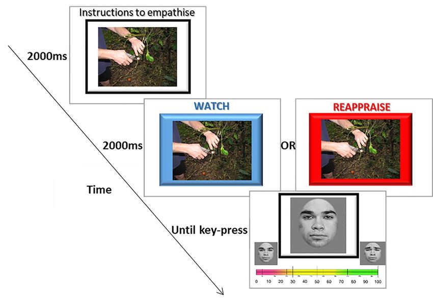
734      Social Cognitive and Affective Neuroscience, 2020, Vol. 15, No. 7

    Traditionally, the study of emotion regulation focused on            magnetic resonance imaging (fMRI) setting. In short, participants
intrinsic and basic emotions (e.g. fear, anger or disgust; Gross,        observed scenarios of painful or non-painful situations. They
2013). Recently, however, growing research interest is being             were then asked to rate the degree of affect in faces that depicted
directed toward more complex emotional situations provoked               either a painful or a happy expression. In half of the trials,
during interpersonal interactions. One such complex emotional            participants were asked to empathize with the scenario, while
situation is the experience of empathy, which is the focus of this       in the other half they were asked to reappraise their empathy.
paper. Empathy is generally defined as an individual’s ability           Empathic engagement with the painful scenario is hypothesized
to vicariously experience the thoughts and feelings of another           to lead to empathy, which will affect the participants’ emotional
person, thus generating connections between individuals. As              state and lead them to judge other people’s levels of pain inaccu-
part of the empathic process, individuals use their own emotions         rately but will not affect the accuracy of their valence judgment
and experiences as a reference point for understanding the               of other emotions. Conversely, the use of reappraisal will down-
mental states of others. Thus, it follows that empathy is                regulate the participant’s own emotional state, resulting in more
influenced by the control individuals exert over their own               accurate empathic judgment.
emotional experiences (Decety, 2010, Naor et al. 2018).                      In addition, we hypothesized that (i) the experience of empa-

                                                                                                                                                Downloaded from https://academic.oup.com/scan/article/15/7/733/5875520 by guest on 11 January 2021
    The tendency to use one’s own emotions while at the same             thy for pain would result in increased activity in the salience
time regulating them is even more relevant in the context of             network, mainly the AI and the ACC; (ii) downregulation of
empathy for pain, i.e. the ability to partake of the pain felt by oth-   empathy for pain via reappraisal would result in increased activ-
ers (Fitzgibbon et al., 2010). Empathy for pain has been the major       ity in regions associated with executive control and decreased
focus of empathy research in social neuroscience and other               activity in limbic networks; and (iii) the degree of activity in the
related fields (Singer and Lamm, 2009), highlighting the impor-          prefrontal-limbic network would affect the degree of cognitive
tance of empathy for pain in daily life. For example, we recently        bias, such that the greater the functional connectivity between
demonstrated that the use of reappraisal to regulate emotions            regions related to emotion regulation and those related to empa-
can influence the empathic process and eliminate biases in               thy, the lesser the bias would be. To this end, in addition to a
judging emotional facial intensity (Naor et al. 2018). The ability       GLM-based fMRI data analysis, we also conducted a generalized
to accurately judge the intensity of emotional facial expressions        psychophysiological interaction (gPPI) analysis to explore the
can be considered to be one type of empathic accuracy (Ickes             functional networks underlying the differences between bias
et al., 1990). The ability to identify others’ emotions based on the     scores after observation of painful scenarios under reappraise
observation of facial expressions has been linked to the ability         and watch conditions. This analysis enabled us to pinpoint the
to share such feelings (Enticott et al., 2008), a key concept in         brain regions that exhibit higher functional coupling during the
empathy (Blais et al., 2012; Singer, 2006). Judgment of morphed          process of downward regulation of empathy.
faces has been used as a measure of empathic accuracy in
previous works, for example in studies that showed participants
                                                                         Methods
dynamic facial expressions and asked them to continuously
judge the intensity of the emotional expressions (e.g. Hall and          Participants
Schmid Mast, 2007; Zaki et al., 2008, 2009). Furthermore, reduction
                                                                         Thirty-three healthy participants were recruited from the stu-
in the ability to make accurate emotional intensity inferences
                                                                         dent population at Ben-Gurion University of the Negev (14 male;
from morphed static face images has been associated with
                                                                         age = 24.65; SD = 1.76) in return for payment. The Ethics Commit-
conditions marked by impairments in empathy, such as cocaine
                                                                         tee at Soroka Medical Centre approved the experiment (Approval
users (Kuypers et al., 2015), patients with ventromedical pre-
                                                                         Number 0114-15-SOR). All participants had normal or corrected-
frontal cortex lesions (Jenkins et al., 2014) and individuals with
                                                                         to-normal vision. Participants were screened for neurological
autistic spectrum disorder (Smith et al., 2010). A recent study
                                                                         or psychiatric history, as well as for any metal implants that
demonstrated a cognitive bias for judgments of pain only when
                                                                         might interfere with the scanning. All participants signed an
these judgments were made after the participant experienced
                                                                         informed consent form prior to participating. Two participants
empathy for pain, yielding exaggerated assessment of emotional
                                                                         were excluded from the final analysis due to technical failures in
intensity compared to the presented intensity. Nevertheless,
                                                                         the scanning session that resulted in the loss of behavioral data.
that bias disappeared when participants used reappraisal to
regulate their empathy (Naor et al. 2018).
    The neural networks underlying the process of modulating             Materials
empathy for pain in the context of emotion regulation have yet           We used a set of 23 matched colored pictures showing hands and
to be explored. Empathy relies heavily on areas of the salience          feet in painful and non-painful scenarios. Each painful scenario
network, namely the anterior insula (AI) and the anterior cin-           was matched with a non-painful scenario that involved all the
gulate cortex (ACC) (Menon and Uddin 2010; Seeley et al., 2007).         same components except the painful element. In addition, the
Conversely, emotion regulation, and mainly reappraisal-based             experiment employed a well-validated set of faces (Blais et al.,
downward regulation, is associated with executive control and            2012; http://mapageweb.umontreal.ca/gosselif/STOIC.rar). Emo-
limbic networks, namely the prefrontal cortex and the amygdala           tional expressions were morphed with neutral ones to create a
(Seeley et al., 2007, Menon and Uddin 2010). An accumulating             sequential morph of 100 pictures. Eight models were used (four
body of research highlights the utility of examining functional          female), each depicting two emotions (happy and painful) at six
connectivity when assessing the relationships between cogni-             levels of intensity (40, 50, 60, 70, 80 and 90%). For more detailed
tive and affective processes, as well as their corresponding brain       information, see Naor et al. (2018).
processes.
    Hence, the current study aimed at exploring the functional
connectivity among the neural networks involved both in upreg-
                                                                         Experimental Procedure
ulating and in downregulating empathy for pain. To this end, we          The behavioral procedure was similar to the procedure used in
employed the task developed by Naor et al. (2018) in an functional       previous research at our lab (Naor et al. 2018). Each experimental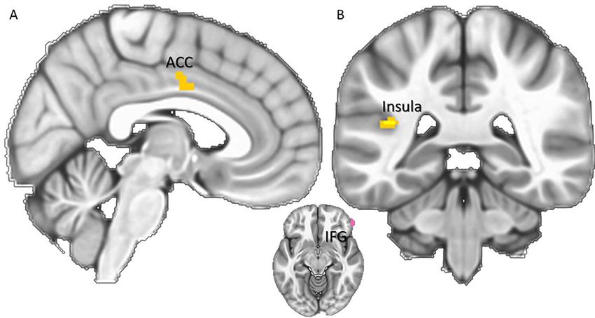
N. Naor et al.         735

                                                                                                                                                                          Downloaded from https://academic.oup.com/scan/article/15/7/733/5875520 by guest on 11 January 2021
Fig. 1. Example of a painful scenario with a neutral face. A picture depicting either a painful or a non-painful scenario appeared for 2000 ms. Then, an instruction to
REAPPRAISE (red frame) or to employ EMPATHIC WATCH (blue frame) appeared for an additional 2000 ms. After participants viewed the scenario, they were shown a
picture depicting an emotional version morphed between 100% neutral and 100% emotion (pain or happy) and were given 6000 ms to assess the emotional intensity
of the presented face.

trial began with a 4000 ms presentation of either a painful                           calculated for each regulation strategy, with the presented sce-
or a non-painful scenario picture taken from the painful/non-                         nario (painful/non-painful) and the presented emotional expres-
painful scenario pictures set. Two thousand ms into the pre-                          sion (happy/painful) as dependent variables and the bias score as
sentation, a colorful frame appeared instructing participants                         the independent variable.
which regulation strategy to employ—REAPPRAISE or EMPATHIC
WATCH. The painful/non-painful scenario picture remained on
the screen with the colorful frame for additional 2000 ms. After                      fMRI data preprocessing
the painful/non-painful scenario picture disappeared, partic-
                                                                                      FMRI data were processed using FEAT (FMRI Expert Analysis
ipants were shown a picture from the facial expression set.
                                                                                      Tool) Version 6.00, a toolbox of FSL (FMRIB’s Software Library,
Participants were then given 6000 ms to judge the intensity of
                                                                                      www.fmrib.ox.ac.uk/fsl). Functional images were registered to
the emotion shown in the facial expression on a scale ranging
                                                                                      high-resolution structural images using Boundary-Based Reg-
from 1 to 100. After 6000 ms, the scale disappeared and a fixation
                                                                                      istration (Greve and Fischl, 2009). The high-resolution struc-
cross appeared from 3000 to 5000 ms before the next trial. A total
                                                                                      tural image was registered to the standard space using FLIRT
of four models (either a male or a female, matching the partici-
                                                                                      (Jenkinson and Smith, 2001; Jenkinson et al., 2002) and then
pant’s sex) were used in the experiment. For each model, six mor-
                                                                                      further refined using FNIRT non-linear registration (Andersson
phed painful faces and six morphed happy faces were selected.
                                                                                      et al., 2013). The following pre-statistical processing was applied:
For statistical power, we ran each stimuli combination twice,
                                                                                      motion correction was carried out using MCFLIRT with options
yielding a total trial count of 96 (4 models × 6 morphed faces × 2
                                                                                      for extended motion parameters (i.e. standard motion param-
emotions × 2 repetitions), divided into two experimental runs
                                                                                      eters plus their derivatives and the squares of their derivatives)
of 48 trials each in the scanner. The complete instructions are
                                                                                      (Jenkinson et al., 2002); non-brain removal using the Brain Extrac-
reported in Appendix 1 (Figure 1).
                                                                                      tion Tool (BET, Smith, 2002). We further scrubbed the volumes’
                                                                                      framewise displacement >0.9 mm. Participants for whom >10%
                                                                                      of the volumes passed this threshold (i.e. >38 volumes) were
Behavioral analysis                                                                   excluded from the analysis. Spatial smoothing using a Gaussian
A three-way repeated measures ANOVA was calculated to exam-                           kernel of FWHM 5 mm; grand-mean intensity normalization
ine the effect of the presented scenario (painful/non-painful),                       of the entire 4D dataset by a single multiplicative factor; and
the presented emotional expression (happy/painful) and the                            high pass temporal filtering (Gaussian-weighted least-squares
regulation strategy deployed (REAPPRAISAL/EMPATHIC WATCH)                             straight line fitting, with sigma = 50.0 s). Denoising was carried
on participants’ judgment of emotional intensity. The dependent                       out with independent component analysis (ICA)-AROMA in FSL
variable in the ANOVA analysis was the calculated bias score,                         by conducting single-subject ICA to remove motion components
which is the averaged difference between the actual observed                          from each participant’s functional data (Pruim et al., 2015a, b).
intensity and the judged intensity.                                                   ICA-AROMA was selected as it was shown to be highly effective
   To further examine the effect of scenario and expression                           in accounting for motion related variance (Ciric et al., 2017;
on bias, a 2 × 2 repeated measures ANOVA was independently                            Pruim et al., 2015a).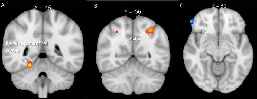
736 Social Cognitive and Affective Neuroscience, 2020, Vol. 15, No. 7
fMRI task within-participant analysis for judgments of painful facial expressions than for those of
happy facial expressions [F(1,30) = 8.759, P = 0.006, ηp2 = 0.226].
Statistical analysis was conducted using FILM with local auto-
In addition, main effects emerged for regulation instructions
correlation correction (Woolrich et al., 2001). The time-series
[F(1,30) = 5.210, P = 0.006 ηp2 = 0.226], scenario [F(1,23) = 5.210,
model included eight EVs to account for the eight contrasts in
P = 0.03 ηp2 = 0.148], and the interaction between them
the experimental design. Each trial lasted ∼10 s (with a 500
[F(1,30) = 10.160, P = 0.003 ηp2 = 0.253]. As predicted, the bias
milliseconds jitter) and included 2 s of initial scenario viewing,
for painful facial expressions was higher when participants
a 2 s regulation period and a 6 s judgment phase. A double
watched the scenario empathically compared to the condition
gamma hemodynamic response function (HRF) was used, and
in which they reappraised their feelings [F(1,30) = 2.112, P = 0.157,
the extended motion parameters served as an indicator function
ηp2 = 0.066]. The bias for painful facial expression was also higher
to model out single TRs identified to have excessive motion
for conditions that followed painful scenarios than for non-
according to a framewise displacement >0.9 mm. The second-
painful ones [F(1,30) = 2.667, P = 0.14, ηp2 = 0.082]. Moreover, all
level analysis, in which contrast estimates were averaged over
three conditions interacted, so that the greatest bias was found
within-subject runs, was conducted using a fixed-effects model
during trials in which judgments of painful facial expressions
by forcing the random effects variance to zero in FLAME (FMRIB’s
Downloaded from https://academic.oup.com/scan/article/15/7/733/5875520 by guest on 11 January 2021
were made following empathic watch of a painful scenario
Local Analysis of Mixed Effects; Beckmann et al., 2003; Woolrich
[F(1,30) = 4.928, P = 0.034, ηp2 = 0.141].
et al., 2004, Woolrich, 2008). Each 10 s trial was modeled in its
Figure 2 portrays the results of further testing the source
entirety to ensure an optimal fit across the data. Specifically, the
of the three-way interaction using a 2 × 2 repeated-measures
first 2-s period was modeled as the observation period, the next
ANOVA with Bonferroni correction for multiple comparisons.
2-s period was modeled as the regulation period and finally the
Under EMPATHIC WATCH, we found a greater bias in judgments
subsequent 6 s were modeled as the judgment time.
of painful facial expressions compared to happy facial expres-
sions [F(1,30) = 12.007, P = 0.002, ηp2 = 0.286], as well as a greater
fMRI task group activity analysis bias in judgments made after exposure to painful scenarios
compared to non-painful ones [F(1,30) = 11.430, P = 0.002, ηp2 =
Group analysis was conducted using FLAME (FMRIB’s Local Anal-
0.276]. In addition, an interaction emerged between facial
ysis of Mixed Effects) stage 1 (Beckmann et al., 2003; Woolrich
expression and scenario [F(1,30) = 7.289, P = 0.011,ηp2 = 0.195].
et al., 2004; Woolrich, 2008). Based on Gaussian random field
Table 1 depicts the follow-up t-tests conducted to examine
theory, Z (Gaussianised T/F) statistic images underwent para-
the source of this interaction. A paired sample t-test revealed
metric thresholding using clusters determined by z > 3.1 and
a greater bias for painful facial expressions in judgments
a corrected cluster significance threshold of P > 0.05 (Wors-
following painful scenarios than in judgments following non-
ley, 2001). We separately examined differences between regula-
painful scenarios (Table 1). A similar Bonferroni corrected
tion following painful scenarios and regulation following non-
ANOVA for REAPPRAISE trials did not yield significant results
painful scenarios for each regulation strategy (i.e. reappraisal
for scenario [F(1,30) = 2.667, N.S.], emotion [F(1,30) = 1.205, N.S.],
and empathic watch). The results of those contrasts were then
or the interactions between them [F(1,30) = 1.386, N.S.]. Moreover,
contrasted themselves to account for the unique effect of each
the bias score for painful expressions following painful scenarios
regulation type.
in EMPATHIC WATCH trials was significantly higher than in
After the whole-brain group analysis, a gPPI analysis (Fris-
REAPPRAISE trials (t = 3.677, df = 30, P = 0.001)1 .
ton et al., 1997; McLaren et al., 2012; O’Reilly et al., 2012) was
conducted in FSL FEAT to examine functional connectivity in
networks involved in the use of reappraisal of empathy for Neuroimaging findings
pain. This analysis examined the interaction between activity
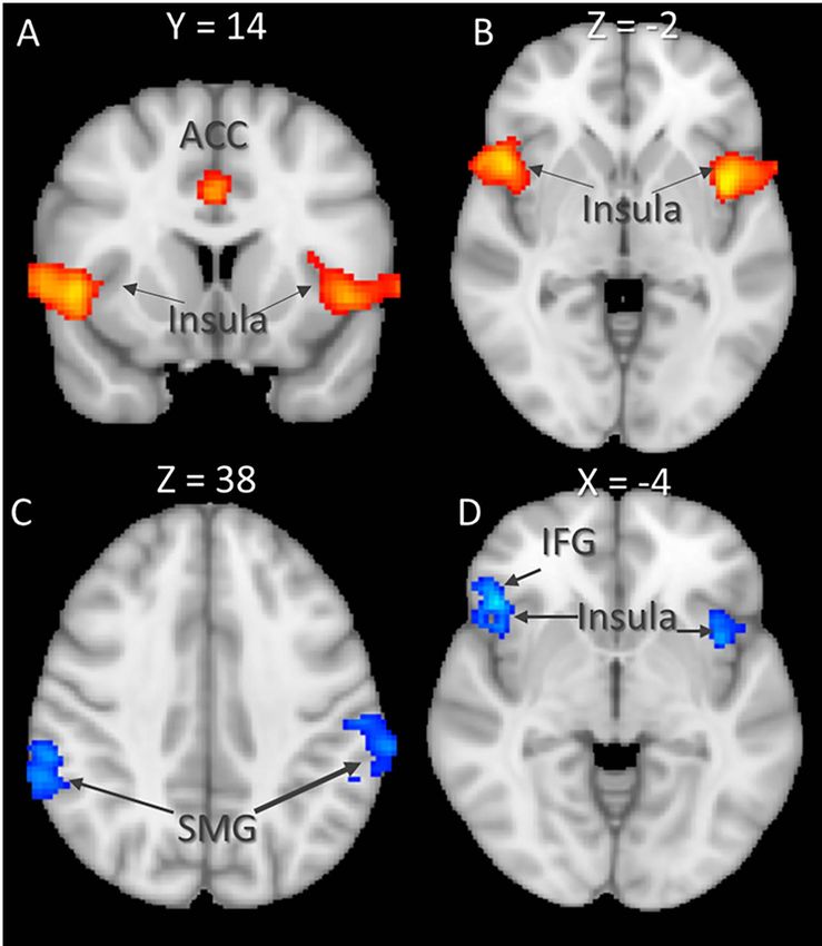
Functional activity results. To examine the effect of emotion
in the seed region—which was selected based on the task anal-
regulation following empathy for pain, we first compared (A)
ysis [i.e. the inferior frontal gyrus (IFG) and the supramarginal
EMPATHIC WATCH trials during exposure to painful scenarios
gyrus (SMG)]—and activity in all other voxels in the brain as
to (B) EMPATHIC WATCH trials during exposure to non-painful
a function of task condition, i.e. REAPPRAISAL (painful/non-
scenarios. Increased activity related to empathy for pain was
painful) and EMPATHIC WATCH (painful/non-painful). The gPPI
found in regions of the salience network, including the bilateral
analysis matrix included all EVs of the original task from the
Insula and the IFG, as well as in the left MFG and the right ACC
group analysis to control for the main effect of task. Judgments
(Table 2). Then, we compared (C) REAPPRAISE trials following
of painful facial expression following painful scenario under
exposure to painful scenarios to (D) REAPPRAISE trials following
the REAPPRAISE and EMPATHIC WATCH conditions were used
exposure to non-painful scenarios. Increased activity was
as the psychological parameter. The physiological parameter
found in regions associated with executive control, including
was the time course of the seed region in the right IFG (based
the left parietal lobule, as well as regions from the salience
on the results of the task analysis, as reported in the Results
network including the bilateral AI. Results of the whole-brain
section). Finally, the mathematical product of the psychological
analysis are reported in Table 2 and Figure 3. To examine the
variable and the physiological variable constituted the interac-
effect of reappraisal, we subtracted the unique activity of both
tion term. A mixed-effects group-level regression was employed
regulation strategies from each other [i.e. (A-B)–(C-D)]. This
using FLAME 1 and the results were thresholded at z > 3.1 and
analysis revealed increased activity during REAPPRAISE trials in
P < 0.05 corrected for multiple comparisons.
the right IFG, whereas for EMPATHIC WATCH, increased activity
was found in left SMG, the right precuneus and the right MFG.
Results
Behavioral findings
A three-way repeated-measures ANOVA with Bonferroni 1 This comparison is not orthogonal and is shown only to demonstrate
correction for multiple comparisons revealed a greater bias the full scope of the effect.N. Naor et al. 737
Downloaded from https://academic.oup.com/scan/article/15/7/733/5875520 by guest on 11 January 2021
Fig. 2. Three-way repeated-measures ANOVA of regulation strategy (reappraise/empathic watch), with scenario (painful/non-painful) and emotion (painful/happy) as
within-subject factors and bias score as a dependent variable, ∗ P < 0.005; ∗∗ P < 0.001.
Table 1. Bias scores, mean differences and t values for WATCH and REAPPRAISE conditions
Condition Scenario Emotion Bias score (SD) Mean difference t-test value
∗∗
Watch Non-painful Pain 0.461 (16.04) 10.671 4.019
Painful 11.132 (7.834)
Non-painful Happy −0.617 (6.946) 1.274 0.556
Painful 0.657 (15.256)
∗
Reappraise Non-painful Pain −0.059 (17.907) 2.781 2.165
Painful 2.722 (13.58)
Non-painful Happy −0.852 (14.38) 0.748 0.507
Painful 0.103 (12.475)
While the interaction between emotion and scenario in EMPATHIC WATCH trials was significant [F(1,30) = 7.289, P = 0.011,ηp2 = 0.195], there was no similar interaction
∗ ∗∗
in REAPPRAISE trials [F(1,30) = 1.386, N.S.]. P < 0.05; P < 0.001.
The results of the whole-brain analysis are reported in Table 3 judgment biases following empathic feelings for the pain of
and Figure 4. others. To this end, we employed a paradigm that measures
biases while participants judge painful facial expressions as a
result of the experience of empathy for pain (Naor et al. 2018).
Functional connectivity results. Two independent gPPI analyses
We compared judgment biases following empathic feelings for
were carried out, employing a seed region in the right IFG based
the pain of others to conditions in which participants regulated
on the effect of reappraisal vs empathic watch. These analyses
their empathy using reappraisal.
showed divergent patterns for empathizing with painful vs non-
Replicating our previous work (Naor et al. 2018), the cur-
painful scenarios as well as for reappraising painful vs non-
rent findings show that the experience of empathy for pain
painful scenarios. Specifically, empathizing with painful scenar-
yields a cognitive bias while judging painful facial expressions
ios was associated with increased connectivity with the mid-
after experiencing empathy for pain. These biases are elimi-
cingulate and ACC, as well as with the bilateral post-central
nated following down-regulation of empathic feelings via reap-
cortex. Conversely, during reappraisal of painful vs non-painful
praisal. Furthermore, these biases are limited to judgments of
scenarios, increased connectivity was found between the IFG
painful expressions following empathy for pain and do not
and the bilateral lateral occipital cortex, as well as with the
occur when participants judge happy facial expressions or when
left IFG, left posterior insula and left parahippocampal gyrus.
they are exposed to non-painful scenarios. These findings sup-
Detailed functional connectivity results are reported in Table 4,
port our previous results and show that when individuals nat-
with key findings illustrated in Figure 5.
urally feel empathy for the pain of another, they develop a
strong bias in their ability to accurately judge the intensity of
the pain felt by that other, leading to their perception of the
Discussion pain as stronger than it really is. We show that reappraisal can
The aim of the present study was to examine the neural net- eliminate this bias in judgment, resulting in accurate emotion
works that facilitate the influence of emotion regulation on judgments.738 Social Cognitive and Affective Neuroscience, 2020, Vol. 15, No. 7
Downloaded from https://academic.oup.com/scan/article/15/7/733/5875520 by guest on 11 January 2021
Fig. 3. (A and B) Results of empathic watch for painful vs non-painful scenarios. (C and D) Results of reappraisal of painful vs non-painful scenarios. Clusters were
derived at z > 3.1 and (corrected) cluster significance P < 0.05. IFG = inferior frontal gyrus; SMG = supramarginal gyrus.
Fig. 4. (A and B) Results of empathic watch larger than reappraisal. (C) Results of reappraisal larger than empathic watch. Clusters were derived at z > 3.1 and (corrected)
cluster significance P < 0.05. SMG = supramarginal gyrus.N. Naor et al. 739
Table 2. A whole-brain analysis of the effects of EMPATHIC WATCH for painful emotion larger than non-painful emotion and the effects
of REAPPRAISE for painful emotion larger than non-painful emotion. Regions were classified using the Harvard–Oxford Atlas, z > 3.1 and
(corrected) cluster significance P < 0.05. Z-MAX values represent peak activity for the cluster. MNI coordinates
EMPATHIC WATCH painful > non-painful (A-B)
Voxels Z-MAX MAX X MAX Y MAX Z R/L
949 4.43 50 14 -2 R Insular Cortex, inferior frontal gyrus, precentral gyrus,
superior temporal gyrus, post-central gyrus, frontal
orbital cortex
923 4.66 -44 2 2 L Insular cortex, inferior frontal gyrus, middle frontal gyrus,
precentral gyrus, superior temporal gyrus, post-central
gyrus, frontal orbital cortex
646 4.07 -62 -22 28 L Superior temporal gyrus, post-central gyrus, superior
parietal lobule, supramarginal gyrus, anterior and
Downloaded from https://academic.oup.com/scan/article/15/7/733/5875520 by guest on 11 January 2021
posterior division, angular gyrus
332 4.13 0 16 32 Superior frontal gyrus, juxtapositional lobule cortex,
paracingulate gyrus, anterior cingulate gyrus
REAPPRAISE painful > non-painful (C-D)
546 4.32 -62 -42 46 L Superior temporal gyrus, posterior division, superior
parietal lobule post-central gyrus, supramarginal gyrus,
anterior and posterior divisions, angular gyrus
466 3.84 60 -46 44 R Superior temporal gyrus, posterior division, post-central
gyrus supramarginal gyrus, anterior and posterior
division, angular gyrus
364 4.21 46 24 -4 R Insular cortex, inferior frontal gyrus, frontal orbital cortex
272 3.93 -38 10 -6 L Insular cortex, inferior frontal gyrus
Table 3. Results of whole-brain analysis for EMPATHIC WATCH effect larger than REAPPRAISE effect, and REAPPRAISE effect larger than
EMPATHIC WATCH effect. Regions were classified using the Harvard–Oxford Atlas, z > 3.1 and (corrected) cluster significance P < 0.05. Z-MAX
values represent peak activity for the cluster. MNI coordinates
EMPATHIC WATCH effect > REAPPRAISE effect [(A-B)—(C-D)]
Voxels Z-MAX X Y Z R/L
109 3.72 -30 -56 50 L Superior parietal lobule, lateral occipital cortex—superior division,
angular gyrus, supramarginal gyrus—posterior division
69 3.58 -6 -98 0 L Occipital pole
61 3.98 22 -46 -16 R Parahippocampal gyrus, posterior division
46 3.53 4 -66 22 Precuneous cortex, cuneal cortex, supracalcarine cortex,
intracalcarine cortex
25 3.39 54 -4 52 R Precentral gyrus, post-central gyrus, middle frontal gyrus
REAPPRAISE effect > EMPATHIC WATCH effect [(C-D)—(A-B)]
45 4.12 56 42 -10 R Inferior frontal gyrus
The current study adds to our previous behavioral research different parts of the network. Empathic watch resulted in
by showing that regulation of empathy for pain via reappraisal increased connectivity with regions involved in processing of
is associated with increased activity in the right IFG. The up- self-pain, while reappraisal resulted in increased connectivity
regulation of empathy for pain via empathic watch led to with regions involved in simulation of others pain, as well as
increased activity in a diffused network of regions known to self-pain processing (Shamay-Tsoory, 2011).
be involved in empathy for pain, including the left SMG, the Activation in the left SMG and the right MFG was found
right precuneus and the right MFG. In addition, neural regions during empathic watch only, suggesting that these two regions
previously associated both with empathy for pain and with reap- play a critical role and are associated with the process of feeling
praisal (e.g. anterior and posterior regions of the left SMG within empathy for the pain of others. The following discussion consid-
the IPL as well as the parietal operculum—empathy for pain: ers the potential role of each of these regions in the regulation
Costantini et al., 2008; Li et al., 2019; Morawetz et al., 2015) were of empathy for pain and its influence on judgment biases.
involved when participants employed reappraisal following The involvement of the right IFG in regulation of empathy
exposure to painful scenarios. A gPPI analysis revealed increased for pain coincides with previous findings outlining a role for the
functional connectivity between the right IFG and regions of the IFG in down-regulation of emotion via reappraisal (Ochsner et al.,
network involved in empathy for pain. Interestingly, different 2012), and specifically in regulation of social emotions (Grecucci
regulation strategies resulted in increased connectivity with et al., 2013). Moreover, the right IFG alongside the putamen and740 Social Cognitive and Affective Neuroscience, 2020, Vol. 15, No. 7
Table 4. Results of gPPI analysis for the effects of EMPATHIC WATCH and REAPPRAISAL, with the time series of a seed in the IFG. Regions were
classified using the Harvard–Oxford Atlas, z > 3.1 and (corrected) cluster significance P < 0.05. Z-MAX values represent peak activity for the
cluster. MNI coordinates
Volume Z-MAX X Y Z R/L
Functional connectivity of the IFG during EMPATHIC WATCH condition
Painful scenario > non-painful scenario
215 3.041 20 −50 14 R Precuneus, supracalcarine cortex
110 3.1602 −28 −50 16 L Precuneus, supracalcarine cortex
105 2.7229 −38 −38 58 L Post-central gyrus
57 2.9896 −32 −24 40 L Post-central gyrus
44 2.8075 −16 20 34 L Middle cingulate gyrus
38 2.8298 2 −18 48 R Mid-cingulate, supplementary motor area
37 2.6564 −64 4 −4 L Superior temporal gyrus
Downloaded from https://academic.oup.com/scan/article/15/7/733/5875520 by guest on 11 January 2021
35 2.7188 −50 −20 28 L Supramarginal gyrus
33 2.473 4 −2 36 R Anterior cingulate gyrus
31 2.7528 −4 −28 −4 L Thalamus
27 2.6531 22 −46 34 R Precuneus
22 2.8328 −54 −4 −28 L Middle temporal gyrus
21 2.7157 54 −40 54 R Supramarginal gyrus
Non-painful scenario > painful scenario
110 −2.9173 56 −34 −6 R Middle temporal gyrus
67 −2.9378 −2 −42 −8 L Cerebellum left I–IV
35 −2.7737 −44 22 12 L Inferior frontal gyrus
34 −2.8068 −10 48 42 L Superior frontal gyrus
Volume Max Int X Y Z R/L
Functional connectivity of the IFG during REAPPRAISAL condition
Painful scenario > non-painful scenario
166 3.0999 −48 −74 20 L Lateral occipital cortex
80 3.1135 32 −88 24 R Lateral occipital cortex
48 2.6958 −60 8 6 L Inferior frontal gyrus
41 2.5736 −40 −38 18 L Insula
40 2.8461 −32 −8 −30 L Parahippocampal gyrus, temporal fusiform cortex
30 2.591 −58 −34 −6 L Middle temporal gyrus
26 2.5268 −60 −16 8 L Planum temporale
24 2.8386 −22 −64 70 L Lateral occipital cortex
21 2.5674 −52 −8 −28 L Inferior temporal gyrus
166 3.0999 −48 −74 20 L Lateral occipital cortex
80 3.1135 32 −88 24 R Lateral occipital cortex
Non-painful scenario > painful scenario
24 −2.7775 70 −40 20 R Supramarginal gyrus
the SMA have been implicated in emotion regulation via motor and elongate a previously exciting emotion (Gross, 2013). Fur-
inhibition, such as in expressive suppression where individuals thermore, among other subregions, the MFG encompasses the
physically suppress emotional facial expressions in order to dorsolateral prefrontal cortex (DLPFC), which is a central region
alter their emotions (Vanderhasselt et al., 2012). These findings in cognitive empathy (Kalbe et al., 2010). Researchers have sug-
suggest that the involvement of the right IFG in the current task gested that empathy can be viewed as a complex system that
following regulation of empathy for pain is related to the motor includes two distinct subsystems: emotional empathy and cog-
qualities of empathy for pain, such as simulating what happens nitive empathy. Whereas emotional empathy represents the
to others or the activation of different muscles in reaction to the ability to share others’ emotion and includes empathy for pain,
pain of others. cognitive empathy allows for the involvement of more cogni-
Empathic watch of painful scenarios was associated with tively complex processes, such as perspective-taking and men-
increased activity in the left SMG, the right precuneus and the talizing (Shamay-Tsoory et al., 2009). Engagement of the DLPFC
right MFG. The MFG and the adjacent precentral gyrus have in emotional empathy is an uncommon finding and may point
been implicated in up-regulation of emotions (Grecucci et al., to the involvement of cognitive empathy in the up-regulation
2013; Frank et al., 2014). Whereas empathic watch, unlike reap- of empathy. An additional indication of this potential role of
praisal, is not a classical regulation strategy, it does possess all the DLPFC in empathy for pain emerges from the functional
the requirements of such a strategy as it is meant to amplify connectivity analysis (see below).N. Naor et al. 741
Downloaded from https://academic.oup.com/scan/article/15/7/733/5875520 by guest on 11 January 2021
Fig. 5. Results of the gPPI functional connectivity analyses, with the time series of a seed in the right IFG (in pink) and the activity during judgments of painful facial
expression when empathically watching painful scenarios compared to neutral scenarios (A) and during judgments of painful facial expression when reappraising
painful scenarios compared to neutral scenarios (B). Clusters were derived at z > 3.1 and (corrected) cluster significance P < 0.05.
We further examined changes in functional connectivity with stimuli because they have been shown to be easy to differentiate
the right IFG, which showed enhanced activity during the task, from painful faces (Naor et al. 2018) and are therefore less likely
using gPPI analysis. One interesting result is that the IFG, a region to skew our results due to mislabeling the perceived emotion,
involved in the simulation of pain, showed higher connectivity even at low intensities. Nevertheless, we cannot rule out the
with the insula as well as with the contralateral IFG during possibility that the observed differences between happy and
reappraisal trials. Whereas during empathic watch trials higher painful expressions stem from other factors, such as processing
connectivity was found with the ACC. While the IFG is related difficulty. Finally, the empathic watch cue was employed as a
to the simulation of pain, both the insula and the ACC are control condition for the reappraisal condition. However, we
part of the empathy for pain network, reportedly responding to cannot rule out the possibility that these instructions enhanced
observed and felt pain (Shamay-Tsoory, 2011). Yet it seems that empathy.
each regulation strategy triggers different parts of that network. This paper is the first to measure and describe the func-
Our findings have implications that go beyond a scientific tional networks underlying biased empathic accuracy following
examination of empathy and its functions. Indeed, our approach empathy for pain. Additional work is required to uncover the
could add to the framework of Research Domain Criteria (RDoC) depth and complexity of the interaction between emotion reg-
project and may serve as a basis for future therapeutic protocols. ulation and prosocial emotions, as well as the neural networks
By validating experimental tasks and protocols the RDoC is that govern these interactions. Recent views on emotional pro-
aiming to change the way mental disorders are being classified cessing maintain that such complex behavior is mediated by
(Morris and Cuthbert, 2012). Gur and Gur (2016) used a simple large cortical and subcortical dynamic brain networks (Pessoa,
emotional faces recognition test to show that individuals with 2017, 2018). This research represents an initial attempt to map
schizophrenia exhibit dysfunctional patterns of facial emotion these networks in the context of regulation of empathy for pain
identification. They concluded that an emotion identification and empathic accuracy. The results also raise questions about
performance index of the RDoC’s social cognition domain should the differences and similarities between the experience of pain
be developed and could be used to improve the diagnostics, and that of empathy for pain. Rütgen et al. (2015) found that
research focus and eventually treatment of schizophrenia. In the experience of empathy for pain relies on the same neural
line with this conclusion, the task developed in our work can responses as well as the same neurotransmitter activity asso-
serve as an implicit tool to examine emotion identification in ciated with the first-hand experience of pain (for more on this
social contexts. Furthermore, it can shed light on the impact of view, see MacDonald and Leary, 2005). Conversely, Singer et al.
emotion regulation on biases in social contexts and the neu- (2004) claimed that the neural networks involved both in self-
ral networks mediating them, among healthy individuals and pain and in empathy for pain go only as far as regions associated
clinical populations. with the affective qualities of pain and not those concerned with
In this study, we sought to describe the neural network its sensory qualities (for more on this view, see Lamm et al., 2011).
involved in regulation of empathy for pain. The study does, how- Whether empathy for pain and first-hand experience of pain
ever, have some potential limitations. First, the stimuli used were share the same neural underpinnings and networks in full, in
artificially morphed images of actors portraying emotional facial part or not at all, it would be interesting to compare how such
expressions rather than real representations of individuals expe- experiences affect empathic accuracy, as well as to examine the
riencing pain. Second, it is possible that the bias in judgment of effect of emotion regulation on the way such experiences bias
painful facial expression resulted from greater sensitivity to the empathic accuracy.
visual features portrayed in these expressions, which may be In this paper, we attempted to map the neural network
linked to empathic accuracy, rather than from their emotional that facilitates the regulation of empathy for pain in order to
value per se. Additionally, happy faces were used as control make accurate empathic judgments. Our results demonstrate742 Social Cognitive and Affective Neuroscience, 2020, Vol. 15, No. 7
the importance of emotion regulation in the formulation of com- Gross, J.J. (Ed.). (2013). Handbook of emotion regulation, Guilford
plex social systems. Although the literature on empathy is based publications.
largely on the premise that adaptive empathic reactions require Gur, R.C., Gur, R.E. (2016). Social cognition as an RDoC domain.
emotion regulation (Jackson and Decety, 2004), little research has American Journal of Medical Genetics Part B: Neuropsychiatric Genet-
directly explored the contribution of emotion regulation to accu- ics, 171(1), 132–41.
rate empathic responses, especially with respect to reappraisal. Hall, J.A., Schmid Mast, M. (2007). Sources of accuracy in the
Indeed, even though empathy is inherently emotional in nature, empathic accuracy paradigm. Emotion, 7(2), 438.
research on empathy seems to remain primarily focused on Ickes, W., Stinson, L., Bissonnette, V., Garcia, S. (1990). Natural-
shared emotions and not on the way these shared emotions are istic social cognition: Empathic accuracy in mixed-sex dyads.
regulated. Understanding the mechanisms underlying empathy Journal of Personality and Social Psychology, 59(4), 730.
regulation is important given that the purpose of empathy is to Jackson, P.L., Decety, J. (2004). Motor cognition: A new paradigm
alleviate the distress of a suffering target. to study self–other interactions. Current opinion in Neurobiology,
14(2), 259–63.
Jenkins, L.M., Andrewes, D.G., Nicholas, C.L., et al. (2014). Social
Funding
Downloaded from https://academic.oup.com/scan/article/15/7/733/5875520 by guest on 11 January 2021
cognition in patients following surgery to the prefrontal cortex.
Psychiatry Research: Neuroimaging, 224(3), 192–203.
There are no funders to report for this submission.
Jenkinson, M., Smith, S. (2001). A global optimisation method
for robust affine registration of brain images. Medical Image
Conf lict of interest Analysis, 5(2), 143–56.
Jenkinson, M., Bannister, P., Brady, M., Smith, S. (2002). Improved
None declared. optimization for the robust and accurate linear registration
and motion correction of brain images. NeuroImage, 17(2),
825–41.
References Kalbe, E., Schlegel, M., Sack, A.T., et al. (2010). Dissociating cogni-
Andersson, J., Jenkinson, M., Smith, S. M. (2013). Non-linear tive from affective theory of mind: a TMS study. Cortex, 46(6),
registration, aka spatial normalisation. FMRIB Analysis Group 769–80.
Technical Reports. TR07JA2. Available: www. fmrib. ox. ac. uk Kuypers, K.P.C., Steenbergen, L., Theunissen, E.L., Toennes, S.W.,
analysis/techrep [October 17, 2017]. Ramaekers, J.G. (2015). Emotion recognition during cocaine
Beckmann, C.F., Jenkinson, M., Smith, S.M. (2003). General multi- intoxication. European Neuropsychopharmacology, 25(11),
level linear modeling for group analysis in FMRI. NeuroImage, 1914–21.
20(2), 1052–63. Lamm, C., Decety, J., Singer, T. (2011). Meta-analytic evidence for
Blais, C., Roy, C., Fiset, D., Arguin, M., Gosselin, F. (2012). The eyes common and distinct neural networks associated with directly
are not the window to basic emotions. Neuropsychologia, 50(12), experienced pain and empathy for pain. NeuroImage, 54(3),
2830–8. 2492–502.
Ciric, R., Wolf, D.H., Power, J.D., et al. (2017). Benchmarking of Li, Y., Zhang, T., Li, W., Zhang, J., Jin, Z., Li, L. (2019). Linking brain
participant-level confound regression strategies for the con- structure and activation in anterior insula cortex to explain the
trol of motion artifact in studies of functional connectivity. trait empathy for pain. Human Brain Mapping.
Neuroimage, 154, 174–87. MacDonald, G., Leary, M.R. (2005). Why does social exclusion
Costantini, M., Galati, G., Romani, G.L., Aglioti, S.M. (2008). hurt? The relationship between social and physical pain. Psy-
Empathic neural reactivity to noxious stimuli delivered to body chological Bulletin, 131(2), 202.
parts and non-corporeal objects. European Journal of Neuro- McLaren, D.G., Ries, M.L., Xu, G., Johnson, S.C. (2012). A general-
science, 28(6), 1222–30. ized form of context-dependent psychophysiological interac-
Decety, J. (2010). The neurodevelopment of empathy in humans. tions (gPPI): a comparison to standard approaches. NeuroImage,
Developmental Neuroscience, 32(4), 257–67. 61(4), 1277–86.
Enticott, P.G., Johnston, P.J., Herring, S.E., Hoy, K.E., Fitzgerald, McRae, K., Ciesielski, B., Gross, J.J. (2012). Unpacking cogni-
P.B. (2008). Mirror neuron activation is associated with facial tive reappraisal: goals, tactics, and outcomes. Emotion, 12(2),
emotion processing. Neuropsychologia, 46(11), 2851–4. 250.
Fitzgibbon, B.M., Giummarra, M.J., Georgiou-Karistianis, N., Enti- Menon, V., Uddin, L.Q. (2010). Saliency, switching, attention and
cott, P.G., Bradshaw, J.L. (2010). Shared pain: from empathy control: a network model of insula function. Brain Structure and
to synaesthesia. Neuroscience & Biobehavioral Reviews, 34(4), Function, 214(5-6), 655–67.
500–12. Morawetz, C., Bode, S., Baudewig, J., Kirilina, E., Heekeren, H.R.
Frank, D.W., Dewitt, M., Hudgens-Haney, M., et al. (2014). Emotion (2015). Changes in effective connectivity between dorsal and
regulation: quantitative meta-analysis of functional activity ventral prefrontal regions moderate emotion regulation. Cere-
and deactivity. Neuroscience & Biobehavioral Reviews, 45, 202–11. bral Cortex, 26(5), 1923–37.
Friston, K.J., Buechel, C., Fink, G.R., Morris, J., Rolls, E., Dolan, Morris, S.E., Cuthbert, B.N. (2012). Research domain criteria: cog-
R.J. (1997). Psychophysiological and modulatory interactions in nitive systems, neural circuits, and dimensions of behavior.
neuroimaging. NeuroImage, 6(3), 218–29. Dialogues in Clinical Neuroscience, 14(1), 29.
Grecucci, A., Giorgetta, C., Bonini, N., Sanfey, A.G. (2013). Reap- Naor, N., Shamay-Tsoory, S.G., Sheppes, G., Okon-Singer, H.
praising social emotions: the role of inferior frontal gyrus, (2018). The impact of empathy and reappraisal on emotional
temporo-parietal junction and insula in interpersonal emotion intensity recognition. Cognition and Emotion, 32(5), 972–987.
regulation. Frontiers in Human Neuroscience, 7, 523. O’Reilly, J.X., Woolrich, M.W., Behrens, T.E., Smith, S.M., Johansen-
Greve, D.N., Fischl, B. (2009). Accurate and robust brain image Berg, H. (2012). Tools of the trade: psychophysiological interac-
alignment using boundary-based registration. NeuroImage, tions and functional connectivity. Social Cognitive and Affective
48(1), 63–72. Neuroscience, 7(5), 604–9.N. Naor et al. 743
Ochsner, K.N., Silvers, J.A., Buhle, J.T. (2012). Functional imaging Zaki, J., Weber, J., Bolger, N., Ochsner, K. (2009). The neural bases
studies of emotion regulation: a synthetic review and evolving of empathic accuracy. Proceedings of the National Academy of
model of the cognitive control of emotion. Annals of the New Sciences, 106(27), 11382–7.
York Academy of Sciences, 1251(1), E1–E24.
Pessoa, L. (2017). A network model of the emotional brain. Trends
in Cognitive Sciences.
Pessoa, L. (2018). Understanding emotion with brain networks. Appendix 1 – Full practice and experimental
Current Opinion in Behavioral Sciences, 19, 19–25. protocol.
Pruim, R.H., Mennes, M., Buitelaar, J.K., Beckmann, C.F. (2015a).
Evaluation of ICA-AROMA and alternative strategies for Slide 1 Intro
motion artifact removal in resting state fMRI. Neuroimage, 112,
I am going to read you the instructions from paper. We are doing
278–87.
that to keep uniformity across all our participants, and as a
Pruim, R.H., Mennes, M., van Rooij, D., Llera, A., Buitelaar, J.K.,
result, you might feel that there is some repetition. We repeat the
Beckmann, C.F. (2015b). ICA-AROMA: A robust ICA-based strat-
instructions to make sure that they are as clear and understood.
Downloaded from https://academic.oup.com/scan/article/15/7/733/5875520 by guest on 11 January 2021
egy for removing motion artifacts from fMRI data. Neuroimage,
In this task you are going to view emotional images. Some of
112, 267–77.
these images might induce extremely negative emotions in you,
Rütgen, M., Seidel, E.M., Silani, G., et al. (2015). Placebo analgesia
while others very little negative emotions, or they might not
and its opioidergic regulation suggest that empathy for pain
induce any emotional reactions.
is grounded in self pain. Proceedings of the National Academy of
For every image you would see you would be asked to imple-
Sciences, 112(41), E5638–46.
ment one of two emotion regulation strategy, reappraisal and
Seeley, W.W., Allman, J.M., Carlin, D.A., et al. (2007). Diver-
watch, in a moment I will explain them to you, and we will
gent social functioning in behavioral variant frontotemporal
practice using them. It is important that you try and remember
dementia and Alzheimer disease: reciprocal networks and
what to do in each of this strategies, as we will ask you about it.
neuronal evolution. Alzheimer Disease & Associated Disorders,
21(4), S50–7.
Shamay-Tsoory, S.G. (2011). The neural bases for empathy. The
Slide 2 - Reappraisal instructions.
Neuroscientist, 17(1), 18–24.
Shamay-Tsoory, S.G., Aharon-Peretz, J., Perry, D. (2009). Two sys- We will start by going over reappraisal, the first emotion reg-
tems for empathy: a double dissociation between emotional ulation strategy you will use to change the way are feeling. In
and cognitive empathy in inferior frontal gyrus versus ventro- reappraisal you are going to try and change the meaning of the
medial prefrontal lesions. Brain, 132(3), 617–27. image. That is, try and think of something to tell yourself about
Singer, T. (2006). The neuronal basis and ontogeny of empathy the image which would help you to reduce any negative feelings
and mind reading: review of literature and implications for you might be experiencing towards it. For example, you could say
future research. Neuroscience and biobehavioral reviews, 30(6), something about the outcome that the situation is going to be
855–63. resolved soon, or that help is on the way. You could focus a detail,
Singer, T., Seymour, B., O’doherty, J., Kaube, H., Dolan, R.J., Frith, or an aspect of the situation that is not as negative. However,
C.D. (2004). Empathy for pain involves the affective but not we want you to stay focused on the image itself, and not just
sensory components of pain. Science, 303(5661), 1157–62. randomly think of other things that might make you feel better.
Singer, T., Lamm, C. (2009). The social neuroscience of empathy. Change something in the meaning of the image that will help
Annals of the New York Academy of Sciences, 1156(1), 81–96. you feel less negative emotions towards it.
Smith, S.M. (2002). Fast robust automated brain extraction. When you are practicing reappraisal it is important that you
Human Brain Mapping, 17(3), 143–55. do not think that the image is fake, or that it is taken out of a
Smith, M.J.L., Montagne, B., Perrett, D.I., Gill, M., Gallagher, L. movie. Rather you should think that you are witnessing a real
(2010). Detecting subtle facial emotion recognition deficits scenario, which you are trying to reappraise. The reason for that
in high-functioning autism using dynamic stimuli of varying is that we are trying to simulate events that happened in real life,
intensities. Neuropsychologia, 48(9), 2777–81. where we can not say about them that they didn’t happen.
Vanderhasselt, M.A., Kühn, S., De Raedt, R. (2012). ‘Put on your
poker face’: neural systems supporting the anticipation for
expressive suppression and cognitive reappraisal. Social Cog- Slide 3 - Reappraisal que.
nitive and Affective Neuroscience, 8(8), 903–10.
A blue frame around the image is an indication you should exer-
Woolrich, M. (2008). Robust group analysis using outlier infer-
cise reappraisal. Do you remember what you should do? Could
ence. NeuroImage, 41(2), 286–301.
you please shortly explain to me how are you implementing
Woolrich, M.W., Ripley, B.D., Brady, M., Smith, S.M. (2001). Tempo-
reappraisal?
ral autocorrelation in univariate linear modeling of FMRI data.
[After participant’s replay] – Good, remember you should
NeuroImage, 14(6), 1370–86.
focus on the image, but think of it in a way that would help you
Woolrich, M.W., Behrens, T.E., Beckmann, C.F., Jenkinson, M.,
reduce any negative emotions you might have.
Smith, S.M. (2004). Multilevel linear modelling for FMRI
group analysis using Bayesian inference. NeuroImage, 21(4),
1732–47.
Slide 4 - Watch instructions.
Worsley, K.J. (2001). Statistical analysis of activity images. Func-
tional MRI: An introduction to methods, 14(1), 251–70. The second strategy you will be asked to practice is watch.
Zaki, J., Bolger, N., Ochsner, K. (2008). It takes two: The interper- During watch you will be asked to observe the image in front
sonal nature of empathic accuracy. Psychological science, 19(4), of you and allow yourself to experience the emotions in an
399–404. interrupted way. That is, try not to block your emotions, and not744 Social Cognitive and Affective Neuroscience, 2020, Vol. 15, No. 7
to alter how you are feeling, but allow thoughts and feeling to Slide 6 – general explanation.
develop freely.
Now you know both strategies – reappraisal and watch, each trial
will begin with the appearance of an image on the screen. A cou-
Slide 5 - Watch que. ple of second after it’s appearance the image will be surrounded
with a frame, either in blue or red. The frame will indicate to
A red frame around the image is an indication you should exer- you which strategy to implement. Do you remember which color
cise watch. Do you remember what you should do? Could you represents which strategy?
please shortly explain to me how are you implementing watch? Shortly after the appearance of the que frame both frame and
[After participant’s replay] – Good, for as long as the image is image would disappear, and am image of a face expressing an
presented look at it and allow for your emotions and thoughts to emotion. Using the response box you will be asked to indicate on
develop freely. a scale of 1 to 100 what is the intensity of the expressed emotion.
Downloaded from https://academic.oup.com/scan/article/15/7/733/5875520 by guest on 11 January 2021You can also read