The rule of brain hematoma pressure gradient and its influence on hypertensive cerebral hemorrhage operation - Nature
←
→
Page content transcription
If your browser does not render page correctly, please read the page content below
www.nature.com/scientificreports
OPEN The rule of brain hematoma
pressure gradient and its
influence on hypertensive cerebral
hemorrhage operation
Guoqing Sun, Tingkai Fu, Zhaoyan Liu, Yuhai Zhang, Xiangtao Chen, Shigang Jin* &
Feng Chi
To comparatively study the size of and variation in the ‘brain-haematoma’ pressure gradient for
different surgical methods for hypertensive intracerebral haemorrhage (HICH) and analyse the
gradient’s influence on surgical procedures and effects of the haemorrhage. Seventy-two patients
with HICH treated from 1/2019 to 12/2019 were randomly divided into two groups, namely, the
keyhole endoscopy and large trauma craniotomy groups, according to different operative methods.
Intraoperative changes in intracranial pressure (ICP) were monitored to calculate intraoperative
alterations in the ‘brain-haematoma’ pressure gradient. Intraoperative characteristics (operative time,
bleeding volume, volume of blood transfusion, and haematoma clearance rate) and postoperative
characteristics (oedema, postoperative activities of daily living (ADL) scores, mortality rate and
rebleeding rate) were compared between the two groups. In the keyhole endoscopy group, ICP
decreased slowly; the ‘brain-haematoma’ pressure gradient was large, averaging 251.1 ± 20.6 mmH2O,
and slowly decreased. The mean operative time was 83.6 ± 4.3 min, the mean bleeding volume was
181.2 ± 13.6 ml, no blood transfusions were given, the average postoperative haematoma clearance
rate was 95.6%, the rate of severe oedema was 10.9%, and the average postoperative ADL score
was 85.2%. In the large trauma craniotomy group, ICP rapidly decreased after craniotomy. When the
haematoma was removed, the ‘brain-haematoma’ pressure gradient was small, averaging 132.3 ± 10.5
mmH2O, and slowly decreased. The mean operative time was 232 ± 26.1 min, the mean bleeding
volume was 412.6 ± 35.2 ml, the average volume of blood transfusion was 281.3 ± 13.6 ml, and the
average postoperative haematoma clearance rate was 82.3%; moreover, the rate of severe oedema
was 72.1%, and the average postoperative ADL score was 39.0%. These differences were statistically
significant (P < 0.05). Neither the death rate (P > 0.05, 2.7% VS 2.8%) nor rebleeding rate (P > 0.05,
2.7% VS 2.8%) showed any obvious changes. The magnitude and variation in the ‘brain-haematoma’
pressure gradient for different surgical methods significantly influence surgical procedures and effects
of HICH. During keyhole endoscopy surgery, this gradient was relatively large and slowly decreased;
the haematoma was therefore easier to remove. Advantages of this approach include a high
haematoma clearance rate, decreased bleeding volume, decreased operative time, reduced trauma,
decreased postoperative brain oedema and improved postoperative recovery of neurological function.
Chinese Clinical Trial Register: ChiCTR1900020655 registration in 12/01/02,019 registration in
28/02/02,020 Number: NCOMMS-20–08,091.
Abbreviations
HICH Hypertensive cerebral hemorrhage
ADL Activity of daily living
Hypertensive intracerebral haemorrhage (HICH) is a common neurosurgical condition with high morbidity1 that
atients2 and is associated with high mortality and a high disability r ate3; in particular,
accounts for 70% of stroke p
61–88% of patients become severely d isabled4,5. The ‘brain-haematoma’ pressure gradient plays an important
Department of Neurosurgery, Rizhao People’s Hospital Affiliated With Jining Medical University, Rizhao 276826,
Shandong Province, People’s Republic of China. *email: sun18963395696@163.com
Scientific Reports | (2021) 11:4599 | https://doi.org/10.1038/s41598-021-84108-w 1
Vol.:(0123456789)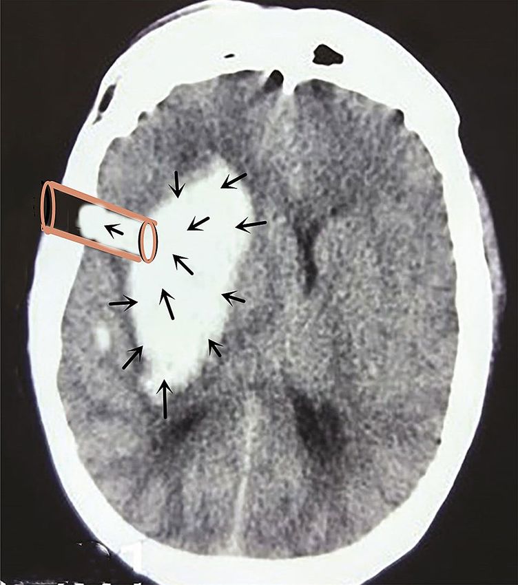
www.nature.com/scientificreports/
role in the process of cerebral haemorrhage and in surgery. Variations in this gradient differ under three distinct
conditions: post-cerebral haemorrhage, keyhole endoscopy and craniotomy. Changes in the “brain-haematoma”
pressure gradient determine movement direction for the brain and haematoma, and differences in these changes
in various operations produce diverse effects on surgical procedures and postoperative results. We studied 72
patients with intracerebral haemorrhage who were treated from 1/2019–12/2019. In addition, for summary and
analysis, we compared changes in the ‘brain-haematoma’ pressure gradient and its influence on surgery and
postoperative effects for HICH patients treated with two different types of operations.
Materials and methods
Ethics. This study was approved by the Medical Ethical Committee of Rizhao People’s Hospital Affiliated with
Jining Medical University. This research was performed in accordance with relevant guidelines and regulations,
and informed consent was obtained from all participants or their legal guardians.
Clinical materials. The patients in the two groups were diagnosed with HICH from 1/2019 to 12/2019
and had a haematoma that was locatable by imaging with a volume greater than 30 ml, all haematoma located
above the tentorium, and they all had a history of hypertension. Patients with cerebral haemorrhage caused by
intracranial aneurysm, intracranial arteriovenous malformation, tumours, haemorrhage after cerebral infarc-
tion, long-term use of anticoagulants, and other disorders were excluded, as were cerebral hernia patients and
patients with severe systemic diseases or dysfunction of other important organs, such as the heart, lung, liver
and kidney. According to operative method, the patients were randomly divided into two groups: the keyhole
endoscopy group and the large trauma craniotomy group by random scale. Both groups of patients were treated
within 24 h after paroxysm. There were 36 patients in the keyhole endoscopy group, including 19 males and 17
females, with an average age of 48.2 ± 6.1 years (range, 31 to 72 years). The preoperative haematoma volume was
46.2 ± 5 ml, and the Glasgow Coma Scale (GCS) score was 7.7 ± 2.1. Seventeen, 13 and 6 patients had haemor-
rhage in the basal ganglia, capsula externa, and brain lobe, respectively. There were 36 patients in the large
trauma craniotomy group, including 21 males and 15 females, aged 47.9 ± 6.5 years (range, 36 to 67 years). The
preoperative haematoma volume was 47.8 ± 5.6 ml, and the GCS score was 7.6 ± 3.1. There were 19, 8 and 9 cases
of haemorrhage in the basal ganglia, capsula externa, and brain lobe, respectively. The general preoperative con-
ditions of the two groups of patients were compared, and there was no significant difference between the groups
(P > 0.05), meaning that they were comparable.
Calculation of the ‘brain‑haematoma’ pressure gradient. Both groups of patients received intracra-
nial pressure (ICP) monitoring before undergoing craniotomy. After general anaesthesia, all patients underwent
puncture of the anterior corner of the lateral ventricle and implanted intracranial pressure sensor (ventricular
catheter type, model 826653, American Johnson & Johnson Company, CODMAN). The site was the frontal
puncture point (2.5 cm before the coronal suture and 2.5 cm beside the midline). The skull was drilled, the
dura was cut, the brain was punctured into the lateral ventricle, and the sensor was implanted at a depth of 6.0–
7.0 cm. Next, the needle core was removed, the cerebrospinal fluid overflow was visualized, an ICP was placed
to monitor the lateral ventricle, and the probe lead of the ICP monitor was extended through the subcutaneous
tunnel. During the operation, the probe was continuously fixed in the ventricle. After the intracranial pressure
monitor was zeroed during the operation, the intracranial pressure was continuously monitored throughout the
operation. Postoperative ICP monitoring was continued, and cerebrospinal fluid drainage was performed for
3–10 days, with an average monitoring of 7.2 days.
The measured ICP was the brain tissue pressure. Data on the craniotomy were recorded, as were intraop-
erative and immediately postoperative data. For the calculation of the haematoma pressure, the haematoma
was located in the brain tissue before and after the craniotomy, such that the pressure in the haematoma cavity
and the brain tissue pressure were in a dynamic equilibrium, meaning the pressure values were equal. At this
time, the ‘brain-haematoma’ pressure gradient = ICP–ICP = 0 m mH2O. When the haematoma was removed, the
haematoma cavity became connected to the external environment, the value of the haematoma cavity pressure
returned to 0 m mH2O, and the ‘brain-haematoma’ pressure gradient = ICP–0 = ICP. Values of ICP and the ‘brain-
haematoma’ pressure gradient during craniotomy, during surgery and at the end of surgery were recorded and
statistically analysed.
Surgical methods. In the keyhole endoscopy group, the keyhole position was located according to the hae-
matoma position, and a milling cutter formed a circular bone window of approximately 2–2.5 cm in diameter.
A brain needle was used to puncture the centre of the haematoma and extract a portion of its contents to reduce
ICP. A special sheath was inserted into the haematoma cavity to form a columnar working channel. Then, the
endoscope was placed, suction was used to remove the haematoma, and bipolar electrocoagulation was used to
stop bleeding. When eliminating a haematoma, one surgeon needed to operate with both hands: the left hand
held the endoscope, and the right hand held a suction device to remove the haematoma. When bleeding was
stopped by bipolar electrocoagulation, a two-person, three-hand operation mode was employed. The assistant
held the endoscope, while the surgeon used the left hand to suction and used the right hand to stop the bleeding.
Finally, the dura mater was sutured, and the bone flaps were attached6.
In the large trauma craniotomy group, a modified pterional approach was employed in the basal ganglia
region, and the size of the bone window was approximately (7–8) cm × (9–12) cm. The lateral cleft was separated
under a microscope to expose the cortex of the insula and form a cortical fistula. Then, in the haematoma cav-
ity, the brain was gently manipulated with a spatula, and the haematoma was removed without touching the
brain parenchyma around the lesion; bipolar electrocoagulation was then used to stop bleeding. With respect to
Scientific Reports | (2021) 11:4599 | https://doi.org/10.1038/s41598-021-84108-w 2
Vol:.(1234567890)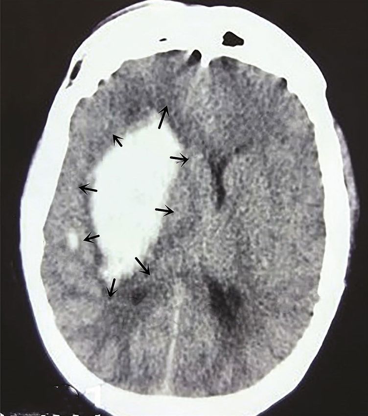
www.nature.com/scientificreports/
Figure 1. Change curves for ICP.
haemorrhage on the surface of the brain lobe, such as the occipital lobe and frontal lobe, a horseshoe or coronary
incision was used to perform a craniotomy according to the location of the haematoma, the haematoma was
located through puncture, a cortical fistula was used to access the haematoma cavity, and the haematoma was
pulled out using a brain spatula. When closing the cranium, the surgeon sutured the dura without increasing its
tension and performed decompressive craniectomy based on the patient’s c ondition6.
Operation evaluation. Operation evaluation was conducted from four aspects for statistical comparative
analysis, namely, operative time including total operative time, time for craniotomy and cranial closure and time
for removal of haematoma, bleeding volume during the operation, the volume of blood transfusion and the
haematoma clearance rate that was mainly calculated based on the 24-h postoperative CT examination (residual
haematoma volume/total haematoma volume).
Postoperative evaluation. Postoperative evaluation was reflected by four aspects: mortality rate, rehaem-
orrhage ratio, postoperative cerebral oedema and activities of daily living (ADL) scores. With regard to post-
operative cerebral oedema, a CT scan was performed 24 h after surgery, and the largest dimension of oedema
was recorded as the maximal diameter of oedema, which was denoted by D. The degree of cerebral oedema
was graded as follows. Level 0 oedema: D = 0; level 1 oedema: D < 2 cm; level 2 oedema: 2 cm < D < 4 cm; level
3 oedema, D > 4 cm. In addition, level 0 oedema and level 1 oedema were regarded as mild oedema, and level
2 oedema and level 3 oedema were regarded as severe oedema. The postoperative follow-up was conducted six
months after surgery, and the ADL scale was employed for evaluation of therapeutic effects. The patients with
ADL grades I-III had good prognoses, and the patients with grades IV-V had poor prognoses.
Statistical methods. The statistical package SPSS 19.0 was used for analysis. The measurement data that
conformed to a normal distribution were denoted as x ± s and were compared using the t-test. The enumeration
data were compared with the chi-square test (χ2). A P value < 0.05 was considered statistically significant.
Results
Changes in ICP during surgery. Before craniotomy, the mean ICP in the keyhole endoscopy and large
trauma craniotomy groups was 271.3 ± 22.3 mmH2O and 267.4 ± 19.8 mmH2O, respectively, with no significant
difference between the groups (P > 0.05). After craniotomy, ICP did not change obviously in the keyhole endos-
copy group, which exhibited an average ICP of 263.8 ± 18.7 mmH2O, whereas in the large craniotomy group,
ICP decreased significantly to an average of 142.2 ± 12.3 mmH2O. The difference between the two groups was
statistically significant (P < 0.05). When the haematoma was revealed after a cortical fistula had been created, ICP
gradually decreased to an average of 251.1 ± 20.6 mmH2O in the keyhole endoscopy group and declined to an
average of 132.3 ± 10.5 mmH2O in the large trauma craniotomy group. This difference was statistically significant
(P < 0.05). When the haematoma was removed, ICP gradually decreased to an average of 212.3 ± 24.3 m mH2O
in the keyhole endoscopy group and decreased in a similarly slow manner to an average of 115.9 ± 11.7 mmH2O
in the large trauma craniotomy group; these average values significantly differed (P < 0.05). After removal of the
haematoma, ICP was reduced to normal in both groups, with an average ICP of 63.6 ± 9.3 mmH2O and 56.8 ± 8.8
mmH2O in the keyhole endoscopy group and the large trauma craniotomy group, respectively; there was no
significant difference in ICP between the two groups (P > 0.05) (Fig. 1).
Changes in the ‘brain‑haematoma’ pressure gradient during surgery. The haematoma was
located in the brain tissue during craniotomy. The pressures in the haematoma cavity and the brain tissue were
dynamically balanced, and the pressure in the haematoma cavity was equal to the value of ICP. At this time, the
Scientific Reports | (2021) 11:4599 | https://doi.org/10.1038/s41598-021-84108-w 3
Vol.:(0123456789)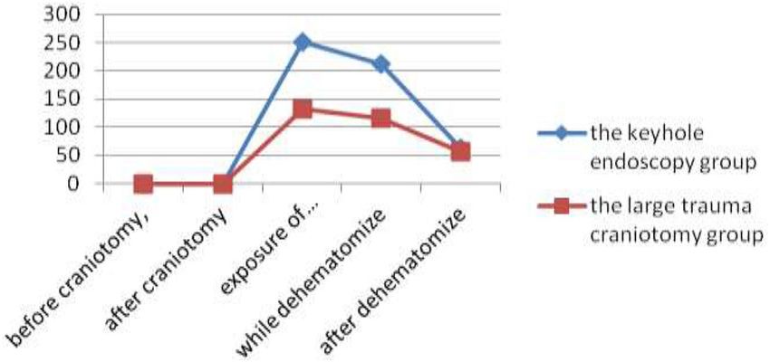
www.nature.com/scientificreports/
Figure 2. Change curves for the ‘brain-hematoma’ pressure gradient.
Average intraoperative blood loss Average intraoperative blood Average hematoma clearance rate
Groups mean operating time (min) (ml) transfusion (ml) (%)
Keyhole endoscopy group 79.5 ± 4.2 173.9 ± 12.4 0 94.8%
Craniotomy group 219.2 ± 5.8 405.6 ± 37.1 272.3 ± 14.1 83.4%
χ2 = 173.39 χ2 = 305.85 χ2 = 25.69 χ2 = 53.97
chi-square test
P < 0.01 P < 0.001 P < 0.01 P < 0.001
Table 1. Intraoperative comparison of the two patient groups.
Groups Death rate Rebleeding rate Severe edema rate Rate of good prognosis in ADL
Keyhole endoscopy group 2.7% 2.7% 11.2% 86.4%
Craniotomy group 2.7% 2.7% 69.9% 38.9%
χ2 = 347.37 χ2 = 347.37 χ2 = 11.25 χ2 = 22.37
chi-square test
P > 0.05 P > 0.05 P < 0.05 P < 0.01
Table 2. Comparison of two groups of patients in postoperative situation.
‘brain-haematoma’ pressure gradient = ICP–ICP = 0 mmH2O. There was no significant difference between the
keyhole endoscopy group and the large trauma craniotomy group.
During the process of revealing and removing the haematoma, the ‘brain-haematoma’ pressure gradient = ICP.
When the haematoma was revealed, the ‘brain-haematoma’ pressure gradient averaged 251.1 ± 20.6 m mH2O and
132.3 ± 10.5 mmH2O in the keyhole endoscopy and large trauma craniotomy groups, respectively. There was a
statistically significant difference between the two groups (P < 0.05). When the haematoma was removed, the
‘brain-haematoma’ pressure gradient was 212.3 ± 24.3 mmH2O in the keyhole endoscopy group and 115.9 ± 11.7
mmH2O in the large trauma craniotomy group. The difference between the two groups was statistically significant
(P < 0.05). After the haematoma was removed, the ‘brain-haematoma’ pressure gradient was 63.6 ± 9.3 m mH2O
in the endoscopy group and 56.8 ± 8.8 m mH2O in the large craniotomy group, with no significant difference
between the groups (P > 0.05) (Fig. 2).
Surgery situation. Compared with that in the large trauma craniotomy group, the average bleeding volume
was reduced in the keyhole endoscope group, as were the volume of blood transfusion, the time needed to clear
the haematoma, and the mean operative time, whereas the average postoperative haematoma clearance rate was
higher. (Table 1).
Postoperative situation. Although the mortality rate and rebleeding rate were not significantly differ-
ent, the average postoperative haematoma clearance rate was high, the rate of severe oedema was low, and the
postoperative ADL score was better in the keyhole endoscopy group than in the large trauma craniotomy group.
(Table 2).
Common complications of intraventricular pressure monitoring include catheter dislocation, catheter block-
age, intracranial haemorrhage, and intracranial infection. In the keyhole endoscopy group, 2 patients had catheter
Scientific Reports | (2021) 11:4599 | https://doi.org/10.1038/s41598-021-84108-w 4
Vol:.(1234567890)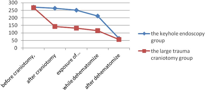
www.nature.com/scientificreports/
blockage, which was solved by irrigation; in the craniotomy group, 1 patient had intracranial infection, which
was cured by anti-infection treatment, and 1 patient had catheter blockage, which was solved by irrigation, and
there was no significant difference in the incidence of complications (P > 0.05, 5.6% VS 5.6%) between the two
groups. Neither the death rate (P > 0.05, 2.8% VS 2.8%) nor rebleeding rate (P > 0.05, 2.8% VS 2.8%) showed any
obvious changes.
Discussion
HICH has high incidence, high mortality, and a high disability rate. Rincon et al. reported the natural mortality
rate of HICH within 30 days after onset to be approximately 45%7. HICH can cause a series of pathophysiologi-
cal changes. First, acute haemorrhage damages brain tissue, neurocytes and nerve conduction bundles, leading
to brain dysfunction. Second, the increasingly large haematoma compresses surrounding brain tissue, forming
pressure gradients between the haematoma and surrounding brain tissue and forcing shifts in brain tissue. Due
to blocked peripheral vascular circulation and cerebral ischaemia and hypoxia of brain tissue, brain oedema
occurs, causing a sharp rise in ICP and even leading to brain stem compression-induced cerebral hernia, which
is life-threatening8. Third, the acute and chronic toxic effects produced by haematoma decomposition cause brain
oedema, degeneration, and necrosis, which can further increase ICP and aggravate neurological d ysfunction9.
Therefore, in the shortest possible time, it is necessary to clear the intracerebral haematoma, relieve compression,
promote the eventual return of displaced brain tissue, reduce ICP, maximize the preservation of nerve function,
and create favourable conditions for the recovery of brain f unction10–12.
Currently, there are many surgical treatments for cerebral haemorrhage, most of which are empirical treat-
ments developed within different medical centres, although no consensus has been reached to d ate13. Common
surgical methods, such as trepanation and drainage, craniotomy evacuation of haematoma, and neuroendoscopic
treatment for cerebral haemorrhage, have their own advantages and disadvantages and respective indications
for adaptation14,15. Haematoma puncture and drainage are quick and simple, and they have been widely used
in primary hospitals. However, this approach has disadvantages of a low haematoma clearance rate and a high
rebleeding rate16. In addition, postoperative injection of urokinase to promote liquefaction of the haematoma
requires long-term intubation and drainage, increasing the probability of intracranial infection17. Craniotomy
for removal of haematoma involves two types of operations: large trauma craniotomy and small bone window
craniotomy. The large trauma craniotomy for haematoma removal has the advantages of a high haematoma
clearance rate and less chance of rebleeding, but it has more limitations, such as a long operative time, serious
intraoperative brain pulling, severe secondary brain injury, heavy trauma, and more intraoperative b leeding13.
Although small bone window craniotomy for haematoma removal relatively shortens the operative time, it can
still cause serious intraoperative brain pulling and severe secondary brain injury. When ICP increases after
surgery, the scope of the bone removal flap is limited, the decompression effect is not obvious, and the visual
field is relatively limited; therefore, greater microscopy skill is required of the surgeon. Intracerebral haematoma
removal under neuroendoscopy results in less surgical trauma and a decreased operative time. However, it is
limited by neuroendoscopy. Therefore, specific surgical instruments are required to perform surgery. In general,
a specific ultrasonic aspirator is used to physically break the lump and remove haematomas14. Due to the dif-
ficulty of mastering this technique, this procedure is difficult to perform at the majority of primary hospitals.
After intracerebral haemorrhage, the mass effect of haematoma caused the haematoma pressure to be higher
than the brain tissue pressure, leading to displacement of the surrounding brain tissue, which eventually stopped
when the haematoma pressure and the brain tissue pressure were balanced (Fig. 3). During intracerebral hae-
matoma removal surgery, as the haematoma cavity was exposed to the environment and the haematoma was
continuously being removed, the pressure in the haematoma cavity was significantly reduced, and a new reverse
pressure gradient was formed between the brain tissue and the haematoma cavity. This gradient promoted the
return of brain tissue to its original state and an outward shift in the haematoma that contributed to haematoma
removal. However, in other surgical approaches, there are significant differences in ICP changes as well as the
size and type of changes in the pressure between the haematoma cavity and brain tissue; these differences have
diverse impacts on the surgical haematoma removal process and on postoperative effects.
When an endoscopic keyhole was employed for craniotomy, ICP was not significantly reduced due to the small
bone hole. However, after a cortical fistula had been created, the ‘brain-haematoma’ pressure gradient formed
by surgery was large. The average ‘brain-haematoma’ pressure gradient was 251.1 ± 20.6 mmH2O, 212.3 ± 24.3
mmH2O and 63.6 ± 9.3 mmH2O at the start of haematoma removal, during removal and after removal, respec-
tively, showing that the ‘brain-haematoma’ pressure difference slowly declined as the haematoma was continu-
ously cleared and ICP slowly declined. Due to the large ‘brain-haematoma’ pressure gradient and its slow decline,
haematomas in the endoscopic channel showed a sustained negative pressure state relative to other haematomas
and brain tissue. Such a high sustained negative pressure contributed to the retraction of brain tissue and forced
the intracerebral haematoma to move to the working channel area (Fig. 4). The haematoma was easily removed
with the help of external absorption; thus, the haematoma removal rate was high, the operative time was short,
intraoperative blood loss was small, and blood transfusion was not required.
Use of the keyhole technique can facilitate rapid craniotomy, remove the intracerebral haematoma with a
high clearance rate in the shortest time, relieve nerve compression and minimize nerve damage. The small bone
hole with a diameter of approximately 2 cm minimized the exposure of brain tissue because lack of exposure is
the best way to protect brain tissue. The endoscopic channel was columnar, and the pressure was transmitted
evenly around the brain. The brain tissue continued to be supported in multiple directions, and there was no
obvious traction on the tissue. The damage to the brain tissue was slight; therefore, severe brain oedema did
not occur after the operation. A continuously high and slowly declining ‘brain-haematoma’ pressure gradient
was utilized to cause automatic flowing of the haematoma to the endoscopic channel. With the haematoma in
Scientific Reports | (2021) 11:4599 | https://doi.org/10.1038/s41598-021-84108-w 5
Vol.:(0123456789)www.nature.com/scientificreports/
Figure 3. The space occupying effect of cerebral hemorrhage makes the pressure of the hematoma higher than
that of the brain tissue and forces the surrounding brain tissue to shift until the pressure of the hematoma equals
that of the brain tissue.
Figure 4. In the keyhole endoscopy group, the hematoma in the endoscopic channel has negative pressure
relative to the remaining hematoma and brain tissue, thus forming a pressure gradient of “brain-hematoma-
endoscopic channel”. Thus, the brain tissue further retracts, forcing the intracerebral hematoma to shift into the
work channel area, and hematoma removal can be completed under synthetic action without any extraneous
tissue exposure.
Scientific Reports | (2021) 11:4599 | https://doi.org/10.1038/s41598-021-84108-w 6
Vol:.(1234567890)www.nature.com/scientificreports/
Figure 5. In the craniotomy group, after removing the flap and opening the dura, because of the pressure
difference of “distant brain-hematoma-brain tissue around bone window”, some brain tissue bulged out from
bone window under the influence of brain tissue pressure and hematoma pressure, significantly decreasing the
brain tissue pressure. Thus, in the process of hematoma removal, the pressure difference of "brain-hematoma "
hardly forms. The hematoma does not retract and is not easy to remove. The only way for removal is the use of a
brain spatula to pull brain tissue and reveal hematoma for clearance, which is also challenging because excessive
pulling can easily damage brain tissue.
the channel removed, the brain tissue shifted and caused the haematoma to swell further. Various endoscope
angles made it easier to efficiently remove haematomas. In addition, with no need for brain pressure plates and
other devices to pull the brain tissue, the keyhole technique can achieve true intraoperative microtraction with
minimal damage to brain tissue, no serious brain oedema after surgery and good recovery of the postoperative
patient’s neurological function.
After a large trauma craniotomy, ICP and brain tissue pressure significantly decreased. The brain tissue first
bulged out under the influence of the ‘brain-haematoma’ pressure gradient and stopped when a balance between
the brain tissue pressure and the haematoma cavity pressure was reached. Lower ICP resulted in a smaller
‘brain-haematoma’ pressure gradient after a cortical fistula had been created. The average pressure gradient was
132.3 ± 10.5 mmH2O at the start of haematoma removal, 115.9 ± 11.7 m mH2O during removal, and 56.8 ± 8.8
mmH2O after removal, demonstrating that ICP further decreased and the ‘brain-haematoma’ pressure gradient
slowly declined as the haematoma was continuously removed. This smaller ‘brain-haematoma’ pressure gradient
meant that there was little pressure on the brain tissue to shift it back to its original location and that the haema-
toma was not easy to retract and remove (Fig. 5). As a result, the haematoma could only be revealed and removed
by pulling the brain tissue through the brain pressure plate, resulting in a long operative time, low haematoma
clearance rate, massive intraoperative blood loss, and the need for blood transfusion therapy. Furthermore,
excessive tension on the brain plate was likely to cause brain tissue damage, causing severe postoperative brain
oedema and leading to poor postoperative neurological recovery.
Although the endoscopically assisted keyhole approach for haematoma removal has the advantages of mild
brain damage and good postoperative neurological recovery, it still has certain limitations, as follows. It is nec-
essary to accommodate the endoscope, suction apparatus and bipolar coagulation device in a relatively small
space to reveal and remove the haematoma and completely stop the bleeding. Such surgery requires surgeons to
have proficient micro-neurosurgery operating skills and experience with endoscopic application skills, as well
as good cooperation with assistants.
In summary, the size of and variation in the ‘brain-haematoma’ pressure gradient for different surgical meth-
ods significantly influence surgical procedures and effects. ICP slowly decreased during endoscopic surgery, and
the ‘brain-haematoma’ pressure gradient was large and slowly decreased. As a result, using this approach relative
to large trauma craniotomy, the haematoma was easier to remove, and there was a high haematoma clearance
rate, less intraoperative blood loss, a shorter operative time, and less intraoperative trauma. In addition, post-
operative brain oedema was mild, and the postoperative nerve recovery rate was high. Large trauma craniotomy
sharply decreased ICP, and the ‘brain-haematoma’ pressure gradient was small. Brain tissue retraction was poor
Scientific Reports | (2021) 11:4599 | https://doi.org/10.1038/s41598-021-84108-w 7
Vol.:(0123456789)www.nature.com/scientificreports/
in this case; therefore, the haematoma was difficult to clear, and it was necessary to use a brain spatula to pull
the haematoma to remove it. In addition, the surgery situation and postoperative situation showed a relatively
low haematoma clearance rate, massive intraoperative bleeding volume, long operative time, large intraoperative
trauma, severe postoperative brain oedema, and a low postoperative nerve recovery rate. However, the number of
patients in this group was still relatively small, and a large sample will be required for further comparative studies.
Received: 15 March 2020; Accepted: 9 February 2021
References
1. Liu, M., Wu, B. & Wang, W. Z. Stroke in China: epidemiology, prevention, and management strategies. Lancet Neurol. 6(5), 456–464
(2007).
2. Morgenstern, L. B., Hemphill, J. C. & Anderson, C. Guidelines for the management of spontaneous intracerebral hemorrhage: a
guideline for healthcare professionals from the american heart association/American stroke association. Stroke 41(9), 2108 (2015).
3. Brodefiek, J., Connolly, S. & Feldmann, E. Guidelines for the management of spontaneous intracerebral hemorrhage in adults.
Neurosurgery 41(9), 328–333 (2007).
4. Asch, C. J. V., Luitse, M. J. & Rinkel, G. J. Incidence, case fatality, and functional outcome of intracerebral haemorrhage over time,
according to age, sex, and ethnic origin: a systematic review and meta-analysis. Lancet Neurol. 9(2), 167–176 (2010).
5. Mayer, S. A. & Rincon, F. Treatment of intracerebral haemorrhage. Lancet Neurol. 4(10), 662–672 (2005).
6. Sun, G. et al. Comparison of keyhole endoscopy and craniotomy for the treatment of patients with hypertensive cerebral hemor-
rhage. Medicine 98(2), e14123. https://doi.org/10.1097/MD.0000000000014123 (2019).
7. Rincon, F. & Mayer, S. A. Intracerebral hemorrhage: getting ready for effective treatments. Curr. Opin. Neurol. 23(1), 59–64 (2010).
8. Gonzalez-Duarte, A., Cantu, C., Ruiz-Sandoval, J. & Barinagarrementeria, F. Recurrent primary cerebral hemorrhage: frequency,
mechanisms, and prognosis. Stroke J. Cereb. Circ. 29(9), 1802–1805 (1998).
9. Zheng, J. et al. Minimally invasive surgery treatment for the patients with spontaneous supratentorial intracerebral hemorrhage
(mistich): protocol of a multi-center randomized controlled trial. BMC Neurol. 14(1), 206 (2014).
10. Li, N. et al. Association of molecular markers with perihematomal edema and clinical outcome in intracerebral hemorrhage. Stroke
44(3), 658–663 (2013).
11. Gaberel, T., Magheru, C. & Emery, E. Management of non-traumatic intraventricular hemorrhage. Neurosurg. Rev. 35(4), 485–495
(2012).
12. Mould, W. A. et al. Minimally invasive surgery plus rt-pa for intracerebral hemorrhage evacuation (mistie) decreases perihema-
tomal edema. Stroke J. Cereb. Circ. 44(3), 627 (2013).
13. Zhang, F. W., Cai, Y. & Zhang, L. Comparison of the efficacy of neuroendoscopy and craniotomy in the treatment of hypertensive
intracerebral hemorrhage. Chin. J. Neurosurg. 1, 19–21 (2015).
14. Chen, C. H., Lee, H. T., Shen, C. C. & Sun, M. H. Aspiration of hypertensive intracerebral hematoma with frameless and fiducial-
free navigation system: technical note and preliminary result. Stereotact. Funct. Neurosurg. 86(5), 288 (2008).
15. Dye, J. A., Dusick, J. R., Lee, D. J., Gonzalez, N. R. & Martin, N. A. Frontal bur hole through an eyebrow incision for image-guided
endoscopic evacuation of spontaneous intracerebral hemorrhage. J. Neurosurg. 117(4), 767–773 (2012).
16. Mendelow, A. D. Surgical trial in intracerebral haemorrhage (s.t.i.c.h.). Acta Neurochir. Suppl. 76(83), 521–522 (1999).
17. Hu, Y. Z., Wang, J. W. & Luo, B. Y. Epidemiological and clinical characteristics of 266 cases of intracerebral hemorrhage in hangzhou,
china. J. Zhejiang Univ. Sci. B (Biomed. Biotechnol.) 14(6), 496–504 (2013).
Author contributions
Conceptualization: G.S., S.J. Data curation: G.S., T.F. Formal analysis: X.C. Investigation: G.S., T.F. Methodology:
S.J. Project administration: G.S., S.J. Resources: G.S., X.C., Software: Z.L., X.C., F.C. Supervision: Y.Z. Validation:
Z.L., Writing original draft: G.S. Writing review & editing: S.J.
Competing interests
The authors declare no competing interests.
Additional information
Correspondence and requests for materials should be addressed to S.J.
Reprints and permissions information is available at www.nature.com/reprints.
Publisher’s note Springer Nature remains neutral with regard to jurisdictional claims in published maps and
institutional affiliations.
Open Access This article is licensed under a Creative Commons Attribution 4.0 International
License, which permits use, sharing, adaptation, distribution and reproduction in any medium or
format, as long as you give appropriate credit to the original author(s) and the source, provide a link to the
Creative Commons licence, and indicate if changes were made. The images or other third party material in this
article are included in the article’s Creative Commons licence, unless indicated otherwise in a credit line to the
material. If material is not included in the article’s Creative Commons licence and your intended use is not
permitted by statutory regulation or exceeds the permitted use, you will need to obtain permission directly from
the copyright holder. To view a copy of this licence, visit http://creativecommons.org/licenses/by/4.0/.
© The Author(s) 2021
Scientific Reports | (2021) 11:4599 | https://doi.org/10.1038/s41598-021-84108-w 8
Vol:.(1234567890)You can also read