The Tail of the Late Jurassic Sauropod Giraffatitan brancai: Digital Reconstruction of Its Epaxial and Hypaxial Musculature, and Implications for ...
←
→
Page content transcription
If your browser does not render page correctly, please read the page content below
ORIGINAL RESEARCH
                                                                                                                                                  published: 29 May 2020
                                                                                                                                           doi: 10.3389/feart.2020.00160

                                             The Tail of the Late Jurassic
                                             Sauropod Giraffatitan brancai:
                                             Digital Reconstruction of Its Epaxial
                                             and Hypaxial Musculature, and
                                             Implications for Tail Biomechanics
                                             Verónica Díez Díaz 1,2* , Oliver E. Demuth 3,4 , Daniela Schwarz 1 and Heinrich Mallison 5,6
                                             1
                                              Museum für Naturkunde Leibniz-Institut für Evolutions- und Biodiversitätsforschung, Berlin, Germany, 2 Humboldt
                                             Universität zu Berlin, Berlin, Germany, 3 Structure and Motion Laboratory, Royal Veterinary College, Hatfield,
                                             United Kingdom, 4 School of Earth Sciences, University of Bristol, Bristol, United Kingdom, 5 Center of Natural History
                                             (CeNak), University of Hamburg, Hamburg, Germany, 6 Palaeo3D, Pöttmes, Germany

                                             Dinosaur locomotion and biomechanics, especially of their pelvic girdles and hindlimbs,
                                             have been analyzed in numerous studies. However, detailed volumetric musculoskeletal
                                             models of their tails are rarely developed. Here, we present the first detailed three-
                           Edited by:
                        Pasquale Raia,
                                             dimensional volumetric reconstruction of the caudal epaxial and hypaxial musculature
  University of Naples Federico II, Italy    of the Late Jurassic sauropod Giraffatitan brancai, and highlight the importance
                       Reviewed by:          and necessity of 3D modeling in musculoskeletal reconstructions. The tail of this
             Peter Lewis Falkingham,
                                             basal macronarian is relatively short compared to diplodocids and other coexisting
    Liverpool John Moores University,
                     United Kingdom          macronarians. The center of mass lies well in front of the hindlimbs, which support only
                      Ryan T. Tucker,        ca. half the body weight. Still, our reconstruction suggests a total weight for the entire tail
 Stellenbosch University, South Africa
                                             of ca. 2500 kg. We conclude that the hypaxial and tail-related hindlimb muscles (most
                   *Correspondence:
                    Verónica Díez Díaz
                                             specifically the M. caudofemoralis longus and its counterpart the M. ilioischiocaudalis) in
         diezdiaz.veronica@gmail.com         Giraffatitan were well developed and robustly built, compensating for the shorter length
                                             of the M. caufodemoralis longus, the main hindlimb retractor muscle, in comparison
                    Specialty section:
          This article was submitted to      with other sauropods. Our methodology allows a better-constrained reconstruction of
                          Paleontology,      muscle volumes and masses in extinct taxa, and thus force and weight distributions
                a section of the journal
                                             throughout the tail, than non-volumetric approaches.
             Frontiers in Earth Science
          Received: 31 January 2020          Keywords: sauropoda, Tendaguru, Giraffatitan, volumetric musculoskeletal modeling, tail
            Accepted: 29 April 2020
            Published: 29 May 2020
                               Citation:     INTRODUCTION
              Díez Díaz V, Demuth OE,
Schwarz D and Mallison H (2020) The          Reconstructions of the musculoskeletal system of dinosaurs have been inferred from the anatomical
   Tail of the Late Jurassic Sauropod        comparison of and the inference of homological structures in closely related or osteologically
             Giraffatitan brancai: Digital   similar animals (e.g., Dilkes, 1999; Carrano and Hutchinson, 2002) based on the extant phylogenetic
          Reconstruction of Its Epaxial
                                             bracket (EPB) (Bryant and Russell, 1992; Witmer, 1995, 1997). Key examples for this are the highly
            and Hypaxial Musculature,
                and Implications for Tail
                                             esteemed and classical publications of Romer (e.g., Romer, 1923), which rely on thorough studies
                          Biomechanics.      of the anatomy of living taxa. In the case of dinosaurs, numerous publications (e.g., Dilkes, 1999;
                Front. Earth Sci. 8:160.     Carrano and Hutchinson, 2002; Hutchinson, 2002; Organ, 2006; Schwarz-Wings, 2009; Allen, 2010)
       doi: 10.3389/feart.2020.00160         have analyzed the muscles of the limbs and the axial skeleton of living archosaurs (i.e., crocodilians

Frontiers in Earth Science | www.frontiersin.org                                     1                                                   May 2020 | Volume 8 | Article 160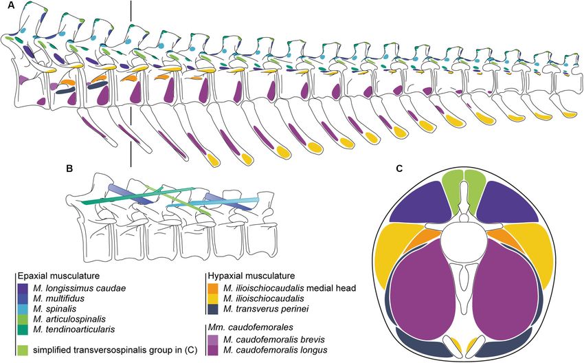
Díez Díaz et al.                                                                                                     Giraffatitan Tail Reconstruction

and birds), and extrapolated this information to infer it on to the       the sacrum. The lack of keystoning in the anterior caudal
preserved osteological remains of extinct taxa.                           vertebrae convinced Janensch (1950b) that the anterior tail of
    Musculature and ligaments often leave characteristic traces           Giraffatitan extended from the hips in a straight, thus caudally
(= osteological correlates, Witmer, 1995) on the bone surface of          descending line, in contrast to the tails of other sauropods, which
all vertebrates. Where such osteological correlates for muscles           emerged from the hips horizontally. This posture led to the
are present on the vertebrae in both sauropods and crocodilians,          tail contacting the ground much further anteriorly in relation
these muscles can be reconstructed as level II inference (Witmer,         to its overall length than in the other species Janensch (1950b)
1995, 1997). However, variation in the soft tissue configuration          mentioned: Dicraeosaurus hansemanni, Diplodocus carnegii and
and uncertainties in the interpretation of osteological correlates        Camarasaurus lentus. During a general renovation of the Berlin
demand a cautious approach in reconstructing soft tissue                  dinosaur exhibition hall (between 2005 and 2007), the mounted
anatomy of extinct taxa (Bryant and Russell, 1992; Witmer, 1995;          skeleton was rebuilt. The new mount differs from the old mount
Carrano and Hutchinson, 2002). Sauropod vertebrae expose a                in several key characteristics (Remes et al., 2011):
complex surface pattern of laminae, fossa, ridges, bulges and
rugosities, which are associated with pneumatic structures and               • Improvements to the models of the presacral vertebrae
attachment sites of muscles, tendons and ligaments (Wedel et al.,              and head,
2000; Wedel, 2003a,b, 2009; O’Connor, 2006; Schwarz-Wings,                   • The posture of the neck, the shape of the torso,
2009). Unambiguous pneumatic structures can be distinguished                 • The orientation of the pectoral girdle and forelimbs, and
from correlates for muscles and ligaments by the presence                    • The posture of the tail, still emerging from the hip caudally
of pneumatic foramina that penetrate deeply into the bone                      descending, but curving slightly dorsally to remain well
(O’Connor, 2006).                                                              clear of the ground.
    Over the last few years, vertebrate paleontology took
advantage of novel techniques and software (Cunningham et al.,                Here, we digitally reconstruct the tail of this sauropod. We
2014; Sutton et al., 2014). Digitization methodologies (e.g.,             applied photogrammetric 3D digitization and 3D modeling tools
CT scanning and photogrammetry, see Mallison and Wings,                   in combination with information provided by dissections of
2014; Fahlke and Autenrieth, 2016) and Computer-Aided-Design              extant crocodilians (Alligator mississippiensis) (Supplementary
(CAD) tools made it possible to capture the morphology                    Figure S1) and an Extant Phylogenetic Bracket (EPB; Witmer,
of the bones upon which three-dimensional reconstructions                 1995) approach by comparing the anatomy of the caudal
of the musculoskeletal system of extinct animals could be                 vertebrae and muscles of Giraffatitan with that of extant
improved. The spatial organization of muscle groups can be                crocodilians. The other side of the bracket, birds, shows a strongly
assessed in 3D and thus intersections of individual muscles               reduced caudal musculoskeletal system, which is adapted for
prevented. These three-dimensional reconstruction methods                 novel functions in flight and display (e.g., Gatesy and Dial,
have overwhelmingly focused on cranial and especially adductor            1996; O’Connor et al., 2013). Bird tails are much shorter, with
musculature (Lautenschlager, 2013; Sharp, 2014; Button et al.,            fewer vertebrae, of which up to half are co-ossified into the
2016; Gignacand Erickson, 2017), while the axial musculature              pygostyle. Just anterior to the pygostyle, there is a maximum of
received little attention. The myological reconstruction of the           six mobile vertebrae present, depending on the species (Gatesy
dinosaur tail in particular is a complex task, as it consists of          and Dial, 1996). Furthermore, changes between the thoracic and
ten individual muscles, partly subdivided into multiple heads.            caudal epaxial musculature are hypothesized to coincide with the
Previous studies on dinosaur tail musculature have primarily              evolution of the synsacrum (a structure highly involved in the
focused on the M. caudofemoralis longus (CFL) (e.g., Mallison,            flight, together with the notarium, see Organ, 2006). For example,
2011; Persons and Currie, 2011, 2012; Persons et al., 2014),              caudal epaxial muscles were decoupled from their locomotor
because of its locomotive importance as the main hindlimb                 function on the evolutionary line to birds (Gatesy and Dial,
retractor muscle. However, none has attempted to reconstruct              1996), and several muscles were lost (e.g., Mm. interspinales and
the complete musculoskeletal system of the tail. Here we present          M. multifidus). In addition, due to the absence of chevrons, the
the reconstructed caudal anatomy of Giraffatitan brancai based            attachments of the tail depressor muscles shifted to the transverse
on the comparison of the caudal vertebral anatomy, which has              processes, among further modifications (Pittman et al., 2013).
previously been described in great detail (e.g., Janensch, 1914,          The absence of chevrons and the truncation of the bird tail
1950a, 1961; Paul, 1988; Taylor, 2009), and muscle attachments            also relate to a reduction of the hypaxial M. caudofemoralis
with those of extant crocodilians.                                        in birds, but also in maniraptoran theropods, as hypothesized
    The axial skeleton of Giraffatitan has been described                 from the lack of a clearly distinguishable fourth trochanter
extensively (Janensch, 1950a), and a reconstruction was also              (Gatesy, 1991a; Rashid et al., 2014). In addition, the origin of
suggested for the mounted skeleton (Janensch, 1950b). Modern              the M. caudofemoralis longus in birds, where present, is on the
anatomical knowledge was later used to establish a new mount of           pygostyle (e.g., Gatesy, 1991a). These changes on the skeleton
the skeleton, on display at the Museum für Naturkunde Berlin              and muscle modifications, especially the ones related to the
(MfN) since 2007 (Remes et al., 2011). In the first mount and             hypaxial musculature, led to a decoupling of the locomotor
descriptions, Janensch (1950b) suggested an anteriorly ascending          structures from each other (Gatesy and Dial, 1996; Dececchi
posed dorsal column, induced by the long forelimbs and high               and Larsson, 2013), therefore in extant birds the reduced tail
anterior trunk, and accordingly a matching tilted position of             muscles have lost their propulsion function and connection

Frontiers in Earth Science | www.frontiersin.org                      2                                           May 2020 | Volume 8 | Article 160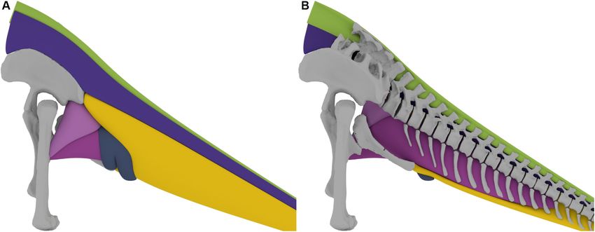
Díez Díaz et al. Giraffatitan Tail Reconstruction
to the hindlimbs. This leaves only extant crocodilians as a (from the twenty-sixth to the thirty-second, missing the
model for the configuration and architecture of the caudal thirty-first) were found associated (Janensch, 1950a). No
musculature for sauropod dinosaurs. Modern crocodilians can chevrons were found next to this caudal series.
be used to infer the set of epaxial and hypaxial tail muscles – Series MB.R.2921 consists of the first 18 caudal vertebrae
present in sauropod dinosaurs, especially given the similarities (MB.R.2921.1-18) and fourteen of their chevrons
in the osteological correlates (see below). The musculoskeletal (MB.R.2921.19-32), found in an articulated sequence
system of the crocodilian tail has been described in detail by behind the last sacral vertebra (Janensch, 1950a).
many researchers (e.g., Hair, 1868; Romer, 1923; Frey, 1982a,b,
1988; Frey et al., 1989; Cong et al., 1998; Hutchinson and Janensch (1950a,b) mentioned severe taphonomic damage to
Gatesy, 2000; Wilhite, 2003; Allen et al., 2014). We focused on series “no” (MB.R.5000), including shrinking of many centra.
these publications for the reconstruction of the tail muscular Additionally, there are some anatomical differences between
anatomy of Giraffatitan brancai. We follow the homologization MB.R.5000 and the other two caudal series, in particular
of different epaxial and hypaxial muscle groups in archosaurs by concerning the presence of pneumatic features (Wedel and
Tsuihiji (2005, 2007). Taylor, 2013). Series MB.R.5000 was therefore used only for
In addition, crocodilians are the sole living large reptiles that comparative purposes for this study, especially considering
can walk with the belly and most of the tail being held off that access to it on the mount is difficult. Series MB.R.2921
the ground. They lift their tail from the ground during high and MB.R.3736 resemble each other in osteology, and were
walk (distal half of tail sometimes on the ground, proximal therefore primarily used for this study. For the analysis of the
half lifted up) and gallop (tail lifted completely), although anatomical features both these caudal series were studied, but
this is not employed over long distances (see e.g., Cott, 1961; only MB.R.2921 (the better preserved series) was used for the
Webb and Gans, 1972; Zug, 1974; Gatesy, 1991b; Hutchinson three-dimensional reconstruction of the musculoskeletal system.
et al., 2019). This makes them the closest available analog to Especially information on the neural arches and the epaxial
sauropod dinosaurs. musculature could be more confidently obtained in the caudal
Institutional abbreviations: MB.R. Museum für Naturkunde series MB.R.2921 and its elements are overall more complete than
Berlin, Berlin, Germany. those of MB.R.3736.
Series MB.R.2921 shows some obvious damage: the left
transverse processes of caudal vertebrae 1–3, 5–7, and 9 are
MATERIALS AND METHODS missing, as is the right transverse processes of caudal vertebra
4, while those of caudal vertebrae 3 and 5 are damaged. The
Material left prezygapophyses of all vertebrae within the entire series are
The lectotype of Giraffatitan brancai (Taylor, 2009, 2011) is well preserved, but the right prezygapophysis is missing in caudal
the partial skeleton SI (MB.R.2180), and the paralectotype vertebra 3. The postzygapophyses are all preserved but for the
is SII (MB.R.2181). The mounted skeleton in the exhibition right one in caudal vertebra 2.
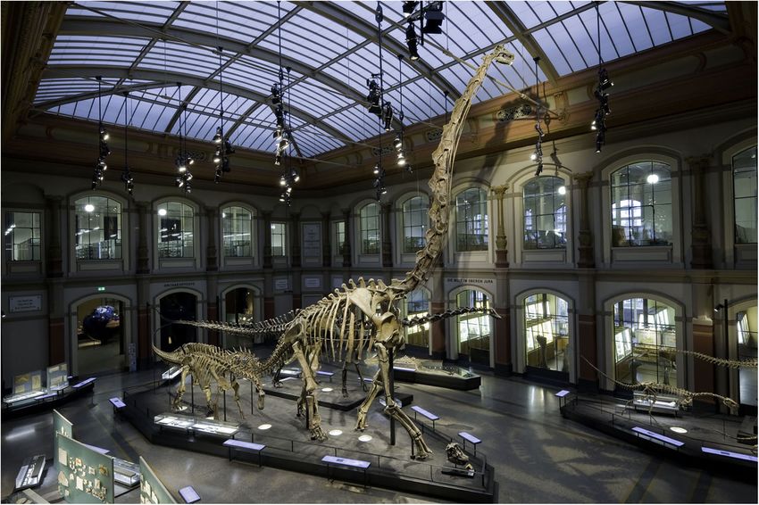
of the Museum für Naturkunde (Figure 1) is a composite The damage in series MB.R.3736 is far more extensive. Only
reconstruction, consisting mostly of MB.R.2181 (SII), and a small number of caudal vertebrae preserve zygapophyses, and
including elements of MB.R.2180 (SI), duplicates of some bones, most neural arches and spines as well as transverse processes
and plaster reconstructions of the missing parts (Janensch, 1950b; are missing or badly damaged. The centra, in contrast, are
Remes et al., 2011). However, neither SI nor SII included caudal mostly well preserved.
vertebrae, even though other skeletal remains of Giraffatitan were One partial sacrum was found in the same quarry “Aa,” as
found in the same quarry “S” in the Tendaguru area. Therefore, were the caudal series MB.R.2921 (Janensch, 1950a, fig. 74).
Janensch used the vertebrae from quarry “no,” MB.R.5000, for the However, this sacrum could not be re-located in the collections
mount (Janensch, 1950b). of the MfN, and it must be assumed that the original fossil
In total, three caudal series are known from the Tendaguru was lost during WWII. A simplified cast of it exists in the
area for Giraffatitan brancai: MB.R.5000 (from quarry skeletal mount. For the development of the three-dimensional
“no”), MB.R.2921 (from quarry “Aa”) and MB.R.3736 musculoskeletal model we used this sacrum cast MB.R.5003,
(from quarry “D”). as well as the right femur MB.R.5016 (quarry number “Ni”),
Giraffatitan brancai had ca. 50–60 caudal vertebrae. right tibia MB.R.2181.84 (quarry number “SII”) and right fibula
MB.R.2181.85 (quarry number “SII”) on display, adjusting their
– The longest caudal series recovered has 50 caudal vertebrae size to match the caudal series.
(MB.R.5000). It consists of the second to fifty-first caudal
vertebrae (Janensch, 1950a). As Janensch (1950a) states,
these caudal vertebrae were found “not articulated, with the Methods
exception of a few at the end, but altogether relatively in The fossils were digitized via photogrammetry, following the
sequence.” protocols of Mallison and Wings (2014), and the updated
– Series MB.R.3736 consists of 29 caudal vertebrae. The first version of Mallison et al. (2017). A digital SLR camera (Canon
23 caudal vertebrae (from the second to the twenty-fourth EOS 70D with Canon 10–18 mm f4.5-5.6 lens) was used
of the series) were found in articulation, and the rest with a LED ring light. Images were processed in Agisoft
Frontiers in Earth Science | www.frontiersin.org 3 May 2020 | Volume 8 | Article 160
Díez Díaz et al.                                                                                                                          Giraffatitan Tail Reconstruction

    FIGURE 1 | The current mount (on display since 2007) of the Late Jurassic sauropod Giraffatitan brancai (foreground) at the main hall of the Museum für Naturkunde
    (Berlin, Germany). Photograph by Antje Dittmann (MfN).

PhotoScan Professional v.1.4.01 , in order to obtain three-                             impact of preconceived ideas (e.g., overall downward position
dimensional models of each bone. High-quality polygon mesh                              of the tail, as in the former reconstruction of the mounted
files were created (approximately 5 million polygons and 250 MB                         skeleton of Giraffatitan). Additionally five cartilaginous neutral
as binary STL files each) for curatorial and museological                               poses (CNP, after Taylor, 2014) were assessed to test the influence
purposes, but also lower resolution color-free STL files (50.000                        of the intervertebral cartilage on the muscle volume.
polygons) for the musculoskeletal modeling and biomechanical                                As previously commented, we only can rely for a 50% EPB
analysis presented here.                                                                level I inference when reconstructing most of the muscles in
    Several chevrons were poorly preserved, some with missing                           terms of insertions and passages, as we only have one part of
parts, e.g., the distal part of the blade, or one of the rami                           the bracket (i.e., crocodilians). However, the general presence of
(MB.R.2921.19-20, 24, 29-30, 31). These elements were digitally                         the reconstructed muscles for tail and femur are present also in
restored in zBrush 4R7 (Pixologic)2 , either by mirroring the                           birds in most cases (e.g., the suite of epaxial muscles – at least
preserved ramus, or by scaling and superimposing the distal part                        until the pygostyle –, and the M. caudofemoralis longus), so a
of the blade of adjacent chevrons. The two missing chevrons, 1                          level II inference would only apply to the M. ilioischiocaudalis.
and 12, were entirely created digitally. Chevrons MB.R.2921.19                          Besides, and as already mentioned, those bones present in birds
(the second) and MB.R.2921.29 (the thirteenth) were used as                             too (i.e., femur, sacrum and first two caudal vertebrae) are very
proxy models and scaled to fit into the sequence, but also to the                       strongly modified in extant taxa, as are the muscles because
articular facets of their corresponding caudal vertebrae.                               of their locomotionary differences. Morphologically, the sacrum
    All 3D models were imported into Rhinoceros 5.0 (McNeel                             and femur of sauropods are more similar to crocodilians than to
Associates)3 and articulated in the osteological neutral pose                           birds, helping to confirm this level II inference.
(ONP, after Stevens and Parrish, 1999, 2005a,b; Mallison,                                   The three-dimensional models of the musculature were
2010a,b) following the protocol described by Mallison (2010a,b):                        created in the software package Autodesk Maya4 . We used a
individual vertebrae were articulated in pairs to minimize the                          polygon-based modeling approach, similar to the box modeling
                                                                                        approach by Rahman and Lautenschlager (2017), to build each
1
  http://www.agisoft.com                                                                muscle individually from the origin to the insertion, on the
2
  http://pixologic.com/features
3                                                                                       4
  www.rhino3d.com                                                                           https://www.autodesk.com/products/maya/overview

Frontiers in Earth Science | www.frontiersin.org                                    4                                                 May 2020 | Volume 8 | Article 160
Díez Díaz et al. Giraffatitan Tail Reconstruction
basis of osteological correlates identified by study of the physical We therefore reassembled the series MB.R.2129 based on
and digital specimen. This allowed us greater control over the centra faces to create a more life-like, less taphonomy-influenced
flow of the muscle shape than in a NURBS-based approach reconstruction (Figure 2C), with higher lateral symmetry, which
(e.g., Persons and Currie, 2011; Persons et al., 2014). Finely we used for the 3D reconstruction of the musculature. This
segmented muscles, e.g., the transversospinalis group, were second articulation consequently suffers from some additional,
simplified into a single body. massive intersection between zygapophyses. While the anterior
Bone volumes were calculated with Rhinoceros 5.0, and rim of the first caudal vertebral centrum is markedly concave in
muscle volumes with Maya. For the muscle mass we use lateral view, the posterior rim is flat in lateral view. In all other
the density value proposed by Méndez and Keys (1960) for vertebrae, both the anterior and posterior rims of the centra are
mammalian muscles (d = 1.06 × 10³ kg/m3 ), which is similar to mostly flat in lateral view, allowing a straightforward assembly
the measurements obtained by Hutchinson et al. (2015) for the of the tail based on the centra faces. Therefore, we were able to
hindlimb muscles of an ostrich (Struthio camelus). create this centra-based articulation without any uncertainty with
For the calculation of bone mass the following factors were regards to the angles between the vertebrae.
taken into consideration. Most caudal vertebrae of Giraffatitan The chevrons were positioned into the existing articulated
have small pneumatic fossa indicating a small amount of caudal vertebral series by matching their anterior and posterior
pneumatisation. Overall, the pattern of pneumatisation in articular facets with the orientation of the ventral articulation
Giraffatitan caudal vertebrae is variable and irregular (Wedel surfaces of each centrum. A short space was retained between the
and Taylor, 2013). As the caudal vertebral centra have a high chevron and the vertebra, representing the volume of the articular
volume to surface ratio, we estimate their cortical portion to be cartilage. We found that the centra faces could be retained in
rather smaller than in girdle bones, ribs, or complexly shaped practically parallel orientation with the chevrons added.
presacral vertebrae, and the trabecular and marrow portion to be Overall, series MB.R.2129 shows a slight curvature in lateral
accordingly larger. The volumetric bone density is typically close view, lifting the posterior end by ca. one full centrum height
to 2 × 10³ kg/m3 for long bones (e.g., Mohiuddin, 2013; Fletcher of caudal vertebra 18 compared to the trend of the first three
et al., 2018) and somewhat lower for marrow-rich bones, due to vertebrae. Repeated “playing” with the bone trios (two vertebrae
the lower density of marrow of ca. 1 × 10³ kg/m3 . We here chose and their chevron) to optimize alignment tended to increase
1.5 × 10³ kg/m3 for overall bone volumetric density, expecting rather than decrease the curvature.
the amount of pneumaticity to be the main driver von average In the skeletal reconstruction of the MB.R.3736 series a slight
density variation in the tail of Giraffatitan. sigmoidal shape is created by centra-only alignment in the
long axis of the tail in lateral view, between the fifteenth and
eighteenth vertebrae, and the terminal section bends slightly
RESULTS ventrally (Figure 2D).
Osteologically Neutral Pose (ONP)
For exact alignment we found the reduced size files with 50.000 Cartilaginous Neutral Poses (CNPs)
polygons insufficiently detailed. We therefore worked with the (Figure 3)
higher-resolution files with five million polygons. Several previous studies analyzed the ONP of the neck of
Initially, we used the prezygapophyses and postzygapophyses sauropods and the effect of the intervertebral cartilage (e.g.,
as proxy for assessing the correct articulation between vertebrae, Taylor and Wedel, 2013a; Taylor, 2014; Vidal et al., 2020a and
assuming that given full zygapophyseal overlap and – as far references therein), but there is scant published data on the
as possible – sub-parallel centra faces, the distance between effects of intervertebral cartilage within the tail. As stated by
vertebral bodies probably reflects the approximate intervertebral Taylor and Wedel (2013a) the thickness of the articular cartilage
cartilage volume (Christian and Preuschoft, 1996; Christian and between the centra of adjacent vertebrae affects posture. In this
Dzemski, 2007). work we also assess how this cartilage thickness affects the mass,
Articulating the zygapophyses of series MB.R.2129 with volume, and extension of the musculature (see below). However,
full overlap (Figure 2E) resulted in a nearly straight line of for extinct taxa we can only make assumptions about the cartilage
vertebrae, but tilted some of the centra around their vertical that existed in life. For that, five CNPs (Taylor, 2014) with
and longitudinal axes (Figures 2A,B). Also, a slight but different cartilage thicknesses (2.5, 5, 10, 15, and 20%) were
noticeable asymmetry and “twisting” around various axes of assessed. Cartilage thicknesses percentages were chosen after the
most neural arches, including the postzygapophyses, induced a calculations of Taylor and Wedel (2013a) and Taylor (2014) for
slight sigmoidal curve, initially to the left, then to the right. The the cervical vertebrae of several extant animals and sauropod
deformations also caused a long-axis rotation in parts of the taxa, as data on caudal series is no available yet. Cartilage
series, tilting the neural spines of caudal vertebrae 3 through thicknesses were calculated as percentages of the centrum lengths
8 noticeably to the left (Figure 2B). Additionally, due to slight (Supplementary Table S1).
taphonomic deformation of some zygapophyses, we could not Intervertebral cartilage volumes were added to the previous
completely avoid small intersections between zygapophyses (e.g., ONP. Taylor (2014) quantified the angle of extension at
between caudal vertebrae 17 and 18) or larger, unrealistic gaps in intervertebral joints when different cartilage thicknesses were
the zygapophyseal joints. included. This extension occurs because the thin zygapophyseal
Frontiers in Earth Science | www.frontiersin.org 5 May 2020 | Volume 8 | Article 160Díez Díaz et al. Giraffatitan Tail Reconstruction FIGURE 2 | Left lateral views (A,C,D) and dorsal view (B) of the three-dimensional skeletal reconstructions of two caudal vertebral series of the Late Jurassic sauropod Giraffatitan brancai. (A–C). MB.R.2921 (D) MB.R.3736. (A,B) show alignment based on best fit of zygapophyses and sub-parallel centra, (C,D) show alignment based on centra faces. (E) Oblique close-up of the sets of zygapopyseal pairs (C 7–8 and C 8–9) from articulation in panels (A,B) aligned for best fit. Only the preserved remains have been illustrated here. cartilage has no or negligible effect on the angle of extension Several sauropod caudal series have been found articulated between vertebrae, while the thick intervertebral one does: or closely associated. Although it is uncertain linking the the angle of elevation at an intervertebral joint is increased intervertebral spaces found in these articulated remains with when cartilage is included. This is true for opisthocoelous the cartilage volume present in the living animal, we have and procoelous vertebrae, but in the caudal series MB.R.2129 checked them looking for hypothetical connections with our of Giraffatitan this dorsal or ventral extension is not present model. For example, the titanosaurian taxa Oversosaurus (Coria when different cartilage thicknesses are included, as these caudal et al., 2013) and Dreadgnothus (Lacovara et al., 2014) preserved vertebrae do not present condyles. Similar poses are obtained the first 20 caudal vertebrae articulated. Some measurements for the ONP and all the CNPs. However, it is important to were made using the published material (figures, and a 3D state that in CNP 15% the zygapophyses start disarticulating model for the Dreadgnothus caudal series), and some interesting from ca. seventeenth caudal onward, and in CNP 20% from data were obtained: for Overosaurus intervertebral spaces of the twelfth. An improved and more accurate model could be 26 to 28% the centrum length were calculated, while for created by articulating the zygapophyses and thickening the Dreadgnothus the values changed along the series, being larger base of the cartilage, resulting in a dorsal extension of the tail in the anterior section (ca. 40 to 44%) and decreasing through from this section. However, the cartilage volume is already thick the series (20 to 22%). One specimen (DFMMh/FV 100) of at CNP 20%, and by increasing it in its base probably would the camarasauromorph Europasaurus presents an articulated lead to disarticulation of the chevrons too. Together with other caudal series of 13 vertebrae (Carballido and Sander, 2014). results (see below) we do not consider this CNP 20% model It was not possible to take accurate measurements as most as a possibility for a living animal. In the other CNPs the of the vertebrae, although articulated, were displaced, but zygapophyses appear articulated, but a dorsal extension of the an intervertebral space of ca. 17–18% was calculated. The tail section that is not preserved could not be ruled out, as also brachiosaurid Padillasaurus preserved the first eight caudal seen in the CNP 15%. Besides, in the CNP 2.5 and 5% the caudal vertebrae in articulation (Carballido et al., 2015). The calculated vertebrae are spaced very tightly, thus limiting movement of the intervertebral spaces were between 26 and 28%. However, this tail, and furthermore making it impossible to correctly articulate specimen presents a dorsal extension, so the calculated cartilage the chevrons. Only the CNP 10% and 15% are therefore deemed thicknesses are probably not comparable to the ones of the possible for this caudal series. living animal. This dorsal extension of the tail is present in Frontiers in Earth Science | www.frontiersin.org 6 May 2020 | Volume 8 | Article 160
Díez Díaz et al.                                                                                                             Giraffatitan Tail Reconstruction

  FIGURE 3 | Comparison between the (A) ONP, (B) CNP at 2.5%, (C) CNP at 5%, (D) CNP at 10%, (E) CNP at 15%, and (F) CNP at 20%. Scale = 50 cm.

Frontiers in Earth Science | www.frontiersin.org                            7                                             May 2020 | Volume 8 | Article 160
Díez Díaz et al.                                                                                                                             Giraffatitan Tail Reconstruction

  FIGURE 4 | (A) Origins and insertions of the caudal musculature of the Late Jurassic sauropod Giraffatitan brancai. (B) Simplified muscle paths of the
  transversospinalis group of the epaxial musculature. (C) Cross-section of the tail at the fourth caudal vertebrae showing the lateral extent of the tail musculature. The
  line in A indicates the location of the cross-section through the musculature. Paired ventral elements represent the ischia.

other sauropod caudal series, as Camarasaurus (Gilmore, 1925)                            Neural Arches
or Spinophorosaurus (Remes et al., 2009). As already seen,                               The lateral faces of the neural spines are slightly concave and
numerous taphonomical factors need to be taken into account,                             show longitudinal (apicoventrally directed) rugosities, parallel
so we cannot use these values with fidelity. Besides, they also                          to each other. At the apical edge of the neural spine both
indicate that cartilage thickness could highly vary through the                          anterior and posterior processus – with different development
caudal series, but also depend on the taxon. In addition, the                            stages depending on the position in the series – can be seen
articulation type between vertebrae (i.e., procoely, opisthocoely,                       in form of the distal tips of both the prespinal and postspinal
etc.) probably affected this thickness too. A recent work on                             laminae. From caudal vertebra 3 on, the middle part of the tip of
the caudal biomechanics of the titanosaur Aeolosaurus maximus                            the neural spine is thickened, developing an apical prominence
suggests a cartilage thickness between 5 and 10% for the anterior                        (slightly posteriorly displaced) from the fourth vertebra on.
section of the tail (Vidal et al., 2020b). More detailed work on                         From caudal vertebra 8 on, this bulge shows sharp posterior
these sauropod articulated caudal series needs to be undertaken                          edges in dorsoventral orientation on the lateral faces of the
to approach more accurately this issue.                                                  neural spines. In addition, three spurs are present in the neural
                                                                                         spine laminae: two on the base of the spinoprezygapophyseal
                                                                                         laminae, and one in the postspinal lamina. The spike present
Muscle Marks and Musculature                                                             on the postspinal lamina appears in the caudal vertebrae 5, 6,
Reconstruction                                                                           8–11, and 14. However, this feature could have been present
The caudal vertebrae and chevrons of Giraffatitan presents                               in all caudal vertebrae with a distinct postspinal lamina. The
numerous osteological correlates related to the origins                                  prominences of the spinoprezygapophyseal laminae appear in
and insertions of the muscles and ligaments of the tail                                  the caudal vertebrae 2, 3, 5–7, although they were probably
(Figure 4 and Supplementary Figure S1). The inferred sets                                present from the first caudal vertebrae to the seventh. These
of epaxial and hypaxial muscles, together with the relevant                              osteological features correspond to the attachment of deep
hindlimb musculature for the correct reconstruction of the tail                          epaxial muscles (mediodistally) and M. multifidus (medially),
musculature (which are directly in or close to contact and/or                            and both the M. spinalis and M. articulospinalis (laterally). The
restrict the volume of the tail musculature) are detailed in                             M. multifidus originates from the distal posterior tip of the neural
Supplementary Table S2.                                                                  spineM. articulospinalis from the distal posterior tip.

Frontiers in Earth Science | www.frontiersin.org                                     8                                                    May 2020 | Volume 8 | Article 160
Díez Díaz et al. Giraffatitan Tail Reconstruction
Zygapophyses (just below the transverse process). In these anterior vertebrae the
The zygapophyses serve as attachment areas for the epaxial distal lateral surface of the centrum serves as attachment for the
muscles including the M. spinalis (zygapophyseal joint capsule) M. caudofemoralis longus. Caudal 11 (right side) and caudal 12
and M. articulospinalis (lateral rugosity on the prezygapophyses). shows the first occurrence of a lateral broadening of the centrum
The M. multifidus inserts in a spur or rugosity on the at half its height, forming an incipient ridge, of which there is
spinoprezygaposeal lamina of the second next vertebra (from no trace in the previous vertebrae. This ridge gradually moves
the first to the seventh vertebrae), or in the subtle dorsal ventrally in the next three vertebrae on both sides, separating two
rugosity on the prezygapophyses from the eighth vertebra lateral surfaces, and merging into the ventrolateral border of the
onward. The M. articulospinalis inserts on a lateral rugosity of centrum. We interpret this weakly developed ridge as the caudal
the prezygapophysis two vertebrae further down the tail. The limit of the M. caudofemoralis longus, constricting the muscle into
development of the mentioned attachment structures also depend a narrow tip well separated from the transverse processes, unlike
on the position in caudal series, being steadily reduced distally. the dorsally directed tapering seen in extant crocodiles. After the
fourteenth caudal the ridge merges into the ventrolateral edge
Vertebral Centra of the centrum; accordingly, the ventrolateral surface disappear
Although the prezygodiapophyseal lamina is not well developed, entirely, indicating that the M. caudofemoralis longus does not
an oblique bulge and rugosity can be seen between the extend distally beyond this point.
prezygapophysis and the transverse process, which is
interpreted as the osteological correlate for the insertion of Chevrons
the M. tendinoarticularis. The lateral surfaces of the chevrons and their general
The transverse process is an important osteological correlate morphology are important osteological correlates for the
for both the epaxial and hypaxial muscles. Its distal tip is an hypaxial musculature and their development. In the lateral
insertion for the hypaxial musculature (dorsal aspect of the surface of the chevrons, more dorsally located in the anterior
M. ilioischiocaudalis). A bulge and rugosity at the junction ones, a weak oblique rugosity appears, for the insertion
between the transverse process and the centrum (proximal tail) of the M. caudofemoralis longus. On the distal tip, in its
and longitudinal ridge (13th vertebra onward) acts as attachment lateral aspect, another rugosity, for the insertion of the
for the bounding septum (Grenzseptum) between the lateral ventral part of the M. ilioischiocaudalis, is apparent. The
epaxial musculature (M. tendinoarticularis and M. longissimus morphology of the distal blade of the chevrons changes
caudae) and is further the insertion for the M. longissimus caudae. along the series: the first three chevrons have a more acute
Transverse processes are present in all the caudal vertebrae distal blade, while the next ones have a more rounded
of MB.R.2921, becoming smaller and rounder distally. In and transversally compressed distal third. From the twelfth
MB.R.3736 the transverse processes disappear by caudal vertebra chevron (thirteenth caudal vertebra) onward, this distal
25 in the series. As they disappear, the mentioned longitudinal third of the blade becomes more posteriorly directed. These
ridge persists close to the junction between the neural arch and differences in morphology reflect a change in the insertions
the centrum, delineating the insertion of both the M. longissimus and development of both M. caudofemoralis longus and
caudae (dorsally) and the M. ilioischiocaudalis (ventrally). M. ilioischiocaudalis.
The major part of the M. caudofemoralis brevis originates from
the medial surface of the postacetabular process of ilium and the
ventral aspect of the last sacral rib. From there, the muscle extents Sizes, Masses and Volumes
distally and also attaches anterolaterally on the centra of caudal From the addition of all individual bone and muscle masses
vertebrae 1 to 3. (Table 1 and Supplementary Tables S3, S4) we here suggest
The first two caudal centra of MB.R.2921 show a lateral hypothetical weights for the preserved caudal series MB.R.2921,
concavity below the transverse processes, which we interpret as depending on the ONP and CNPs.
a space for cryptic diverticula (see below). However, between
these depressions and the transverse process in caudal vertebra ONP
2 there is a shallow shelf, there are subtle bumps on caudal The caudal series MB.R.2921 presents a total length (from the
vertebra 3 to 5 at this position, which we regard to be the first caudal vertebra to the last) of 280.82 cm when articulated
attachment site of the medial head of the M. ilioischiocaudalis in ONP. All the caudal bones (vertebrae and chevrons) weighted
(see Supplementary Figure S2). Ventral to the lateral depression 106.92 kg (1775.67 kg with the pelvis and sacrum), and all
in caudal vertebra 2 and approximately at the same position the muscle (right and left) groups 950.07 kg. So the preserved
on caudal vertebra 3 there is a faint ridge, which serves caudal series MB.R.2921 of Giraffatitan approximately weighted
as the origin for the M. transversus perinei. The muscle 1056.99 kg in total (2725.74 kg with the pelvis and sacrum). The
extends laterally, wrapping around the M. caudofemoralis total volume of the caudal series is 967.57 liters (2080.07 liters
longus and the ischial ramus of the M. ilioischiocaudalis to with the pelvis and sacrum). It is important to keep in mind
insert on the distal lateral ischium and on the aponeurosis that not all the vertebrae were completely preserved (e.g., some
surrounding the cloaca. of them were missing parts of the transverse processes), and
The ventral half of the centrum is convex in the anteriormost some epaxial muscles have been simplified, but this should not
nine vertebrae, and then becomes gradually more concave affect the volume.
Frontiers in Earth Science | www.frontiersin.org 9 May 2020 | Volume 8 | Article 160Díez Díaz et al. Giraffatitan Tail Reconstruction
TABLE 1 | Comparison of the calculated volumes (l) and masses (kg) for each reconstructed muscle group of the caudal series MB.R.2921 of the Late Jurassic
sauropod from Tanzania Giraffatitan in CNP and CNPs.
ONP CNP_2.5% CNP_5% CNP_10% CNP_15% CNP_20%
Volume (l)
Muscle TSP 22.669 20.802 21.312 22.325 23.340 24.354
LC 76.924 70.681 72.407 75.853 79.303 82.750
IIC 121.276 114.303 117.115 122.738 128.359 133.982
IICmed 10.139 9.235 9.451 9.885 10.319 10.753
CFB 36.952 35.003 35.406 36.213 37.018 37.825
CFL 170.262 156.311 159.894 167.583 175.097 182.611
TRPR 9.923 8.972 9.210 9.684 10.159 10.633
Total 448.144 415.307 424.795 444.281 463.595 482.908
Mass (Kg)
Muscle TSP 24.029 22.050 22.591 23.665 24.740 25.815
LC 81.539 74.922 76.751 80.404 84.061 87.715
IIC 128.553 121.161 124.142 130.102 136.061 142.021
IICmed 10.747 9.789 10.018 10.478 10.938 11.398
CFB 39.169 37.103 37.530 38.386 39.239 40.095
CFL 180.477 165.690 169.488 177.638 185.603 193.568
TRPR 10.518 9.510 9.763 10.265 10.769 11.271
Total 475.032 440.225 450.283 470.938 491.411 511.882
The muscle volumes were calculated with the software Maya. The muscle masses were calculated using the density value proposed by Méndez and Keys (1960) for
mammalian muscles (d = 1.06 × 10³ kg l/m3 ). CFB, m. caudofemoralis brevis; CFL, m. caudofermoralis longus; IIC, m. ilioischiocaudalis; IICmed, m. ilioischiocaudalis
medial head; LC, m. longissimus caudae; TRPR, m. transversus perinei; TSP, Transversospinalis group. See Supplementary Figure S4 for the individual segments.
CNPs DISCUSSION
The calculated lengths (from the first caudal vertebra to the last)
and masses for the caudal series MB.R.2921 when articulated in ONP and CNPs
the five CNPs are detailed in Table 2. In light of the steep position of the tail base on the mounted
A length difference of 44.76 cm is calculated from the lowest skeleton, a slight upward turn in the distal part of the anterior
to the highest length values. With our model we suggest that tail section as suggested for MB.R.2129 is not surprising, seeing
the chosen intervertebral cartilage volume could affect between how it is required to keep a full tail of ca. 50 vertebrae from
14.5 and 17% the total length of the tail. A muscular mass dragging on the ground. Overall, the good fit of the alignment
difference of 143.32 kg can be stated between the CNP with with subparallel centra faces and an overall rather straight long
the lowest cartilage thickness value (2.5%) and the model with axis matches other sauropods and in fact most dinosaurs well
the highest value (20%). With our current model we can (Upchurch et al., 2004). We therefore find no indication that
hypothesize that the chosen cartilage thickness could affect the the strong anterior uptilt of the hip of Giraffatitan in any major
total mass of the reconstructed muscles between ca.12 and way influenced the overall biomechanical organization of the
14% (ca. 5% when we also take into account the mass of the tail. The total lack of keystoning, already mentioned in relation
pelvis and sacrum). The increase of the muscle volumes and to overall tail articulation by Janensch (1950a,b) is a marked
masses is proportional to the increase in cartilage thickness contrast to e.g., the basal sauropodomorph Plateosaurus, in which
(r2 = 1, p-value < 5.00E-07) for each muscle individually and all the addition of chevrons to the digital mount forced a wedge-
combined (see Supplementary Table S3). shaped gap between the caudal vertebral centra that induced a
straight tail axis (Mallison, 2010a). Without the chevrons, i.e.,
with parallel centra faces, the tail of Plateosaurus would show a
TABLE 2 | Total lengths (cm) and masses (kg) calculated for the caudal series
MB.R.2921 of the Late Jurassic sauropod from Tanzania Giraffatitan in CNP and
significant ventral curvature (Wellnhofer, 1993; Moser, 2003). In
CNPs. Giraffatitan, the distance between chevron and vertebra, caused
by the articulation process described, is consistent throughout the
Total length Mass (w/o pelvis) Mass (w/pelvis)
tail. N.B.: The articulation in the 3D model is noticeably tighter
ONP 280.82 1056.99 2725.74 than on the mounted skeleton, where the support rod for the
CNP_2.5% 262.15 987.37 2656.12 tail runs between centra and chevrons, making an anatomically
CNP_5% 268.55 1007.49 2676.24 correct articulation impossible.
CNP_10% 281.34 1048.8 2717.55 A curvature similar to that seen in MB.R.3736 is also discer-
CNP_15% 294.12 1089.74 2758.5 nible in the hypothetical caudal vertebral series of the titanosau-
CNP_20% 306.91 1130.69 2799.43 rian sauropod Lirainosaurus (Vidal and Díez Díaz, 2017).
Frontiers in Earth Science | www.frontiersin.org 10 May 2020 | Volume 8 | Article 160Díez Díaz et al. Giraffatitan Tail Reconstruction
However, MB.R.3736 is noticeably straighter, probably because for determining the distal end of the M. caudofemoralis longus.
of the articulation morphology between the caudal vertebrae. Persons and Currie, 2011 found that the process expands beyond
Lirainosaurus has highly procoelous caudal vertebrae, with the distal end of the M. caudofemoralis longus in some squamates.
anterior and posterior articular surfaces highly inclined In Alligator mississippiensis the M. caudofemoralis longus ends at
(keystoned centra, see Vidal and Díez Díaz, 2017, fig. 6B). the fourteenth caudal vertebra, the first one without transverse
When comparing the calculated values for the ONP and the processes (Frey, 1988; Mallison, 2019).
CNPs, it can be observed that the most similar lengths and masses
are obtained for the ONP and the CNP with an intervertebral – The ventrolateral surfaces of the centra appear from the
cartilage thickness of 10% of the centrum length. third to the fourteenth caudal in MR.2921.
Taylor (2009) calculated a volume of 1520 liters (∼1216 kg, – The dorsolateral rugosity is present from chevrons 4 to 11
although bone and muscle densities cannot be separated from (caudal vertebrae fifth to twelfth).
these calculations) for the tail of Giraffatitan, after a modified
reconstruction of Paul (1988). Most of the mass of the tail Therefore, we inferred the extent of the M. caudofemoralis
was probably located at its base, where the largest parts of the longus from the first caudal vertebra onward and not beyond
muscles are placed together with the heaviest bones. It is therefore the fifteenth caudal vertebra in Giraffatitan. While the maximal
reasonable to suggest that in Giraffatitan at least half of the extent of the M. caudofemoralis longus is important for volume
weight of the tail would have been placed in the first 20 caudal calculation (and thus maximal force estimates) the bulk of the
vertebrae. When following our reconstruction and results, the tail muscle is located in the anterior region, thus the influence
of Giraffatitan could have weighted ca. 2500 kg (not including the of a slightly longer or shorter muscle (±1 vertebra) are only
pelvis and sacrum), doubling Taylor’s calculations. minor on the total volume. However, a correct reconstruction of
M. transversus perinei is more important for an accurate estimate
of hip joint moments than an exact determination of the taper
Reconstruction of the Mm. point of M. caudofemoralis longus. The M. transversus perinei
caudofemorales acts as a lateral constraint on the M. caudofemoralis longus.
The development of the Mm. caudofemorales, especially the If no M. transversus perinei is reconstructed, there is a high
M. caudofemoralis longus, has always been a major issue risk of overestimation of the M. caudofemoralis longus volume
when reconstructing the tails of extinct animals. The general at is base, where even minimal changes to the lateral extent
morphology of the Mm. caudofemorales (as well as their induce large volume changes, and accordingly a misestimation of
origins and insertions) seems to be highly conservative within its overall power.
crocodilians (see e.g., Gatesy, 1991a; Ibiricu et al., 2014). Previous In basal saurischians (e.g., Eoraptor and Guaibasaurus) and
studies suggested that in crocodilians the M. caudofemoralis non-avian theropods the M. caudofemoralis brevis originates
longus originates from the sides of the centrum and ventral from the brevis fossa, a transitional structure located on the
surface of the transverse processes of caudal vertebrae 3–15 ventromedial surface of the ilium (Carrano and Hutchinson,
(Romer, 1923; Galton, 1969). However, Wilhite (2003) confirmed 2002; Holtz and Omólska, 2004; Langer, 2004; Makovicky and
that this muscle additionally originates from the lateral surface Norell, 2004; Makovicky et al., 2004; Norell and Makovicky,
of the first 13 chevrons, but only runs along the underside of 2004; Omólska et al., 2004; Tykoski and Rowe, 2004). However,
the transverse processes, from which it is separated by a layer of in sauropods the origin of the M. caudofemoralis brevis is
connective tissue and, in well-fed individuals, by a layer of fat. somewhat ambiguous, as they lack a brevis shelf and fossa
Therefore, chevron morphology may be indicative of the size, (Upchurch et al., 2004), similar to the condition in extant
shape, and extent of M. caudofemoralis longus in fossil archosaurs. crocodilians. In Giraffatitan a concave surface appears medially
This hypothesis is also followed by Otero and Vizcaíno (2006, in the postacetabular process, below the last sacral rib and in the
2008). The development and morphology of the transverse junction with the ilium. This surface is inferred as the origin of
processes and the lateral and ventral surfaces of the centra are the M. caudofemoralis brevis, in combination with the anterior
therefore important indicators for the size, shape and extent of lateral surface of the centra of caudal vertebra 1 to 3.
M. caudofemoralis longus in sauropod dinosaurs. Several previous The Mm. caudofemorales insert on the fourth trochanter of
studies highlight the importance of the lateroventral surfaces the femur. In sauropods the trochanter appears as a longitudinal
of the anterior caudal centra for the origin and development ridge, without any differential sites for the insertion of both
of the Mm. caudofemorales in titanosaurian sauropods (Borsuk- M. caudofemoralis longus and brevis. Otero and Vizcaíno (2008)
Bialynicka, 1977; Salgado and García, 2002; Salgado et al., 2005), went as far as suggesting a common tendon for both muscles. In
and Gallina and Otero (2009) suggest that the development the case of Giraffatitan the trochanteric ridge is well-developed;
of the M. caudofemoralis brevis and M. caudofemoralis longus therefore, we modeled the insertion of each muscle separately,
occurs in relation with the anterior caudal transverse processes the M. caudofemoralis longus medially and the M. caudofemoralis
morphological variation along the tail. Several osteological brevis posterolaterally located to the fourth trochanter. In extant
correlates are indicative of the development and extent of the crocodiles the insertion is often highly complex, with the
M. caudofemoralis longus in Giraffatitan: tendon of the longus portion wrapping around the very short
The transverse processes disappear by caudal 25 in MB.R.3736. tendon of the brevis portion, and often inserting into it (Allen,
However, the absence of transverse processes cannot be used 2015; HM, 2017). However, this complexity leaves no trace on
Frontiers in Earth Science | www.frontiersin.org 11 May 2020 | Volume 8 | Article 160Díez Díaz et al. Giraffatitan Tail Reconstruction
FIGURE 5 | (A) 3D tail muscle reconstruction of the Late Jurassic sauropod Giraffatitan brancai. (B) Sagittal section of the tail musculature, showing the dorsal and
ventral extent of the musculature in relation to the vertebrae.
the bone, so that the exact paths and interactions cannot be analyzing the general trajectory of these muscles. As muscles
reconstructed reliably. are normally not arbitrarily constrained in width by other
soft-tissue, it was assumed that they follow a straight line from
the origin to the insertion and not in a concave trajectory. In the
Extent of the Caudal Musculature proximal region the M. caudofemoralis longus is constrained in
(Figures 5, 6) all directions. Medially it is confined by the muscles originating
The three-dimensional approach enabled the inference of the on the ischium, namely the Mm. adductores femores and the
size and the spatial arrangement of the musculature. Individual M. flexor tibialis internus 3. The Mm. adductores femores
muscles are not only constrained by their origins and insertions, originate on the obturator plate and the middle ischial shaft,
but also by the neighboring musculature. It is therefore for M. adductor femoralis 1 and M. adductor femoralis 2,
insufficient to model only the tail muscles. Consequently respectively. They run down medially on the hindlimb and
the nearby limb musculature was also modeled and taken insert on the posterior surface of the femur distal to the fourth
into account. Nevertheless, some uncertainties regarding the trochanter. The Mm. adductores femores are laterally covered by
development and proportions, lengths and volumes of the caudal the M. flexor tibialis internus 3, which originates on a shallow
musculature remain. In particular the ventral, lateral and dorsal depression on the proximal lateral ischium and inserts on the
extensions of the muscles are difficult to reconstruct, e.g., proximal tibia, and runs between the Mm. adductores femores
Persons (2009), Persons and Currie (2011), and Mallison (2011) and the M. caudofemoralis longus, thus limiting the latter
demonstrated that the soft tissues in the tails of crocodilians and medially. Dorsally and laterally the M. caudofemoralis brevis
many squamates extend significantly beyond the bones dorsally wraps around the M. caudofemoralis longus, and thus limits
and especially ventrally and laterally. Previous studies on caudal the extent in these directions. The M. caudofemoralis longus is
musculature in dinosaurs have predominantly used the extent of further constrained laterally by the M. flexor tibialis externus,
the bones as the extension for their soft tissue reconstructions which spans from the postacetabulur process of the ilium to the
(Carpenter et al., 2005, fig. 17.5; Arbour, 2009, fig. 9; Hutchinson proximal tibia and restricts both M. caudofemoralis longus and
et al., 2011, fig. 5), however, this minimal extension does not M. caudofemoralis brevis. Ventrally the M. caudofemoralis longus
appear in any living animal. Persons (2009) correctly pointed is constrained by the M. ilioischiocaudalis and the M. transversus
out that the lateral width of the transverse processes is often a perinei, as both encompass M. caudofemoralis longus and insert
poor indicator of the lateral extent of M. caudofemoralis longus on the distal ischium, thus preventing a ventral extent below the
across a wide range of reptiles. Lacertilians such as the leopard distal tip of the ischium, see Figure 5.
gecko (Eublepharis macularius), the tokay gecko (Gekko gecko) In Alligator mississippiensis, the M. tendinoarticularis is
and the green anole (Anolis carolinensis) possess tail muscles that only mildly developed in the tail base, corresponding to an
extend beyond the bones (Ritzman et al., 2012; Gilbert et al., 2013, increase in cross section of M. longissimus caudae (Frey, 1988).
fig. 1.B; Sanggaard et al., 2012, fig.2.C, SI Movie_S2). In these Because of the lack of osteological correlates for the contact
lizards, and in the crocodilian Alligator mississippiensis (see Frey, between the two muscles any reconstruction must remain
1988; Mallison, 2011) the hypaxial muscles are greatly expanded speculative, and this relative thinning of the M. tendinoarticularis
ventrally and laterally. compared to M. longissimus caudae has not been represented
The lateral extent of the ventral muscles, the in previous muscle volume modeling attempts (e.g., Arbour,
M. caudofemoralis longus and the M. ilioischiocaudalis, in 2009; Persons, 2009; Persons and Currie, 2011; Persons et al.,
the mid-tail region of Giraffatitan was determined in comparison 2014). However, in the caudal series of Giraffatitan, the lateral
with the extant crocodilian Alligator mississippiensis and in oblique bulge between the prezygapophysis and the transverse
Frontiers in Earth Science | www.frontiersin.org 12 May 2020 | Volume 8 | Article 160Díez Díaz et al. Giraffatitan Tail Reconstruction
of the M. ilioischiocaudalis and the lateral expansion of the
M. caudofemoralis brevis. Anatomically, and in terms of volume,
the M. longissimus caudae (epaxial), the M. caudofemoralis group
and the M. ilioischiocaudalis (hypaxial) seem to be the most
important muscles of the base of the tail. Then, from the fifth
caudal vertebra, when both rami of the M. ilioischiocaudalis meet
and totally enclose the M. caudofemoralis longus, all the muscular
groups occupy similar volumes.
Internal Structure of Epaxial Muscles
While we propose that the suite of epaxial and hypaxial tail
muscles of Giraffatitan, including the Mm. caudofemorales,
are generally comparable in terms of its general extensions,
origins and insertions to crocodilians, here are some key
differences between extant archosaurs and our inference for the
tail musculature in Giraffatitan. Extant crocodilians possess a
very specialized internal muscle architecture (e.g., Frey, 1988;
Salisbury and Frey, 2001). Whereas the medial epaxial muscles,
in particular M. multifidus, M. spinalis, and M. articulospinalis,
form a system of counter-running (criss-crossing) tendons, the
lateral epaxial muscles M. tendinoarticularis and in particular
M. longissimus form large myoseptal sheets and cones. The
tendons of the epaxial muscles are connected firmly not
FIGURE 6 | 3D reconstruction of the tail and hindlimb musculature of the Late only to the vertebrae, but also to the osteoderms. The close
Jurassic sauropod Giraffatitan brancai used to constrain the extent of the Mm. association between the myosepts and tendons of the epaxial
caudofemorales muscles. The m. ilioischiocaudalis and m. flexor tibialis muscles in crocodilians is important as it forms a part of
externus are semi-transparent to show the underlying musculature. In color their bracing system (Salisbury and Frey, 2001). Besides, in
(A) and labeled (B). ADD1, m. adductor femoris 1; ADD2, m. adductor
femoris 2; CFB, m. caudofemoralis brevis; CFL, m. caudofemoralis longus;
contrast to extant birds and sauropods, extant and fossil
FTE, m. flexor tibialis externus; FTI1, m. flexor tibialis internus 1; FTI3, m. flexor crocodilians do not possess postcranial skeletal pneumaticity
tibialis 3; IIC, m. ilioischiocaudalis; IICmed, m. ilioischiocaudalis medial head; (e.g., Gower, 2001).
IT2, m. iliotibialis 2; IT3, m. iliotibialis 3; ILFB, m. iliofibularis; LC, m. Additionally, birds also have a highly modified internal
longissimus caudae; TC, m. truncocaudalis; TRPR, m. transversus perinei; muscular construction, and thus their anatomy cannot simply
TSP, Transversospinalis group.
be extrapolated onto non-avian dinosaurs. In birds, the presacral
epaxial muscles form muscles slips that attach only to small
process (the inferred osteological correlate for the insertion areas of the bone (e.g., Boas, 1929; Zweers et al., 1987; Vanden
of the M. tendinoarticularis) becomes more prominent from Berge and Zweers, 1993). In combination with that, extant
the seventh vertebra onward (Supplementary Figure S3), bird skeletons are highly pneumatic, which means that the
disappearing again in the seventeenth caudal vertebra, which vertebrae are interspersed by a large number of pneumatic
formed the basis for the reconstruction and proportions of diverticula that occupy parts of the vertebral surface and
both the M. tendinoarticularis and the M. longissimus caudae, additionally resolve the vertebral surface to create pneumatic
confirming that the M. tendinoarticularis was less developed foramina (Gier, 1952; Duncker, 1971; Hogg, 1984a; Witmer,
at the base of the tail. However, the tendons and fascicles 1997; O’Connor, 2004), and muscle attachment areas are
of the epaxial musculature are highly intertwined and thus generally small.
muscle divisions are not easily distinguishable (e.g., Frey et al., For sauropod dinosaurs, a similar slip-like internal muscular
1989; Tsuihiji, 2005; Organ, 2006; Schwarz-Wings et al., 2009). architecture of the epaxial muscles as in extant birds has been
In our model we simplified the epaxial musculature by only hypothesized, based on the presence of unambiguous osteological
modeling two elements: one gathering all the dorsomedial correlates at the presacral vertebrae (see Wedel and Sanders,
muscles (the deep musculature, the M. multifidus, and the 2002; Taylor and Wedel, 2013b). Another similarity to birds is
Transversospinalis group), and the lateral M. longissimus caudae. the presence of vertebral pneumaticity at least in the presacral
However, for biomechanical analyses, and considering all the vertebral column of most neosauropods (Wedel, 2003a,b, 2009);
osteological correlates, individual musculo-tendon units are and additionally saltasaurine titanosaurs possess pneumatic
easier to create and model. sacral and anterior caudal vertebrae (Cerda et al., 2012; Zurriaguz
As indicated above, the lateral, ventral and dorsal extent of and Cerda, 2017).
the tail is difficult to estimate in extinct animals. The base of These similarities and differences need to be kept in mind
the tail is laterally delimited for three main muscles: dorsally by when reconstructing the internal structure of musculature of
the M. longissimus caudae, and ventrally by the dorsal ramus non-avian dinosaurs. However, our model is a simplification, and
Frontiers in Earth Science | www.frontiersin.org 13 May 2020 | Volume 8 | Article 160You can also read