AMSER Rad-Path Case of the Month: 55 year old female with a vulvar mass Courtney Wiley, MS4 - AGH Radiology Case ...
←
→
Page content transcription
If your browser does not render page correctly, please read the page content below
AMSER Rad-Path Case of the
        Month:

  55 year old female with a vulvar mass

                Courtney Wiley, MS4
         Medical University of South Carolina

         Dr. Jeanne Hill and Dr. Laura Spruill
         Medical University of South Carolina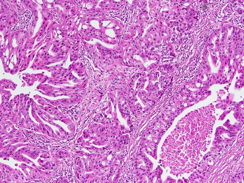
Patient Presentation
Clinical History: Pt is a 55 year old female who presented with a
palpable lump in her groin and a vulvar mass that had been present for
3 years. She noted discomfort with certain clothes and while sitting but
denied pruritus, bleeding, abnormal discharge and weight loss. After
initial treatment with mupirocin ointment failed, she had a lymph node
biopsy and imaging done.

PE: 5cmx3cm firm mass in the left labia majora with no discoloration or
ulceration of the overlying skin. There was also a 3cm palpable left
inguinal lymph node.
CCT Abdomen/Pelvis with Contrast (unlabeled)
CCT Abdomen/Pelvis with Contrast (labeled)

  Contrast enhanced coronal CT               Contrast enhanced axial CT better visualizes
  demonstrates inguinal lymphadenopathy      the entire vulvar mass, which measured
  (white arrow) and a partially visualized   2.7cm
  mass in the left labia majora (yellow
  arrow).
PET CT (unlabeled)
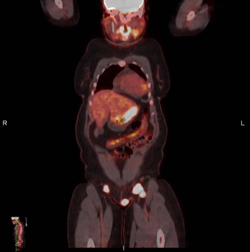
PPET CT (labeled)

Corresponding PET CT shows
hypermetabolic activity in the vulvar
mass (yellow arrow) with a max
SUV of 16.9) and bilateral inguinal
lymph nodes (white arrows).
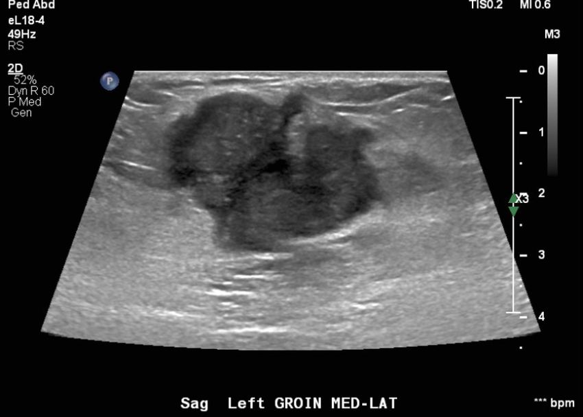
Ultrasound of Left Inguinal Lymph Node
              (unlabeled)
Ultrasound of Left Inguinal Lymph Node
(labeled)
Enlarged, round lymph node with irregular borders and heterogeneous echogenicity.DDX (Based on Imaging) • Vulvar cancer (adenocarcinoma, squamous cell carcinoma, melanoma, extramammary Paget disease) • Lipoma • Leiomyoma • Bartholin cyst • Mucinous cyst
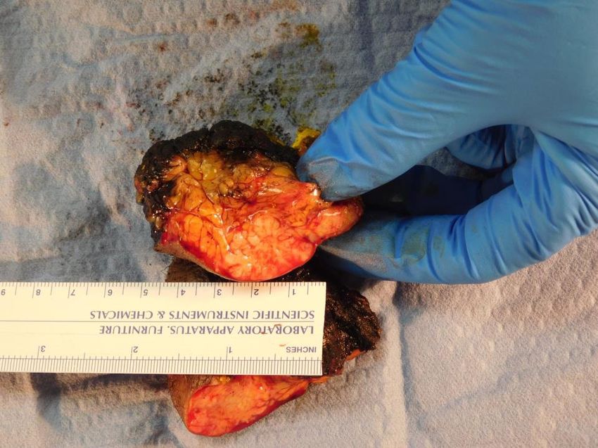
Gross Specimen

5cm firm mass
with no
ulceration of
overlying skin
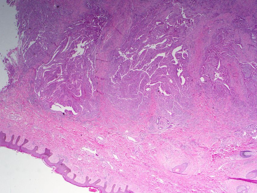
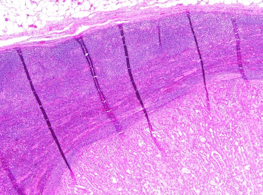
Histology

H&E stain with intact epidermis and dermis. There is a
glandular mass in the subcutaneous layer (arrow).
Histology

H&E stain demonstrating tumor cells with               H&E stain of lymph node showing normal lymph
irregular shaped nuclei and papillary architecture.    tissue and tumor invasion (arrow).
Final Dx:

Vulvar Adenocarcinoma
Case Discussion
• Majority of primary vulvar malignancies are squamous cell carcinomas, with adenocarcinomas representing
  less than 10%
• The pathology results noted the adenocarcinoma arose from an anogential mammary-like gland
• Originally thought to represent remnants of the milk ridges, they are now favored to be a normal cell type in
  this region
• These glands differ from normal sweat glands in that they are hormone receptor (estrogen and
  progesterone) positive.
• Differences between breast mammary glands and mammary-like glands of vulva include:
     • Different acinar epithelium
     • Higher concentration of glands than if it were from rudimentary mammary tissue
     • Glands are organized in rows which may suggest cloacal origin vs breast tissue which has linear
       orientation
     • Mammary ridge is not believed to extend all the way to the vulva
Case Discussion
• Age at diagnosis is usually 50s-80s
• Often present with a unifocal, sometimes pruritic lesion on the labia majora
• Lesion can also be on the labia minora, mons pubis or clitoris
• Other symptoms can include bleeding, dysuria, and lymphadenopathy
• Diagnosis is confirmed by biopsy of lesion
• Treatment options for local disease include wide local excision and
hemivulectomy and radical vulvectomy with or without lymph node dissection for
extensive diseaseReferences: 1. van der Putte, SC. Mammary-like glands of the vulva and their disorders. Int J Gynecol Pathol. 1994 Apr;13(2):150-60. doi: 10.1097/00004347-199404000-00009. PMID: 8005737. 2. Berek, JS and Karam, A. Vulvar cancer: Epidemiology, diagnosis, histopathology, and treatment. In: UpToDate, Post, TW (Ed), UpToDate, Waltham, MA, 2020.
You can also read