APPROCCIO TERANOSTICO E NUOVO APPROCCIO ALLE IMMAGINI NEI NET - Prof DESIREE DEANDREIS
←
→
Page content transcription
If your browser does not render page correctly, please read the page content below
APPROCCIO TERANOSTICO E
 NUOVO APPROCCIO ALLE
   IMMAGINI NEI NET

             Prof DESIREE DEANDREIS
Neuroendocrine tumors

Rare tumors
              Several sites

                              Heterogenous
                              disease        Different
                                             treatments
                                             available
Clinical presentation (site, functioning)

                Histology (Grade, Ki67)

NET diagnosis   Laboratory (CgA, NSE); Molecular test

                Morphological imaging (endoscopy,
                CT,MRI)

                Nuclear Medicine (Molecular Imaging)
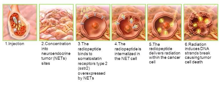
Theranostic approach:
from molecular imaging to therapy
«THERANOSTIC APPROACH

✓ Earlier detection and characterisation of disease
  (“molecular signature”)

✓ Understanding of underlying biology

✓ Selection of specific treatment option for targeted
  therapy
Molecular imaging

 Define the Target
SPECT/CT             PET/CT                 PET/MRI

           Detect and quantify the target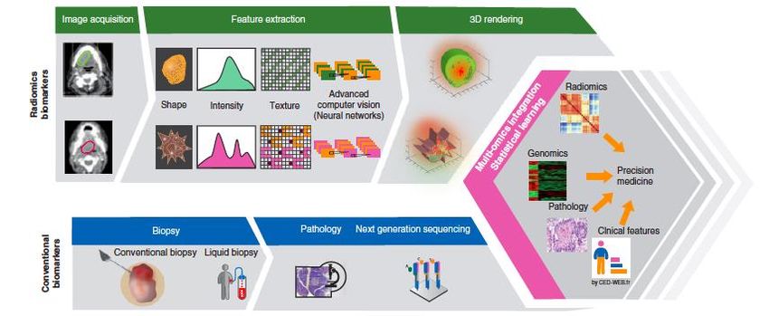
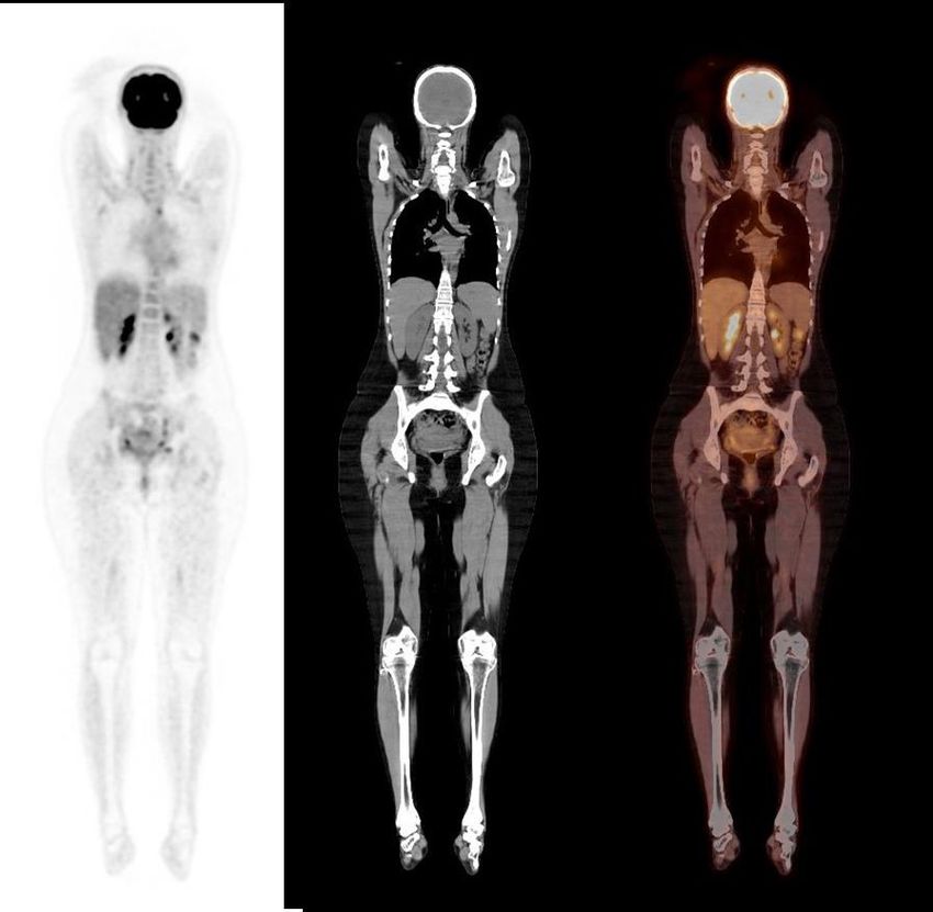
CURRENT COMMONLY UTILIZED NUCLEAR TECHNIQUE
CONVENTIONAL SCINTIGRAPHY                        PET/CT
111In-pentetreotide   123I-MIBG   18F-DOPA   68Ga-SSA-peptides   18F-FDG
CURRENT COMMONLY UTILIZED NUCLEAR TECHNIQUE
CONVENTIONAL SCINTIGRAPHY                      PET/CT
111In-pentetreotide   131I-MIBG   18F-DOPA   177Lu-DOTATATE   18F-FDG
177Lu-DOTATATE
Median Follow up: 14 mesi

                            177Lu-Dotatate

                            Median PFS: NR

                                             Strosberg et al 2017
PRECISION MEDICINE
1. PRECISION DIAGNOSTIC:
- TRACER
2. PRECISION THERAPY:
- 177Lu-DOTATATE
- OthersPRECISION IN NUCLEAR MEDICINE
PATIENT SELECTION
PRECISION THERAPY
RESPONSE EVALUATION
CRITERIAMolecular
imaging
Define the target
Hofland et al 2008TARGET: SST-R
PET
Octreoscan
Dotatoc 68Ga DOTA(0)-Phe(1)-Tyr(3)-octreotide
111In DTPA octreotide Dotanoc 68Ga DOTA(0)-Phe(1)-NaI(3)-Octreotide
Dotatate 68Ga DOTA(0)-Phe(1)-Tyr3-octreotate
68 Ga DOTA WHAT?BIODISTRIBUTION
Scintigraphy PET PET PET
POTENTIAL FALSE POSITIVE
Physiologic activity in the pancreatic uncinate
process
Inflammation: reactive nodes, prostatitis,
post radiotherapy change
Osteoblastic activity: degenerative bone
disease, fracture or vertebral hemangioma
Benign meningioma
Epiphyseal growth plates
Intra-pancreatic accessory spleenMolecular Imaging is strictly dependent on Differentiation grade and Proliferation Index
Rozenblum et al 2019DUAL TRACER CONCEPT
NEN G1 NEN G2 NEN G3
❖ PET/CT with 68Ga-DOTA-peptides ❖ PET/CT with 68Ga-DOTA-peptides; ❖ PET/CT with 18F-FDG
❖ PET/CT with 18F-FDG if ki67 is high and
imaging with 68Ga-DOTA-peptides is
negative.
G1 G2 G3
68Ga-DOTA-peptide 8F-FDG 68Ga-DOTA-peptides 8F-FDG 18F-FDGNET PET score
Detect the target: hybrid machine

Octreoscan   SSTR PET/CT
Image quality
               Affinity for SST- R
                                                      and resolution

Advantages
  of PET

             Reduced time            Hybrid machine         Semiquantitative
                                        with CT                  analysis
IMAGE QUALITY, RESOLUTION and reduced TIME
Advantages of SSTR PET/CT over conventional
scintigraphy
+ + Sensitivity: < 5 mm lesion characterization
Fully tomographic (3D)
Multi-slice CT
- Uptake time: 45-60 min vs 24-48 hours
- Imaging time: 12-15 min vs 45-60 min
Quantitative
In-house on demand production
(SPECT) obtained after injection of 111In- pentetreotide
reveals tumour lesions in over 60–80% of patients vs
Octreoscan around 90% for PET.
SSTR PET/CT
Hofland et al 200868Ga-SSA-peptides
localization, therapy selection, evaluation of response and restaging
RECCOMENDED INDICATIONS FOR
68Ga-SSA-peptides PET/CT SCAN IN NET
Exclude more advanced disease prior to surgical intervention
Localize primary tumor in patients with biochemical suspicion of NET
Identify primary tumor in patients with known metastatic NET
Confirm diagnosis of NET in patients with anatomic lesions that are
suspicious for NET
Identify patients who are likely to benefit from octreotide hormonal
therapy or PRRT with 177Lu- or 90Y-DOTATATEPET/CT findings resulted in managment changes in
44% of the patientsQuantify the target : SEMI QUANTITATIVE EVALUATION
- Metabolic data:
SUVmax
SUVmean
SUL peak
-Volume data:
FV
SRETV
-Integrated data:
TLSRE68-Ga-DOTA-PET : Predictive value
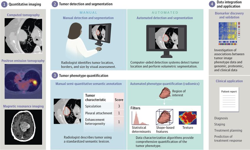
RADIOMICS
     •bioinformatic approaches (statistical methods and machine learning);
     •extrapolates the quantitative variables of the analyzed imaging
     (geometry, intensity and texture)
     •identifies the characteristics of the abnormal tissue.

PURPOSE: associate the
variables extracted from
the imaging with clinical or
biological endpoint (whole-
body histology, response to
therapy, survival, etc.) →
PERSONALIZED MEDICINE.           Limkin et al. Promises and challenges for the implementation of computational medical
                                 imaging (radiomics) in oncology. Annals of Oncology. 2017. 28: 1191-1206.
On a simple level, it is recognized that malignant tumours show heterogeneity of molecular and
cellular features, including cellular density and proliferation, necrosis, fibrosis, metabolism, hypoxia,
angiogenesis and receptor expression, factors that have been independently associated with a poor
treatment response and more aggressive tumour behavioR. This variation, defined by
histopathological appearance, may in turn reflect the degree of genetic clonal variation. These
biological processes can be crudely determined using functional and molecular imaging methods on
a global scale, and there is some evidence that several of these adverse biological features may
be reflected in medical images
Imaging DATA ANALYSIS and INTERPOLATION = DIAGNOSISSollini et al EJNM 2019
Visvikis et al EJNM 2019
AIM
    texture variables
         robust
68Ga-DOTATOC PET/CT

   DIFFERANTIATE
BIOLOGICAL BEHAVIOR
  OF TUMOR and MTS

 PERSONALIZED
   MEDICINE
Valle d’Aosta
IRMET
Citta della Salute
MaurizianoComplessità organizzativa
biologia chimica
molecolare
Imaging e medicina
farmacologia terapia molecolare
«Theranostic»
radiofarmacia
ingegneria
fisica
Courtesy of Dr Carlo PotiPROGETTO CLINICAL RESEARCH/PET CENTER
Nuclear Medicine Division
PET CENTER
Nuclear Medicine Division
THERANET Department of Medical Sciences
University of Turin
Umberto Parini Hospital, Aosta
Dr Carlo Poti
Città della Salute e della Scienza Hospital
(Prof. G. Bisi, Prof. D. Deandreis)
PET CENTER/
ENDOCRINE ONCOLOGY IRMET Torino
-Department of Medical Sciences Dr Vincenzo arena
University of Turin
Città della Salute e della Scienza Hospital
( Prof E. Arvat)
-Endocrinology Service
Mauriziano Umberto I Hospital
( D.ssa P. Razzore)
Rete University of Turin
Oncologica PATHOLOGY
ONCOLOGY Department of Oncology,
Department of Oncology University of Turin
University of Turin -AOU Città della Salute e della Scienza
- Città della Salute e della Scienza (Prof. M. Papotti)
University of Turin -S. Luigi Gonzaga Hospital
(Dr. L Ciuffreda, Dr. M. Airoldi) (Prof M. Volante)
- S. Luigi Gonzaga Hospital 68Ga
(D.ssa MP Brizzi)
DOTATOC Lutathera
GASTROENTEROLOGY
Department of Medical Sciences DOTATATE
University of Turin
Città della Salute e della Scienza Hospital
(Prof C. De Angelis)
Endocrino Alessandria
Laura Rossi
RADIOMETABOLIC POLES
-Mauriziano Umberto I Hospital, Turin
(Dr R. Pellerito)
Imaging platform sharing -SS Antonio e Biagio e C. Arrigo Hospital, Dosimetry tool
UniTO start up Alessandria
Software
(Dr A. Muni)PERCORSO PAZIENTE
Discussione
GIC
struttura
inviante
Compilazione
Programmazione scheda valutativa
terapia pre screening rete
Oncologica
Invio Scheda a
Medicina Nucleare di
Programmazione Visita riferimento ( Mauriziano
Medico Nucleare o Alessandria)
Visita Medico
NucleareSCHEDA DI PRE-INCLUSIONE DATA E GIC INVIANTE 1. DATI PAZIENTE: Nome Cognome Sesso F M Data di nascita ECOG Peso: Altezza Comorbidità: Terapie in atto : SSA SI/NO data ultima somministrazione
SCHEDA DI PRE-INCLUSIONE

2. DATI TNE

Sede di primitivo ( GEP NET):
Tumore funzionante SI/NO in compenso SI/NO
Tipologia sindrome: s. carcinoide tipica SI/NO atipica SI/NO
Grading su primitivo: G1-G2; (Ki67%)
Grading su metastasi: G1-G2; (Ki67%)
Data di diagnosi primitivo
Data di diagnosi della prima metastasi:
Sede: Linfonodi Fegato Polmone Osso :
Terapie precedenti: 1.analoghi 2. Chemioterapia ( linee 1.2.3)
Inoperabile (GIC): SI NO
Progressivo SI NO (3 classi 1.>12 mesi;2. < 12 mesi; 3. < 8 mesi ) (GIC)
Data Progressione: tipologia di indagine utilizzata per definire progressione
(TC/RMN)
SCHEDA DI PRE-INCLUSIONE

 3. PATTERN METABOLICO

 Data PET con 68Ga DOTATOC/DOTATATE:
 PET con 68Ga DOTATOC/DOTATATE: positiva: SI NO
 GRADO: 1.2.3.4.
 SUVmax della lesione più captante:
 Numero totale di lesioni:
 Coinvolgimento epatico :1.assente 2. limitato.3. massivo

 PET con FDG Disponibile (G2) SI NO;
 Data PET FDG :
 PET FDG: Positiva SI NO

 Classe 1. DOTATOC > FDG; 2. DOTATOC =FDG; 3. DOTATOC< FDG
SCHEDA DI PRE-INCLUSIONE

 4. EMATOCHIMICI:

 Emocromo nella norma SI NO ( allegare)
 Funzionalità renale mantenuta SI NO
 Creatinina : ………. (Data) allegare
 Funzionalità epatica nella norma SI NO
 Bilirubina :……(Data) allegare
Cortesia di E Richetta
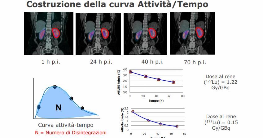
Calcolo della dose erogata post trattamento
                          Attività somministrata fissa: 7.4 GBq x 4 cicli
                PET-CT 68Ga                     PET-CT 68Ga

Cortesia di E Richetta   PRE TERAPIA               POST TERAPIA
METODO DOSIMETRICO AO Ordine Mauriziano
METODO DOSIMETRICO

                     [cortesia F. Fioroni]
DOSE al target tumorale Lesioni
epatiche
117- 153 Gy
6,0
y = 5,2025e-0,005x
5,0
4,0
R² = 0,8764 ossea
3,0
2,0
43 Gy
1,0
0,0
0,0 50,0 100,0 150,0
2,0
1,5
paraesofagea
1,0 y = 1,8704e-0,003x
R² = 0,9893 18 Gy
0,5
0,0
0,0 50,0 100,0 150,0
4,0
3,0
2,0
y = 2,6188e-0,012x
R² = 0,8834
pericardica
1,0 5 Gy
0,0
0,0 50,0 100,0 150,0Perché una ATTIVITA’ PERSONALIZZATA
potrebbe essere meglio di una ATTIVITA’ FISSA?
[cortesia F. Fioroni]Challenge for next years … liquid biopsy
PRRT predictive quotient (PPQ)CONCLUSION :
Tumor
Tailored Define
therapy the target
Quantify
the targetCondivisione e distribuzione delle risorse Fare network HOT POINTS Stabilire un iter diagnostico terapeutico condiviso Facilities: Radiofarmacia, Fisica, Tecnologia Potenziare ricerca nel campo : progetti unito, partnership Sviluppo per altre patologie attualmente in fase sperimentale e non rari Sviluppo di nuovi radiofarmaci Sviluppo nuovi trials
You can also read