Book Chapter Unique Polyhalogenated Peptides from the Marine Sponge Ircinia sp.
←
→
Page content transcription
If your browser does not render page correctly, please read the page content below
Updates in Pharmacology

Book Chapter

Unique Polyhalogenated Peptides from
the Marine Sponge Ircinia sp.
Rogelio Fernández1, Asep Bayu2, Tri Aryono Hadi3, Santiago
Bueno1, Marta Pérez1*, Carmen Cuevas1 and Masteria Yunovilsa
Putra2
1
  Natural Products Department, PharmaMar S.A., Pol. Ind. La
Mina Norte, Spain
2
  Research Center for Biotechnology, Indonesian Institute of
Sciences, Indonesia
3
  Research Center for Oceanography, Indonesian Institute of
Sciences, Indonesia

*Corresponding Author: Marta Pérez, Natural Products
Department, PharmaMar S.A., Pol. Ind. La Mina Norte, Avda.
de los Reyes 1, 28770 Colmenar Viejo (Madrid), Spain

Published March 03, 2021

This Book Chapter is a republication of an article published by
Marta Pérez, et al. at Marine Drugs in July 2020. (Fernández, R.;
Bayu, A.; Aryono Hadi, T.; Bueno, S.; Pérez, M.; Cuevas, C.;
Yunovilsa Putra, M. Unique Polyhalogenated Peptides from the
Marine Sponge Ircinia sp. Mar. Drugs 2020, 18, 396.
https://doi.org/10.3390/md18080396)

How to cite this book chapter: Rogelio Fernández, Asep Bayu,
Tri Aryono Hadi, Santiago Bueno, Marta Pérez, Carmen Cuevas,
Masteria Yunovilsa Putra. Unique Polyhalogenated Peptides
from the Marine Sponge Ircinia sp. In: Nosheen Akhtar, editor.
Updates in Pharmacology. Hyderabad, India: Vide Leaf. 2021.

© The Author(s) 2021. This article is distributed under the terms
of the Creative Commons Attribution 4.0 International
License(http://creativecommons.org/licenses/by/4.0/),      which

1                                             www.videleaf.com
Updates in Pharmacology

permits unrestricted use, distribution, and reproduction in any
medium, provided the original work is properly cited.

Author Contributions: Sample collection: T.A.H, A.B. and S.
B.; Conceptualization and supervision: C.C. and M.Y.P;
Methodology and writing: RF; Investigation: A.B; Original draft
preparation and writing: M.P; Review and editing: C.C, M.P and
M.Y.P. All the authors read, reviewed, and agreed with the
structure and content of the manuscript.

Funding: The present research was financed in part by Grants
from Ministerio de Ciencia, Innovación y Universidades of
Spain (AGL2015-63740-C2-2-R and RTC-2016 4611-1,
Inmunotop project), cofunded by the FEDER Programme from
the European Union.

Acknowledgments: We gratefully acknowledge the help of our
PharmaMar colleagues C. Crespo for her excellent technical
assistance, E. Millán for performing the ESI(+) and MSe
experiments, J. M. Dominguez for the biological assays and S.
Munt for revision of the manuscript.

Conflicts of Interest: The authors declare no conflict of interest.

Abstract
Two new bromopyrrole peptides, haloirciniamide A (1) and
seribunamide A (2), have been isolated from an Indonesian
marine sponge of the genus Ircinia collected in Thousand Islands
(Indonesia). The planar structure of both compounds was
assigned on the basis of extensive 1D and 2D NMR spectroscopy
and mass spectrometry. The absolute configuration of the amino
acid residues in 1 and 2 was determined by the application of
Marfey’s method. Compound 1 is the first dibromopyrrole
cyclopeptide having a chlorohistidine ring while compound 2 is
a rare peptide possessing a tribromopyrrole ring.           Both
compounds failed to show significant cytotoxicity against four
human tumor cell lines and neither compound was able to inhibit
the enzyme topoisomerase I or impair PD1-PDL1 interaction.

2                                              www.videleaf.com
Updates in Pharmacology

Keywords
Marine Sponge; Ircinia sp.; Polyhalogenated Peptides; Marfey’s
Analysis

Introduction
Indonesia is located at the centre of a biodiversity hotspot and
around 750 structures from Indonesian waters have been
published in the last 50 years [1]. The structural diversity and
bioactive properties of the compounds isolated from this region
encouraged us to continue to investigate this area, which still
remains largely unexplored. Thus, one of the recent PharmaMar
expeditions was carried out in Thousand Islands, in collaboration
with Indonesian Institute of Sciences (LIPI). Thousand Islands
archipelago is located about 25 miles from the coast to the
northeast of Jakarta and the collection site is an area full of
gentle rock and coral slopes, and is potentially a highly
productive area, both in terms of the quantity and the nature of
the biodiversity. In this paper we describe the isolation of two
new peptides isolated from an Ircinia specimen from this area.

Of all the marine organisms investigated, sponges (Porifera) are
the most primitive multicellular animals with ample time to
evolve into more complex living organisms. In fact, marine
sponges are recognized as the richest sources of MNP,
contributing to nearly 30% of all marine natural products
discovered so far [2]. Previous reports revealed that Marine
sponges of the genus Ircinia are known as a rich source of varied
bioactive natural products, including fatty acids [3], steroids [4],
terpenes [5], macrolides [6], and peptides [7], many of which
have biological activities. This structural diversity could be due
to the fact that sponges harbour diverse microorganisms and in
numerous cases, bacteria isolated from sponges or symbiotic
bacteria are the true producers of the compounds found in their
extracts [8]. Specifically, an intriguing group of Ircinia derived
peptides are assumed to be of microbial origin due to the
presence of both D-amino acids and unusual amino acids as
illustrated by the cyclic hexapeptide waiakeamide from Ircinia

3                                               www.videleaf.com
Updates in Pharmacology

dendroides [9] and the cyclotheonamides E4 and E5, cyclic
pentapeptides also from Ircinia species [10].

Undeniably, among the compounds isolated from marine
sources, linear and cyclic peptides are recognized as an
important class with great structural diversity and a wide range
of bioactivities, and these include the antimalarial carmabin A
[11], the antiproliferative jaspamides [12] and the cytotoxic
patellamides [13]. Furthermore, two marine peptide-derived
products have reached the market, ziconotide [14] for analgesic
use and a synthetic derivative of dolastatin 10 [15] linked to an
antibody for the treatment of Hodgkin´s lymphoma. PharmaMar
has also developed a marine natural peptide Aplidin, originally
found in the Ascidian Aplidium albicans, which has recently
been approved for commercialization in Australia for the
treatment of multiple myeloma. Recent studies suggest that
Aplidin may also have antiviral properties and a clinical trial to
treat patients with COVID-19 has been initiated.

In the course of our screening program to isolate novel
compounds with antitumor properties from marine sources, we
have isolated two unique peptides haloirciniamide A (1) and
seribunamide A (2) from an Ircinia specimen, which was
collected off the coast of Thousand Islands. It is worth
mentioning that the number of known peptides with a
halogenated pyrrole ring is limited, with only cyclocinamides
and corticiamide A [16] as well as gunungamide A [17] having
been described as possessing chlorinated pyrrole rings.
Although there are dozens of dibromopyrrolecarboxamide
derivatives from porifera like nagelamide [18] and carteramine
[19], mainly from Agela and Stylissa species, compound 1 is the
first example of a cyclopeptide containing a halogenated pyrrole
ring in its structure. Indeed, haloirciniamide A represents a
structurally unique depsipeptide, since it also has an
unprecedented chlorohistidine moiety. Furthermore, there are
only two examples of tribromopyrrole rings derived from natural
sources, 2,3,4-tribromopyrrole itself, which was isolated from
the marine Poychaete Polyphysia crassa [20] and
tribromopyrrol-2-methylphenol isolated from a coralline algal-

4                                              www.videleaf.com
Updates in Pharmacology

associated Pseudoalteromone [21], with compound 2 being the
first of its class.

Details of the isolation and structural elucidation of the new
halogenated peptides 1 and 2 are provided. The results of
antitumor, PD1 and TOPO I screening are also described.

Results and Discussion
Isolation and Structure Elucidation

The sponge Ircinia sp. was collected by hand while diving in
Thousand Islands (Indonesia). The specimen was repeatedly
extracted using CH2Cl2:MeOH (1:1 v/v). The combined
concentrated extracts, after vacuum liquid chromatography
(VLC) and semipreparative reverse-phase HPLC separations, led
to the isolation of the two pure compounds shown in Figure 1.

Figure 1: Chemical structures of the compounds 1 and 2 isolated from Ircinia
sp.

Compound 1 was isolated as an amorphous white solid. The
isotopic distribution observed in the (+)-LRESI mass spectrum
with four protonated ion [M+Na]+ peaks at m/z 830, 832, 834
and 836 in the ratio 3:6:3:1 respectively, showed the presence of
two bromine and a chlorine atom in the molecule. The presence
of these halogens in the structure was confirmed by (+)-HRESI-
TOFMS analysis, with the ion peak observed at m/z 830.0400
[M+Na]+       corresponding     to    the    molecular   formula
C25H3279Br235ClN11O8Na (calcd. 830.0383). Interpretation of the

5                                                     www.videleaf.com
Updates in Pharmacology mono NMR data (1H, 13C and 1D-TOCSY) compiled in Table 1 and two-dimensional NMR spectra (gHSQC, gCOSY, gHMBC, and 2D-TOCSY) in CD3OD, led to identification of 6 spin systems. Taking into consideration the seven carbonyl carbon resonances (C 161.4 – 175.8) and the number of -amino acid proton signals (H 4.06 – 4.84), the peptide nature of compound 1 was expected. This hypothesis was confirmed by the NH signals observed in the 1H NMR spectrum in CD3OH (H 7.13 – 12.68) and DMSO-d6 (H 7.37 – 12.03), with the latter solvent being chosen for full structural elucidation. The COSY correlations observed between methines at H 4.23/C 50.5 and H 4.49/C 51.5 with the diastereotopic methylenes at H 2.87; 3.13/C 49.2 and H 3.22; 4.04/C 40.5 respectively, indicated the presence of two 2,3-diaminopropionic acid units (Figure 2). Both amino acids were directly connected, based on the HMBC correlation of the NH signals at H 9.11 for Dap1 and H 7.72 for Dap2 with the same carbonyl carbon at C 169.5. The next amino acid present in the peptide core was an isoserine with a methylene group at H 2.75; 3.47/C 42.8, a methine carbon at H 4.13/C 67.8 and an NH signal at H 8.23. Isoserine was placed linked to the Dap2 by the correlation between NH at H 8.23 and the carbonyl carbon of Dap2 at C 170.6. In addition, an HMBC correlation of the methine carbon at H 4.13 and the NH signal at H 7.13, that belongs to a unit of isoasparagine (H 4.58/C 48.4, H 2.80; 3.09/C 35.0 ), with the carbonyl group at C 170.6, allowed the sequence of amino acids to be continued. Finally, the peptide ring was closed by an NMe-histidine (H 3.99/C 65.6, H 3.05/C 25.0), whose N-methyl group showed an HMBC correlation with carbons belonging to the carbonyl group of iAsn at C 173.5 and its own methine at C 65.6., with an additional HMBC correlation between the -aminoacid proton signal of NMe-histidine and the NH signal at H 9.11 of Dap1 with the carbonyl carbon at C 169.0 (Figure 2). NMe-histidine ring shifts at C 110.5, C 128.2 and H 6.76/C 109.5, revealed that the non- protonated carbon at C 128.2 bore one of the three halogen atoms. 6 www.videleaf.com
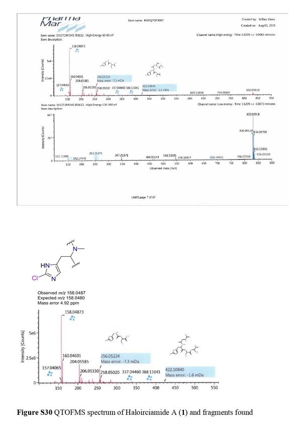
Updates in Pharmacology Figure 2: Selected key COSY (bold), HMBC (red) and ROESY (blue) correlations for 1 and 2. To complete the structure elucidation, the two remaining doublets with a small coupling constant value of 2.7 Hz were assigned to a sp2 methine at H 6.30/C 110.4 and a significant downfield NH signal at H 12.68. An HMBC correlation of these two protons with three non-protonated sp2 carbons (C 96.9, 118.1 and 123.2), demonstrated the existence of a trisubstituted pyrrol moiety, with two of these three positions bearing halogens. The placement of this heterocycle was established by the HMBC correlation of the methylene group of Dap2 with a carbonyl group at C 158.6. To confirm the direct connection between the pyrrol moiety and this carbonyl group, a new gHMBC experiment with J = 3 Hz was conducted. The position of the sp2 methine in the pyrrol unit and the bond to the ring with the cyclopeptide, was settled by the HMBC correlation between the methine proton at H 6.30 and the carbonyl group at C 158.6. Although the chlorine and two bromine atoms were undoubtedly located on the three free positions of the heterocycle ring and the carbon shifts (C 96.9 and 123.2) suggested that both bromine atoms were on the pyrrol moiety, this evidence was insufficient to fully confirm this proposal. Fortunately, this could be resolved by detailed study of the peptide structure by (+)-HRESI-TOFMS and QTOF (Figure 3), which showed significant cluster ions at m/z 158.0487/160.0460 in a 3:1 ratio corresponding to the iAsn moiety. These m/z values, the mass error observed and the 7 www.videleaf.com
Updates in Pharmacology
isotopic distribution clearly confirmed the presence of a chlorine
atom on the NMeHis amino acid.
Figure 3: Fragment found for 1 by QTOF.
The absolute stereochemistry of compound 1 was established on
the basis of Marfey’s analysis with the 1-fluoro-2,4-di-
nitrophenyl-5-L-alanine amide (L-FDAA) [22]. Compound 1
was hydrolyzed in strong acid conditions and derivatization of
the free aminoacids with L-FDAA allowed an exhaustive
analysis by HPLC-MS. A comparison of the retention times of
the derivatized amino acids present in 1 and the suitably
derivatized pure amino acid standards, unambiguously
demonstrated the absolute configuration as L-Dap, L-iSer and D-
Asn. The absolute configuration of NMeClHis could not be
determined due to the absence of the standard amino acid.
Table 1: NMR spectroscopy data for 1 (1H NMR MHz, 13C NMR 125 MHz).
Pos C, multa H, mult (J in C, multb H, mult (J in
Hz)a Hz)b
Dap1 1 169.5, C 172.3, C
2 50.3, CH 4.23 ddd (6.2, 52.2, CH 4.54 dd (5.6, 2.9)
6.2, 2.8)
3 49.2, CH2 2.87 d (13.9, 50.5, CH2 3.17 dd (14.6,
6.2) 5.6)
3.13 d (13.9, 3.54 d (14.2)
2.8)
NH 9.11 d (6.2) 8.97 d (6.6)*
8 www.videleaf.com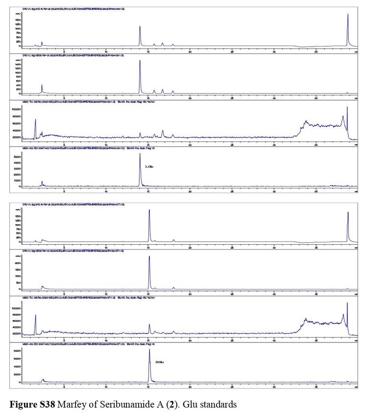
Updates in Pharmacology

            NH2
    NMeC    1      169.0, CO                       170.9, CO
    lHis
            2      65.6, CH    3.99 dd (10.6,      67.0, CH    4.06 dd (10.1,
                               3.9)                            4.5)
            3      24.9, CH2   3.05 m              25.9, CH2   3.19 dd (15.2,
                                                               10.1)
                                                               3.30 dd (15.2,
                                                               4.5)
            4      110.5, C                        135.6, C
            5      109.5, CH   6.76 s              120.8, CH   6.96 s
            6      128.2, C                        131.0, C
            NMe    39.6, CH3   2.84 s              40.4, CH3   3.02 s
    iAsn    1      173.5, C                        175.8, C
            2      35.0, CH2   2.80 dd (16.6,      36.2, CH2   3.02 m
                               2.7)
                               3.09 dd (16.6,                  3.27 m
                               5.8)
            3      48.4, CH    4.58 ddd (8.0,      50.3, CH    4.84 m
                               5.8, 2.7)
            4      172.2, CO                       173.8, CO
            NH                 7.13 d (8.0)                    7.61 d (6.3)*
            NH2
    iSer    1      171.9, C                        173.3, C
            2      67.8, CH    4.13 dd (9.0,       69.5, CH    4.40 dd (9.5, 4.0)
                               4.2)
            3      42.8, CH2   2.75 ddd (9.0,      44.2, CH2   3.00 m
                               9.4, 5.4)
                               3.47 m                          3.75 dd (13.2,
                                                               4.0)
            NH                 8.23 t (5.4)                    8.25 s*
    Dap2    1      170.5, C                        172.8, C
            2      51.5, CH    4.49 ddd (9.2,      53.4, CH    4.84 m
                               9.2, 6.3)
            3      40.1, CH2   3.22 m              41.6, CH2   3.57 dd (13.8,
                                                               8.6)
                               4.04 ddd (12.9,                 4.22 dd (13.8,
                               6.3, 6.3)                       5.4)
            NH-1               7.72 d (9.2)                    7.98 d (9.5)*
            NH-2               7.20 t (6.3, 6.3)               7.37 t (6.0)*
    Br2Py   1      158.6, CO                       161.4, CO
            2      118.0, C                        120.2, C
            3      110.4, CH   6.30 d (2.7)        112.0, CH   6.15 s
            4      96.9, C                         99.4, C
            5      123.2, C                        124.2, C
            NH                 12.68 d (2.7)                   12.03 s

a
    In DMSO-d6. b In CD3OD (*CD3OH).

9                                                     www.videleaf.com
Updates in Pharmacology
Compound 2 was isolated as an amorphous white solid. Its (+)-
LRESI showed a m/z=825 [M+H]+ with a characteristic cluster
corresponding to the presence of three bromine atoms. The
molecular formula C29H4379Br3N6O7 was established by (+)-
HRESI-TOFMS analysis of the [M+H]+ at m/z 825.0806 (calcd.
825. 0816). The peptide nature of 2 was evident from its 1H and
13
C NMR spectra (Table 2). 1H NMR in DMSO showed the
characteristic -proton resonances of four -amino acids in the
range H 5.35 to 4.10 ppm, five interchangeable protons at H
12.53, 8.56, 8.08, 7.19 and 6.77 ppm and two NMe signals at H
3.61 and 3.04 ppm. 13C NMR data displayed six carbonyl signals
between C 173.5 and 159.1 ppm, four adjacent methine carbons
in the range C 59.1-51.5 ppm, and two NMe signals at C 35.7
and 30.5 ppm. Extensive 2D NMR analysis, including COSY,
TOCSY, HSQC and HMBC was used to determine the identity
of the four amino acids and to assign the NMR signals. As a
result of these studies, the amino acids were found to be one Ile,
one NMe-Leu, one Pro and one Asn unit. A long-range
correlation between protons at H 7.19/6.77 and 2.15/2.11 ppm
with the carbonyl group at C 173.5 ppm and the observation of
ROESY cross-peaks between protons at H 7.19/6.77 ppm and
the CH2 of position 4 at H 2.15/2.11 ppm established the
presence of a Gln. A N-methyl-2,3,4-bromopyrrol unit was
inferred by the presence of four aromatic non-protonated carbons
at C 128.5, 107.9, 100.7 and 93.4 ppm with chemical shifts
similar to those described for bromopseudoceratines [23].
Table 2: NMR spectroscopy data for 2 (1H NMR 500 MHz, 13
C NMR 125
MHz).
Pos H, mult C, multa H, mult C, multb
(J in (J in Hz)b
Hz)a
Br3Py 1 - 161.8, CO - 159.1, CO
2 - 128.6, C - 128.5, C
3 - 102.7, C - 100.7, C
4 - 101.3, C - 93.4, C
5 - 110.8, C - 107.9, C
NMe 3.76, s 36.7, CH3 3.61, s 35.7, CH3
Ile 1 - 174.3, CO - 171.4, CO
2 4.84, m 55.7, CH 4.65, dd, 53.7, CH
8.35, 8.5
10 www.videleaf.comUpdates in Pharmacology
3 1.96, m 38.0, CH 1.87, m 35.8, CH
4 1.73, m, 25.9, CH2 1.57, m; 24.2, CH2
1.22, m 1.21, m
5 0.95, t, 11.2, CH3 0.83, t, 7.4 10.7, CH3
7.4
6 0.99, d, 15.7, CH3 0.85, d, 15.1, CH3
6.8 6.9
NH 8.24, d, - 8.56, d, -
8.1 8.2
NMeLeu 1 - 171.8, CO - 168.8, CO
2 5.51, dd, 54.3, CH 5.35, dd, 51.7, CH
10.3, 4.7 10.1, 4.3
3 1.77, m; 38.0, CH2 1.59, m; 36.7, CH2
1.61, m 1.42, m
4 1.56, m 25.7, CH 1.43, m 23.9, CH
5 0.97, d, 23.6, CH3 0.87, d, 23.1, CH3
6.2 6.2
6 0.93, d, 22.3, CH3 0.83, d, 21.8, CH3
6.1 6.2
NMe 3.21, s 31.8, CH3 3.04, s 30.5, CH3
Pro 1 - 174.5, CO - 171.4, CO
2 4.41, m 61.6, CH 4.31, dd, 59.1, CH
8.3, 4.1
3 2.23, m; 30.5, CH2 2.02, m; 28.9, CH2
2.00, m 1.80, m
4 2.08, m; 26.0, CH2 1.90, m; 24.4, CH2
1.92, m 1.77, m
5 3.75, m; 48.8, CH2 3.53, m; 46.7, CH2
3.69, m 3.50; m
Gln 1 - 174.7, 12.53, brs 173.3,
CO2H CO2H
2 4.41, m 52.8, CH 4.10, ddd, 51.5, CH
8.6, 8.5,
5.2
3 2.27, m; 30.5, CH2 1.93, m; 27.0, CH2
1.92, m 1.74, m
4 2.41, m; 32.6, CH2 2.15, m; 31.3, CH2
2.32, m 2.11, m
5 - 177.9, 7.19, s; 173.5,
CONH2 6.77, s CONH2
NH - - 8.08, brs -
a
In CD3OD. b In DMSO-d6.
The sequencing for compound 2 was carried out using a
combination of HMBC and ROESY data. Long-range
correlations from α-protons, NH and NMe to carbonyl carbons
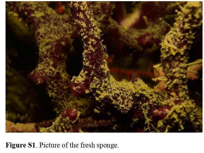
11 www.videleaf.comUpdates in Pharmacology of adjacent amino acids plus ROESY correlations between α- protons, NH and NMe protons of adjacent amino acids (see Figure 2) allowed us to establish the sequence as Br3Py-Ile- NMeLeu-Pro-Gln. The absolute configurations of the aminoacids were determined by comparing the hydrolysis products of 2 (6 N HCl, 110 °C, 18 h) after derivatization with Marfey’s reagent (N-(3-fluoro-4,6- dinitrophenyl)-L-alaninamide, L-FDAA), with appropriate amino acid standards using HPLC-MS chromatography. As a result, all the amino acids were determined to be L. Materials and Methods General Experimental Procedures Optical rotations were determined using a Jasco P-1020 polarimeter. UV spectra were performed using an Agilent 8453 UV−vis spectrometer. IR spectra were obtained with a Perkin- Elmer Spectrum 100 FT-IR spectrometer with ATR sampling. NMR spectra were recorded on a Varian “Unity 500” spectrometer at 500/125 MHz (1H/13C). Chemical shifts were reported in ppm using residual CD3OH (δ 3.31 ppm for 1H and 49.0 ppm for 13C) and DMSO-d6 (δ 2.50 ppm for 1H and 39.5 ppm for 13C) as an internal reference. HRESITOFMS was performed on an Agilent 6230 TOF LC/MS chromatograph spectrometer. (+)-ESIMS were recorded using an Agilent 1100 Series LC/MSD spectrometer. HRESITOFMS was performed on an Agilent 6230 TOF LC/MS chromatograph spectrometer. ESI(+) and MSe were performed on an Waters UHPLC-QTOF Acquity I-Class + Xevo G2-XS. Biological Material The sponge Ircinia sp. (158 g) was collected by hand using a diving rebreather system in Thousand Islands (Indonesia). The sponge was immediately frozen and kept under these conditions until extraction. The specimen was identified by María Jesús Uriz at CEAB, Blanes, Spain. A voucher specimen (ORMA155272) is deposited at PharmaMar facilities (Madrid, Spain). 12 www.videleaf.com
Updates in Pharmacology
Extraction and Isolation
The sponge Ircinia sp. (158 g) was triturated and exhaustively
extracted with MeOH:DCM (1:1, 3 × 500 mL). The combined
extracts were concentrated to yield a crude mass of 7.9 g. The
crude product was subjected to VLC on Lichroprep RP-18 with a
stepped gradient from H2O to MeOH to CH2Cl2. The fractions
eluting with H2O:MeOH (3:1, 606 mg) and H2O:MeOH (1:1,
89.6 mg) were subjected to semipreparative HPLC (Symmetry
Prep C18 5 μm, 10 × 150 mm; 3 min isocratic H2O + 0.04 %
TFA: CH3CN + 0.04 % TFA 95:5 and then gradient from 5% to
68% CH3CN + 0.04 % TFA in 25 min, flow 3 mL/min, UV
detection) to obtain 4.3 mg of compound 1. The fraction eluting
with H2O:MeOH (1:3, 43.9 mg) was subjected to
semipreparative HPLC (Symmetry Prep C18 5 μm, 10 × 150 mm;
3 min. isocratic H2O + 0.04 % TFA: CH3CN + 0.04 % TFA
90:10 and then gradient from 10% to 75% CH3CN + 0.04 %
TFA in 25 min, flow 3 mL/min, UV detection) to obtain 3.0 mg
of compound 2.
Haloirciniamide A (1): amorphous white solid; []25D -62.7º (c
0.1, MeOH); IR υmax 3314, 2920, 2850, 1644, 1523, 1416,
1311, 1239, 1199, 1041 cm-1; UV (MeOH) λmax 198, 268 nm. 1H
NMR (500 MHz) and 13C NMR (125 MHz) see Table 1; (+)-
HREI-TOFMS m/z 830.0400 [M+Na]+ (calcd for
79
C25H32 Br2N11O8Na m/z 830.0383).
Seribunamide A (2): amorphous white solid; []25D -38.4º (c 0.2,
MeOH); IR υmax 3352, 2932, 2850, 1658, 1515, 1320, 1236,
1035 cm-1; UV (MeOH) λmax 197, 266 nm. 1H NMR (500 MHz)
and 13C NMR (125 MHz) see Table 2; (+)-HREI-TOFMS m/z
825.0806 [M+H]+ (calcd for C29H4479Br3N6O7 m/z 825.0816).
Absolute Configuration
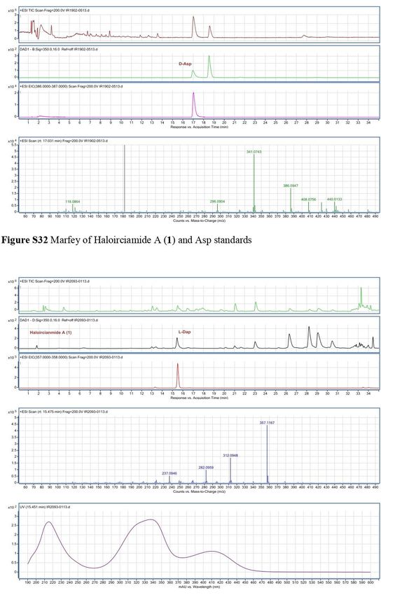
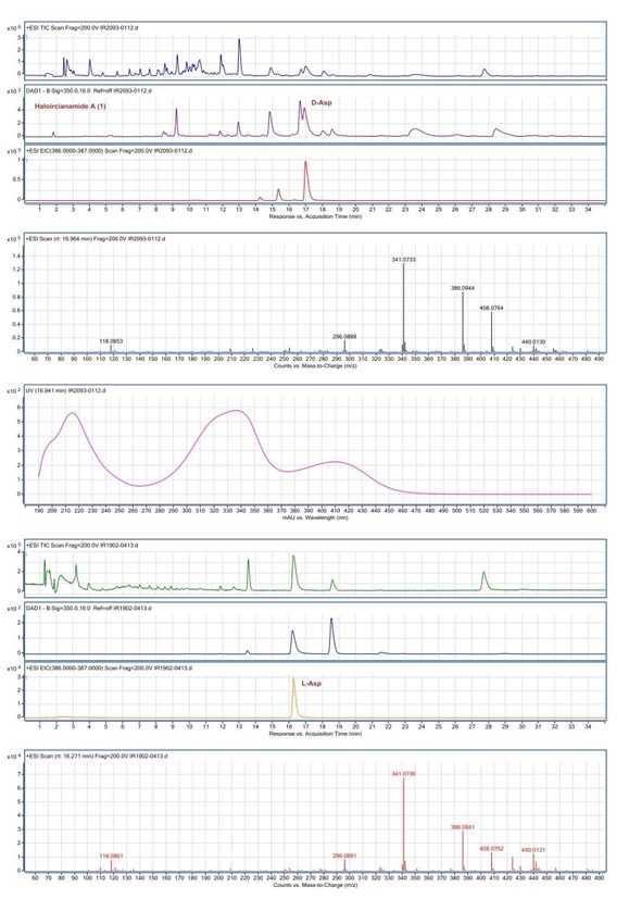
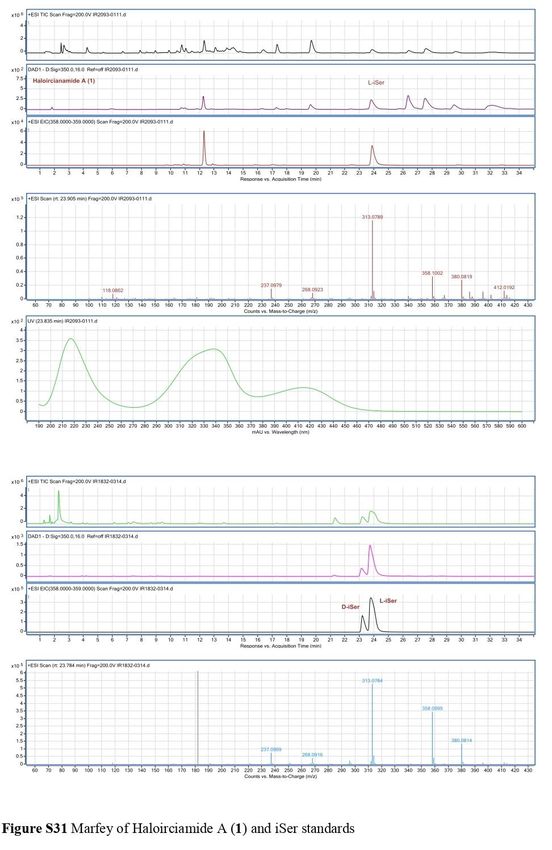
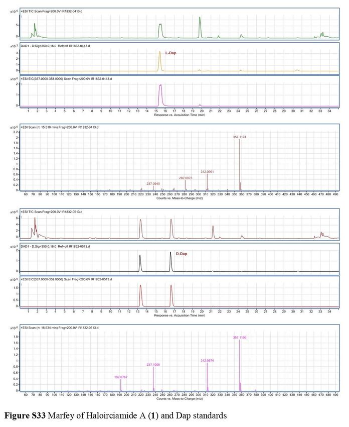
Absolute Configuration of 1: 0.5 mg of haloirciniamide A was
hydrolyzed in 0.5 mL of 6 N HCl at 110 °C for 15 h. The excess
aqueous HCl was removed under a N2 stream, and a solution of
700 µg of L-FDAA (N-(3-fluoro-4,6-dinitrophenyl)-L-alanine-
amide) in acetone (160 µL), H2O (100 µL) and NaHCO3 1N (50
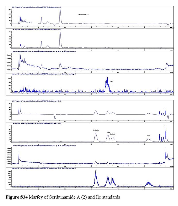
13 www.videleaf.comUpdates in Pharmacology µL) was added to the dry hydrolysate. The resulting mixture was heated at 40 ºC for 1 h, before being cooled to 23 ºC, quenched by addition of 2N HCl (20 µL), dried, and dissolved in H2O (800 μL). The resultant aqueous solution was subjected to reversed-phase LC/MS (column: Waters Symmetry 4.6x150 mm, 3.5 µm, flow rate 0.8 mL/min) in three different gradient. Gradient 1 for iSer (mobile phase CH3CN + 0.04% formic acid /H2O + 0.04% formic acid, using a linear gradient from 5 to 20% CH3CN in 10 min and them from 20% to 35% CH3CN in 25 min): the retention time was 23.9 min for L-iSer. Gradient 2 for Asp (mobile phase CH3CN + 0.04% formic acid/H2O + 0.04% formic acid, using a linear gradient from 5 to 30% CH3CN in 10 min and them from 30% to 50% CH3CN in 30 min): the retention time was 17.0 min for D-Asp. Gradient 3 for Dap: mobile phase CH3CN + 0.04% formic acid/H2O + 0.04% formic acid, using a linear gradient from 5 to 10% CH3CN in 5 min and them from 10% to 35% CH3CN in 25 min): the retention times was 15.5 min for L-Dap. Retention times for the derivatized amino acids standards were as follows: gradient 1 (23.2 min for D-iSer and 23.8 min for the L-iSer); gradient 2 (16.2 min for L-Asp and 17.0 min for D-Asp) and gradient 3 (15.5 min for L-Dap and 16.6 min for the D-Dap). Absolute Configuration of 2: 0.3 mg of seribunamide A was hydrolyzed in 0.4 mL of 6 N HCl, 110 °C for 15 h. The excess aqueous HCl was removed under a N2 stream, and a solution of 400 µg of L-FDAA (N-(3-fluoro-4,6-dinitrophenyl)-L-alanine- amide) in acetone (160 µL), H2O (100 µL) and NaHCO3 1N (50 µL) was added. The vial was heated at 40 ºC for 1 h, and the contents neutralized with 2N HCl (20 µL) after cooling to room temperature. The resulting solution was dried in vacuum and reconstituted in H2O (600 μL) before being analyzed by HPLC- MS using two different methods. Ile was analyzed using: Lux Cellulose-4, 5 µm, flow 1 mL/min, H2O/AcN + 0.04%TFA isocratic 65:35 in 60 min. The retention 14 www.videleaf.com
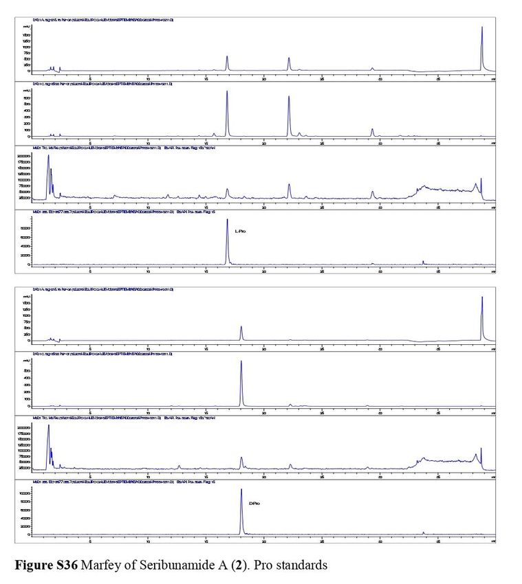
Updates in Pharmacology

time of the L-FDAA amino acid in the hydrolysate of 2 were
established as L-Ile 36.1 min. Retention times for the derivatized
amino acids standards were as follows: L-allo-Ile 31.7 min, L-Ile
36.1 min, D-allo-Ile 38.1 min and D-Ile 51.4 min.

Pro, NMeLeu and Glu were analyzed using: Symmetry 4.6x150
mm, 3.5 µm, flow 0.8 mL/min, H2O+ 0.04%TFA/ CH3CN +
0.04%TFA from 20 to 50% in 30 min. The retention time of the
L-FDAA amino acids in the hydrolysate of 2 were established as
L-Glu 14.0 min, L-Pro 16.8 min and NMe-L-Leu 28.5 min.
Retention times for the derivatized amino acids standards were
as follows: L-Glu 14.1 min, D-Glu 15.2 min, L-Pro 16.8 min, D-
Pro 14.01 min, NMe-L-Leu 28.5 min and NMe-D-Leu 30.1 min.

Biological Activity

The cytotoxic activity of 1 and 2 was tested against four human
tumour cell lines, lung (A-549), colon (HT-29), breast (MDA-
MB-231) and pancreas PSN-1 and both compounds displayed an
GI50 >1.2 E-5 M. Compound 1 was further tested for the capacity
to inhibit the enzyme topoisomerase I, but showed no inhibition
of the enzyme at 1.0E-5 M, and, was therefore not considered
active in inhibiting this enzyme. Likewise, compounds 1 and 2
were unable to impair the interaction between the programmed
cell death protein PD-1 and its natural ligand PD-L1 as
demonstrated by their lack of effect in a cell-based assay whose
final readout was dependent on the interaction between the two
proteins (Table 3).

Table 3: % PD-1 and TOPO-I Inhibition for compounds 1 and 2.

 Compound       Target     %Inhibition at 1 E-5 M

 1              Top-I      3
 1              PD-1       0.3
 2              PD-1       -3.5

Conclusions
In summary, two new peptides bearing unprecedented
halogenated moieties, haloirciniamide A (1) and seribunamide

15                                                   www.videleaf.com
Updates in Pharmacology

A (2) were isolated from a marine sponge belonging to the
Irnicia genus which was selected for further studies. The sample
was collected around Thousand Islands (Indonesia) by the
Pharmamar expedition team in collaboration with the Research
Center for Oceanography, Indonesian Institute of Sciences
(RCO-LIPI). The planar structures of the novel compounds were
determined by a combination of extensive NMR and HPLC-MS
experiments. The absolute configuration was achieved by
Marfey’s analysis after acid hydrolysis. Cytotoxic activity in the
four cancer cell lines tested was not observed for 1 and 2. In
addition, neither compound was able to impair PD1-PDL1
interaction, and compound 1 failed to inhibit the enzyme
topoisomerase I. This work is the first example of the isolation
and structural elucidation of novel compounds with unique
structural features from an Ircinia sponge, which highlights this
gender and its microbiota as a distinctive source of novel
structures.

References
1. Hanif N, Murni A, Tanaka Ch, Tanaka J. Marine Natural
   Products from Indonesian Waters. Mar. Drugs. 2019; 17:
   364.
2. Mehbub MF, Lei J, Franco C, Zhang W. Marine Sponge
   Derived Natural Products between 2001 and 2010: Trends
   and Opportunities for Discovery of Bioactives. Mar. Drugs.
   2014; 12: 4539-4577.
3. Kawakami A, Miyamoto T, Higuchi R, Uchiumi T, Kuwano
   M, et al. Structure of a novel multidrug resistance modulator,
   irciniasulfonic acid, isolated from a marine sponge Ircinia
   sp. Tetrahedron Lett. 2001; 42: 3335–3337.
4. Sica D, Piccialli V, Pronzato R. Sterols from the sponges
   Ircinia pipetta and Dysidea avara identification of
   cholestatrienol. Comp. Biochem. Physiol. 1987; 88: 293–
   296. Venkateswarlu Y, Reddy MVR, Rao MN. A new epoxy
   sterol from the sponge Ircinia fasciculata. J. Nat. Prod. 1996;
   59: 876–877.
5. Issa HH, Tanaka J, Higa T. New cytotoxic
   furanosesterterpenes from an Okinawan marine sponge
   Ircinia sp. J. Nat. Prod. 2003; 66: 252–254. , Lai YY, Lu

16                                              www.videleaf.com
Updates in Pharmacology

      MC, Wang LH, Chen JJ, Fang LS, et al. New scalarane
      sesterterpenoids from the Formosan sponge Ircinia felix.
      Mar. Drugs. 2015; 13: 4296–4309.
6.    Rashid MA, Gustafson KR, Boyd MR. New chondropsin
      macrolide lactams from marine sponges in the genus Ircinia.
      Tetrahedron Lett. 2001; 42: 1623–1626. , Chevallier C,
      Bugni TS, Feng X, Harper MK, Orendt AM, et al.
      Tedanolide C: A potent new 18-membered-ring cytotoxic
      macrolide isolated from the Papua New Guinea marine
      sponge Ircinia sp. J. Org. Chem. 2006; 71: 2510–2513.
7.    Feng Y, Caroll AR, Pass DM, Archbold JK, Avery VM, et
      al. Polydiscamides B−D from a marine sponge Ircinia sp. as
      potent human sensory neuron-specific G protein coupled
      receptor agonists. J. Nat. Prod. 2008; 71: 8–11.
8.    Mohamed AM, Rao V, Hamann MT, Kelly M, Hill RT.
      Monitoring Bacterial Diversity of the Marine Sponge Ircinia
      strobilina upon Transfer into Aquaculture. Appl. Environ.
      Microbiol. 2008; 74: 4133–4143.
9.    Mau C, Nakao Y, Yoshida WY, Scheuer PJ, Kelly-Borges
      M. Waiakeamide, a Cyclic Hexapeptide from the Sponge
      Ircinia dendroides. J. Org. Chem. 1996; 61: 6302-6304.
10.   Murakami Y, Takei M, Shindo K, Kitazume C, Tanaka J, et
      al. Cyclotheonamide E4 and E5, New Potent Tryptase
      Inhibitors from an Ircinia Species of Sponge. J. Nat. Prod.
      2002; 65: 259-261.
11.   Hooper GJ, Orjala J, Schatzman RC, Gerwick WH.
      Carmabins A and B, New Lipopeptides from the Caribbean
      Cyanobacterium Lyngbya majuscula. J. Nat. Prod. 1998;
      614: 529-533.
12.   Ebada SS, Wray V, De Voogd NJ, Deng Z, Lin W, et al.
      Two New Jaspamide Derivatives from the Marine Sponge
      Jaspis splendens. Mar. Drugs. 2009; 7: 435-444.
13.   Fu X, Do T, Schmitz FJ, Andrusevich V, Engel MH. New
      Cyclic Peptides from the Ascidian Lissoclinum patella. J.
      Nat. Prod. 1998; 61: 1547-1551.
14.   McIntosh M, Cruz LJ, Hunkapiller MW, Gray WR, Olivera
      BM. Isolation and structure of a peptide toxin from the
      marine snail Conus magus. Arch. Biochem. Biophys. 1982;
      218: 329-334.

17                                             www.videleaf.com
Updates in Pharmacology
15. Pettit GR, Kamano Y, Herald CL, Tuinman AA, Boettner FE,
et al. The isolation and structure of a remarkable marine
animal antineoplastic constituent: dolastatin 10. J. Am.
Chem. Soc. 1987; 109: 6883-6885.
16. Laird DW, LaBarbera DV, Feng XD, Bugni TS, Harper MK,
et al. Halogenanted Cyclic Peptides Isolated from the
Sponge Corticium sp. J. Nat.Prod. 2007; 70: 741-746.
17. Tarazona G, Fernández R, Cruz PG, Pérez M, Rodríguez J,
et al. Combining JBCA and Marfey’s methodology to
determine the absolute configuration of threonines: the case
of gunungamide A, a new cyclic depsideptide containing
chloropyrrole from the sponge Discodermia sp. Org. Chem.
Front. 2019; 6: 15-21.
18. Yasuda T, Araki A, Kubota T, Ito J, Mikami Y, et al.
Bromopyrrole Alkaloids from Marine Sponges of the Genus
Agelas. J. Nat. Prod. 2009; 72: 488-491.
19. Kobayashi H, Kitamura K, Nagai K, Nakao Y, Fusetani N,
et al. Carteramine A, an inhibitor of neutrophil chemotaxis,
from the marine sponge Stylissa carteri. Tetrahedron Lett.
2007; 48: 2127-2129.
20. Emrich R, Weyland H, Weber K. 2,3,4-Tribromopyrrole
from the marine Polychaete Polyphysia crassa. J. Nat. Prod.
1990; 53: 703-705.
21. Tebben J, Motti Ch, Tapiolas D, Thomas-Hall P, Harder T.
A Coralline Algal-Associated Bacterium,
Pseudoalteromonas Strain J010, Yields Five New
Korormicins and a Bromopyrrole. Mar. Drugs. 2014; 12:
2802-2815.
22. Marfey P. Determination of D-amino acids. II. Use of a
bifunctional reagent, 1,5-difluoro-2,4-dinitro- benzene.
Carlsberg Res. Commun. 1984; 49: 591−596.
23. Parra LLL, Berthonha AF, Severo IRM, Aguiar ACC, de
Souza GE, et al. Isolation, Derivative Synthesis, and
Structure−Activity Relationships of Antiparasitic
Bromopyrrole Alkaloids from the Marine Sponge Tedania
brasiliensis J. Nat. Prod. 2018; 81: 188-202.
18 www.videleaf.comUpdates in Pharmacology Supplementary Materials 19 www.videleaf.com
Updates in Pharmacology

20                        www.videleaf.com
Updates in Pharmacology

21                        www.videleaf.com
Updates in Pharmacology

22                        www.videleaf.com
Updates in Pharmacology

23                        www.videleaf.com
Updates in Pharmacology

24                        www.videleaf.com
Updates in Pharmacology

25                        www.videleaf.com
Updates in Pharmacology

26                        www.videleaf.com
Updates in Pharmacology

27                        www.videleaf.com
Updates in Pharmacology

28                        www.videleaf.com
Updates in Pharmacology

29                        www.videleaf.com
Updates in Pharmacology

30                        www.videleaf.com
Updates in Pharmacology

31                        www.videleaf.com
Updates in Pharmacology

32                        www.videleaf.com
Updates in Pharmacology

33                        www.videleaf.com
Updates in Pharmacology

34                        www.videleaf.com
Updates in Pharmacology

35                        www.videleaf.com
Updates in Pharmacology

36                        www.videleaf.com
Updates in Pharmacology

37                        www.videleaf.com
Updates in Pharmacology

38                        www.videleaf.com
Updates in Pharmacology

39                        www.videleaf.com
Updates in Pharmacology

40                        www.videleaf.com
Updates in Pharmacology

41                        www.videleaf.com
Updates in Pharmacology

42                        www.videleaf.com
You can also read