Characterization of a Divergent Sec61β Gene in Microsporidia
←
→
Page content transcription
If your browser does not render page correctly, please read the page content below
doi:10.1016/j.jmb.2006.04.028                                                                J. Mol. Biol. (2006) 359, 1196–1202

Characterization of a Divergent Sec61β
Gene in Microsporidia
Claudio H. Slamovits, Lena Burri and Patrick J. Keeling⁎

Canadian Institute for                   The general secretory (Sec) pathway is the main mechanism for protein
Advanced Research, Botany                secretion and insertion into endoplasmic reticulum and plasma membrane
Department, University of                in prokaryotes and eukaryotes. However, the complete genome of the
British Columbia, 3529-6270              highly specialized microsporidian parasite Encephalitozoon cuniculi appears
University Boulevard                     to lack a gene for Sec61β, one of three universally conserved proteins that
Vancouver, BC                            form the core of the Sec translocon. We have identified a putative, highly
Canada V6T 1Z4                           divergent homologue of Sec61β in the genome of another microsporidian,
                                         Antonospora locustae, and used this to identify a previously unrecognized
                                         Sec61β in E. cuniculi. The identity of these genes is supported by evidence
                                         from secondary structure prediction and gene order conservation. Their
                                         functional conservation is confirmed by expressing both microsporidian
                                         homologues in yeast, where they are localized to the endoplasmic reticulum
                                         and rescue a yeast Sec61β deletion mutant.
                                                                                     © 2006 Elsevier Ltd. All rights reserved.
                                         Keywords: general secretory pathway; SecYEG; Sec61; ER protein
*Corresponding author                    translocation; secretion

Introduction                                                         stimulatory effects and is not essential in Escherichia
                                                                     coli2,3 or yeast.4,5 Both γ and β proteins are small,
   Most protein translocation across biological                      ranging from 70 to 110 residues, and have a lower
membranes in prokaryotes and eukaryotes occurs                       level of sequence conservation than the α subunit.
through the general secretory (Sec) pathway.                         Consistent with their central role in cell biology,
Although some differences exist between the                          genes for these three components of the Sec
bacterial, archaeal and eukaryotic Sec pathways,                     machinery are known from all prokaryotic and
they share a common conserved central core of                        eukaryotic organisms with completely sequenced
three proteins named SecY, SecE and SecG in                          genomes.6
bacteria and Sec61α, β and γ in eukaryotes.                             Microsporidia is a diverse group consisting of
These proteins assemble into a heterotrimeric                        over 1300 described species, all of which are highly
complex that functions as the translocon and is                      derived, obligate intracellular parasites.7 Many
intimately involved in co- and post-translational                    characteristics of this enigmatic group appear
modes of membrane translocation.1 The α subunit                      primitive, which led investigators to think that
is well conserved and by far the largest component                   they were among the earliest lineages of eukar-
of the translocon. It consists of ten transmembrane                  yotes.8 However, it is now known that they are
domains (TMD) that fold up to form the aqueous                       not primitive, but rather extremely specialized and
pore that allows the passage of peptides across the                  highly reduced relatives of fungi.9–12 The severe
plasma membrane in prokaryotes and the endo-                         reduction that typifies microsporidia is exemplified
plasmic reticulum (ER) in eukaryotic cells. The γ                    by their genomes, which are among the smallest
subunit is considered to be an integral part of the                  known in eukaryotes. The genome of the human
pore and, like the α subunit, is essential for                       parasite Encephalitozoon cuniculi has been complete-
viability. The β subunit, on the other hand, has                     ly sequenced10 and is characterised by a compact
                                                                     and highly reduced complement of genes. Genome
                                                                     size in this parasite is just below three million
  Abbreviations used: TMD, transmembrane domains;                    base-pairs divided among11 small linear chromo-
ORF, open reading frame; EST, expressed sequence tag;                somes ranging from 217 to 315 kb with a typical
GFP, green fluorescent protein.                                      eukaryotic shape. However, gene density is about
  E-mail address of the corresponding author:                        one gene per kb, twice as high as in Saccharomyces
pkeeling@interchange.ubc.ca                                          cerevisiae and about the same as in typical bacterial

0022-2836/$ - see front matter © 2006 Elsevier Ltd. All rights reserved.
Microsporidian Sec61β 1197
genomes. Compaction has proceeded in these This sequence was compared to a database of A.
genomes to an extreme that basic transcription is locustae expressed sequence tag (EST) sequences,13
severely affected.13 Two other striking features of revealing two exact matches. Both ESTs encode the
microsporidian genomes are that gene order is complete ORF, which suggests that it is an
evolving relatively slowly, while the genes them- expressed functional gene, except that both also
selves are evolving very rapidly at the sequence encode a portion of the upstream gene. Many A.
level.14 The high level of sequence divergence can locustae transcripts have been shown to encode
make it difficult to recognise many microsporidian fragments of multiple genes and it can be difficult,
genes by sequence homology alone, but this is as in this case, to determine which gene is actually
partly mitigated by the high degree of synteny, expressed.13 The deduced peptide sequence was
which can be a useful tool for identifying compared to all annotated proteins in the E.
homologous genes in different microsporidian cuniculi genome with no success. However, when
genomes.15 TBLASTN was used to compare the deduced A.
A significant portion of the proteome of E. locustae protein sequence to the Genbank non-
cuniculi is devoted to essential, basic cellular redundant nucleotide database, the single signifi-
processes, such as replication, transcription, and cant match (E = 3e−09) corresponded to an inter-
translation.10 However, many generally conserved genic region on chromosome IX of E. cuniculi.
and widespread proteins have not been identified Examination of the region surrounding this posi-
in the E. cuniculi genome, either because they have tion of E. cuniculi chromosome IX (between nucleo-
been lost and their functions supplied by the host, tides 116,300−116,600) revealed a 260 bp ORF in
or they are too divergent to be recognised by frame with the TBLASTN match with a predicted
sequence similarity. Interestingly, divergent but size of 87 amino acid residues, only one shorter
clearly recognisable homologues of Sec subunits α than the A. locustae predicted protein. An align-
and γ are present, but the β subunit is absent. It is ment of the predicted gene products from E.
possible that the E. cuniculi Sec complex is cuniculi and A. locustae to other Sec61β homologues
simplified in a unique way and lacks the β subunit, (Figure 1(a)) shows significant divergence between
or alternatively it is present but unrecognised due the two microsporidia and other eukaryotes, and
to an unprecedented level of sequence divergence. that the microsporidian proteins are more similar
Determining between the two is of interest because to one another than any other Sec61β. Most of the
the secretion pathway is likely a major route for similarity resides between positions 80−110 (num-
parasite−host interactions in this system: any bered according to Figure 1(a)), which corresponds
protein secreted by a microsporidian is actually to a hydrophobic TMD whose sequence and
released into the parasitophorous vacuole or the position is conserved among eukaryotes.6 The
host cytoplasm, and infection by many microspor- microsporidian sequences are missing a substantial
idia is known to produce a dramatic effect on the N-terminal domain and have an additional C-
organisation and activity of the host cell.16–18 To terminal domain not found in other eukaryotes
determine if microsporidian Sec complexes include (Figure 1(a)).
Sec61β, we used a genome sequence survey of a To confirm that the A. locustae expressed gene
second microsporidian species, Antonospora locus- and this un-annotated region of the E. cuniculi
tae, and based on genomic position, secondary genome are indeed orthologous, we investigated
structure analysis, as well as localisation and their relative genomic contexts. The E. cuniculi
complementation in yeast, found that a highly ORF is situated on chromosome 9 between the
divergent form of Sec61β is indeed present in both genes for a syntaxin-like protein (09-0940) and a
microsporidia. hypothetical protein (09-0950), with genes oriented
as shown in Figure 1(b). The intergenic regions on
either side of the putative Sec61β would be only
Results 70 bp each. The A. locustae genome sequence
survey fragment extends 517 bp upstream from
Identification of Sec61β homologues in the ATG codon of Sec61β, where it shows
A. locustae and E. cuniculi similarity to the 3′ region of the syntaxin-like
protein with a 97 bp intergenic space between the
The well-annotated E. cuniculi genome does not termination codon of the syntaxin and the
encode a Sec61β homologue, but in the course of a initiation codon of the putative Sec61β. This is
genome sequence survey of the distantly related confirmed by the EST clones, both of which
microsporidian, A. locustae, we detected a fragment contain part of syntaxin, the complete intergenic
with some similarity to Sec61β using BLASTX region, and terminate with poly(A) tails just
against public sequence databases. The sequence downstream of Sec61β. The genomic clone ends
shared a low overall similarity with known Sec61β just downstream of the Sec61β gene as well, so
sequences (E = 0.06), but corresponded to an open we compared our sequences with the ongoing A.
reading frame (ORF) with a similar predicted size locustae genome project†, which confirmed the
of 88 amino acid residues and a central stretch of
27 amino acid residues highly similar to the † http://www.gmod.mbl.edu/perl/site/antonospora01?
conserved TMD of eukaryotic Sec61β sequences. page=intro1198 Microsporidian Sec61β
Figure 1. New E. cuniculi and A. locustae ORFs are putative Sec61β genes. (a) Protein sequence alignment of
eukaryotic Sec61β homologues and the two microsporidian genes. Shading represents conservation of identical (black) or
similar (grey) amino acids, and dashes represent gaps. Below the alignment, white boxes represent transmembrane
domains (TMD), inside of which broken lines show positions of α-helices. Hs, Homo sapiens; Ce, Caenorhabditis elegans;
Dm, Drosophila melanogaster; At, Arabidopsis thaliana; Sp, Schizosaccharomyces pombe; Sc, Saccharomyces cerevisiae; Ec, E.
cuniculi; Al, A. locustae. (b) Genomic contexts of AlocSec61β and EcuSec61β (grey) between a syntaxin-like protein and a
conserved hypothetical ORF. Arrows represent relative gene orientations.
order of syntaxin and Sec61β, and also confirmed philic tendency in the N-terminal half (Figure 2). In
that the gene lying 148 bp downstream of Sec61β contrast, the microsporidian sequences are pre-
is homologous to the hypothetical E. cuniculi ORF dicted to have two peaks of hydrophobicity,
09_0950 (Figure 1(b)). Overall, the three genes are suggesting two TMDs (Figures 1(a) and 2). The
found in the same order with two single-gene first peak corresponds to the TMD region con-
inversions, arguing strongly that the E. cuniculi served in other eukaryotes, and the second is the
intergenic region encodes a protein homologous to C-terminal region unique to E. cuniculi and A.
the Sec61β that we identified in the same region locustae (Figure 1(a)). In spite of not being espe-
of the A. locustae genome. cially similar at the sequence level, both microspo-
ridian sequences shared a similar general pattern of
Prediction of secondary structure secondary structure and hydropathy (Figures 1
and 2).
The primary sequence of the putative Sec61β
proteins from both A. locustae and E. cuniculi are Subcellular localisation and complementation in
quite different from homologues in other eukar- S. cerevisiae
yotes, but often the three-dimensional structure of
homologous proteins is maintained with little Sequence comparisons of the microsporidian
sequence similarity. Accordingly, we compared proteins suggest homology with eukaryotic
some structural properties of known Sec61β with Sec61β, they are divergent and analysis of their
the predicted microsporidian proteins. Homolo- secondary structure and physicochemical profiles
gues from diverse eukaryotes have a distinctive exhibit similarity but also differences with the
and well conserved structural pattern characterized otherwise conserved structure. We therefore tested
by a TMD and an alpha-helix located very close to whether the biological properties of the E. cuniculi
the C terminus (Figure 1(a)) resulting in a similar and A. locustae proteins conform to the expectations
hydropathy profile with a strong peak spanning for a genuine Sec61β. Since this peptide is involved
the C-terminal third of the protein and an hydro- in protein translocation to the ER, it is embedded inMicrosporidian Sec61β 1199
Figure 2. Microsporidian Sec61β proteins exhibit a distinct pattern of secondary structure. Hydropathy plots of
several eukaryotic Sec61β proteins, showing a distinct additional hydrophobic domain at the C terminus of the two
microsporidian proteins.
the ER membrane and is expected to encode an ER- disruption is non-lethal, but the strain is tempera-
signal peptide. Available applications for predict- ture-sensitive. It grows normally at 30 °C but is
ing subcellular localization produced confusing unable to grow at 38 °C.5 Growth of the transfor-
results when applied to these proteins: while mants was screened at both temperatures and
some methods (e.g. Psort II) predict ER or plasma Sec61β expressing cells were compared with a
membrane localization (55.6% for E. cuniculi and vector-transformed control (Figure 4). All strains
30.3% for A. locustae), others (e.g. Subloc, PrediSi, were indistinguishable from the wild-type (wt) at
TargetP, SignalP) failed to predict location. In the permissive temperature. However, at 38 °C growth
absence of any genetic system for expressing is significantly compromised in the deletion strain
proteins in microsporidia, we used a related fungal (Figure 4: 38 °C, vector), but the mutant phenotype
system, S. cerevisiae, to determine whether the is completely suppressed in seb1/seb2 mutants
putative proteins from A. locustae and E. cuniculi overexpressing either AlocSec61β or EcuSec61β,
localise to the ER. We overexpressed N-terminal showing that both microsporidian proteins can
green fluorescent protein (GFP) fusions for both functionally replace the autologous SEB1 and SEB2
genes in S. cerevisiae cells and observed their proteins from yeast.
subcellular distribution. Figure 3 shows that both
GFP-Sec61β fusions label the nuclear membrane
and membrane structures underlying the plasma Discussion
membrane, characteristic for a typical ER staining
pattern in yeast.19 Like many other highly specialised intracellular
We also tested the function of microsporidian parasites, microsporidia have diverged consider-
Sec61β in yeast using a second construct, containing ably from their closest relatives. At the molecular
the coding sequence from either A. locustae or E. level, this is most obviously reflected in highly
cuniculi under the control of a MET25 promoter divergent genes that are conserved in most
introduced into a yeast strain in which both versions organisms. Currently, the identity of about 50%
of Sec61β (SEB1 and SEB2) are disrupted. This of the predicted protein-coding genes in the E.
cuniculi genome remain unknown. While some of
these likely represent a set of genes that are
unique to microsporidia, others probably encode
common cellular components that are simply
difficult to identify due to the high sequence
divergence. At the same time, however, the E.
cuniculi genome is also missing many genes
expected in a eukaryotic genome, and some
complete pathways that are essential for a free-
living eukaryote, while other pathways are sim-
plified or reduced.10 Some of these proteins and
pathways probably have been lost, but in other
cases the major components may all be there but
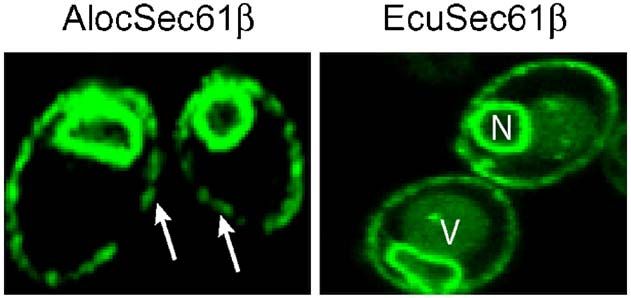
Figure 3. Both A. locustae and E. cuniculi Sec61β
are unidentified due to their divergent nature. The
proteins are localized in the ER. Yeast cells expressing Sec pathway constitutes the most common mech-
GFP-Sec61β were viewed by confocal microscopy. The anism to translocate proteins across membranes,
image shows a single confocal section through represen- and no prokaryotic or eukaryotic organism is
tative yeast cells. Both fusions target GFP to the ER known to lack it, although some other, non-
(arrows, peripheral membrane; N, nucleus; V, vacuole). universal mechanisms of translocation exist.20 It1200 Microsporidian Sec61β
Figure 4. A. locustae and E. cuniculi Sec61β can functionally replace their yeast homologues. The yeast strains H973
(wild-type) and H3235 (Δseb1Δseb2) were transformed with 2μ URA3 plasmids containing AlocSec61β or EcuSec61β
genes. A control transformation was performed with the vector lacking an insert. Serial, fivefold dilutions were spotted on
selective plates and incubated at 30 °C and 38 °C.
was therefore curious that only two of the three Materials and Methods
components of the Sec translocon (Sec61α and
Sec61γ) could be found in E. cuniculi, but we have
now demonstrated that the third component, Sequence analyses
Sec61β, is also present.
The microsporidian Sec61β is very unusual A. locustae DNA sequences come from genomic and
EST libraries described previously.13,14 Genome se-
compared with homologues in other eukaryotes,
quence searches were done using BLAST against NCBI
both in primary sequence and predicted secondary nucleotide and protein databases as well as local
structure, so it is interesting that it functions in the genomic and EST databases. The extended genomic
heterologous background of yeast cells. The trun- context of Sec61β in A. locustae was determined using
cated N terminus is unusual, but many microspor- genomic and EST data, and using data retrieved from
idian proteins are shorter than homologues in other the A. locustae genome project at MBL‡. Predictions of
eukaryotes, which is hypothesised to be the result of sub cellular localisation were made with the programs
attenuated interaction networks in their relatively PSORT II21, Subloc v1.0,22 PrediSi,23 TargetP v1.124 and
simple proteome.10 However, the microsporidian SignalP.25 Protein alignments were done using ClustalX
Sec61β proteins also contain an additional domain, and illustrated with Boxshade 3.21. Secondary struc-
ture predictions were carried out with the WHAT
the hydrophobic C-terminal region downstream of
program.26 Accession numbers for sequences reported
the conserved TMD. This domain is conserved in in this study are DQ415516 (A. locustae) and BK005765
both microsporidian proteins, but it is difficult to (E. cuniculi).
predict whether or how such structural differences
(i.e. an additional TMD) might affect the function in
microsporidian Sec complexes. These structural Plasmids, yeast strains, and media
differences apparently do not significantly alter the
association of microsporidian Sec61β with the rest of DNA fragments corresponding to A. locustae and E.
cuniculi Sec61β were amplified by PCR using primers that
the Sec complex, since both proteins rescue a yeast
generated in-frame restriction sites. PCR products were
mutant, suggesting functional association with the cloned under the control of the MET25 promoter behind
yeast Sec61α and Sec61γ. GFP-S65T for analysis by confocal microscopy, with the
In conclusion, we have shown that the microspor- URA3 gene for selection.27 The wild-type strain H973 (his4-
idian parasites A. locustae and E. cuniculi both encode 619 ura3-52) and the temperature-sensitive strain H3235
divergent homologues of Sec61β, despite its appar- (seb1∷KanMx seb2∷HphMx his4-619 ura3-52 GAL+) were
ent absence from the annotated genome of E. used for the complementation assay and the diploid strain
cuniculi. Evidence is based on sequence homology, JK9-3da/α (leu2-3,122/leu2-3,122 ura3-52/ura3-52 rme1/
conserved genomic context, and localisation and rme1 trp1/trp1 his4/his4 GAL+/GAL+ HMLa/HMLa) for
functional complementation in yeast. In spite of the confocal analysis. Yeast strains were generously provided
by Jussi Jäntti.
extreme reduction characteristic of microsporidian
cell biology, these parasites retain a complete core
complex of the Sec-dependent protein translocation ‡ http://www.gmod.mbl.edu/perl/site/antonospora01?
pathway. page=introMicrosporidian Sec61β 1201
Strains of S. cerevisiae were grown at 30 °C or 38 °C on 9. Thomarat, F., Vivares, C. P. & Gouy, M. (2004).
selective media (2% (w/v) glucose and 0.67% (w/v) Phylogenetic analysis of the complete genome se-
yeast nitrogen base supplemented with the relevant quence of Encephalitozoon cuniculi supports the fungal
amino acids). origin of microsporidia and reveals a high frequency
of fast-evolving genes. J. Mol. Evol. 59, 780–791.
10. Katinka, M. D., Duprat, S., Cornillot, E., Metenier,
Fluorescence microscopy G., Thomarat, F. Prensier, G. et al. (2001). Genome
sequence and gene compaction of the eukaryote
Fluorescence images were captured by using a parasite Encephalitozoon cuniculi. Nature, 414,
Nikon C1 confocal microscope. Cells were grown to 450–453.
mid log phase at 30 °C in selective media before 11. Keeling, P. J., Luker, M. A. & Palmer, J. D. (2000).
analysis. Evidence from beta-tubulin phylogeny that micro-
sporidia evolved from within the fungi. Mol. Biol. Evol.
17, 23–31.
12. Keeling, P. J. (2003). Congruent evidence from alpha-
tubulin and beta-tubulin gene phylogenies for a
Acknowledgements zygomycete origin of microsporidia. Fungal Genet.
Biol. 38, 298–309.
13. Williams, B. A., Slamovits, C. H., Patron, N. J., Fast,
We thank Jussi Jäntti for providing the yeast N. M. & Keeling, P. J. (2005). A high frequency of
strains H973 and H3235. This work was supported overlapping gene expression in compacted eukary-
by a grant from the Canadian Institutes for Health otic genomes. Proc. Natl Acad. Sci. USA, 102,
Research (MOP-42517). The Antonospora locustae 10936–10941.
Genome Project, Marine Biological Laboratory at 14. Slamovits, C. H., Fast, N. M., Law, J. S. & Keeling, P. J.
Woods Hole, is funded by NSF award number (2004). Genome compaction and stability in micro-
0135272. L.B. is supported by fellowships from sporidian intracellular parasites. Curr. Biol. 14,
CIHR and the Michael Smith Foundation for Health 891–896.
Research. P.J.K. is a Fellow of the Canadian Institute 15. Polonais, V., Prensier, G., Metenier, G., Vivares, C. P. &
Delbac, F. (2005). Microsporidian polar tube proteins:
for Advanced Research, and new investigator
highly divergent but closely linked genes encode
awards from CIHR and MSFHR. PTP1 and PTP2 in members of the evolutionarily
distant Antonospora and Encephalitozoon groups.
Fungal. Genet. Biol. 42, 791–803.
References 16. Leitch, G. J., Shaw, A. P., Colden-Stanfield, M.,
Scanlon, M. & Visvesvara, G. S. (2005). Multinucleate
1. Stephenson, K. (2005). Sec-dependent protein translo- host cells induced by Vittaforma corneae (Microspor-
cation across biological membranes: evolutionary idia). Folia Parasitol. (Praha), 52, 103–110.
conservation of an essential protein transport path- 17. Scanlon, M., Leitch, G. J., Shaw, A. P., Moura, H. &
way (review). Mol. Membr. Biol. 22, 17–28. Visvesvara, G. S. (1999). Susceptibility to apoptosis
2. Nishiyama, K., Mizushima, S. & Tokuda, H. (1993). A is reduced in the Microsporidia-infected host cell. J.
novel membrane protein involved in protein translo- Eukaryot. Microbiol. 46, 34S–35S.
cation across the cytoplasmic membrane of Escherichia 18. Scanlon, M., Shaw, A. P., Zhou, C. J., Visvesvara, G. S.
coli. EMBO J. 12, 3409–3415. & Leitch, G. J. (2000). Infection by microsporidia
3. Nishiyama, K., Hanada, M. & Tokuda, H. (1994). disrupts the host cell cycle. J. Eukaryot. Microbiol. 47,
Disruption of the gene encoding p12 (SecG) reveals 525–531.
the direct involvement and important function of 19. Burri, L., Varlamov, O., Doege, C. A., Hofmann, K.,
SecG in the protein translocation of Escherichia coli Beilharz, T. Rothman, J. E. et al. (2003). A SNARE
at low temperature. EMBO J. 13, 3272–3277. required for retrograde transport to the endoplas-
4. Toikkanen, J. H., Miller, K. J., Soderlund, H., Jäntti, J. & mic reticulum. Proc. Natl Acad. Sci. USA, 100,
Keranen, S. (2003). The beta subunit of the Sec61p 9873–9877.
endoplasmic reticulum translocon interacts with the 20. Mori, H. & Ito, K. (2001). The Sec protein-translocation
exocyst complex in Saccharomyces cerevisiae. J. Biol. pathway. Trends Microbiol. 9, 494–500.
Chem. 278, 20946–20953. 21. Nakai, K. & Horton, P. (1999). PSORT: a program for
5. Toikkanen, J., Gatti, E., Takei, K., Saloheimo, M., detecting sorting signals in proteins and predicting
Olkkonen, V. M. Soderlund, H. et al. (1996). Yeast their subcellular localization. Trends. Biochem. Sci. 24,
protein translocation complex: isolation of two genes 34–36.
SEB1 and SEB2 encoding proteins homologous to the 22. Hua, S. & Sun, Z. (2001). Support vector machine
Sec61 beta subunit. Yeast, 12, 425–438. approach for protein subcellular localization predic-
6. Cao, T. B. & Saier, M. H. Jr (2003). The general protein tion. Bioinformatics, 17, 721–728.
secretory pathway: phylogenetic analyses leading to 23. Hiller, K., Grote, A., Scheer, M., Munch, R. & Jahn, D.
evolutionary conclusions. Biochim. Biophys. Acta, 1609, (2004). PrediSi: prediction of signal peptides and
115–125. their cleavage positions. Nucl. Acids Res. 32,
7. Wittner, M. & Weiss, L. M. (1999). The Microsporidia W375–W379.
and Microsporidiosis, ASM Press, Washington, DC. 24. Emanuelsson, O., Nielsen, H., Brunak, S. & von
8. Vossbrinck, C. R., Maddox, J. V., Friedman, S., Heijne, G. (2000). Predicting subcellular localization
Debrunner-Vossbrinck, B. A. & Woese, C. R. (1987). of proteins based on their N-terminal amino acid
Ribosomal RNA sequence suggests microsporidia sequence. J. Mol. Biol. 300, 1005–1016.
are extremely ancient eukaryotes. Nature, 326, 25. Nielsen, H., Engelbrecht, J., Brunak, S. & von Heijne,
411–414. G. (1997). A neural network method for identification1202 Microsporidian Sec61β
of prokaryotic and eukaryotic signal peptides and transmembrane topology for a single protein se-
prediction of their cleavage sites. Int. J. Neural. Syst. 8, quence. J. Mol. Microbiol. Biotechnol. 3, 501–502.
581–599. 27. George, R., Beddoe, T., Landl, K. & Lithgow, T. (1998).
26. Zhai, Y. & Saier, M. H. Jr (2001). A web-based The yeast nascent polypeptide-associated complex
program (WHAT) for the simultaneous prediction of initiates protein targeting to mitochondria in vivo.
hydropathy, amphipathicity, secondary structure and Proc. Natl Acad. Sci. USA, 95, 2296–2301.
Edited by J. Karn
(Received 6 March 2006; received in revised form 4 April 2006; accepted 14 April 2006)
Available online 25 April 2006You can also read


