CIRCULATING SARS-COV-2 SPIKE N439K VARIANTS MAINTAIN FITNESS WHILE EVADING ANTIBODY-MEDIATED IMMUNITY
←
→
Page content transcription
If your browser does not render page correctly, please read the page content below
Article

Circulating SARS-CoV-2 spike N439K variants
maintain fitness while evading antibody-mediated
immunity
Graphical Abstract                                                        Authors
                                                                          Emma C. Thomson, Laura E. Rosen,
                                                                          James G. Shepherd, ..., Davide Corti,
                                                                          David L. Robertson, Gyorgy Snell

                                                                          Correspondence
                                                                          david.l.robertson@glasgow.ac.uk
                                                                          (D.L.R.),
                                                                          gsnell@vir.bio (G.S.)

                                                                          In Brief
                                                                          Epidemiological, clinical, molecular, and
                                                                          structural characterization of the N439K
                                                                          mutation in the SARS-CoV-2 spike
                                                                          receptor binding motif demonstrates that
                                                                          it results in similar viral fitness compared
                                                                          to wild-type while conferring resistance
                                                                          against some neutralizing monoclonal
                                                                          antibodies and reducing the activity of
                                                                          some polyclonal antibody responses.

Highlights
d   The receptor-binding motif (RBM) is a highly variable region
    of SARS-CoV-2 spike

d   RBM mutation N439K has emerged independently in
    multiple lineages

d   N439K increases spike affinity for hACE2; viral fitness and
    disease are unchanged

d   N439K confers resistance to several mAbs and escapes
    some polyclonal responses

           Thomson et al., 2021, Cell 184, 1171–1187
           March 4, 2021 ª 2021 The Authors. Published by Elsevier Inc.
           https://doi.org/10.1016/j.cell.2021.01.037                                                      ll
ll
                                                                                                                        OPEN ACCESS

Article
Circulating SARS-CoV-2 spike N439K variants
maintain fitness while evading
antibody-mediated immunity
Emma C. Thomson,1,2,29 Laura E. Rosen,3,29 James G. Shepherd,1,29 Roberto Spreafico,3,29 Ana da Silva Filipe,1
Jason A. Wojcechowskyj,3 Chris Davis,1 Luca Piccoli,4 David J. Pascall,5 Josh Dillen,3 Spyros Lytras,1
Nadine Czudnochowski,3 Rajiv Shah,1 Marcel Meury,3 Natasha Jesudason,1 Anna De Marco,4 Kathy Li,1 Jessica Bassi,4
Aine O’Toole,6 Dora Pinto,4 Rachel M. Colquhoun,6 Katja Culap,4 Ben Jackson,6 Fabrizia Zatta,4 Andrew Rambaut,6
Stefano Jaconi,4 Vattipally B. Sreenu,1 Jay Nix,7 Ivy Zhang,8,9 Ruth F. Jarrett,1 William G. Glass,8 Martina Beltramello,4
Kyriaki Nomikou,1 Matteo Pizzuto,4 Lily Tong,1 Elisabetta Cameroni,4 Tristan I. Croll,10 Natasha Johnson,1 Julia Di Iulio,3
Arthur Wickenhagen,1 Alessandro Ceschi,11,12,13 Aoife M. Harbison,14 Daniel Mair,1 Paolo Ferrari,15,16 Katherine Smollett,1
Federica Sallusto,17,18 Stephen Carmichael,1 Christian Garzoni,19 Jenna Nichols,1 Massimo Galli,20 Joseph Hughes,1

                                                                                                         (Author list continued on next page)

1MRC-University     of Glasgow Centre for Virus Research, University of Glasgow, Glasgow G61 1QH, UK
2Department    of Clinical Research, London School of Hygiene and Tropical Medicine, London WC1E 7HT, UK
3Vir Biotechnology, San Francisco, CA 94158, USA
4Humabs Biomed SA, a subsidiary of Vir Biotechnology, 6500 Bellinzona, Switzerland
5Institute of Biodiversity, Animal Health and Comparative Medicine, Boyd Orr Centre for Population and Ecosystem Health, University of

Glasgow, Glasgow G61 1QH, UK
6Institute of Evolutionary Biology, University of Edinburgh, Edinburgh EH9 3FL, UK
7Molecular Biology Consortium, Advanced Light Source, Lawrence Berkeley National Laboratory, Berkeley, CA 94720, USA
8Computational and Systems Biology Program, Sloan Kettering Institute, Memorial Sloan Kettering Cancer Center, New York, NY 10065, USA
9Tri-Institutional PhD Program in Computational Biology and Medicine, Weill Cornell Graduate School of Medical Sciences, New York, NY

10065, USA
10Cambridge Institute for Medical Research, Department of Haematology, University of Cambridge, Cambridge CB2 0XY, UK
11Faculty of Biomedical Sciences, Università della Svizzera italiana, 6900 Lugano, Switzerland
12Division of Clinical Pharmacology and Toxicology, Institute of Pharmacological Sciences of Southern Switzerland, Ente Ospedaliero

Cantonale, 6900 Lugano, Switzerland
13Department of Clinical Pharmacology and Toxicology, University Hospital Zurich, 8091 Zurich, Switzerland
14Department of Chemistry and Hamilton Institute, Maynooth University, Maynooth, Ireland
15Department of Nephrology, Ospedale Civico Lugano, Ente Ospedaliero Cantonale, 6900 Lugano, Switzerland

                                                                                                        (Affiliations continued on next page)

SUMMARY

SARS-CoV-2 can mutate and evade immunity, with consequences for efficacy of emerging vaccines and anti-
body therapeutics. Here, we demonstrate that the immunodominant SARS-CoV-2 spike (S) receptor binding
motif (RBM) is a highly variable region of S and provide epidemiological, clinical, and molecular characteriza-
tion of a prevalent, sentinel RBM mutation, N439K. We demonstrate N439K S protein has enhanced binding
affinity to the hACE2 receptor, and N439K viruses have similar in vitro replication fitness and cause infections
with similar clinical outcomes as compared to wild type. We show the N439K mutation confers resistance
against several neutralizing monoclonal antibodies, including one authorized for emergency use by the US
Food and Drug Administration (FDA), and reduces the activity of some polyclonal sera from persons recov-
ered from infection. Immune evasion mutations that maintain virulence and fitness such as N439K can
emerge within SARS-CoV-2 S, highlighting the need for ongoing molecular surveillance to guide development
and usage of vaccines and therapeutics.

INTRODUCTION                                                         as of the end of 2020. Molecular epidemiology studies across
                                                                     the world have generated over 330,000 viral genomic se-
SARS-CoV-2, the cause of COVID-19, emerged in late 2019 and          quences, shared with unprecedented speed via the GISAID
expanded globally, resulting in over 82 million confirmed cases      Initiative (https://gisaid.org). These data are essential for

                                               Cell 184, 1171–1187, March 4, 2021 ª 2021 The Authors. Published by Elsevier Inc. 1171
                                   This is an open access article under the CC BY license (http://creativecommons.org/licenses/by/4.0/).
ll
      OPEN ACCESS                                                                                                                Article

Agostino Riva,20 Antonia Ho,1 Marco Schiuma,20 Malcolm G. Semple,21,22 Peter J.M. Openshaw,23 Elisa Fadda,14
J. Kenneth Baillie,24,25 John D. Chodera,8 The ISARIC4C Investigators,26 the COVID-19 Genomics UK (COG-UK)
Consortium,27 Suzannah J. Rihn1 Samantha J. Lycett,24 Herbert W. Virgin,3,28 Amalio Telenti,3 Davide Corti,4
David L. Robertson,1,* and Gyorgy Snell3,30,*
16Prince   of Wales Hospital Clinical School, University of New South Wales, Sydney, NSW 2052, Australia
17Institute  for Research in Biomedicine, Università della Svizzera italiana, 6500 Bellinzona, Switzerland
18ETH Institute of Microbiology, ETH Zurich, 8093 Zürich, Switzerland
19Clinic of Internal Medicine and Infectious Diseases, Clinica Luganese Moncucco, 6900 Lugano, Switzerland
20III Division of Infectious Diseases, ASST Fatebenefratelli Sacco, Luigi Sacco Hospital, 20157 Milan, Italy
21NIHR Health Protection Research Unit in Emerging and Zoonotic Infections, Institute of Infection, Veterinary and Ecological Sciences,

Faculty of Health and Life Sciences, University of Liverpool, Liverpool L69 7BE, UK
22Respiratory Medicine, Alder Hey Children’s Hospital, Liverpool L12 2AP, UK
23National Heart and Lung Institute, Imperial College London, London SW3 6LY, UK
24The Roslin Institute, University of Edinburgh, Edinburgh EH25 9RG, UK
25Intensive Care Unit, Royal Infirmary Edinburgh, Edinburgh EH16 4SA, UK
26ISARIC4C Investigators
27https://www.cogconsortium.uk
28Washington University School of Medicine, Saint Louis, MO 63110, USA
29These authors contributed equally
30Lead contact

*Correspondence: david.l.robertson@glasgow.ac.uk (D.L.R.), gsnell@vir.bio (G.S.)
https://doi.org/10.1016/j.cell.2021.01.037

monitoring virus transmission and spread (Meredith et al., 2020).         and spread by chance. Moreover, the full impact of immune se-
Of special interest is the evolution of the SARS-CoV-2 surface            lection, which can drive variant selection, has not yet influenced
protein, spike (S), which is responsible for viral entry via its inter-   the pandemic, because herd immunity has not been attained. As
action with the human angiotensin-converting enzyme 2 (hACE2)             population immunity increases and vaccines are deployed at
receptor on host cells. The S protein is the target of neutralizing       scale, this will very likely change. The potential for circulating
antibodies generated by infection (Jiang et al., 2020) or vaccina-        viral variants to derail promising vaccine or antibody-based pro-
tion (Folegatti et al., 2020; Jackson et al., 2020; Keech et al.,         phylactics or treatments, even in the absence of selective pres-
2020) as well as monoclonal antibody (mAb) drugs currently in             sure from the drug or vaccine, is demonstrated by the failure of a
clinical trials and/or approved for Emergency Use Authorization           phase III clinical trial of a mAb targeting the respiratory syncytial
(EUA) by the US Food and Drug Administration (FDA) (Chen                  virus (Simões et al., 2020) and the need for new influenza vac-
et al., 2021; Hansen et al., 2020; Jones et al., 2020; Pinto              cines on a yearly basis. It is therefore critical to understand
et al., 2020).                                                            whether and how SARS-CoV-2 may evolve to evade antibody-
   A SARS-CoV-2 S amino acid change, D614G, is now dominant               dependent immunity.
in most places around the globe (Korber et al., 2020). Studies               Here, we examine the immunodominant SARS-CoV-2 recep-
in vitro indicate that this mutation confers greater infectivity while    tor binding motif (RBM), the primary target of the neutralizing
molecular epidemiology correlates it with an increase in trans-           Ab response within the RBD (Piccoli et al., 2020), and find it to
missibility with no evidence to date for increased virulence              be a highly variable region of the S protein in circulating viruses.
(Hou et al., 2020; Hu et al., 2020; Korber et al., 2020; Volz et          To understand the implications of this structural plasticity, which
al., 2021; Zhang et al., 2020). Amino acid 614 is located outside         could allow the RBD to accommodate amino acids changes
the receptor binding domain (RBD) of S, the domain targeted by            that could contribute to immune evasion, we defined the clinical
90% of neutralizing antibody activity in serum of SARS-CoV-2              and epidemiological impact, molecular features, and immune
survivors (Piccoli et al., 2020). Initial studies suggest that            response to the RBM mutation N439K. This amino acid replace-
D614G viruses exhibit increased sensitivity to neutralizing anti-         ment has arisen independently multiple times, and in two cases
bodies, likely due to the effect of the mutation on the molecular         formed lineages of more than 500 sequences. As of January 6,
dynamics of the S protein (Hou et al., 2020; Weissman et al.,             2021, it was observed in 34 countries and was the second
2021; Yurkovetskiy et al., 2020). Therefore, this now dominant            most commonly observed RBD mutation worldwide, and the
variant is unlikely to jeopardize natural or vaccine-derived anti-        sixth most common S mutation. We find that the N439K mutation
body-mediated immunity generated in response to D614 S                    results in enhanced RBD affinity for hACE2, it is associated with a
protein.                                                                  similar clinical spectrum of disease and slightly higher viral loads
   The low numbers of novel mutations reaching high frequency             in vivo compared to viruses with the wild-type (WT) N439 resi-
in sequenced SARS-CoV-2 genomes relates to the moderate                   due, and it results in immune escape from polyclonal sera
intrinsic error rate of SARS-CoV-2 RNA replication (Li et al.,            from a proportion of recovered individuals and some neutralizing
2020c; Robson et al., 2020). Nevertheless, the increasing num-            mAbs.
ber of infected individuals and the large reservoir of hosts sus-            N439K provides a sentinel example of immune escape, indi-
ceptible to infection increase the likelihood that novel variants         cating that RBM variants must be evaluated when considering
that impact vaccine and therapeutic development will emerge               vaccines and the therapeutic or prophylactic use of mAbs.

1172 Cell 184, 1171–1187, March 4, 2021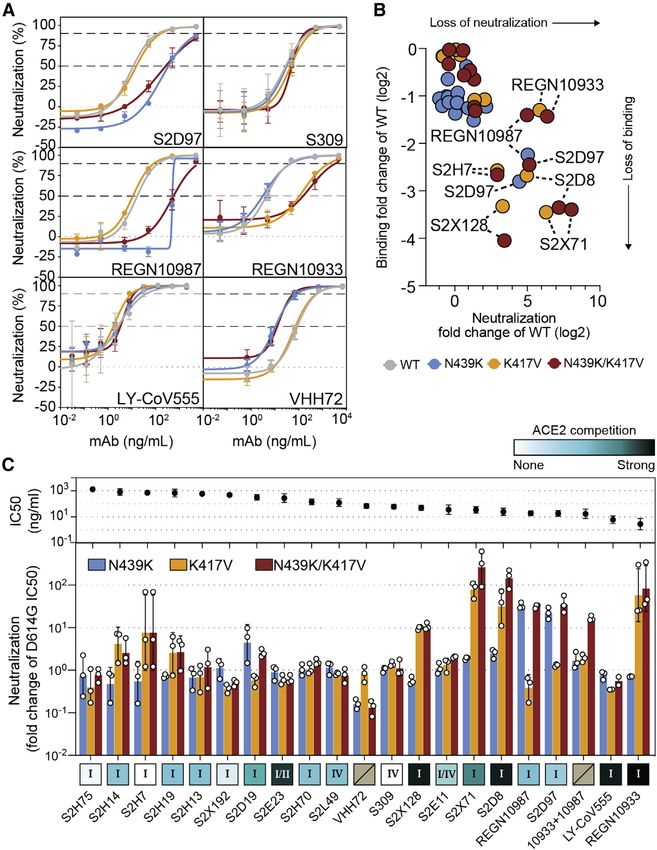
ll
Article                                                                                                                                   OPEN ACCESS

                                                                                                            Figure 1. The RBM exhibits significant natu-
                                                                                                            ral diversity in circulating SARS-CoV-2 vi-
                                                                                                            ruses
                                                                                                           SARS-CoV-2 variants (retrieved from CoV-
                                                                                                           GLUE) are based on 209,239 high-quality se-
                                                                                                           quences downloaded from GISAID on November
                                                                                                           30, 2020.
                                                                                                           (A) Structure of the SARS-CoV-2 RBD-hACE2
                                                                                                           complex (PDB: 6M0J) highlighting the RBM
                                                                                                           (blue) and residue N439 (yellow).
                                                                                                           (B) Thirty-four residues (the size of the RBM)
                                                                                                           were randomly sampled without replacement
                                                                                                           50,000 times from the mature S protein
                                                                                                           (excluding the RBM). Median entropies were
                                                                                                           computed for each draw. The resulting 50,000
                                                                                                           median entropies were used to build the entropy
                                                                                                           distribution of residues other than the RBM. The
                                                                                                           top 10% medians are highlighted in red. The
                                                                                                           median entropy of RBM residues was compared
                                                                                                           with the non-RBM entropy distribution to deter-
                                                                                                           mine the variability of the RBM relative to non-
                                                                                                           RBM residues. To allow for a fair comparison,
                                                                                                           sampling was performed without enforcing res-
                                                                                                           idue contiguity, as the RBM is not contiguous in
                                                                                                           sequence space. Therefore, in any given sam-
                                                                                                           ple, residues are unlikely to share any functional
                                                                                                           relationship.
                                                                                                           (C) Per-residue entropies of the mature S protein
                                                                                                           were smoothed by plotting medians of a 25-aa
                                                                                                           center-aligned sliding window. Smoothing al-
                                                                                                           lows visualizing local peaks of variability. The
                                                                                                           RBM residues and the NTD, RBD, and S2 do-
mains are highlighted. Due to the non-contiguous nature of the RBM in sequence space, the sliding window median at RBM residues is diluted by
neighboring non-RBM residues.
(D) Boxplot of per-residue entropies in four S domains (or full mature S protein). The lower and upper hinges correspond to the first and third quartiles. The
lower/upper whiskers extend from the hinge to the smallest/largest value no further than 1.5 times the inter-quartile range. Outliers beyond the end of the
whiskers are not plotted but are retained for statistical testing. Pairwise comparisons by Mann-Whitney U tests. p value thresholds are 0.05 (*), 0.01 (**) and
0.001 (***); ns, not significant.
See also Figures S1 and S2.

Long-term control of the pandemic with vaccines will require                     off (Figures 1A–1D and S1A, 2). We evaluated SARS-CoV-2
systematic monitoring of immune escape variants and may                          genomic sequences deposited in GISAID as of November
require new vaccine preparations that address the variants                       30th, 2020 and observed a high number of variants occurring
circulating globally.                                                            in the RBM. To understand how the variability of the RBM com-
                                                                                 pares to the variability of the entire RBD and the whole S protein,
RESULTS                                                                          we evaluated well-defined S protein domains: within S1, the
                                                                                 N-terminal domain (NTD) and the RBD (further split into RBM
The RBM is a variable region of the SARS-CoV-2 S                                 and non-RBM), and the S2 domain. Analysis of entropy, which
protein                                                                          estimates sequence variability at a given position in a protein
Competing pressures influence the evolution of the S RBM. First,                 alignment, identified the RBM as a highly variable region of the
the RBM mediates viral entry (Shang et al., 2020; Walls et al.,                  RBD and of the entire S protein (Figures 1B–1D), with a median
2020; Wrapp et al., 2020b) and therefore must maintain sufficient                entropy within the top 10% of equivalently sized sets of randomly
affinity to engage the entry receptor hACE2. Second, it is a major               sampled residues (Figure 1B). This result is confirmed by an anal-
target of neutralizing antibodies (Piccoli et al., 2020; Robbiani                ysis of sequence variability that is not weighted by total counts
et al., 2020; Rogers et al., 2020; Wec et al., 2020) and so would                of each variant, thereby capturing the diversity of circulating
be a primary location for the emergence of immune escape mu-                     variants with mitigated bias toward oversampled variants
tations. We set out to understand these competing pressures                      (Figure S1A).
by evaluating the landscape of RBM sequence divergence                              To understand constraints on RBM variability, we evaluated
observed in circulating SARS-CoV-2 variants and in other vi-                     the published deep mutational scanning (DMS) dataset of the
ruses of the Sarbecovirus lineage.                                               RBD (Starr et al., 2020b) and compared it to sequences of circu-
   We used re-refined published X-ray structures of SARS-CoV                     lating viruses. The DMS data define the effect of each possible
and SARS-CoV-2 RBD:hACE2 complexes (Lan et al., 2020; Li                         single amino acid change on both expression of the RBD and
et al., 2005) to define the RBM residues using a 6 Å distance cut-              its capacity to bind hACE2. For each position in the RBM, we

                                                                                                             Cell 184, 1171–1187, March 4, 2021 1173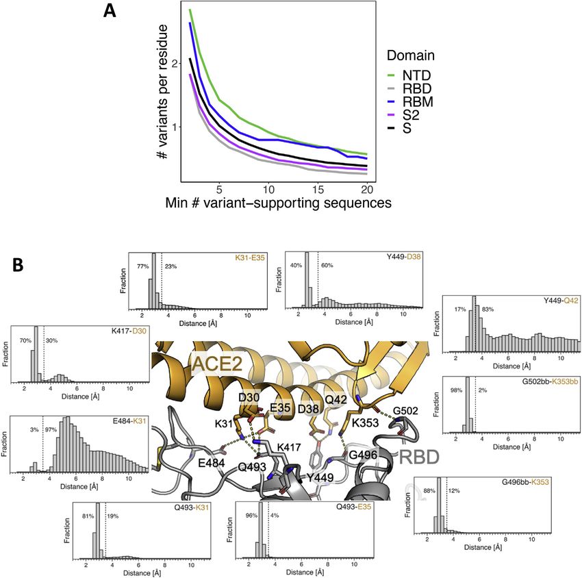
ll
      OPEN ACCESS                                                                                                                                 Article

                                                                                     compared the DMS results for all amino acid replacements at
                                                                                     that position versus only changes that have been observed in
                                                                                     circulating SARS-CoV-2 variants (Figure 2). A subset of residues
                                                                                     shows the largest loss of hACE2 binding on mutation (top 1/3 of
                                                                                     RBM residues in Figure 2) and, as would be expected, few nat-
                                                                                     ural occurrences of mutations at these residues have been
                                                                                     observed to be circulating. However, for the majority of the
                                                                                     RBM (bottom 2/3 of RBM residues in Figure 2), variation in
                                                                                     circulating virus sequences confirms the tolerance to mutation
                                                                                     predicted by the DMS data.
                                                                                        To further assess the ability of the RBM to accommodate mu-
                                                                                     tations without disrupting hACE2 binding, we examined the
                                                                                     structural dynamics and energetics of the RBM:hACE2 binding
                                                                                     interface. We performed an approximate, residue-level decom-
                                                                                     position of binding free energy based on the RBD:hACE2
                                                                                     complex X-ray structure (green in Figure 2) as well as molecular
                                                                                     dynamics simulations of the complex, resulting in 118 ms of
                                                                                     aggregate simulation data (Figure S1B). Consistent with expec-
                                                                                     tation, the two residues with the highest variant frequency (S477
                                                                                     and N439) contribute weakly to the binding energy (Figure 2).
                                                                                     Surprisingly, the two RBM residues with the strongest interac-
                                                                                     tions with hACE2 based on the X-ray structure (K417 and
                                                                                     E484, dark green in Figure 2) were not highly conserved (variant
                                                                                     % in red, Figure 2), with 10-fold more variants for E484. This
                                                                                     could be explained by results from the molecular dynamics
                                                                                     simulation: K417 formed close interactions with hACE2 70% of
                                                                                     the simulation time, while E484 only 3% of the time (Figure S1B).
                                                                                     The low percent for E484 is also consistent with the non-conser-
                                                                                     vative amino acid replacements observed for circulating variants
                                                                                     (e.g., the most common E484 substitution is currently E484K),
                                                                                     with a positively charged lysine substituting for the negatively
                                                                                     charged glutamate. Overall, these results demonstrate that the
                                                                                     RBM has a high degree of structural plasticity whereby it is
                                                                                     able to accommodate amino acid changes without disrupting
                                                                                     hACE2 binding.
                                                                                        Evolutionary analysis of the Sarbecovirus subgenus provides
                                                                                     further support for RBM plasticity (Boni et al., 2020; Li et al.,
                                                                                     2020b; Rambaut et al., 2020). The SARS-CoV RBM is highly
                                                                                     divergent from the SARS-CoV-2 RBM (Figures S2A and S2B)
                                                                                     while maintaining hACE2 binding affinity. Additionally, there are
                                                                                     many sequence changes in the RBM across a panel of related
                                                                                     coronaviruses from animal isolates (Figures S2A and S2B; Table
Figure 2. RBM functional constraints compared to RBM natural
diversity                                                                            S1). To determine the ability of members of the Sarbecovirus
Each residue in the RBM is annotated by several metrics, depicted as a               lineage to bind hACE2, we produced nine recombinant RBD pro-
heatmap. DMS scores: outlined in black boxes (center) are summaries of               teins corresponding to seven animal isolates, SARS-CoV-2, and
hACE2 binding and RBD expression deep mutational scanning (DMS) exper-               SARS-CoV, and evaluated their binding to recombinant hACE2
imental results (Starr et al., 2020b). DMS score is the binding or expression fold   (Figure S2C). We found that three of the RBDs from animal iso-
change of a variant over WT on a log10 scale (red indicating improvement and
                                                                                     lates showed strong affinity for hACE2: GD Pangolin, which
blue indicating loss as compared to WT). In the ‘‘mutagenesis’’ columns, DMS
results are given for each residue as either the minimum (most disruptive
                                                                                     has a highly similar RBM to SARS-CoV-2, GX Pangolin, which
variant) or the average score across all possible variants of a residue, except      has a more divergent RBM, and Bat CoV WIV1 which is highly
for the reference residue and the stop codon. In the ‘‘observed variants’’
columns, minimum and average scores are computed only across variants
that have been observed in GISAID (same set of sequences as used for Fig-            purple indicating more countries). Binding energy: a re-refined SARS-CoV-2
ure 1). When no natural variants have been observed, cells are gray. Data were       RBD:hACE2 complex X-ray structure (PDB: 6M0J) was used to determine the
sorted on the leftmost DMS column. Frequency: each RBM position is anno-             approximate, decomposed binding free energy associated with each RBM
tated with the frequency of non-reference amino acids in deposited sequences         residue. Results for each RBM residue are expressed as a percentage of the
(darker red indicating higher frequency; at least 1 supporting sequence per          total binding interface interaction energy (darker green indicating stronger
25,000 deposited sequences is required to call a variant). The number of             contribution to the binding energy).
countries in which variants have been observed is also annotated (darker             See also Figures S1 and S2.

1174 Cell 184, 1171–1187, March 4, 2021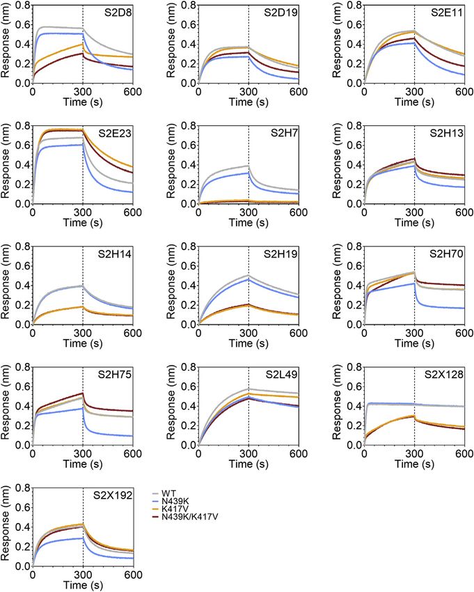
ll
Article                                                                                                                                    OPEN ACCESS

                                                                                                             Figure 3. The N439K RBM mutation has
                                                                                                             arisen independently multiple times, twice
                                                                                                             forming significant lineages
                                                                                                            (A) Phylogenetic tree (de-duplicated and down-
                                                                                                            sampled) showing the relationship among repre-
                                                                                                            sentative global SARS-CoV-2 variants, with N439K
                                                                                                            variants highlighted in color. Two significant
                                                                                                            N439K lineages, one in Scotland (>500 sequences,
                                                                                                            blue circles) and one in 32 countries (>6,000 se-
                                                                                                            quences, yellow circles) were detected as of
                                                                                                            January 6, 2021. The N439K mutation has also
                                                                                                            emerged independently on at least seven occa-
                                                                                                            sions (red circles show four of these) bringing the
                                                                                                            total country count to 34. Vertical bars indicate
                                                                                                            global lineage, the presence of N439K (same
                                                                                                            colors as tree), D614G (orange) or D614N (dark
                                                                                                            gray). The scale bar corresponds to a single
                                                                                                            nucleotide polymorphism (SNP).
                                                                                                            (B) Frequency of N439K variants relative to sam-
                                                                                                            pling time and their geographical area of occur-
                                                                                                            rence (see key): Africa (Morocco, Nigeria), Amer-
                                                                                                            icas (Brazil, USA), Asia (Japan, Singapore, South
                                                                                                            Korea), the European countries Denmark, England,
                                                                                                            Republic of Ireland and Scotland and other Euro-
                                                                                                            pean countries (Belgium, Bosnia-Herzegovina,
                                                                                                            Croatia, Czech Republic, Faroe Islands, Finland,
                                                                                                            France, Germany, Hungary, Italy, Luxembourg,
                                                                                                            Netherlands, Northern Ireland, Norway, Poland,
                                                                                                            Romania, Slovakia, Sweden, Switzerland, Wales),
                                                                                                            and Oceania (Australia, New Zealand). The prom-
inent light gray bars correspond to other European countries. See Table S2 for total numbers for each country.
(C) Frequency of the two N439K lineages (same colors as A) over time relative to all sequences for that country (gray) and their normalized contributions (lower
panels) in Scotland, England, Republic of Ireland, and Denmark.
See also Figure S3.

divergent (Figures S2A and S2B). This further indicates that the                 countries (Figures 3A–3C). N439K lineages i and ii have recently
RBM is structurally plastic, retaining binding with hACE2 as a re-               received the lineage designations B.1.141 and B.1.258, respec-
ceptor despite changes to sequence. Given this plasticity, we                    tively (Rambaut et al., 2020). We also observe at least seven in-
next considered whether an RBM variant can lead to immune                        stances of the N439K mutation that have arisen independently of
evasion while retaining virulence.                                               these two large lineages, including again in the United States in
                                                                                 at least four linked infections, and in Brazil and Nigeria where no
Phylogenetic analysis of the prevalent SARS-CoV-2                                lineage ii/B.1.258 has been observed, resulting in a total of 34
RBM mutation N439K                                                               countries where N439K has been detected to date (Figures 3A
N439K is a prevalent RBM mutation (the second most common                        and 3B).
mutation in the RBD through the end of 2020) which was first                        Sequence counts are heavily influenced by sampling fre-
sampled in March 2020 in Scotland from lineage B.1 (Rambaut                      quency, which varies widely between countries, and N439K as
et al., 2020) on the background of D614G. Using phylogenetic                     a percentage of total sequences appears low: as of January 6,
analysis, we determined that the earliest reported N439K se-                     2021, there have been 6,868 N439K observations in GISAID,
quences represented a single SARS-CoV-2 lineage (Figure 3A)                      2% out of 290,000 SARS-CoV-2 genome sequences for the
that increased in frequency to 542 sequences in Scotland by                      34 countries where this mutation has been detected (Table
June 20, 2020 (10% of the available Scottish viral genome se-                   S2). Nevertheless, when comparing the percentage of N439K
quences for this time period). Subsequently, numbers of N439K                    sequences over time in countries with sufficient data, the propor-
and all other variants decreased in Scotland concurrent with                     tion can be significant: 10% in Scotland from March to June
control of the pandemic after initiation of stringent public health              2020 and 10% in Denmark from August to December 2020,
measures, with this specific N439K lineage (designated here as                   both countries with high sequencing rates, and 13% in Ireland
lineage i) not being detected since June 2020 (Figures 3B and                    from July to December 2020, where regional coverage is reason-
3C). However, the N439K mutation appears in >6,000 additional                    able, but the sequencing rate is lower (Figure 3C). Importantly, on
sequences in the GISAID database as of January 6, 2021. Our                      the scale of a pandemic, small proportions correspond to large
analysis demonstrates that the majority of these sequences                       numbers of infections. If the proportion of N439K sequences in
represent a second, independent lineage (designated lineage ii)                  each country predicts what proportion of its confirmed infections
which was first sampled in Romania on May 13, 2020, then Nor-                    are associated with N439K variants, then N439K variants corre-
way on June 23, 2020, and is now detected to be circulating in 32                spond to 764,000 of the confirmed SARS-CoV-2 infections as

                                                                                                              Cell 184, 1171–1187, March 4, 2021 1175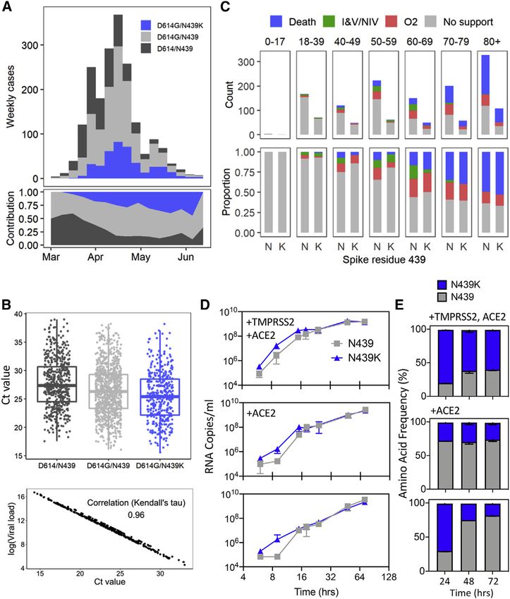
ll
     OPEN ACCESS                                                                                                                     Article

A                                      D                                                         Figure 4. N439K creates a new RBD:hACE2
                                                                                                 salt bridge and enhances RBD:hACE2 affinity
                                                                                                 (A–C) X-ray structures of the SARS-CoV (A), SARS-
                                                                                                 CoV-2 WT (B), and SARS-CoV-2 N439K (C) RBD in
                                                                                                 complex with hACE2 (based on 2AJF, 6M0J, and
                                                                                                 current work, respectively). Select interface resi-
                                                                                                 dues are shown as sticks. hACE2 is shown in or-
                                                                                                 ange and RBD in gray. The inset in (C) shows the
                                                                                                 2Fo-Fc electron density contoured at 1s for the
                                                                                                 K439-E329 salt bridge.
B                                                                                                (D) Binding affinity of RBD and Spike variants for
                                                                                                 hACE2 measured by surface plasmon resonance.
                                                                                                 Monomeric hACE2 is injected successively at 11,
                                                                                                 33, 100, and 300 nM onto surface-captured spike
                                                                                                 extracellular domain (ECD) or RBD; alternately,
                                                                                                 RBD is injected successively at 3.1, 12.5, and 50 nM
                                                                                                 onto surface-captured hACE2. All spike ECD
                                                                                                 contain the D614G mutation. Bar graph: affinity
                                                                                                 measurements (averages of 3–4 replicates) ex-
                                                                                                 pressed as a fold change relative to WT binding
C                                                                                                within each experiment format, where >1 indicates
                                                                                                 improved binding (smaller KD) relative to WT. WT KD
                                                                                                 values measured as: 95 ± 1.6 nM (Spike surface),
                                                                                                 63 ± 1.0 nM (RBD surface), 19 ± 3.3 nM (hACE2
                                                                                                 surface); errors are SEM.
                                                                                                 See also Table S3.

                                                                                               land, its growth rate is similar to the median
                                                                                               N439/D614 or N439/D614G WT growth
                                                                                               rates, with no evidence for a faster growth
of January 6, 2021 (Table S2). If detected cases represent 5%–           conferred by the N439K mutation (Figure S3A).
33% of true infections, as has been estimated for the United
States (Wu et al., 2020b), then a very rough approximation of            N439K RBD forms a new interaction with hACE2 and has
the actual cumulative number of N439K-associated infections              enhanced hACE2 affinity
would be in the range of 2–15 million.                                   In addition to its frequency and repeated emergence, the N439K
    Overall, the spread of N439K to at least 34 countries is con-        mutation stood out from other circulating RBM mutations as hav-
cerning, as is its repeated independent emergence. At the nucle-         ing a plausible mechanism for maintenance of viral fitness. The
otide level, all N439K variants to date have arisen from the same        equivalent position to N439K in the SARS-CoV RBM is also a
mutation: a C-to-A transversion in the third codon position. Inter-      positively charged amino acid (R426), which forms a salt bridge
estingly, 4,209 of sequences in lineage ii/B.1.258 also carry the S      with hACE2 (Li et al., 2005) (Figure 4A). We therefore hypothe-
69-70 deletion that has occurred independently multiple times in         sized that the N439K SARS-CoV-2 variant may form a similar
the pandemic and most notably with the Y453F amino acid                  salt bridge at the RBD-hACE2 interface (RBD N439K:hACE2
replacement associated with mink infections (Oude Munnink                E329) (Figure 4B). We determined the X-ray structure of the
et al., 2021). In both cases the 69-70 deletion mutation has arisen      N439K RBD in complex with hACE2 at 2.8 Å resolution and
subsequent to the RBM mutation and then been retained in all             observed that this new interaction does indeed form (Figure 4C;
subsequent variants. This deletion has also been recently re-            Table S3). Because salt bridges can be strong non-covalent
ported to provide an escape for NTD-specific neutralizing anti-          bonds, and therefore the N439K mutation plausibly adds a
bodies (McCarthy et al., 2021). Very recently, this deletion has         strong interaction at the binding interface, we hypothesized
also been observed to co-occur with another RBM mutation,                that the N439K variant has enhanced binding for hACE2.
N501Y (Volz et al., 2021).                                                  To test this hypothesis, we used surface plasmon resonance
    Because there is concern that mutations with high prevalence         (SPR) to evaluate binding of recombinant N439K S or RBD pro-
may have increased virus transmissibility, we next evaluated             tein to recombinant hACE2. We also evaluated the N439R and
whether any difference could be detected in the rate of spread of        K417V variants, each of which are found in SARS-CoV at these
the N439K lineages as compared to other lineages. Because Scot-          positions, and the latter of which would remove a salt bridge at
land has a high sampling frequency for its population size (Table S2),   the RBD:hACE2 interface. Across multiple assay formats, we
it is possible to calculate a growth rate for N439K lineage i based on   found that the N439K and N439R variants exhibited an 2-fold
a comparison with other Scottish lineages (see STAR methods and          enhanced binding affinity for hACE2 as compared to the original
http://sars2.cvr.gla.ac.uk/RiseFallScotCOVID/). We find that while       N439 variant (termed herein WT) (Figure 4D). The magnitude of
the N439K/D614G lineage is one of the largest to emerge in Scot-         this enhancement was paralleled by an 2-fold loss of binding

1176 Cell 184, 1171–1187, March 4, 2021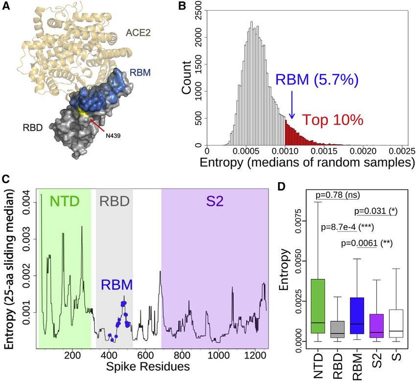
ll
Article                                                                                                                    OPEN ACCESS

                                                                                             Figure 5. Clinical outcomes and virological
                                                                                             evaluation of N439K lineage i indicate main-
                                                                                             tenance of fitness relative to WT virus
                                                                                             (A) Epidemiological growth of the N439/D614,
                                                                                             N439/D614G, or N439K/D614G virus in the Na-
                                                                                             tional Health Service (NHS) Greater Glasgow and
                                                                                             Clyde (GGC) Health Board area relative to sam-
                                                                                             pling time in epidemiological (epi) weeks (top) and
                                                                                             their relative contributions (bottom) for 1,918 pa-
                                                                                             tients whose diagnostic samples were sequenced.
                                                                                             (B) Top: real-time PCR data for N439/D614, N439/
                                                                                             D614G, and N439K/D614G groups, same patient
                                                                                             population as in (A). The N439K genotype was
                                                                                             associated with marginally lower Ct values than the
                                                                                             N439 genotype (posterior mean Ct value difference
                                                                                             between N439K/D614G and N439/D614G: 0.65,
                                                                                             95% CI: 1.22, 0.07). Bottom: correlation be-
                                                                                             tween Ct and quantitative viral load.
                                                                                             (C) Severity of disease within NHS GGC for a
                                                                                             subset of 1,591 patients. Ordinal scale scored by
                                                                                             requirement for supplementary oxygen: (1) no
                                                                                             respiratory support, (2) supplemental oxygen, (3)
                                                                                             invasive or non-invasive ventilation or oxygen
                                                                                             delivered by high-flow nasal cannula, and (4)
                                                                                             death. Ordinal regression analysis indicated that
                                                                                             the N439K viral genotype was associated with
                                                                                             similar clinical outcomes compared to the N439
                                                                                             genotype (posterior mean of N439K/D614G ge-
                                                                                             notype effect: 0.06, 95% CI: 1.21, 1.33).
                                                                                             (D) Growth curves for GLA1 (N439/D614G) or
                                                                                             GLA2 (N439K/D614G) virus isolates in Vero
                                                                                             E6 cells with ACE2 and TMPRSS2 over-
                                                                                             expression (+TMPRSS2 +ACE2), ACE2 over-
                                                                                             expression (+ACE2), or no overexpression. Error
                                                                                             bars are SD from three replicates.
                                                                                             (E) Competition of GLA1 and GLA2 virus isolates
                                                                                             for growth in Vero E6 cells with ACE2 and
                                                                                             TMPRSS2 overexpression (+TMPRSS2 +ACE2),
                                                                                             ACE2 overexpression (+ACE2), or no over-
                                                                                             expression, after inoculation at a matched MOI.
                                                                                             Quantification of each virus was performed by
                                                                                             tracking the frequency of N439K within the spike
                                                                                             gene using metagenomic NGS. Error bars are SD
                                                                                             from three replicates.
                                                                                             See also Figure S3 and Tables S4–S6.

affinity for the K417V variant relative to WT. Our data are in line   N439K SARS-CoV-2 maintains fitness and virulence
with the DMS results (Starr et al., 2020b), which show a 2-fold       The enhanced hACE2 affinity conferred by the N439K mutation,
loss of binding for K417V and no change for N439K/R, as the           its geographical emergence as independent lineages, as well as
two assays are inherently different and the DMS data are much         its prevalence among circulating viral isolates is consistent with
higher-throughput but lower sensitivity. We also tested the effect    no effect on viral fitness. We set out to directly examine N439K
of the N439K/R and K417V mutations in combination. These              impact on viral fitness by evaluating clinical data and outcomes
double mutants swap one salt bridge at the hACE2 binding inter-       associated with virus carrying the N439K mutation versus WT
face at RBD position 417 for one at position 439; we found they       N439, as well as by direct in vitro viral growth and competition.
had an hACE2 affinity similar to the WT (Figure 4D).                  Clinical data including age, gender, date of diagnosis, hospitali-
   Overall, these data indicate that acquisition of the N439K mu-     zation status, and mortality were collected prospectively, and
tation enhances hACE2 binding, which could have implications          sequencing was carried out in real time, as part of the Scottish
in vivo in the context of infection and transmission. At a minimum,   strategy for COVID-19 surveillance.
we found no evidence for any decreased success of N439K line-            We used qPCR to evaluate viral load (as measured by cycle
age i relative to other lineages present in Scotland at the same      threshold [Ct]) in 1,918 Scottish patients whose positive samples
time (Figure S3A). The enhanced affinity could compensate for         had been sequenced (Figures 5A and 5B). Variants were either
other mutations that would otherwise decrease binding (e.g.,          N439K/D614G (n = 406), N439/D614G (n = 978), or ancestral
K417V), further highlighting the plasticity of the RBM and the        (N439/D614) (n = 534). Our analysis found strong evidence that
need for surveillance.                                                the N439K/D614G genotype was associated with marginally lower

                                                                                             Cell 184, 1171–1187, March 4, 2021 1177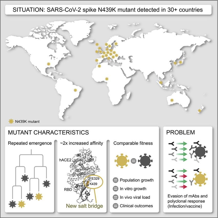
ll
     OPEN ACCESS                                                                                                              Article

Ct than the N439/D614G genotype, even after controlling for con-        The N439K mutation promotes evasion of antibody-
founders: age, sex, viral co-ancestry, and epidemic stage (mean Ct      mediated immunity
value difference between N439K/D614G and N439/D614G:                    Having established that the N439K mutation has no detectable
  0.65, 95% confidence interval [CI]: 1.22, 0.07) (Figure 5B; Ta-       effect on virus replication, we sought to test whether it promotes
ble S4). Assuming the PCR was 95% efficient, then a mean Ct dif-        evasion of antibody-mediated immunity by evaluating recogni-
ference of 0.65 would represent an RNA copy number increase of          tion of N439K RBD by mAbs and by polyclonal immune serum
1.54-fold in N439K/D614G relative to N439/D614G. Because Ct             from 442 recovered individuals, including six donors who were
measurements were from multiple locations in Scotland, a sub-           infected by the SARS-CoV-2 N439K variant. 6.8% of the tested
analysis of viral load using RNA standards was carried out with         sera showed a >2-fold reduction in binding to N439K RBD as
available samples. This analysis showed a near-complete correla-        compared to WT (Figures 6A, 6B, and S4; Data S1). In some in-
tion with Ct values (Figure 5B). D614G has previously been associ-      dividuals, the >2-fold reduction diminished the RBD ED50
ated with higher viral loads/lower Ct values (Korber et al., 2020;      response below 30 (Figure 6A; Data S1), a threshold previously
Lorenzo-Redondo et al., 2020; Mueller et al., 2020; Volz et al.,        determined to be a cutoff for specific binding (Piccoli et al.,
2021); although our data suggest a similar trend in a naive analysis,   2020). Thus, the response to the RBD can be significantly influ-
when controlling for confounders (given above), we could not            enced by the N439K mutation in a number of individuals infected
detect this effect (Table S4).                                          by WT SARS-CoV-2. The majority of serum samples for which
   Clinical outcomes were also obtained for a subset of these pa-       there was a loss of binding were those that had overall lower
tients (n = 1,591), who were scored for severity of disease based       Ab titers against WT RBD. The sera from the six individuals
on oxygen requirement: (1) no respiratory support, (2) supple-          known to have recovered from infection with SARS-CoV-2
mental oxygen, (3) invasive or non-invasive ventilation or high         N439K virus all showed 2-
   We next experimentally tested growth of two representative           fold reduction of RBD binding in response to the N439K mutation
SARS-CoV-2 isolates, GLA1 (N439) and GLA2 (N439K), both                 (Figures 6C, 6D, and S5; Data S1). For comparison, we also eval-
with the D614G background (Table S6). Culture was carried               uated the K417V and N439K/K417V mutations. A similar per-
out for 72 h in Vero E6 cells with either hACE2 and TMPRSS2             centage, 9.7% for K417V and 14.6% for N439K/K417V, lost
overexpression, hACE2 overexpression, or no overexpression.             >2-fold binding to these variants (Figures 6C, 6D, and S5; Data
There was no significant difference between the growth of these         S1). Of note, some mAbs demonstrated a larger loss of binding
isolates after inoculation at multiplicities of infection (MOIs) of     to the double mutant as compared to either single mutant (Fig-
0.005 and 0.01. The N439K variant replicated slightly faster            ures 6C, 6D; and S5; Data S1). The reduced binding of mAbs
initially after inoculation (Figure 5D). These experimental data        to these RBD mutants was also confirmed by bio-layer interfer-
indicate that the N439K mutation does not exhibit positive or           ometry analysis (Figures 6E and S6). The mAb panel was evalu-
negative effects on viral growth. To further assess fitness for         ated by RBD-binding competition experiments with hACE2 as
replication in cultured cells, we carried out a cross-competition       well as with three structurally characterized antibodies defining
assay using inoculation of cells at a matched MOI followed by           distinct epitopes on the RBD: S304/site II, S309/site IV, and
quantitation of N439 and N439K by metagenomic sequencing                S2H14/site I, the latter significantly overlapping with the RBM
over time (Figure 5E). N439K demonstrated similar fitness as            (Piccoli et al., 2020). The majority of the panel were site I,
the WT N439 variant, with a slight fitness advantage for N439K          hACE2-blocking mAbs; the mAbs with sensitivity to N439K
in cells expressing TMPRSS2. Collectively, these results indicate       were enriched for site I mAbs with moderate or weak/no
that the N439K mutation results in viral fitness that is similar or     hACE2 blockade, consistent with the positioning of N439K at
possibly slightly improved relative to the WT N439 virus. These         the edge of the RBM (Figures 1A and 6F; Data S1).
results may relate to the improved hACE2 affinity measured for              To define the potential biological importance of these muta-
the N439K RBD in the SPR binding assays, or could relate to             tions for evasion of antibody-mediated neutralization, we tested
additional mechanisms, such as changes to S density on the viral        mAbs against pseudoviruses expressing S variants N439K,
particle surface or changes to the conformational dynamics of           K417V, and N439K/K417V (Figures 7A–7C and S7; Data S1).
the S protein.                                                          Neutralization of pseudoviruses containing these mutations

1178 Cell 184, 1171–1187, March 4, 2021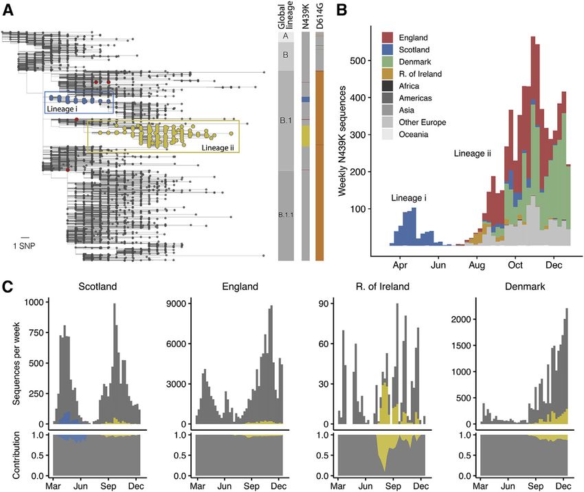
ll
Article                                                                                                                                        OPEN ACCESS

Figure 6. RBM variants exhibit escape from monoclonal antibodies and sera binding
(A and B) Binding of serum and plasma samples from 442 SARS-CoV-2 infected individuals against WT and N439K RBD plotted as (A) ELISA ED50 for each RBD
(cut-off for positive binding to WT set at 30) and (B) fold change relative to WT. Data shown are the average of two independent replicates (source data given in
Data S1). Blue dots indicate sera with at least 2-fold loss of binding to the N439K RBD variant as compared to WT in both replicates. Purple dots indicate sera from
individuals infected with SARS-CoV-2 N439K variant.
(C and D) Binding of 140 mAbs from SARS-CoV-2 infected individuals and four clinical-stage or EUA-approved mAbs against WT, N439K, K417V, and N439K/
K417V RBD, plotted as (C) ELISA AUC for each RBD and (D) fold change relative to WT. Data shown are the average of two independent replicates (source data
given in Data S1). For all, the colored dots indicate mAbs demonstrating at least 2-fold loss of binding to the variant RBD as compared to WT (counted if the
average of both replicates is at least 2-fold and each individual replicate is at least 1.7-fold).
(E) Kinetics of binding to RBD variants by Octet of six representative mAbs (representative of n = 2 independent experiments).
(F) Distribution of the 144 mAbs based on binding to RBD variants (expressed as fold-change over WT) and hACE2 competition (expressed as the mAb con-
centration blocking 80% of hACE2 binding, BC80, also indicated as a blue gradient; source data in Data S1). Higher BC80 values (lighter blue) correspond to less
hACE2 competition, with mAbs indicated at the top of the panels (white) showing no competition at all.
See also Figures S4, S5, and S6.

was significantly diminished for certain mAbs, including some                      reactive camelid nanobody, VHH-72, which has enhanced po-
that are currently in use in patients under EAU. As predicted by                   tency for SARS-CoV as compared to SARS-CoV-2, predicted
its non-RBM epitope (Pinto et al., 2020), S309 was capable of                      to be partially due to a contact with R426 in SARS-CoV RBD,
neutralizing each of these variants. We also evaluated a cross-                    the same position as 439 in SARS-CoV-2 RBD (Wrapp et al.,

                                                                                                                Cell 184, 1171–1187, March 4, 2021 1179
ll
OPEN ACCESS Article
Figure 7. Neutralization of four RBM vari-
ants by a panel of antibodies and a nano-
body
(A) Neutralization of four VSV-pseudovirus variants
by six of the mAbs tested. Data shown are repre-
sentative of n = 3 biological replicates, bars = SD of
technical duplicate (Data S1).
(B) Correlation of ELISA-binding fold change and
neutralization fold change for each variant relative
to WT.
(C) Top: neutralization IC50 of the D614G virus
determined as the geometric mean of three bio-
logical replicates. Bottom: neutralization results for
all mAbs tested, expressed as a fold-change
relative to D614G (all variants are in the back-
ground of D614G) (Data S1). The individual values
of the three replicates are shown as open circles,
their geometric mean as colored bars and the
geometric SD as error bars. Each antibody is an-
notated according to its hACE2 competition (as
shown in Figure 6F) as well as its epitope (site I, II,
or IV) (Data S1). Gray boxes with a slash indicate
not tested for hACE2 competition or epitope
analysis.
See also Figure S7.
losing fitness relative to WT. The success
of variants with the N439K mutation is evi-
denced by their repeated emergence by
convergent evolution on at least nine oc-
casions, spread to 34 countries as of
January 2021, significant representation
in sampled genome sequences (indica-
tive of high infection rates), the fact that
the N439K RBD retains a high-affinity
interaction with the hACE2 receptor, and
efficient replication of N439K virus in
cultured cells. Additionally, we observed
no evidence for change in disease
severity in a large cohort of individuals in-
fected with N439K virus as compared to
WT N439 virus, although we acknowledge
2020a). Consistent with this prediction, VHH-72 showed some limitations in the data collection, including variations in
enhanced potency against N439K SARS-CoV-2 pseudovirus testing guidelines and availability of testing during the course
compared to WT N439 (Figures 7A and 7C), highlighting the pos- of the study (da Silva Filipe et al., 2021).
sibility that a single mutation can impact antibody efficacy posi- The success of the N439K mutation is consistent with our find-
tively as well as negatively. Sensitivity of a few neutralizing mAbs ings that the RBM is a highly variable region of S. It demonstrates
to mutations at positions 417 and 439 have also been reported in the ability of SARS-CoV-2 to accommodate mutations at the
other studies (Baum et al., 2020; Gaebler et al., 2021; Greaney et RBM while retaining efficient hACE2 binding. This ability could
al., 2021; Li et al., 2020a; Starr et al., 2020a; Weisblum et al., have emerged by chance or in response to immune pressure
2020), although combinations of mutations have typically not from neutralizing Ab responses in viral hosts. There is precedent
been evaluated. Overall, our results demonstrate that mutations for the most immunogenic region of a viral surface protein to be
compatible with equivalent viral fitness to WT can result in im- highly divergent despite harboring the receptor binding site; for
mune evasion from both monoclonal and polyclonal antibody example, the immunogenic globular head domain of the influ-
responses. enza virus hemagglutinin surface protein, which contains the
sialic acid receptor binding site, evolves faster than the stalk re-
DISCUSSION gion (Doud et al., 2018; Kirkpatrick et al., 2018). The ability to
readily accommodate mutations in the RBM indicates a high like-
Here, we describe an example of a circulating RBM mutation, lihood that potentially immune-evading SARS-CoV-2 variants
N439K, which can evade antibody-mediated immunity without will continue to emerge, with implications for reinfection,
1180 Cell 184, 1171–1187, March 4, 2021ll
Article OPEN ACCESS
vaccines, and both monoclonal and polyclonal antibody fitness as it parallels SARS-CoV RBM:hACE2 interactions (salt
therapeutics. bridge at SARS-CoV RBD position R426 and no salt bridge at
A few other circulating RBM mutations have become promi- V404) (Figure 4A). Current SARS-CoV-2 mutations have arisen
nent since N439K first emerged. S477N appeared in the in the absence of pressure from significant population immunity.
sequence databases in March 2020 but did not become the However, as immunity to the WT virus becomes more wide-
most prevalent RBD mutation until the summer (as of January spread, immune escape mutations can be expected to increas-
2021, it has >19,000 counts). Consistent with the high preva- ingly circulate. In the final weeks of 2020, SARS-CoV-2 variants
lence, position 477 is the RBM position where mutations are carrying multiple mutations in the S protein, in both the RBM and
predicted to be the most well-tolerated with respect to hACE2 Domain A, have been observed (Volz et al., 2021) including one
binding (Figure 2). Studies across multiple mAb panels have variant carrying three simultaneous RBM mutations (K417N,
not found this mutation to be conferring resistance (Gaebler et E484K, and N501Y) (Tegally et al., 2020). Accumulation of multi-
al., 2021; Greaney et al., 2021; Tortorici et al., 2020; Weisblum ple changes may increase the risk of immune escape from vac-
et al., 2020). In contrast, mutations at position 484 in the RBM cines that are based on early SARS-CoV-2 sequences.
have been reported to confer resistance to many mAbs across Mutations in the RBM will also impact the prophylactic or ther-
multiple studies (Baum et al., 2020; Gaebler et al., 2021; Greaney apeutic use of mAbs. In our profile of immune escape from the
et al., 2021; Tortorici et al., 2020; Weisblum et al., 2020) and also N439K variant, we observed resistance to a mAb which is part
appeared in a persistent (>150 days) infection in an immunocom- of a two-mAb cocktail that recently received EAU. The promise
promised individual who did not respond to treatment with a of using cocktails of mAbs is that they should significantly lower
two-mAb cocktail (Choi et al., 2020). The variant count at posi- the likelihood of drug-induced selection of resistant viruses
tion 484 has been steadily increasing (>500 as of January (Baum et al., 2020). However, if circulating viral variants already
2021), and the possibility for no fitness consequences for these carry resistant mutations to one antibody in the cocktail, this
variants is notable (Figures 2 and S1B) (Starr et al., 2020b), as is a could cause the cocktail to be reduced to a monotherapy. Addi-
recent study identifying this position as particularly important for tionally, we observed that two mutations together (N439K/
escape from polyclonal serum antibodies (Greaney et al., 2021a) K417V) conferred resistance in vitro to the two-mAb cocktail
and the appearance of this mutation in a new, fast-growing viral (Figure 7C).
lineage (Tegally et al., 2020). The Y453F mutation has become Two approaches will be critical for minimizing the impact of
noteworthy recently for its association with virus circulating in mAb escape mutations. One is to develop mAbs with epitopes
mink farms and its transmission back to humans (Oude Munnink that are highly resistant to viral escape. This may include epi-
et al., 2021) and the DMS measurement indicating it confers topes outside of the RBM and/or epitopes that are cross-reac-
significantly increased hACE2 binding (Starr et al., 2020b). To tive across SARS-CoV and SARS-CoV-2, indicating conserved
date, we know of only one example of published immune escape epitopes with a low tolerance for mutation (Garrett Rappazzo
documented for Y453F (Baum et al., 2020), but more examples et al., 2021; Pinto et al., 2020; Wec et al., 2020; Wrapp et al.,
may arise as this new mutation is investigated further. Last, the 2020a). A comparison of epitopes of RBM-targeting mAbs with
N501Y mutation has gained notice in the final weeks of 2020 the most conserved regions of the RBM (Figure 2) may also iden-
for its association with high rates of infection (Tegally et al., tify RBM mAbs with a higher barrier to escape. The second will
2020; Volz et al., 2021), although further research is needed to be to screen patients, likely at the population level, for the pres-
determine the impact on immune escape. ence of potential resistance variants prior to drug administration.
SARS-CoV-2 appears to be evolving relatively slowly consis- The availability of multiple different mAb therapeutics in the clinic
tent with its low mutation rate, the highly susceptible human pop- could provide the opportunity to tailor the choice of therapeutics
ulation, and its generalist nature (Conceicao et al., 2020) and, at to local circulating variants.
present, evidence indicates it will be controllable by vaccines In general, given that access to therapeutic mAbs is expand-
based on early SARS-CoV-2 genome sequences (Dearlove ing, and as more people develop immune responses to the WT
et al., 2020). Additionally, for the majority of our tested sera virus via infection or vaccination, monitoring the evolution of
and mAbs, a single amino acid change in the RBM was not suf- SARS-CoV-2 for escape mutants will be critical. Although we
ficient to confer resistance. Nevertheless, our data indicate that only report on evasion of antibody-mediated immunity here, it
individuals with a mild antibody response to vaccination or first would be surprising if similar changes are not observed that
infection could be at risk from a virus carrying a mutation in the confer evasion of T cell immunity and innate immunity.
RBM. Furthermore, considering the high level of structural plas-
ticity of the RBM demonstrated in the present study, there could Limitations of study
be many combinations of RBM mutations, including some This study presents the finding that the RBM is a highly variable
requiring compensatory changes, that are compatible with region of the SARS-CoV-2 S protein, and we provide a thorough
high viral fitness. Some of these combinations will contribute to characterization of the N439K RBM amino acid replacement,
efficient immune escape. For example, our data show that and the ability of this mutation to confer immune evasion without
N439K can compensate for a mutation (K417V) that otherwise attenuating (or enhancing) fitness or disease. When this study
decreases receptor binding affinity (Figure 4D) and that several was initiated in June 2020, the general consensus was that the
mAbs were more sensitive to these mutations in combination slow rate of evolution of SARS-CoV-2 would result in no immedi-
versus individually (Figure 6D; Data S1). This particular combina- ate threat to vaccines or therapies. N439K was the first RBM
tion of mutations is plausibly compatible with maintained viral amino acid replacement, relative to the ancestral SARS-CoV-2
Cell 184, 1171–1187, March 4, 2021 1181ll
OPEN ACCESS Article
variant used in vaccine preparations, to increase to high fre- SUPPLEMENTAL INFORMATION
quency and so can be viewed as a sentinel mutation for SARS-
Supplemental information can be found online at https://doi.org/10.1016/j.cell.
CoV-2 antigenic drift. Since initial submission to Cell in late
2021.01.037.
October 2020, the emergence of multiple highly transmissible
variants carrying other RBM mutations of significance has CONSORTIA
brought the study of immune evasion variants to the forefront
of SARS-CoV-2 research. Future retrospective studies will The members of the COG-UK consortia are Thomas R. Connor, Nicholas J. Lo-
confirm whether these new RBM mutations fall into the same man, Samuel C. Robson, Tanya Golubchik, M. Estee Torok, William L. Hamil-
category as N439K: mutations that do not attenuate viral fitness ton, David Bonsall, Ali R. Awan, Sally Corden, Ian Goodfellow, Darren L. Smith,
Martin D. Curran, Surendra Parmar, James G. Shepherd, Matthew D. Parker,
or disease but cause immune evasion. Our results on the plas-
Catherine Moore, Derek J. Fairley, Matthew W. Loose, Joanne Watkins,
ticity of the RBM also anticipated the emergence of the RBM mu- Matthew Bull, Sam Nicholls, David M. Aanensen, Sharon Glaysher, Matthew
tations present in the SARS-CoV-2 variants of concern. Bashton, Nicole Pacchiarini, Anthony P. Underwood, Thushan I. de Silva, Den-
nis Wang, Monique Andersson, Anoop J. Chauhan, Mariateresa de Cesare,
Catherine Ludden, Tabitha W. Mahungu, Rebecca Dewar, Martin P. McHugh,
STAR+METHODS
Natasha G. Jesudason, Kathy K. Li, Rajiv N. Shah, Yusri Taha, Kate E. Temple-
ton, Simon Cottrell, Justin O’Grady, Andrew Rambaut, Colin P. Smith,
Detailed methods are provided in the online version of this paper Matthew T.G. Holden, Emma C. Thomson, Samuel Moses, Meera Chand,
and include the following: Chrystala Constantinidou, Alistair C. Darby, Julian A. Hiscox, Steve Paterson,
Meera Unnikrishnan, Andrew J. Page, Erik M. Volz, Charlotte J. Houldcroft,
d KEY RESOURCES TABLE Aminu S. Jahun, James P. McKenna, Luke W. Meredith, Andrew Nelson, Sar-
d RESOURCE AVAILABILITY ojini Pandey, Gregory R. Young, Anna Price, Sara Rey, Sunando Roy, Ben
B Lead contact Temperton, Matthew Wyles, Stefan Rooke, Sharif Shaaban, Helen Adams,
B Materials availability Yann Bourgeois, Katie F. Loveson, Áine O’Toole, Richard Stark, Ewan M. Har-
rison, David Heyburn, Sharon J. Peacock, David Buck, Michaela John, Dorota
B Data and code availability
Jamrozy, Joshua Quick, Rahul Batra, Katherine L. Bellis, Beth Blane, Sophia T.
d EXPERIMENTAL MODEL AND SUBJECT DETAILS Girgis, Angie Green, Anita Justice, Mark Kristiansen, Rachel J. Williams, Rado-
B Cell lines slaw Poplawski, Garry P. Scarlett, John A. Todd, Christophe Fraser, Judith
B Sample donors Breuer, Sergi Castellano, Stephen L. Michell, Dimitris Gramatopoulos, Jona-
d METHOD DETAILS than Edgeworth, Gemma L. Kay, Ana da Silva Filipe, Aaron R. Jeffries, Sascha
B Structural analysis Ott, Oliver Pybus, David L. Robertson, David A. Simpson, Chris Williams, Cres-
sida Auckland, John Boyes, Samir Dervisevic, Sian Ellard, Sonia Goncalves,
B RBM variability across SARS-CoV-2 sequences
Emma J. Meader, Peter Muir, Husam Osman, Reenesh Prakash, Venkat Si-
B Evaluation of deep mutational scanning (DMS) data
vaprakasam, Ian B. Vipond, Jane A.H. Masoli, Nabil-Fareed Alikhan, Matthew
B Molecular dynamics simulations Carlile, Noel Craine, Sam T. Haldenby, Nadine Holmes, Ronan A. Lyons, Chris-
B System solvation and parametrization topher Moore, Malorie Perry, Ben Warne, Thomas Williams, Lisa Berry, An-
B System equilibration drew Bosworth, Julianne Rose Brown, Sharon Campbell, Anna Casey,
B Folding@home simulations Gemma Clark, Jennifer Collins, Alison Cox, Thomas Davis, Gary Eltringham,
Cariad Evans, Clive Graham, Fenella Halstead, Kathryn Ann Harris, Christo-
B Simulation analysis
pher Holmes, Stephanie Hutchings, Miren Iturriza-Gomara, Kate Johnson, Ka-
B RBM variability across Sarbecoviruses
tie Jones, Alexander J. Keeley, Bridget A. Knight, Cherian Koshy, Steven Lig-
B Recombinant glycoprotein production gett, Hannah Lowe, Anita O. Lucaci, Jessica Lynch, Patrick C McClure, Nathan
B Crystallization, data collection, structure determina- Moore, Matilde Mori, David G. Partridge, Pinglawathee Madona, Hannah M.
tion, and analysis Pymont, Paul Anthony Randell, Mohammad Raza, Felicity Ryan, Robert
B Binding measurements using surface plasmon reso- Shaw, Tim J. Sloan, Emma Swindells, Alexander Adams, Hibo Asad, Alec
nance (SPR) Birchley, Tony Thomas Brooks, Giselda Bucca, Ethan Butcher, Sarah L.
Caddy, Laura G. Caller, Yasmin Chaudhry, Jason Coombes, Michelle Cronin,
B hACE2 binding measurements using bio-layer interfer-
Patricia L. Dyal, Johnathan M. Evans, Laia Fina, Bree Gatica-Wilcox, Iliana
ometry (BLI) Georgana, Lauren Gilbert, Lee Graham, Danielle C. Groves, Grant Hall, Ember
B Epidemiological and genome surveillance Hilvers, Myra Hosmillo, Hannah Jones, Sophie Jones, Fahad A. Khokhar, Sara
B Phylogenetic and phylodynamic analysis Kumziene-Summerhayes, George MacIntyre-Cockett, Rocio T. Martinez Nu-
B Evaluation of clinical samples nez, Caoimhe McKerr, Claire McMurray, Richard Myers, Yasmin Nicole Pan-
B qPCR of clinical samples chbhaya, Malte L. Pinckert, Amy Plimmer, Joanne Stockton, Sarah Taylor, Ali-
cia Thornton, Amy Trebes, Alexander J. Trotter, Helena Jane Tutill, Charlotte A.
B Viral growth curve
Williams, Anna Yakovleva, Wen C. Yew, Mohammad T. Alam, Laura Baxter,
B Competition assay
Olivia Boyd, Fabricia F. Nascimento, Timothy M. Freeman, Lily Geidelberg, Jo-
B Ab discovery and recombinant expression seph Hughes, David Jorgensen, Benjamin B. Lindsey, Richard J. Orton,
B Enzyme-linked immunosorbent assay (ELISA) Manon Ragonnet-Cronin, Joel Southgate, Sreenu Vattipally, Igor Starinskij,
B Antibody binding measurements using bio-layer inter- Joshua B. Singer, Khalil Abudahab, Leonardo de Oliveira Martins, Thanh Le-
ferometry (BLI) Viet, Mirko Menegazzo, Ben E.W. Taylor, Corin A. Yeats, Sophie Palmer, Carol
B Blockade of RBD binding to ACE2 M. Churcher, Alisha Davies, Elen De Lacy, Fatima Downing, Sue Edward, Nikki
Smith, Frances Bolt, Alex Alderton, Matt Berriman, Ian G. Charles, Nicholas
B RBD epitope mapping (blockade of binding assay)
Cortes, Tanya Curran, John Danesh, Sahar Eldirdiri, Ngozi Elumogo, Andrew
B VSV pseudovirus generation Hattersley, Alison Holmes, Robin Howe, Rachel Jones, Anita Kenyon, Robert
B Pseudovirus neutralization A. Kingsley, Dominic Kwiatkowski, Cordelia Langford, Jenifer Mason, Alison E.
d QUANTIFICATION AND STATISTICAL ANALYSIS Mather, Lizzie Meadows, Sian Morgan, James Price, Trevor I. Robinson, Giri
1182 Cell 184, 1171–1187, March 4, 2021You can also read