Clinical Signs and Pathology Associated With Domoic Acid Toxicosis in Southern Sea Otters (Enhydra lutris nereis)
←
→
Page content transcription
If your browser does not render page correctly, please read the page content below
ORIGINAL RESEARCH
                                                                                                                                               published: 26 May 2021
                                                                                                                                      doi: 10.3389/fmars.2021.585501

                                            Clinical Signs and Pathology
                                            Associated With Domoic Acid
                                            Toxicosis in Southern Sea Otters
                                            (Enhydra lutris nereis)
                                            Melissa A. Miller 1,2* , Megan E. Moriarty 1,2 , Pádraig J. Duignan 2,3 , Tanja S. Zabka 4 ,
                                            Erin Dodd 1 , Francesca I. Batac 1 , Colleen Young 1 , Angelina Reed 1 , Michael D. Harris 1 ,
                                            Katherine Greenwald 1 , Raphael M. Kudela 5 , Michael J. Murray 6 , Frances M. D. Gulland 2 ,
                           Edited by:
                                            Peter E. Miller 7† , Kendra Hayashi 5 , Catherine T. Gunther-Harrington 8 , Martin T. Tinker 9
                    Debra Lee Miller,       and Sharon Toy-Choutka 1
         The University of Tennessee,       1
                                             Marine Wildlife Veterinary Care and Research Center, California Department of Fish and Wildlife, Santa Cruz, CA,
             Knoxville, United States
                                            United States, 2 Karen C. Drayer Wildlife Health Center, One Health Institute, School of Veterinary Medicine, University
                       Reviewed by:         of California, Davis, Davis, CA, United States, 3 The Marine Mammal Center, Sausalito, CA, United States, 4 Genentech, Inc.,
                      Wesley Siniard,       South San Francisco, CA, United States, 5 Department of Ocean Sciences, Institute for Marine Sciences, University
         The University of Tennessee,       of California, Santa Cruz, Santa Cruz, CA, United States, 6 Monterey Bay Aquarium, Monterey, CA, United States, 7 Institute
             Knoxville, United States       of Marine Sciences, University of California, Santa Cruz, Santa Cruz, CA, United States, 8 Department of Medicine
                 Annie Page-Karjian,        and Epidemiology, School of Veterinary Medicine, University of California, Davis, Davis, CA, United States, 9 Nhydra
           Florida Atlantic University,     Ecological Consulting, Head of Saint Margarets Bay, NS, Canada
                        United States
                 *Correspondence:
                                            The marine biotoxin domoic acid (DA) is an analog of the neurotransmitter glutamate
                       Melissa A. Miller
         melissa.miller@wildlife.ca.gov     that exerts potent excitatory activity in the brain, heart, and other tissues. Produced by
                   † Presentaddress:        the diatom Pseudo-nitzschia spp., DA accumulates in marine invertebrates, fish, and
                      Peter E. Miller,      sediment. Southern sea otters (Enhydra lutris nereis) feed primarily on invertebrates,
United States Agency for International
      Development, Washington, DC,          including crabs and bivalves, that concentrate and slowly depurate DA. Due to their
                       United States        high prey consumption (25% of body weight/day), sea otters are commonly exposed
                                            to DA. A total of 823 necropsied southern sea otters were examined to complete
                   Specialty section:
         This article was submitted to      this study; first we assessed 560 subadult, adult, and aged adult southern sea
                    Marine Megafauna,       otters sampled from 1998 through 2012 for DA-associated pathology, focusing mainly
               a section of the journal
          Frontiers in Marine Science
                                            on the central nervous system (CNS) and cardiovascular system. We applied what
            Received: 20 July 2020
                                            was learned to an additional cohort of necropsied sea otters of all demographics
        Accepted: 17 February 2021          (including fetuses, pups, juveniles, and otters examined after 2012: n = 263 additional
           Published: 26 May 2021           animals). Key findings derived from our initial efforts were consistently observed in
                              Citation:     this more demographically diverse cohort. Finally, we assessed the chronicity of DA-
                Miller MA, Moriarty ME,
      Duignan PJ, Zabka TS, Dodd E,         associated pathology in the CNS and heart independently for 54 adult and aged adult
           Batac FI, Young C, Reed A,       sea otters. Our goals were to compare the temporal consistency of DA-associated
Harris MD, Greenwald K, Kudela RM,
 Murray MJ, Gulland FMD, Miller PE,
                                            CNS and cardiovascular lesions and determine whether multiple episodes of DA
  Hayashi K, Gunther-Harrington CT,         toxicosis could be detected on histopathology. Sea otters with acute, fatal DA toxicosis
Tinker MT and Toy-Choutka S (2021)          typically presented with neurological signs and severe, diffuse congestion and multifocal
          Clinical Signs and Pathology
        Associated With Domoic Acid         microscopic hemorrhages (microhemorrhages) in the brain, spinal cord, cardiovascular
     Toxicosis in Southern Sea Otters       system, and eyes. The congestion and microhemorrhages were associated with
                 (Enhydra lutris nereis).
            Front. Mar. Sci. 8:585501.
                                            detection of high concentrations of DA in postmortem urine or gastrointestinal content
    doi: 10.3389/fmars.2021.585501          and preceded histological detection of cellular necrosis or apoptosis. Cases of chronic

Frontiers in Marine Science | www.frontiersin.org                                  1                                              May 2021 | Volume 8 | Article 585501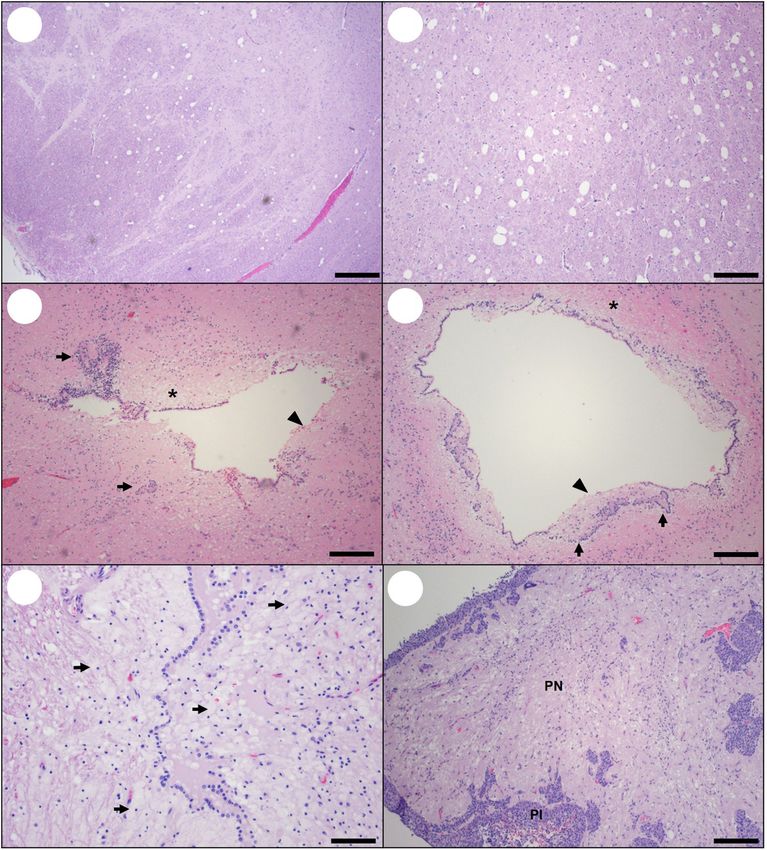
Miller et al.                                                                                                             Domoic Acid Toxicosis in Otters

                                            DA toxicosis often presented with cardiovascular pathology that was more severe
                                            than the CNS pathology; however, the lesions at both sites were relatively quiescent,
                                            reflecting previous damage. Sea otters with fatal subacute DA toxicosis exhibited
                                            concurrent CNS and cardiovascular pathology that was characterized by progressive
                                            lesion expansion and host response to DA-associated tissue damage. Acute, subacute,
                                            and chronic cases had the same lesion distribution in the CNS and heart. CNS
                                            pathology was common in the hippocampus, olfactory, entorhinal and parahippocampal
                                            cortex, periventricular neuropil, and ventricles. The circumventricular organs were
                                            identified as important DA targets; microscopic examination of the pituitary gland,
                                            area postrema, other circumventricular organs, and both eyes facilitated confirmation
                                            of acute DA toxicosis in sea otters. DA-associated histopathology was also common
                                            in cardiomyocytes and coronary arterioles, especially in the left ventricular free wall,
                                            papillary muscles, cardiac apex, and atrial free walls. Progressive cardiomyocyte loss
                                            and arteriosclerosis occurred in the same areas, suggesting a common underlying
                                            mechanism. The temporal stage of DA-associated CNS pathology matched the DA-
                                            associated cardiac pathology in 87% (n = 47/54) of cases assessed for chronicity,
                                            suggesting that the same underlying process (e.g., DA toxicosis) was the cause of these
                                            lesions. This temporally matched pattern is also indicative of a single episode of DA
                                            toxicosis. The other 13% of examined otters (n = 7/54) exhibited overlapping acute,
                                            subacute, or chronic DA pathology in the CNS and heart, suggestive of recurrent DA
                                            toxicosis. This is the first rigorous case definition to facilitate diagnosis of DA toxicosis in
                                            sea otters. Diagnosing this common but often occult condition is important for improving
                                            clinical care and assessing population-level impacts of DA exposure in this federally
                                            listed threatened subspecies. Because the most likely source of toxin is through prey
                                            consumption, and because humans, sea otters, and other animals consume the same
                                            marine foods, our efforts to characterize health effects of DA exposure in southern sea
                                            otters can provide strong collateral benefits.
                                            Keywords: biotoxin, brain and circumventricular organs, heart and cardiomyopathy, domoic acid toxicosis,
                                            harmful algal bloom (HAB), pathology, Pseudo-nitzschia, sea otter (Enhydra lutris)

INTRODUCTION                                                                    Kvitek et al., 2008), and identified factors influencing Pseudo-
                                                                                nitzschia bloom dynamics and DA production (Bargu et al., 2010;
Domoic acid (DA) is an amino acid analog of kainic acid and                     Lewitus et al., 2012; McKibben et al., 2017).
glutamate that acts as a potent excitotoxin when ingested by                        Laboratory research has provided insight into the
humans and other animals. Along the North American Pacific                      pathophysiology of DA (Tryphonas et al., 1990; Pulido
Coast, DA is produced during blooms of the diatom Pseudo-                       et al., 1995; Sobotka et al., 1996; Giordano et al., 2007).
nitzschia spp., including P. australis (Scholin et al., 2000; Trainer           Early observational studies focused on neurotoxicity (Tryphonas
et al., 2000; Bargu et al., 2010). Domoic acid can accumulate                   et al., 1990; Pulido et al., 1995; Silvagni et al., 2005), while later
in food webs (Lefebvre et al., 2002; Trainer et al., 2002; Kvitek               investigations documented cardiovascular pathology and other
et al., 2008) and persist in marine sediments (Sekula-Wood et al.,              health impacts (Kreuder et al., 2005; Gill et al., 2007; Zabka et al.,
2009), enhancing exposure risks for humans and wildlife. Domoic                 2009; Moriarty et al., 2021a,b). Acute, subacute, and chronic
acid-associated human deaths were first recognized along the east               effects of DA exposure are now recognized in people, wildlife,
coast of North America in 1987 (Perl et al., 1990a,b; Teitelbaum                and laboratory animals (Sutherland et al., 1990; Goldstein et al.,
et al., 1990). In California, DA was initially reported as a cause              2008; Pulido, 2008; Buckmaster et al., 2014). Severe, intermittent,
of avian mortality (Fritz et al., 1992; Work et al., 1993) and                  or chronic sublethal DA exposure can cause cognitive and
then as a cause of marine mammal mass-stranding (Scholin                        functional deficits such as impaired memory, poor spatial
et al., 2000). Subsequent studies have confirmed health impacts                 navigation, and temporal lobe epilepsy (Zatorre, 1990; Cendes
in diverse marine wildlife (Shumway et al., 2003; Bejarano et al.,              et al., 1995; Sobotka et al., 1996; Goldstein et al., 2008; Thomas
2008; Lefebvre et al., 2010; McHuron et al., 2013), clarified food              et al., 2010; Buckmaster et al., 2014; Ramsdell and Gulland, 2014;
web exposure patterns (Lefebvre et al., 2002; Goldberg, 2003;                   Grattan et al., 2018).

Frontiers in Marine Science | www.frontiersin.org                           2                                        May 2021 | Volume 8 | Article 585501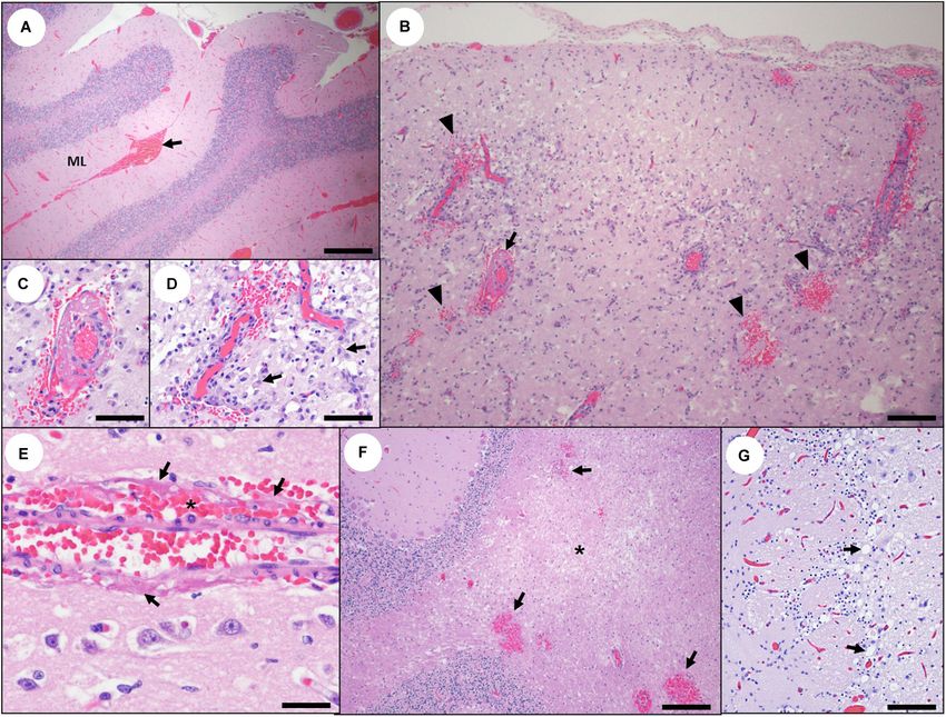
Miller et al.                                                                                                          Domoic Acid Toxicosis in Otters

    Sequestration of DA in fetal fluids can prolong maternal and              Tables 1, 2). The total sample size for all three phases of this
fetal toxicosis (Ramsdell and Zabka, 2008; Goldstein et al., 2009;            study was 823 southern sea otters that were necropsied from
Lefebvre et al., 2018). Permanent neurobehavioral deficits from               1998 through 2018.
in utero or early postnatal DA exposure have been described in                    When available, antemortem clinical data were reviewed
California sea lions (Zalophus californianus) (Goldstein et al.,              to assess findings associated with DA toxicosis. Standardized
2008; Ramsdell and Zabka, 2008; Thomas et al., 2010; Cook                     protocols were utilized for gross necropsy, tissue and body fluid
et al., 2015, 2016), primates (Burbacher et al., 2019), and rodents           collection, histopathology, and diagnostic testing. The abdomen,
(Sobotka et al., 1996; Doucette et al., 2004; Levin et al., 2006;             thorax, brain, and heart were photographed during necropsy
Lefebvre et al., 2017). Domoic acid has also been detected in                 to facilitate comparison among sampled sea otters. All major
milk from California sea lions, indicating lactational exposure               organs and tissues were examined grossly and microscopically,
(Rust et al., 2014).                                                          including the brain, pituitary gland, trigeminal ganglia, heart,
    Despite awareness of the effects of DA in Pacific coastal marine          aorta, kidneys, eyes, lung, liver, spleen, pancreas, adrenal glands,
ecosystems (Lefebvre et al., 2016), little is known about the                 gallbladder, stomach, intestines, rectum, omentum, lymph nodes,
health and population impacts on California’s smallest marine                 salivary glands, skeletal muscles, tongue, soft palate, tonsil,
mammal: the southern sea otter (Enhydra lutris nereis). This                  esophagus, thymus, thyroid, parathyroid, bladder, skin, gonads,
federally listed threatened subspecies has a current population               and reproductive tract. Formalin-fixed tissues were trimmed
of approximately 3,000 individuals distributed along 500 km of                using a standard protocol (Supplementary Data Sheets 1, 2
the central California coast (Hatfield et al., 2019). Toxic Pseudo-           and Supplementary Images 1A,B) so that lesions could be
nitzschia blooms are common throughout southern sea otter                     compared among cases.
habitat (McCabe et al., 2016; McKibben et al., 2017), and cases                   Central nervous system (CNS) trimming of the brain for
of DA-associated fatal neurotoxic and cardiovascular disease have             microscopic examination was initiated by a single coronal slice
been reported in sea otters (Kreuder et al., 2005; Miller et al., 2020;       through the left and right cerebral hemisphere and diencephalon
Moriarty et al., 2021b).                                                      just caudal to the mammillary bodies (Supplementary Data
    The primary objective of this study was to establish a case               Sheet 1 and Supplementary Image 1A). Coronal slices were
definition for DA toxicosis in southern sea otters using insight              cut sequentially from this point both rostrally and caudally
gained from necropsy and microscopic examination of 823                       at approximately 4 mm thickness throughout the cerebrum,
individuals necropsied from 1998 through 2018. Our secondary                  cerebellum, and brainstem, and standardized sections were
objectives were to describe acute, subacute, and chronic lesions              collected for histopathology to facilitate comparison. The
associated with DA toxicosis, estimate the DA post-exposure                   pituitary gland was bisected along the sagittal midline for
intervals based on lesion characteristics and stereotypical host              histological examination (Supplementary Data Sheet 1 and
tissue response patterns, and compare the temporal patterns                   Supplementary Image 1B). Microscopic examination included
in the CNS and cardiovascular system. Improved recognition                    the left and right rostral and caudal hippocampus, olfactory and
of DA toxicosis can inform clinical care, facilitate estimates                entorhinal cortex, parahippocampal cortex, temporal cerebral
of population-level health impacts, and aid assessment of                     cortex, rhinencephalon at the level of the lateral olfactory
disease prevalence in relation to climate change and other                    tract, pituitary gland, cerebellum, pons, medulla, ventricles,
environmental perturbations.                                                  meninges, choroid plexus, and trigeminal ganglia. For some
                                                                              cases, the olfactory bulbs, spinal cord, brainstem, diencephalon,
                                                                              mesencephalon, and additional circumventricular organs
MATERIALS AND METHODS                                                         (CVOs), including the area postrema, pineal gland, and median
                                                                              eminence, were also examined.
Sample Population and Comprehensive                                               Cardiac trimming and histopathology were standardized
Postmortem Evaluations                                                        to include the left and right atrioventricular free wall and
The sample population consisted of minimally decomposed                       valves, papillary muscles, interventricular septum, cardiac apex,
southern sea otters from the central California coast that                    and thoracic aorta (Supplementary Data Sheet 1). One or
were necropsied from 1998 through 2018 (Supplementary                         both eyes were bisected mid-sagittally at the optic nerve and
Table 1). Criteria for diagnosing DA toxicosis were developed                 examined microscopically (Supplementary Data Sheet 1). Trim
initially using a subgroup of 560 subadults, adults, and aged                 sheets were completed so that each tissue could be identified
adults that were part of an in-depth review of sea otter                      during microscopic examination (Supplementary Data Sheet 2).
mortality patterns from 1998 through 2012 (Miller et al.,                     Epidemiological assessment of DA-associated mortality patterns
2020). Provisional diagnostic criteria were assessed and further              and comprehensive DA testing of body fluids and tissues from
refined via examination of additional otters of all age classes               necropsied southern sea otters are summarized in separate
(fetuses, pups, immatures, subadults, adults, and aged adults),               publications (Miller et al., 2020; Moriarty et al., 2021b; Tinker
and animals necropsied through 2018 (n = 263; Supplementary                   et al., in Press; Miller et al., In final preparation).
Table 1). Finally, we independently assessed and compared the
chronicity of gross and microscopically apparent DA-associated                Assessment of DA-Associated Pathology
pathology in the CNS and heart of 54 adult and aged adult                     The potential for DA toxicosis as a cause of death for each
southern sea otters with optimal tissue sampling (Supplementary               sea otter was determined using a preponderance of evidence

Frontiers in Marine Science | www.frontiersin.org                         3                                       May 2021 | Volume 8 | Article 585501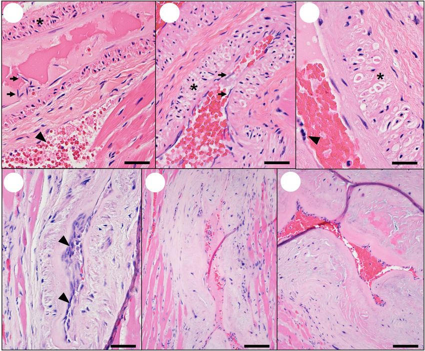
Miller et al.                                                                                                     Domoic Acid Toxicosis in Otters

approach that included review of available case history, gross           pathology from the most definitive acute DA cases were
and microscopic assessment of the brain, heart, eyes, and                compared with non-DA cases. We also identified lesions
other tissues, and biochemical tests (liquid chromatography-mass         in sea otters similar to those that have been reported
spectrometry and enzyme-linked immunosorbent assay) for DA               in other species with DA toxicosis. Due to the complex
in tissues and body fluids. Publications on the pathology of             systemic effects of DA toxicosis, the possibility of repeated
DA in other species aided lesion assessment (Teitelbaum et al.,          sublethal DA exposure, and potential interactions among
1990; Tryphonas et al., 1990; Bruni et al., 1991; Pulido et al.,         concurrent health conditions, lesion patterns were reported
1995; Silvagni et al., 2005; Goldstein et al., 2008; Zabka et al.,       conservatively as DA-associated pathology. Confirming the
2009; Lefebvre et al., 2010; Vranyac-Tramoundanas et al., 2011;          pathophysiological mechanisms underlying lesion development
McHuron et al., 2013; Buckmaster et al., 2014). Also considered,         was beyond the scope of this descriptive, necropsy-based,
when available, were data on confirmed toxic Pseudo-nitzschia            retrospective study.
bloom events, DA-associated public safety seafood harvest
closures, and DA-associated stranding of sympatric wildlife.
   Variation in the level of detail available for each case (a
                                                                         Estimating the Interval Between DA
common challenge for retrospective studies of free-ranging               Exposure and Death Based on Lesion
wildlife) affected the precision of diagnostic assessment. More          Chronicity
informative cases had antemortem data, a shorter time interval           For a prior analysis of southern sea otter mortality patterns
between death and necropsy, biochemical testing for DA, and              (Miller et al., 2020), estimates of DA lesion chronicity were
comprehensive necropsy and histopathology. Less precise cases            pooled as acute/subacute for CNS lesions and subacute/chronic
had more limited clinical history, DA test results, histopathology       for cardiovascular lesions because more in-depth temporal
and/or reduced sample quality due to minor postmortem                    assessment was not considered possible in wild sea otters
scavenging or autolysis; however, diagnostic assessment was              with unknown DA exposure timing, frequency, and dose.
both possible and informative. Domoic acid case classification           However, as we gained an understanding of patterns of DA
(Miller et al., 2020) reflected the level of confidence in               lesion development, progression, and healing responses, we
the likelihood of DA toxicosis as a primary or contributing              were able to estimate acute, subacute, and chronic post-DA
cause of death: probable cases had the highest confidence                exposure intervals. These intervals were based on the gross and
for DA toxicosis as a cause of death, while possible cases               microscopic appearance of DA-associated CNS, cardiovascular,
had indications of DA exposure but insufficient detail to                and ocular pathology, and uniform patterns of host response
confirm whether DA toxicosis contributed to mortality. Negative          following toxic injury (Kumar et al., 2014). Estimates of lesion
cases were considered unlikely to have DA toxicosis as                   chronicity and post-DA exposure intervals were also guided by
a cause of death.                                                        DA concentrations in postmortem tissues and body fluids, case
   Prior epidemiological studies have identified DA exposure             histories, and descriptions of DA-associated pathology and lesion
and systemic protozoal disease due to Toxoplasma gondii                  progression in other animals (Bruni et al., 1991; Pulido et al.,
and/or Sarcocystis neurona as significant risk factors for fatal         1995; Silvagni et al., 2005; Goldstein et al., 2008; Zabka et al.,
cardiomyopathy in southern sea otters (Kreuder et al., 2005;             2009; Lefebvre et al., 2010; Vranyac-Tramoundanas et al., 2011;
Miller et al., 2020; Moriarty et al., 2021b). Kreuder et al.             Buckmaster et al., 2014). Based on the above criteria, acute lesions
(2005) pooled cases with moderate to severe non-suppurative              were estimated to have developed within a few hours to a few
myocarditis and those with significant non-inflammatory cardiac          days prior to death, subacute lesions likely developed within a
pathology. For the current study, a range of lesions including           few days to approximately 6 months prior to death, and chronic
myocardial congestion, hemorrhage, cardiomyocyte vacuolation,            pathology was considered the result of DA toxicosis occurring
apoptosis, necrosis or loss, interstitial edema, non-suppurative         >6 months to years before death. Because the duration between
inflammation, vascular pathology, fibrosis, and microscopic              DA exposure and death is unknown for wild sea otters, and
hemorrhage (microhemorrhage) were assessed independently to              recurrent DA exposure is possible given the pervasiveness of
distinguish cardiac lesions associated with DA toxicosis from            this toxin in the marine environment, these temporal criteria are
those attributable to protozoal infection or other etiologies.           estimates that should be further validated and refined through
Based on prior research (Miller et al., 2020), southern sea              prospective research.
otters with purely suppurative myocarditis or endocarditis were              After identifying CNS and cardiac lesion patterns
grouped separately due to suspected or confirmed bacterial               characteristic of each DA post-exposure chronicity category
etiology. Cases were not classified based on the presence/absence        (acute, subacute, and chronic), we assessed the chronicity of
of cardiac dilation, because experience from >20 years of                gross and microscopic DA-associated pathology in the CNS
southern sea otter postmortem examinations suggests that                 and heart of 54 adult and aged adult southern sea otters with
these differences represent stages within a continuum of                 optimal tissue sampling. The relative chronicity of DA-associated
cardiomyopathy expression.                                               pathology and the presence/absence of any mixed temporal
   Microscopic assessment of the CNS, heart, and eyes was                patterns (e.g., acute pathology superimposed on pre-existing
performed in relation to the other tissues to document                   chronic lesions) were noted independently for the CNS and
the systemic effects of DA toxicosis and identify criteria               heart prior to comparison. Strong temporal associations, such
that could facilitate diagnosis. Gross and microscopic                   as the presence of subacute pathology in both organ systems,

Frontiers in Marine Science | www.frontiersin.org                    4                                       May 2021 | Volume 8 | Article 585501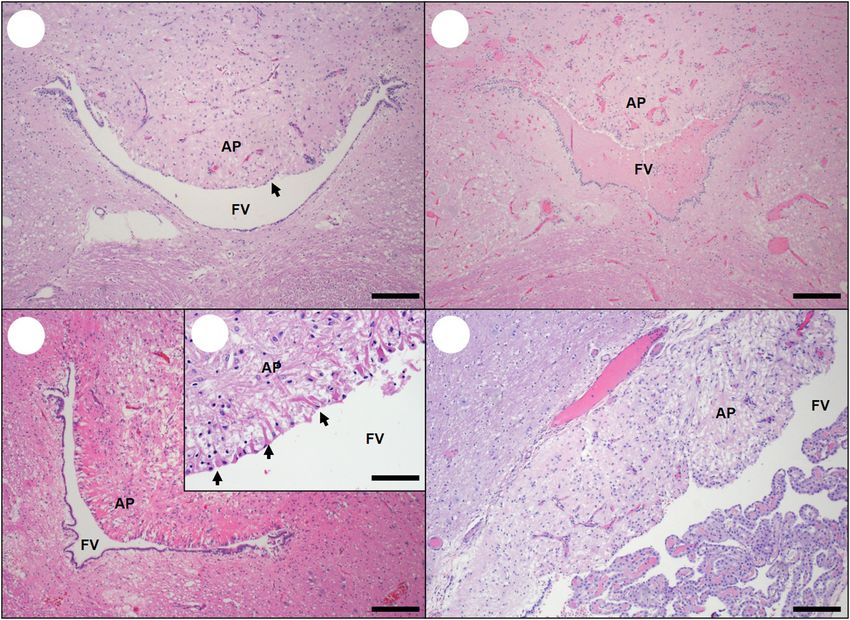
Miller et al.                                                                                                        Domoic Acid Toxicosis in Otters

were considered indicative of a single event of DA toxicosis               Supplementary Presentation 1, where the figures are arranged
that concurrently affected the CNS and cardiovascular system.              by 1) organ system (e.g., brain, heart, etc.), then 2) tissue subtype
The presence of concurrent acute, subacute, and/or chronic                 within each organ system (e.g., hippocampus, atrium, etc.),
DA-associated pathology in the CNS, heart, or both tissues was             3) from gross, broad-scale pathology findings to progressively
also evaluated, given its importance as potential evidence for             higher magnification microscopic views that show lesions
recurrent DA toxicosis.                                                    in detail, and 4) from acute to chronic with respect to
                                                                           the estimated post-exposure interval for DA toxicosis, so
Documentation of Pseudo-nitzschia                                          that the reader can visually assess lesion progression and
Uptake by Ingested Prey in Sea Otters                                      resolution through time. The figures are arranged this way so
Acutely Exposed to DA                                                      that the most definitive findings for confirming a diagnosis
                                                                           of DA toxicosis are illustrated first, while helpful but less
Prey items from the gastrointestinal (GI) tracts of two sea
                                                                           diagnostically definitive findings are found at the close of
otters were assessed for Pseudo-nitzschia diatom ingestion.
                                                                           each section (e.g., in the CNS, diagnostic hippocampal lesions
Both sea otters died during toxic Pseudo-nitzschia blooms, had
                                                                           are presented prior to DA-associated cerebellar pathology.)
histopathology indicative of acute DA toxicosis, and had very
                                                                           Side-by-side images within figures also depict normal tissues
high DA concentrations (5,000–56,200 ppb) in postmortem
                                                                           along with progressive lesion development through time (acute,
urine and gastric content. Minimally digested sand crabs
                                                                           subacute, and chronic). To facilitate comparison between the
(Emerita analoga) were recovered from the stomach of both
                                                                           lesion descriptions in each table and corresponding images, the
sea otters at necropsy. The GI tracts of these sand crabs
                                                                           Supplementary Presentation Figures are also cross-referenced
were dissected, pooled for each sea otter, and cleaned using a
                                                                           in column 1 of the Supplementary Presentation Tables 1 and
modified KMnO3 /HCl oxidation method to remove soft tissue
                                                                           2.
(Miller and Scholin, 1998). Cleaned cells were resuspended in
distilled water and filtered onto a Millipore filter (Millipore
Sigma, Burlington, MA, United States). The dried filter was                Domoic Acid-Associated Clinical
mounted to an aluminum stub, sputter-coated with gold-
palladium, and viewed on a scanning electron microscope
                                                                           Findings
                                                                           Sea otters stranding alive with acute DA toxicosis were often
(SEM: model ISI WB-6; International Scientific Instruments,
                                                                           in good to excellent nutritional condition (Figures 1A,B),
New Delhi, India) for the presence of Pseudo-nitzschia frustules
                                                                           with full GI tracts (Figure 2A). Clinically these animals
(silica-based exoskeletons of diatoms). Free-living sand crabs
                                                                           often exhibited tremors, seizures, obtundation, hyper- or
were also collected during a toxic Pseudo-nitzschia bloom that
                                                                           hypothermia, and visual deficits (Table 1). Other clinical signs
was associated with marine mammal mass-mortality; the GI
                                                                           included somnolence or unusual tameness, difficulties with
tracts of these crabs were also processed to confirm Pseudo-
                                                                           ambulation and food prehension, and abnormal vocalization.
nitzschia ingestion as described above. Finally, an in vitro culture
                                                                           These neurological signs could be caused by DA-associated
of Pseudo-nitzschia australis diatoms was cleaned, dried on a
                                                                           neurotoxicity and/or the observed regional or systemic
glass coverslip, and mounted to an aluminum stub prior to
                                                                           vascular pathology leading to fluid leakage, brain swelling,
coating and SEM; this served as a positive control for Pseudo-
                                                                           and cerebellar herniation.
nitzschia frustules.
                                                                              In subacute and chronic cases, clinical signs could be
                                                                           continuous or episodic. Chronically affected individuals often
RESULTS                                                                    exhibited abrupt cessation of normal behaviors (such as
                                                                           eating), remaining immobile and unresponsive for several
A bimodal pattern of DA-associated clinical signs and pathology            minutes before resuming normal activity. Periods of reduced
was observed in southern sea otters. Otters with acute DA                  activity were sometimes accompanied by tremors. A few sea
toxicosis typically stranded with severe CNS signs, chronic                otters displayed unusual aggression or persistent biting and
cases presented predominantly with severe cardiovascular                   chewing on an extremity and were euthanized as potential
signs, and subacute cases showed either or both patterns.                  rabies suspects; brain tissue screening for rabies was negative
Detailed descriptions of clinical, gross, and histopathology               in all cases. Abrasions or scrapes on the nose, face, and
findings are described below. Clinical, biochemical, and gross             paws, asymmetrical whisker wear, and self-trauma to the
necropsy findings are summarized by chronicity category in                 paws, penis, or tail were suggestive of significant neurological
Table 1, and DA-associated histopathology is summarized in                 impairment, paresthesia, and/or ambulatory deficits. Some
Table 2. To optimize the utility of the summary tables and                 otters had clinical signs or gross pathology suggestive of
pathology image content of this manuscript for diagnosing                  spinal cord damage, including hind limb paraparesis, fecal
potential DA cases, all figures and Tables 1, 2 are also                   impaction, and urine retention, all of which have been
available as separate, large, high-resolution, downloadable                reported for other DA-affected species (Jian Wang et al., 2000;
files in Supplementary Presentation 1 in the Supplementary                 Pulido, 2008).
Materials. It is important to note that the figure order                      Live-stranded sea otters with subacute to chronic
and numbering differs between the text of the manuscript,                  cardiomyopathy had radiographic evidence of cardiomegaly
where the figures are cited in order of discussion, and                    and enhanced pulmonary density, and clinical indications

Frontiers in Marine Science | www.frontiersin.org                      5                                        May 2021 | Volume 8 | Article 585501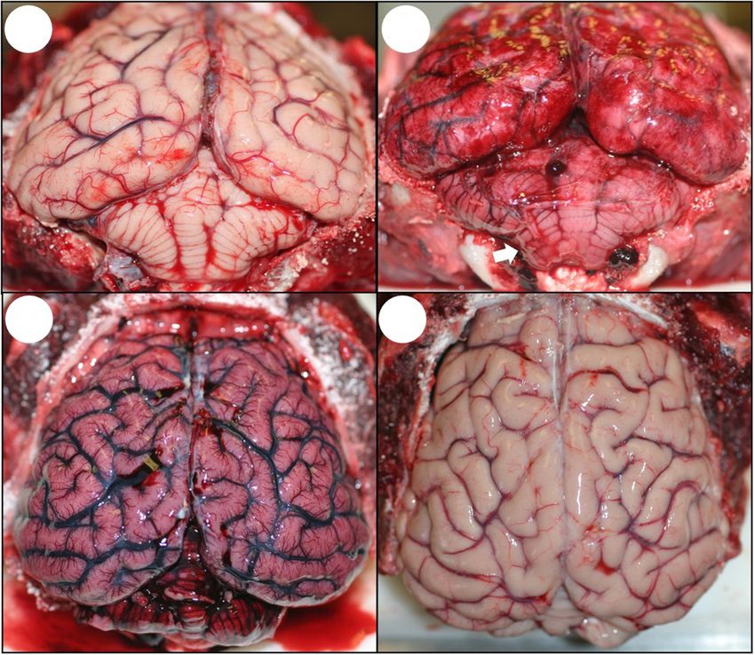
Frontiers in Marine Science | www.frontiersin.org

                                                                                                                                                                                                                                                                              Miller et al.
                                                    TABLE 1 | Clinical, biochemical and gross findings suggestive of domoic acid toxicosis and DA-associated cardiomyopathy in southern sea otters (Enhydra lutris nereis).

                                                                                                                Acute (Hours to days)                                Subacute (Days to 2 ml)
                                                                                                • Myocardium may look brown, wet,                        • Myocardium pale & mottled or streaked,                  • Variable (mild to severe) cardiac & venous
                                                                                                  translucent                                              especially apex, ventricles (L > R), brown                dilation +/− venous shunts (chest, abdomen)
                                                                                                • +/− Heart, systemic veins & viscera                      discoloration mild/absent.                              • Myocardium pale, tan or white spots or
                                                                                                  appear mildly dilated & "full of blood", but           • Atrial walls increasingly opaque                          streaks, atrial walls often opaque
                                                             Cardiovascular System                organs not abnormally enlarged                         • +/− Mild myocardial hemorrhage                          • Severe congestion, cardiomegaly,
                                                                                                • +/− Grossly apparent hemorrhage in                     • Variable (none-severe) cardiac dilation                   hepatomegaly, chronic passive congestion
                                                                                                  papillary muscles, ventricular myocardium              • +/− Pleural, pericardial, peritoneal effusion           • Moderate-severe peritoneal, pleural &
                                                                                                • +/− Pleural, pericardial and peritoneal effusion                                                                   pericardial effusion, hepatic capsular fibrin
                                                                                                  (mild to moderate)                                                                                               • Pulmonary edema +/− interstitial fibrosis
May 2021 | Volume 8 | Article 585501

                                                                                                                                                                         • +/− Depressed, thin or transparent areas, especially near apex (myofiber loss)

                                                                                                                                                                                                                                                                              Domoic Acid Toxicosis in Otters
                                                                                                                                                                                                 • +/− Hepatic vein thrombosis
                                                                                                                                            • +/− Arrhythmias, acute death, heart failure, pulmonary edema, dyspnea, cyanosis

                                                                                                                                                                                                                                                                (Continued)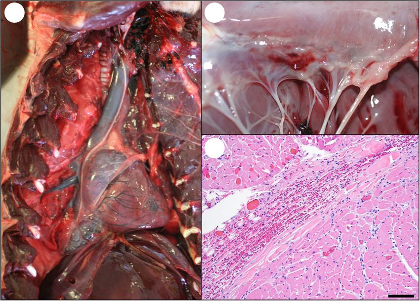
Frontiers in Marine Science | www.frontiersin.org
Miller et al.
TABLE 1 | Continued
Assessment Type Acute (Hours to days) Subacute (Days to < 6 months) Chronic (≥6 months to years)
• Volume mildly increased (Normal = 1.5-2 • Volume moderately/markedly increased (∼4-8 • Volume markedly increased (often >8 ml)
Pericardial Fluid∗ ml), may be strongly DA positive ml), may be DA-positive
• +/− pale brownish/reddish tinge
• +/− fibrin strands interspersed in pericardial fluid
• +/− Blindness/visual deficits • +/− Blindness/visual deficits
Eyes • Hyphema (Unilateral or bilateral) • +/− Hyphema (Unilateral or bilateral)
• Hepatomegaly (mild/moderate), hepatic passive • Hepatomegaly (moderate/marked), hepatic passive
congestion, +/− mild hemosiderosis congestion, hemosiderosis +/− Liver surface has
Liver • Moderate diffuse congestion • Serous ascites +/− fibrinous capsulitis irregular "cobblestone" appearance
• +/− Grossly apparent fibrin thrombi in hepatic vein • Hemorrhagic ascites, fibrinous capsulitis
& hepatic parenchymal necrosis • +/− Hepatic vein thrombi, hepatic necrosis
Kidneys and Bladder • Variable congestion • Moderate-marked congestion
• Moderate/marked diffuse congestion • +/− Dilated, urine-distended bladder
• Often food in GI tract, including minimally • +/− Partially digested food in GI tract • GI tract often empty, atrophic
Stomach and Intestines digested food in stomach
• +/− Ileus, intussusception, torsion
7
• +/− Serosal & mucosal congestion
• +/− Recent abortion, acute death while • +/− Recent abortion, death while pregnant, • +/− Cardiac decompensation during
pregnant, stillborn fetus, pre-term fetus stillborn fetus, +/− uterine torsion pregnancy, recent abortion, stillborn fetus
• +/− Signs of forced copulation during • +/− Signs of forced copulation during • +/− Pregnant but emaciated, or marked
non-estrus period (while pregnant, non-estrus period (due to neurological nutritional mismatch (ample systemic adipose
possibly due to neurological disease) disease) but severe muscle catabolism)
Female-Specific • Uterus, placenta, fetus: Congestion, • +/− Fetus: Congestion, edema, brain or • +/− Fetus: Emaciation, congestion, edema,
edema, brain or myocardial hemorrhage, myocardial hemorrhage, meconium stains meconium stains (female attempted to reproduce
meconium stains +/−uterine torsion • Variable mismatch between systemic adipose despite progressive cardiac disease)
• None to mild mismatch between systemic stores & muscle catabolism
adipose stores & muscle catabolism
• History of "rebooting" reproductive cycle, poor pup care or preweaning pup
• Perimortem fight trauma
Male-Specific • +/− Mild paraphimosis or self-trauma • No paraphimosis or self-trauma
May 2021 | Volume 8 | Article 585501
• No testicular atrophy • +/− Testicular atrophy • Testicular atrophy
Domoic Acid Toxicosis in Otters
Definitions: GI, gastrointestinal; PPB, parts per billion.
Bold text, most helpful criteria for DA case diagnosis and staging, when present. +/−, findings that are less consistently observed or less definitive.
*Longer postmortem interval, blood loss & dehydration can decrease yield.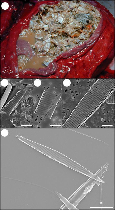
Frontiers in Marine Science | www.frontiersin.org
Miller et al.
TABLE 2 | Histopathology findings suggestive of domoic acid (DA) toxicosis and DA-associated cardiomyopathy in southern sea otters (Enhydra lutris nereis).
Acute (Hours to days) Subacute (Days to < 6 months) Chronic (≥6 months to years)
Key Tissues Central nervous system (CNS) signs and May present with severe CNS and/or Cardiovascular signs and lesions dominate.
lesions dominate. Occasional deaths from cardiovascular deficits. CNS signs may be present, but are often less
acute cardiovascular impacts. < ——————————————– > severe than cardiovascular pathology.
• Pyramidal neurons: Necrosis, apoptosis, • Mild to complete segmental loss of pyramidal
hyperchromic, shrunken, pale, elongated, neurons (especially in CA2), mild gliosis,
dropout, especially cornu ammonis 2 malacia. Smooth curve of CA pyramidal
region (CA2), entorhinal cortex, neurons becomes angular
parahippocampal gyrus. Dentate neurons • Inflexion of dentate neurons often “points”
relatively spared. “Red dead” neurons toward area of most severe segmental
relatively uncommon pyramidal neuron damage (consistent tissue
• Lesions commonly segmental, spread up trimming is required)
and down CA over time (spans CA1-4 in • Retraction of severely affected CA segments
• Marked diffuse congestion and severe cases), with most severe damage toward dentate neurons, flattened medial
multifocal microhemorrhages, around CA2. Pyramidal neuron loss and hippocampus, ventricle dilation
• +/− spongiosis gemistocytic astrocyte proliferation. • Hippocampi may appear shrunken, flattened,
Hippocampus, Entorhinal Cortex, +/− Perivascular fibrinoid changes in • Perivascular and laminar spongiosis of asymmetrical, with enlarged lateral ventricle
Parahippocampal Gyrus hippocampus and elsewhere without molecular layer between CA and dentate • Ventricle walls appear irregular, “moth-eaten”,
evidence of sepsis or endotoxemia granule cells with gliosis with discontinuous ependyma
• No/mild neuronal necrosis, shrinkage, • Lesions often asymmetrical • Chronic, mild, sublethal cases can be
8
dropout, astrogliosis on histopathology • Progressive neuropil vacuolation, +/− challenging to confirm, especially when
malacia near blood vessels and ventricles autolysis or concurrent pathology are present
• Segmentally decreased pyramidal neuron • For severe chronic lesions, may see neuropil
density, focal retraction toward dentate, loss, cavitation, extracellular vacuoles; lesions
and flattening of pyramidal neuron curve, often bilateral but may be asymmetrical
outer hippocampus, dilation of lateral • No severe congestion or microhemorrhage
ventricle • Minimal/no discernible neuronal necrosis,
• Ventricle walls appear irregular, “moth- shrinkage, dropout. Astrogliosis may persist
eaten”, with discontinuous ependyma
• Progressive decrease in congestion,
microhemorrhage, fibrinoid change
• Moderate to marked congestion in • +/− Vascular congestion in pars distalis and • No abnormal pituitary congestion, edema,
Pituitary pars distalis and pars nervosa with pars nervosa with relative sparing of pars hemorrhage
relative sparing of pars intermedia. intermedia, +/− edema and microhemorrhages • +/− Scarring in pars nervosa
May 2021 | Volume 8 | Article 585501
Microhemorrhages in pars nervosa in pars nervosa
Area Postrema, Other Circumventricular • Mild to moderately congested, spongiotic, • Adjacent ventricle may appear scarred,
Domoic Acid Toxicosis in Otters
Organs (CVOs) • Congested, spongiotic, +/− +/− neuronal necrosis or apoptosis, “moth-eaten”, with discontinuous ependyma
microhemorrhages astrogliosis, damaged ventricle wall, • +/−Scarring, gliosis, vacuolation
microhemorrhage
(Continued)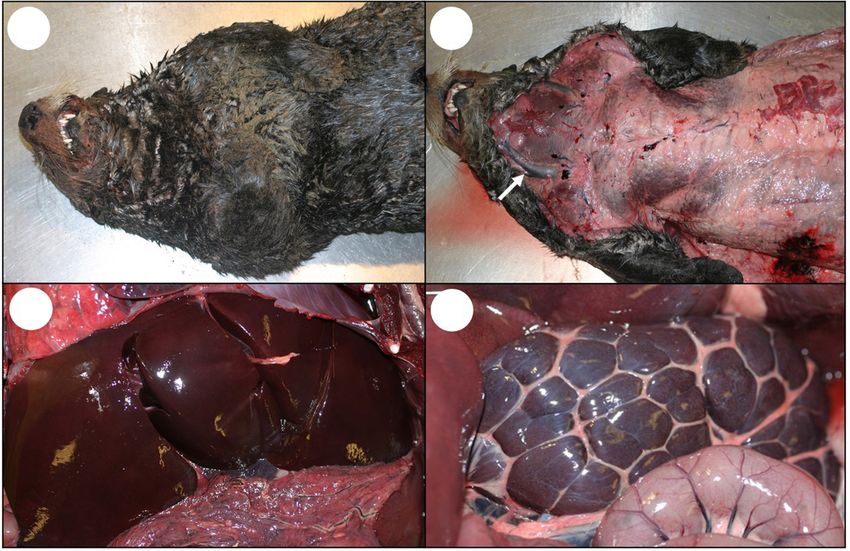
Frontiers in Marine Science | www.frontiersin.org
Miller et al.
TABLE 2 | Continued
Key Tissues Acute (Hours to days) Subacute (Days to < 6 months) Chronic (≥6 months to years)
• Congestion of choroid plexus • Ventricles may contain cell debris and mild • +/− Mild pallor, spongiosis and astrogliosis of
• Ventricles may contain cell debris and to severe hemorrhage sub-ventricular neuropil
Choroid Plexus, Ependyma, Ventricles, acute hemorrhage • Sub-ependymal neuropil: Congestion, • Ventricles variably dilated with irregular
Sub-Ependymal Neuropil • Sub-ependymal neuropil: microhemorrhage, spongiosis, astrogliosis contours, “moth-eaten” appearance
+/− Congestion, microhemorrhage, • Ventricles: Irregular contour, “moth-eaten” • +/− Ventricles discontinuously lined by
spongiosis (especially lateral and third appearance, patchy ependymal lining, ependyma and acellular fibrils, bands of
ventricles) ependymal cells trapped below surface ependymal cells trapped below surface
• Ependyma and choroid: +/−acute • Ventricle edges may adhere, forming scars • Ventricle edges may adhere, forming scars
necrosis/apoptosis, cells rounded, swollen, • Ependyma, choroid: Lessening congestion,
eosinophilic, sloughing necrosis, apoptosis
• Marked vascular congestion, • Progressive development of multifocal
Diencephalon (Thalamus, Hypothalamus) microhemorrhage less prominent large, well-defined vacuoles, especially • Multifocal large, sharp-walled, well-defined
• +/− Perivascular spongiosis near ventricles and neuronal nuclei vacuoles, especially near ventricles and neuronal
• Minimal/no discernible neuronal • Congestion progressively fades nuclei
necrosis, dropout, astrogliosis • Perivascular spongiosis • Periventricular neuropil and ependyma may appear
• Sparse neuronal necrosis/apoptosis, scarred, “moth-eaten”
shrinkage, dropout, astrogliosis • +/− Decreased neuronal density in some nuclei,
• Periventricular neuropil and ependyma +/− mild astrogliosis
may look spongiotic, scarred,
“moth-eaten”
9
• Marked diffuse vascular congestion,
Cerebellum, Pons, Medulla especially in meninges and molecular • Marked diffuse vascular congestion, • Periventricular neuropil and ependyma may appear
layer especially in molecular layer scarred, “moth-eaten”
• Sparse scarring and vacuolation
• Purkinje cell layer: +/− mild laminar swelling of astrocytes and Purkinje cell loss
• Marked congestion, mild/moderate
spongiosis and laminar swelling of • Mild/moderate spongiosis and laminar
Olfactory Tracts, Olfactory Bulbs astrocytic foot processes, swelling of astrocytic foot processes • +/− Sparse, well-defined vacuoles along laminae
+/− microhemorrhages • Marked congestion +/− multifocal (Swollen astrocytic processes)
• Neurons and glia: Mild-moderate microhemorrhage (progressively fades) • +/− Mild gemistocytic astrogliosis
cytoplasmic vacuolation, minimal/no • Sparse neuronal necrosis, shrinkage,
necrosis, shrinkage, dropout, dropout, gemistocytic astrogliosis
astrogliosis
• Severe diffuse congestion, multifocal • Dilation of central canal, ependymal loss,
May 2021 | Volume 8 | Article 585501
microhemorrhage minimal intraluminal hemorrhage • +/− Dilation and mural scarring of central
• +/− Dilation of central canal, ependymal • +/− Patchy symmetrical neuronal necrosis canal, variable ependymal loss
Domoic Acid Toxicosis in Otters
Spinal Cord loss, minimal intraluminal hemorrhage • +/− Microhemorrhage, gemistocytic • +/− Sparse axonal loss, ballooning of axon
• +/− Perivascular and periventricular astrocytes, sparse dilated axon sheaths sheaths, mildly decreased neuronal density
spongiosis and microhemorrhage • +/− Multifocal large, well-defined clear spaces,
especially near ventricles
(Continued)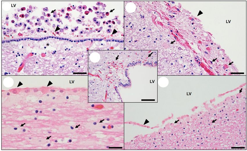
Frontiers in Marine Science | www.frontiersin.org
Miller et al.
TABLE 2 | Continued
Key Tissues Acute (Hours to days) Subacute (Days to < 6 months) Chronic (≥6months to years)
• Marked diffuse myocardial congestion • Cardiomyocytes: Cytoplasmic vacuolation, • Moderate to severe multifocal cardiomyocyte
and multifocal microhemorrhages: eosinophilia, swelling, necrosis, apoptosis, loss, intersecting bands of stromal collapse,
Perivascular, epi- and endocardium, myophagia fatty replacement, stromal fibrosis
coronary groove, papillary muscles • Multifocal cardiomyocyte loss with stromal • Large expanses of atrium may contain few
• Coronary endothelial and vascular collapse, fatty replacement, mild fibrosis, cardiomyocytes, severe fatty replacement,
Cardiovascular and Pulmonary smooth muscle necrosis/apoptosis/ non-suppurative inflammation no/mild inflammation
vacuolation, mural edema, hemorrhage • Coronary arterioles: Smooth muscle • Walls of coronary arterioles hyalinized, hypocellular,
• +/− Proteinacious interstitial edema necrosis/apoptosis. Walls of coronary thickened, sparsely mineralized Endothelium may
• +/− Bands of bunched cardiomyocytes, arterioles increasingly hyalinized, hypocellular be bunched and rounded
hyper-eosinophilic, glassy appearance +/− sparsely mineralized • Cardiomyocyte necrosis/apoptosis/cytoplasmic
• +/−Mild cardiomyocyte swelling, edema, • Decreasing congestion, interstitial edema vacuolation less prominent
vacuolation, necrosis, or apoptosis • Most severe lesions: apex, left ventricular free wall, • Lesions most severe apex, left ventricular free wall,
• Coronary vasculature: No hyalinization septum, and papillary muscles atrium, papillary muscles, septum
• Ventricles: Often sub-endocardial/sub-epicardial
lesion distribution
• Moderate diffuse congestion • Hepatic passive congestion, atrophy of hepatic • Hepatic passive congestion, atrophy of hepatic
10
Hepatobiliary •+/− Mild acute venous dilation cords (especially centrilobular) cords (centrilobular to diffuse)
• +/− Patchy hepatic necrosis and thrombosis • +/− Patchy hepatic necrosis and thrombosis
•Hyphema and severe diffuse vascular congestion (bilateral) • +/− Blindness/visual deficits
Eyes • +/−Congestion, hyphema due to congestive heart
failure
• +/− Bands and patches of myometrial
hyper-eosinophilia, necrosis, apoptosis • Bands and patches of myometrial
Female Reproductive System • +/− Myometrial arteries: Endothelial hyper-eosinophilia, necrosis, apoptosis
and smooth muscle necrosis or • Myometrial vessels: Endothelial and smooth • Placental congestion, hemorrhage, necrosis
apoptosis, degeneration, edema, muscle cell necrosis or apoptosis, (associated with congestive heart failure)
hemorrhage degeneration, edema, hemorrhage
• Placental congestion, hemorrhage, • Placental congestion, hemorrhage, necrosis
necrosis
Renal • Moderate to marked diffuse congestion • Variable congestion • Moderate to marked congestion
May 2021 | Volume 8 | Article 585501
Gastrointestinal • +/− Moderate vascular congestion
Definitions: CA, cornu ammonis of hippocampus.
Domoic Acid Toxicosis in Otters
Bold text, most helpful criteria for DA case diagnosis and staging, when present.
+/−, findings that are less consistently observed or less definitive.Miller et al. Domoic Acid Toxicosis in Otters
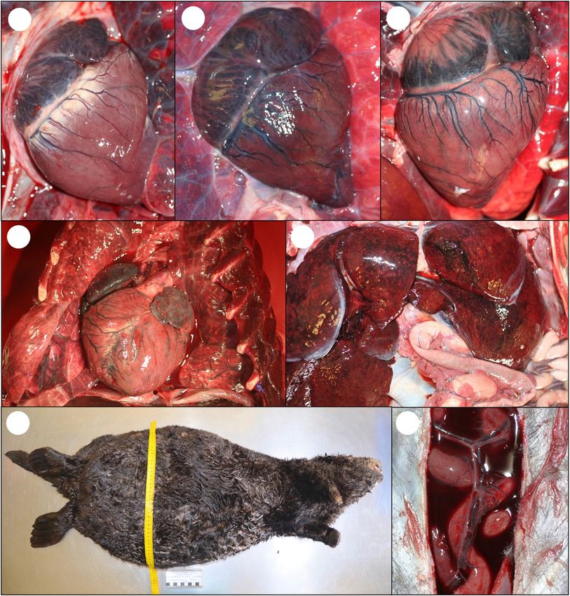
FIGURE 1 | Gross presentation for acute domoic acid (DA) toxicosis in southern sea otters. (A) Adult male sea otter that died from acute DA toxicosis. The otter was
in excellent nutritional condition with red-tinged, seromucoid fluid stains around the nose and mouth. (B) Subcutis of the same sea otter showing abundant adipose
and mildly distended jugular veins (arrow). The liver (C) and kidneys (D) were diffusely congested but not abnormally enlarged.
of congestive heart failure (Table 1): dyspnea, lethargy, (CSF) (≤2 ml is normal at necropsy). Severe congestion was
anorexia, copious white or pink oronasal froth, and pleural, sometimes accompanied by diffuse brain swelling and herniation
pulmonary, pericardial, and peritoneal edema (Moriarty of the caudal cerebellar vermis through the occipital foramen
et al., 2021a). Affected otters were emaciated with a poorly (Figures 3B, 4A), along with grossly apparent meningeal,
groomed pelage. Murmurs, pulse deficits, and abnormalities neuropil, and ventricular hemorrhage (Figures 3B,C, 4A).
on electrocardiographic examination, such as reduced P wave
amplitude, atrial and ventricular ectopy, and first- and second- On histopathology, severe diffuse congestion was observed
degree atrioventricular block were also noted (Moriarty et al., in the meninges, hippocampus, diencephalon, entorhinal,
2021a), similar to reports for humans and other mammals with olfactory and parahippocampal cortex, and the olfactory
DA toxicosis (Perl et al., 1990a; Vranyac-Tramoundanas et al., bulbs (Table 2 and Figure 4B). Domoic acid-associated
2011; Vieira et al., 2016). cerebellar congestion was easiest to see in the molecular layer
(Figure 5A). Severe congestion was often accompanied by
multifocal microhemorrhage (Table 2 and Figures 4B,C, 6B–
Domoic Acid-Associated Pathology of D, 7A,B, 8A,B). In contrast, neuronal or glial necrosis or
the CNS and Cardiovascular Systems degeneration were minimal in sea otters with acute DA toxicosis
Acute DA Toxicosis (Figures 6C,D, 7A).
Central nervous system Early vascular and perivascular CNS lesions included
The earliest, most common, and most striking gross lesion in the mural and perivascular edema, vacuolation and necrosis
CNS of sea otters with high DA concentrations (>500 ppb) in or apoptosis of arteriolar mural smooth muscle cells and
urine or GI content was severe diffuse brain, spinal cord, and endothelium, and occasional sloughed endothelium (Table 2).
meningeal congestion (Table 1 and Figures 3B,C, 4A; please Microhemorrhages were common near small arterioles or
also see Figure 3A as a normal comparison image). Affected engorged capillary beds (Figures 6C,D; please also see Figure
brain tissue was pink, moist, and mildly translucent; postmortem 6A as a normal comparison image), especially in the cornu
cisternal taps commonly yielded >2 ml of cerebrospinal fluid ammonis (CA) of the hippocampus. Intramural and perivascular
Frontiers in Marine Science | www.frontiersin.org 11 May 2021 | Volume 8 | Article 585501Miller et al. Domoic Acid Toxicosis in Otters
FIGURE 2 | Pseudo-nitzschia frustule detection in sand crabs (Emerita analoga) ingested by southern sea otters with acute domoic acid (DA) toxicosis. Sea otters
with acute DA toxicosis often have full gastrointestinal (GI) tracts. Examination of the GI tracts of these prey items can reveal the presence of pennate frustules
(Continued)
Frontiers in Marine Science | www.frontiersin.org 12 May 2021 | Volume 8 | Article 585501Miller et al. Domoic Acid Toxicosis in Otters FIGURE 2 | Continued (silica-based exoskeletons of diatoms), confirming that the prey consumed Pseudo-nitzschia diatoms that could have contained DA prior to predation by sea otters. (A) Full stomach from a sea otter that died of acute DA toxicosis during a toxic Pseudo-nitzschia bloom; postmortem stomach content and urine were strongly positive for DA (≥10,240 ppb). The stomach contained a mixture of small crabs, including sand crabs (Emerita analoga). The masses of tiny, bright orange spheres are crab eggs. Sea otters may preferentially consume crabs with eggs because their nutritional value is higher; these reproductive cycles can coincide with Pseudo-nitzschia blooms. (B) Scanning electron microscopy (SEM) micrograph of fragment of Pseudo-nitzschia frustule from cleaned GI tracts of free-living sand crabs collected during a toxic Pseudo-nitzschia bloom event (Bar = 4 µm). (C) SEM micrograph from the stomach content shown in (A). A fragment of Pseudo-nitzschia frustule was obtained from the gastrointestinal tracts of sand crabs in this sample (Bar = 4 µm). (D) SEM micrograph of stomach content from a second sea otter that died from acute DA toxicosis. Postmortem stomach content and urine were strongly positive for DA (≥5,000 ppb) and a fragment of Pseudo-nitzschia frustule was obtained from the GI tracts of sand crabs recovered from the stomach of this sea otter (Bar = 4 µm). (E) Reference Pseudo-nitzschia australis frustules showing the characteristic pennate shape and complex ribbed morphology (Bar = 20 µm). FIGURE 3 | Gross appearance of the southern sea otter central nervous system following domoic acid (DA) toxicosis. (A) Normal sea otter brain; the outer meninges (dura and arachnoid) were removed to show the brain surface. (B) Brain (with dura and arachnoid) from a sea otter that died from acute DA toxicosis showing severe diffuse meningeal congestion, multifocal acute hemorrhage, and mild deformation of the caudal cerebellar vermis (arrow) due to diffuse brain swelling that resulted in perimortem occipital herniation. (C) Brain (without dura and arachnoid) from a sea otter that died from acute DA toxicosis; there was severe diffuse congestion, multifocal hemorrhage, and mild to moderate diffuse swelling. (D) Brain (without dura and arachnoid) from a sea otter that died from chronic DA-associated cardiomyopathy (chronic DA post-exposure case); the brain is mildly atrophied with flattened gyri and pale tan neuropil. Frontiers in Marine Science | www.frontiersin.org 13 May 2021 | Volume 8 | Article 585501
Miller et al.                                                                                                                       Domoic Acid Toxicosis in Otters

  FIGURE 4 | Gross and microscopic brain lesions associated with acute domoic acid (DA) toxicosis in southern sea otters. (A) Marked congestion, multifocal
  hemorrhage, diffuse brain swelling, and mild deformation of the caudal cerebellar vermis due to perimortem occipital herniation (bottom center).
  (B) Parahippocampal cortex of a sea otter with acute DA toxicosis. There is diffuse severe congestion, acute meningeal hemorrhage (asterisk), and multifocal
  microhemorrhage (arrows) (Bar = 200 µm). (C) Cerebrum from a fetus that died in utero from acute DA toxicosis. Severe diffuse congestion is accompanied by
  multifocal microhemorrhage (arrows) (Bar = 200 µm). (D) Cerebrum from a fetus that died in utero from acute DA toxicosis. Severe congestion is accompanied by
  marked venodilation in the choroid plexus (Bar = 200 µm).

hemorrhage, fibrin deposition, and malacia were also observed                        while the pars intermedia was relatively unremarkable
in some acute and subacute DA cases (Figures 5B–F). Severe                           (Table 2 and Figures 11B–D). Multifocal microhemorrhage
congestion of the choroid plexus was common (Figure 4D),                             and spongiosis were also common in the pars nervosa
and necrosis of the choroidal epithelium and ventricular                             (Figures 11C,D, 12F). Pituitary congestion was often
ependyma was suspected (Figures 9A–E), although postmortem                           so extreme that it was visible on subgross examination.
autolysis precluded confirmation. Ependymal swelling,                                Although congestion and spongiosis were also observed in
cytoplasmic eosinophilia, and sloughing were consistent                              the pineal gland and area postrema (Figures 10B–D), the
findings throughout the ventricles (Figures 9A–E) and spinal                         pattern was less visually striking when compared with the
cord central canal.                                                                  pituitary gland.
   Severe     congestion,    spongiosis,    and     occasional
microhemorrhage were apparent in all circumventricular                               Cardiovascular system
organs (CVOs) examined microscopically (Table 2), including                          Sea otters with acute DA toxicosis commonly had mild diffuse
the area postrema (Figure 10B; please also see Figure 10A                            venodilation (Figure 1B), and the atria had a mildly dilated
as a normal comparison image), median eminence, pineal,                              appearance (Table 1 and Figures 13B, 14A; please also see
and pituitary gland (Figures 11B–D; please also see Figure                           Figure 13A as a normal comparison image) grossly. The
11A as a normal comparison image). The microscopic                                   ventricular myocardium was diffusely wet, brown, and mildly
appearance of the pituitary gland was especially striking in                         translucent (Figures 13B, 14A), accompanied by a mild increase
acute DA cases: when bisected along the median sagittal plane,                       in pericardial fluid (≤2 ml is normal at necropsy) that had a
the pars distalis and pars nervosa were severely congested,                          slight brownish tinge.

Frontiers in Marine Science | www.frontiersin.org                               14                                            May 2021 | Volume 8 | Article 585501
Miller et al.                                                                                                                          Domoic Acid Toxicosis in Otters

  FIGURE 5 | Acute and subacute cerebral and cerebellar pathology associated with domoic acid (DA) toxicosis in southern sea otters. (A) Diffuse, severe cerebellar
  congestion in a sea otter with acute DA toxicosis. The congestion is often most visible in the molecular layer (ML), often accompanied by multifocal meningeal
  hemorrhage (arrow) (Bar = 200 µm). (B) Parahippocampal gyrus in a sea otter with subacute DA toxicosis. Malacia, astrogliosis, perilesional microhemorrhage
  (arrowheads), and vascular mural and perivascular fibrin (arrow) are occasionally observed in the brain of sea otters with subacute DA toxicosis (Bar = 100 µm).
  (C,D) Higher magnification of (B) showing perivascular fibrin deposition, hemorrhage, malacia, and perivascular gliosis (arrows) (C,D: Bar = 40 µm). (E) Vascular
  mural and perivascular fibrin deposition (arrows) and hemorrhage (asterisk) in the parahippocampal gyrus of a sea otter with subacute DA toxicosis (Bar = 20 µm).
  (F) Spongiosis and malacia (asterisk), and multifocal microhemorrhage (arrows) in the cerebellar white matter of a sea otter with subacute DA toxicosis
  (Bar = 200 µm). (G) Olfactory lobe of a sea otter with acute or subacute DA toxicosis. There is diffuse congestion, mild patchy spongiosis, and linear bands of
  swollen, pale cytoplasmic vacuoles (presumptive astrocytic processes; arrows) (Bar = 60 µm).

   Acute cardiac pathology was subtle on histopathology                                Figures 14B,C, 15B,C), especially the perivascular interstitium,
and affected mainly the myocardium, coronary vasculature,                              the papillary, epicardial and, endocardial myocardium, the
and, to a lesser extent, the Purkinje fibers. Severe diffuse                           apex, and along the atrioventricular border near the base of
congestion (Figures 14C, 15A) was often accompanied by patchy                          the valve leaflets. Acute inflammation was usually minimal
cardiomyocyte hyper-eosinophilia, swelling and vacuolation,                            or absent.
linear bands of contracted cardiomyocytes, and swelling
and cytoplasmic vacuolation of Purkinje fibers (Table 2).                              Subacute DA Toxicosis
Small and medium-sized arterioles in areas of cardiomyocyte                            Central nervous system
damage often had mural smooth muscle cell swelling and                                 Gross CNS lesions were often less visually striking in sea
cytoplasmic hyper-eosinophilia, accompanied by mural and                               otters with subacute DA toxicosis when compared with acute
perivascular edema and microhemorrhage (Figures 16A–C).                                DA cases (Table 1). Subacute DA cases were characterized
Perivascular and interstitial microhemorrhage and edema were                           by less severe vascular congestion and the appearance of
apparent in areas of cardiomyocyte pathology (Table 2 and                              significant neuronal, glial, and stromal histopathology (Table 2).

Frontiers in Marine Science | www.frontiersin.org                                 15                                             May 2021 | Volume 8 | Article 585501
You can also read