Coronavirus Disease 2019 and the Thyroid - Progress and Perspectives - ScienceOpen
←
→
Page content transcription
If your browser does not render page correctly, please read the page content below
REVIEW
                                                                                                                                                published: 24 June 2021
                                                                                                                                       doi: 10.3389/fendo.2021.708333

                                           Coronavirus Disease 2019 and the
                                           Thyroid - Progress and Perspectives
                                           Hidefumi Inaba 1,2* and Toru Aizawa 3
                                           1 The First Department of Medicine, Wakayama Medical University, Wakayama, Japan, 2 Department of Diabetes,

                                           Endocrinology, and Metabolism, Japanese Red Cross Wakayama Medical Center, Wakayama, Japan, 3 Diabetes Center,
                                           Aizawa Hospital, Matsumoto, Japan

                                           SARS-CoV-2 infection (COVID-19) is currently a tremendous global health problem.
                                           COVID-19 causes considerable damage to a wide range of vital organs most
                                           prominently the respiratory system. Recently, clinical evidence for thyroidal insults
                                           during and after COVID-19 has been accumulated. As of today, almost all non-
                                           neoplastic thyroid diseases, i.e., Graves’ disease, Hashimoto’s thyroiditis, subacute,
                                           painless and postpartum thyroiditis, have been reported as a complication of COVID-
                                           19, and causality by the virus has been strongly implicated in all of them. Similar thyroid
                                           problems have been reported in the past with the SARS-CoV outbreak in 2002. In this
                                           review, we briefly look back at the reported evidence of alteration in thyroid functionality
                                           and thyroid diseases associated with SARS-CoV and then proceed to examine the issue
                         Edited by:        with COVID-19 in detail, which is then followed by an in-depth discussion regarding a
                   Akira Sugawara,         pathogenetic link between Coronavirus infection and thyroid disease.
            Tohoku University, Japan

                      Reviewed by:         Keywords: coronavirus, SARS-CoV, SARS-CoV-2, thyroid gland, autoimmunity, hyperinflammatory syndrome,
                         Kenji Ohba,       cytokine storm
              Hamamatsu University
          School of Medicine, Japan
                    Fausto Bogazzi,        INTRODUCTION
              University of Pisa, Italy
                    Tetsuya Tagami,        Coronavirus disease 2019 (COVID-19): caused by SARS-CoV-2 infection had broken out in China
       National Hospital Organization      in December 2019 and then rapidly spread all over the world creating a global pandemic (1).
        Kyoto Medical Center, Japan
                                           Patients with COVID-19 suffer from high mortality due not only to respiratory failure but also to
                *Correspondence:           other complications such as cardiovascular collapse and disseminated intravascular coagulation (2,
                    Hidefumi Inaba         3). In addition, there have been co-morbidities including autoimmune diseases associated with
         inaba@wakayama-med.ac.jp
                                           COVID-19 such as Guillain–Barre’s syndrome (4), autoimmune hemolytic anemia (5), and
                                           autoimmune thrombocytopenic purpura (6). Recently, evidence has been accumulated for
                   Specialty section:
                                           changes in thyroid function and thyroid diseases associated with COVID-19 (7–29). Review
         This article was submitted to
               Thyroid Endocrinology,
                                           articles on this topic have also been rapidly published (30–32). However, as far as we are aware,
               a section of the journal    none of these have provided a thorough and comprehensive clinical picture and systematic
            Frontiers in Endocrinology     etiological view on these thyroid complications. In this review, therefore, we performed a
            Received: 11 May 2021
                                           literature survey and displayed a balanced overview on the mechanisms involved in the thyroidal
           Accepted: 07 June 2021          complications associated with COVID-19. The literature search was conducted up until March
           Published: 24 June 2021         30th, 2021, by using the keywords [COVID-19], [SARS-CoV-2], and [coronavirus] in combination
                            Citation:      with [thyroid], [thyroiditis], [thyrotoxicosis], [hyperthyroidism], and [hypothyroidism] through
        Inaba H and Aizawa T (2021)        MEDLINE and PubMed. Only articles in English were cited. The review will begin with
  Coronavirus Disease 2019 and the
Thyroid - Progress and Perspectives.       Abbreviations: COVID-19, coronavirus disease 2019; SARS-CoV-2, severe acute respiratory coronavirus 2; TSH, thyrotropin;
       Front. Endocrinol. 12:708333.       T3, triiodothyronine; T4, thyroxine; SAT, subacute thyroiditis; GD, Graves’ disease; HT, Hashimoto’s thyroiditis; PT, painless
   doi: 10.3389/fendo.2021.708333          thyroiditis; PTT, postpartum thyroiditis.

Frontiers in Endocrinology | www.frontiersin.org                                   1                                           June 2021 | Volume 12 | Article 708333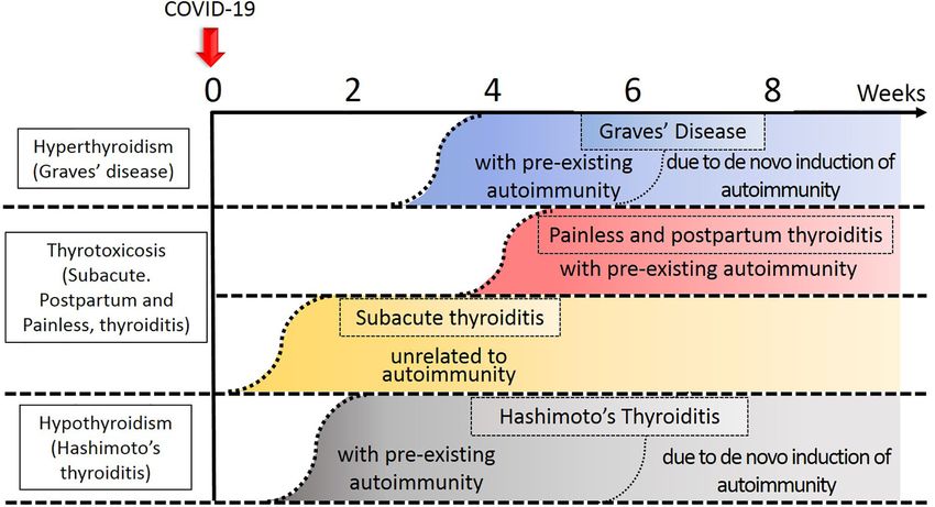
Inaba and Aizawa                                                                                                            COVID and the Thyroid

confirmation of what we have learned in the 2002 outbreak of                C-reactive protein (CRP) and erythrocytes sedimentation rate
severe acute respiratory syndrome (SARS) coronavirus (SARS-                (ESR), together with focal hypoechogenic areas in the thyroid
CoV) which is a platform for understanding the thyroidal                   gland are characteristic laboratory findings of SAT (39). Damage
problems associated with COVID-19.                                         to the thyroid per se and suppression of TSH by thyrotoxicosis
    In this communication, the terms “to provoke and provocation”          together cause extremely low thyroidal uptake of radioiodine (39,
were used to let the autoimmune abnormalities occur in the subjects        40). Nonetheless, thyrotoxicosis in SAT commonly subsides
without history of autoimmune thyroid disorders (AITD). On the             within 3 months (40). Viral infection such as human foamy
other hand, “to activate and activation” were used to mean to arouse       virus (HFV), mumps, coxsackie, adenovirus, Epstein-Barr
clinically silent, otherwise dormant, autoimmune abnormalities in          viruses (EBV), measles, chickenpox, cytomegalovirus (CMV),
those with history of AITD.                                                influenza, and rubella have all been implicated as a cause of SAT
                                                                           (40, 41). An association of HLA and SAT was also reported (40),
                                                                           and will be discussed in section Etiologic Background for
ALTERATION IN THYROID                                                      Thyroidal Insults in COVID-19.
                                                                               To date, a total of 13 cases of SAT with COVID-19 have been
FUNCTIONALITY DURING                                                       reported (Supplementary Tables 2 and 3) (7–16). Most reports
SARS-COV INFECTION                                                         (7/13, 54%) were from Italy (Supplementary Table 3). Patients
                                                                           were distributed across all generations (18-69 years old) with
Coronaviruses (CoVs), a family of single-stranded RNA
                                                                           overwhelming female predominance (11/13, 85%). The onset of
viruses, typically cause signs and symptoms resembling common
                                                                           SAT ranged from ‘7 weeks before’ to ‘7 weeks after’ the diagnosis
cold. The CoVs are subdivided into four genera2 such as
                                                                           of COVID-19. Fever was a common symptom in the patients
Alphacoronavirus, Betacoronavirus (bCoV), Gammacoronavirus,
                                                                           with COVID-19 irrespective of the presence or absence of SAT
and Deltacoronavirus (33).
                                                                           so that it cannot be taken as a sign of SAT in this situation.
    Since both SARS-CoV and SARS-CoV2 belong to the same b-
                                                                           Accordingly, thyroidal pain and tenderness are diagnostic clues
Coronavirus group, sharing the key clinical manifestations in
                                                                           for SAT with the combination of thyrotoxic signs and symptoms
common (33).
                                                                           such as palpitations, finger tremor, hyperhidrosis, and soft stool.
    Alteration of the thyroid functionality has been documented
                                                                           The patients usually show increased serum levels of FT3 and FT4
in patients with SARS during the 2002 outbreak. We have
                                                                           concurrent with a decreased level of TSH. Izumi et al. previously
summarized the similarities and dissimilarities between SARS
                                                                           reported that the mean (SD) value for the serum FT3 to FT4 ratio
and COVID-19 in Supplementary Table 1. Transient subclinical
                                                                           was 0.399 (0.089) in patients with GD, 0.335 (0.057) in those with
thyrotoxicosis, central hypothyroidism, and primary
                                                                           SAT, and 0.304 (0.072) in painless thyroiditis (PT), respectively,
hypothyroidism were previously reported in patients with
                                                                           so that the lower serum FT3/FT4 in the presence of
SARS (34). Wei et al. reported that thyroid-stimulating
                                                                           thyrotoxicosis can be a marker for thyroiditis, not Graves’
hormone (TSH) and adrenocorticotropin (ACTH) staining of
                                                                           hyperthyroidism (42). The mean value for FT3/FT4 in patients
thyrotrophs and corticotrophs, respectively, was significantly
                                                                           with SAT associated with COVID-19 ranged from 0.22-0.32
attenuated in the pituitary gland of patients with SARS upon
                                                                           (Cases 1, 2, 4, 5, 6, 7, 8, 10), suggesting that “the Izumi
autopsy (35). Apoptosis of the thyroid follicular cells was seen in
                                                                           hypothesis” was applicable in these patients (Supplementary
SARS (36), but the information on the thyroid function of the
                                                                           Table 3). The serum levels of thyroglobulin (Tg) were increased
patients was not provided (36). SARS-CoV virus has not been
                                                                           in 4 patients (Cases 1, 2, 6, 8) and CRP levels were increased in
found in the thyroid gland (37) and, therefore, primary
                                                                           all of the patients tested (Supplementary Table 3). A
hypothyroidism in this patient may or may not have been a
                                                                           hypoechogenic area in the thyroid, either focal or diffuse, was
consequence of the direct viral attack on the thyroid follicular
                                                                           found in the majority of them and echogenic evidence for
cells. In addition, the non-thyroidal-illness syndrome (NTIS) due
                                                                           increased vascularization was absent in all of the patients
to severe acute respiratory distress in SARS-CoV infection has
                                                                           tested (except for case 12) (Supplementary Table 3). Thyroid
been implicated (38) (Supplementary Table 1).
                                                                           scintigrams showed decreased uptake in all of the cases tested
                                                                           (Cases 2, 6, 8, 12) (Supplementary Table 3). Thyroid
                                                                           autoantibodies (TSH receptor autoantibody (TRAb), TgAb,
THYROIDAL INSULTS OBSERVED IN                                              and TPOAb) were seldom positive.
COVID-19 AND THEIR MANAGEMENT                                                  Muller et al. reported a high frequency of atypical SAT in their
                                                                           patients with COVID-19 (29) (Supplementary Tables 2 and 3).
We divide the consequences of COVID-19 into major and minor                They reported that 13 out of consecutive 85 (15%) COVID-19
influences on the thyroid gland and its function (Figure 1).                patients admitted to their high-intensive care units (HICU20) had
                                                                           overt thyrotoxicosis: the corresponding value was 1% in the HICU
Major Sequelae                                                             patients without COVID-19 (HICU19) and 2% among patients
Destructive Thyroiditis                                                    with COVID-19 in the low-intensive care units (LICU20). As an
Subacute Thyroiditis (SAT)                                                 entire group, the mean (SD) FT4 level in the patients in HICU20
This is an inflammatory disorder, usually with a painful goiter,            and LICU20 was, 18.7 (5.4) pmol/l and 13.5 (4.6) pmol/l,
fever, palpitation, and fatigue (39). Elevated serum levels of             respectively: it was significantly higher in the former (P = 0.016).

Frontiers in Endocrinology | www.frontiersin.org                       2                                    June 2021 | Volume 12 | Article 708333
Inaba and Aizawa COVID and the Thyroid
FIGURE 1 | Temporal profile of development of thyroid diseases in relation to COVID-19. Approximate timing of onset of each disease was indicated by the
curvilinear broken lines. Thyroid disease process related to autoimmunity tend to occur earlier in the subjects with preexisting autoimmunity to the thyroid gland. The
darker the color, the degree of thyroidal insults stronger.
The authors named these patients ‘atypical’ because thyroidal by Bartalena et al. in cases of destructive thyroiditis (43).
pain and swelling were absent. Despite such ‘atypical’ physical Moreover, IL-6 has been reported to be involved with various
findings, focal hypoechogenic areas, decreased 99mTc uptake, and autoimmune and inflammatory diseases (44). Han et al. revealed
negativity of thyroid autoantibodies (except for one patient) were that serum IL-6 levels can predict disease severity in patients with
all present as in the typical subacute thyroiditis, which made COVID-19 (45).
them propose the new concept, ‘atypical SAT’ (29). Lania et al.
reported that 58 out of consecutive 287 (20%) patients with Management of SAT and Atypical SAT in COVID 19
COVID-19 hospitalized in non-ICU beds developed SAT is treated with 16-40 mg/day prednisolone followed by
thyrotoxicosis possibly provoked by systemic inflammation or tapering within 4-6 weeks (7–16). Atypical SAT is a self-limiting
immune activation induced by COVID-19 (24) (Supplementary disorder, and therefore can be observed without specific
Table 4). Out of the 58 thyrotoxic patients, thyrotoxicosis was pharmacological treatment (29).
clinically overt in 31 (53%). On the other hand, 15 out of the 287
(5%) had hypothyroidism. Importantly, in the entire group, there Autoimmune Thyroid Disease (AITD)
was a strong, inverse correlation between the serum level of IL-6 Painless Thyroiditis (PT) and Painless Postpartum
and TSH (Spearman rho = -0.41, P < 0.001), indirectly suggesting Thyroiditis (PPT)
at least a partial contribution of inflammatory destruction of the These disorders may belong to destructive thyroiditis, and also
thyroid to the elevation of the serum thyroid hormone levels. may be subtypes of autoimmune thyroid disease (AITD) (40, 46,
Moreover, the patients with thyrotoxicosis (TSH < 0.34 mIU/l) 47) although painless SAT may also occur if the inflammatory
had significantly higher levels of the mean serum IL-6 than those response is mild. In general, a majority of the patients with PT and
without thyrotoxicosis (P < 0.05) (most of them > 10 pg/ml with PPT initially experience a mild thyrotoxic phase with elevated
the reference range < 6.4 pg/ml), suggesting inflammation due to serum thyroid hormone levels and depressed TSH, which was
COVID-19 infection, as indexed by elevated IL-6, was a driving followed by depressed thyroid function and then recovery to
force for the thyroiditis. They concluded that COVID-19 may normal within several months, i.e., spontaneous resolution of
have provoked thyrotoxicosis in the 31 cases described above. the thyroid dysfunction. We hypothesize that autoimmune-
In summary, these reports (24, 29) indicated that associated thyroiditis with COVID-19 may preferentially be
thyrotoxicosis may occur in 10-20% of the patients with observed in the subjects who possess susceptibility to AITD,
COVID-19. Increased levels of serum IL-6 were also reported because the patients who experienced PT and PPT in
Frontiers in Endocrinology | www.frontiersin.org 3 June 2021 | Volume 12 | Article 708333Inaba and Aizawa COVID and the Thyroid
association with COVID-19 often developed thyroid autoantibody GD (26) (Supplementary Table 2). A 21-year-old woman
positivity 1-1.5 months later (28). Such patients may share presented with tachycardia, palpitation, anxiety, and finger
increased HLA genotypes with patients with AITD (See section tremor 17 days after the diagnosis of COVID-19. Her mother
Etiologic Background for Thyroidal Insults in COVID-19). had hypothyroidism. A diffuse, non-tender, moderate-sized
PPT patients who had both TPOAb and TgAb often have an goiter was present. Elevated thyroid hormone and suppressed
increased percentage of activated T cells, such as HLA-DR+ and TSH levels, and the positivity of TRAb indicated the diagnosis of
CD3+ cells, in the peripheral circulation (48). Thus, alteration in GD. Graves’ ophthalmopathy or dermopathy was absent. The
the T-cell population may be predisposed to or associated with three patients with GD responded to thiamazole and b-
the development of PPT. Along with this evidence, Elefsiniotis blocker uneventfully.
et al. encountered the development of PPT in 4 out of 16 chronic A 45-year-old man with COVID-19 who presented with
hepatitis C virus (HCV)-infected women, proposing “viral- fatigue and muscle weakness was diagnosed as HT, based on
triggered PPT” as a subtype of the thyroiditis (49). Altered T the hypothyroidism with the positivity of TPOAb and
cell populations in patients with HCV infection have been successfully treated with 25 mg/day L-thyroxine (27)
considered as a reflection of Th1/Th2 imbalance (50). (Supplementary Table 2). As can be seen, in the patients with
AITD (25–27), depending upon the different background thyroid
Graves’ Disease (GD) and Hashimoto’s autoimmunity, a variety of organ-specific autoimmune
Thyroiditis (HT) abnormalities may be provoked or activated upon SARS-CoV-
The AITD’s are a constellation of thyroid-specific autoimmune 2 infection.
diseases, and Graves’ disease (GD) and Hashimoto’s thyroiditis Liu et al. reported that 25 out of a consecutive 191 (13%)
(HT) are the two major disorders included in this entity (51, 52). patients with COVID-19 showed abnormal results in thyroid
GD is characterized by TSH receptor antibodies (TRAb) which function tests (17) (Supplementary Table 4). Ten patients had
stimulate the thyroid follicular cells leading to hyperthyroidism isolated low TSH, suggestive of subclinical thyrotoxicosis: one of
(51). HT is characterized by the positivity of the serum for them was positive for TPOAb and TRAb, and two of them were
thyroglobulin autoantibody (TgAb) and thyroperoxidase positive for TRAb. Therefore, the three might have had
autoantibody (TPOAb) (52). Hypothyroidism in HT is due to subclinical GD. Apart from the ten patients, there was a
T-cell mediated damage of thyrocytes and interstitial fibrosis. patient with isolated high FT4 and another with high FT4 and
The serum thyroid autoantibodies such as TgAb and TPOAb are low FT3, who were also positive for TRAb leaving the possibility
often also positive in patients with GD (53). Clinically, of mild GD. Patients with abnormal thyroid function in this
transitions of patient from GD to HT and vice versa are not study contained additional three patients: the first one with
uncommon (52, 54), and a positive family history for GD often isolated high FT4 with TgAb positivity, the second one with
overlaps with that for HT (55). Provocation or activation of isolated TSH elevation with positive TPOAb and TgAb, and the
AITD by COVID-19 toward the seemingly opposite direction, to third one with low TSH and FT3. The remaining ten patients
GD or HT, can be understood from these perspectives with isolated low FT3 were compatible with non-thyroidal illness
(Supplementary Figure 1). syndrome, and one patient was positive for TRAb and TPOAb
The association of viral and non-viral infection and AITD has (see section below on ‘Non-Thyroidal Illness Syndrome (NTIS)’
often been suggested (56–58). For example, serological evidence below). Overall, 14/191 (7%) had features of thyrotoxicosis,
of infection with human herpesvirus-6 (HHV-6) (56), and diagnosed by low TSH and/or raised FT4. The authors re-
Toxoplasma gondii (57), HCV (58) was obtained from patients examined 10 of the 25 patients a median of 28 days after the
with AITD at, or around, the time of diagnosis of AITD. initial thyroid function test and found normalization, permanent
However, it remains to be determined whether the infection hypothyroidism, and various stages of thyroiditis in evolution,
was causal for the development of the thyroid diseases or just with no uniform recovery.
innocent bystanders (41). The relationship between SARS and
AITD also has not been described with certainty. Management of AITD in COVID-19
Two cases of GD with COVID-19 were reported by Mateu- PT and PPT are self-limiting disorders, and therefore can be
Salat et al: one case was a relapse of hyperthyroidism in a 60- observed without specific pharmacological treatment (29).
year-old woman in whom GD had been in the state of drug-free Regarding the management of GD, treatment of thyrotoxicosis
remission for longer than 30 years (25). The other was the by thionamide drugs is usually safe, but should be performed
development of GD in a 53-year-old woman without a known with caution. This is because the signs and symptoms of COVID-
history of thyroid disease (25) (Supplementary Table 2). 19 are indistinguishable from those of antithyroid drug-induced
Cervical pain was absent and Graves’ ophthalmopathy was agranulocytosis. On the other hand, hypothyroidism due to HT
equivocal in both of these cases. Despite such ambiguity in can be treated by a regular L-T4 supplement.
signs and physical findings, the serum thyroid hormone levels
were indeed elevated and TSH suppressed, thyroid iodine
scintigram uptake increased and TRAb was positive, so that Minor Sequelae
they were diagnosed as GD. The timing of the diagnosis of GD Non-Thyroidal Illness Syndrome (NTIS)
was 1 and 2 months after the onset of COVID-19 in the former A systemic disease of any kind, if it is critical, causes the non-
and the latter, respectively. Harris et al. reported another case of thyroidal-illness syndrome (NTIS), characterized by low T3
Frontiers in Endocrinology | www.frontiersin.org 4 June 2021 | Volume 12 | Article 708333Inaba and Aizawa COVID and the Thyroid
levels as a result of changes in type 1 deiodinase activity (38, 59). patients with hypothyroidism receiving thyroid hormone
Patients in the ICU typically present with decreased levels of replacement among 3,703 patients with COVID-19 (251/3703,
serum T3, normal or low T4, and normal or slightly decreased 7%), and concluded that hypothyroidism was not associated with
levels of TSH (38). Zou et al. reported that 41 out of 149 (28%) an increased risk of hospitalization, mechanical ventilation, or
patients with COVID-19 fitted the diagnosis of NTIS (FT3 death in patients with COVID-19 (18) (Supplementary Table 4).
levels < 2.3 pg/ml) (19) (Supplementary Table 4). Similarly, Unfortunately, the impact of COVID-19 on the pre-existing
Gao et al. showed that FT3 levels were significantly lower in thyroid-related cellular and humoral immunity was not
patients with severe COVID-19 (66 out of 100: 66%) than in evaluated in this study. It is, however, possible on the basis of
mild COVID-19 patients (34 out of 100: 34%) (20) other reports (50, 62), that viral infection is likely to exaggerate the
(Supplementary Table 4). Th1/Th2 imbalance and reduce T regulatory cell (Treg) action
In a consecutive evaluation of deceased (N = 113) and (63) causing it to initiate a flare-up otherwise clinically dormant
recovered (N = 161) COVID-19 patients, the serum levels of AITD explaining some of the cases described but there remains no
TSH and FT3 on admission were significantly lower in the evidence that it exacerbates already established disease. In fact,
former (21) (Supplementary Table 4). Taken together, a low impairment of Treg has also been shown in AITD (64).
level of FT3 is commonly associated with, or predictive, of an
intractable form of COVID-19. Another group found that total
T3 levels were inversely correlated with the severity of COVID-
ETIOLOGIC BACKGROUND FOR
19 (22) (Supplementary Table 4). Nonetheless, the data should
be interpreted carefully because a significant proportion of the THYROIDAL INSULTS IN COVID-19
patients (31/50: 62%) were taking glucocorticoid at the time of
Major Sequelae
the hormone measurements (22).
Hyperinflammatory Syndrome
Lui et al. (17) evaluated 191 COVID-19 patients cross-
Increased proinflammatory/Th1 cytokine production has been
sectionally and found that 1) lower SARS-CoV-2 PCR cycle
associated with respiratory failure due to lung damage from
threshold levels and elevated CRP levels were associated with low
inflammation of the pulmonary tissue in SARS (65). Huang et al.
levels of TSH, 2) elevated CRP was associated with low FT3,
found that COVID-19, especially in its severe form, is associated
3) elevated levels of ESR associated with lower FT3 to FT4 ratio,
with a hyperinflammatory syndrome characterized by similar
and 4) lowering of FT3 was associated with increasing COVID-19
hypercytokinemia with multiorgan failure as seen in SARS (2)
severity. Somewhat differently, Khoo et al. reported that 289 out
(Figure 2). They have also reported that, in COVID-19,
of 334 (87%) patients with COVID-19 were euthyroid (23)
proinflammatory/Th1 cytokine production is increased and Th2
(Supplementary Table 4). They recognized that patients with
cytokines increased as well, which was a different picture from SARS
COVID-19 had lower levels of TSH and FT4 compared to those
(2). A pathogenetic role of cytokines in the development of
who did not have COVID-19. They also reported that the serum
thyroiditis and flare-up of the thyroid autoimmunity has been
TSH and FT4 concentration were both depressed upon
implicated (66). We hypothesize that dormant AITDs such as GD
admission for the treatment of COVID-19 compared to the
and HT become clinically overt by Th2-mediated autoantibody
pre-hospital baseline levels. They also reported that patients
production and Th1-mediated cellular immunity, respectively, in
who were admitted to the ICU had lower TSH levels than
COVID-19 and exaggerated further by Treg dysfunction
those treated at the non-emergency ward. There was a
(Supplementary Figure 1 and Figure 2).
significant inverse correlation between the serum cortisol and
As described above, Lania et al. reported a significant positive
TSH levels and between CRP and TSH levels, and a positive
correlation between serum IL-6 and the degree of thyrotoxicosis
correlation between CRP and FT4 levels. The findings suggested
in patients with COVID-19 (24). The finding suggested that
stress-induced suppression of TSH in COVID-19. At least partial
elevation of IL-6 and/or cytotoxic effects of T-cells during the
recovery of TSH levels toward the baseline was observed at the
hyperinflammatory syndrome might be causal for thyroidal
follow-up at several months later (23). Elevated serum levels of
destruction of the thyroiditis in COVID-19 (2, 24, 28, 29).
cortisol in patients with COVID-19 were reported (60), and
They further postulated that the thyroiditis may not subside
hypercortisolism has been reported to suppress TSH levels (23,
as long as the storm persists. Such patients may end up with
61). This change in TSH may be likely due to the changes in
permanent hypothyroidism or thyrotoxicosis years later (24). As
deiodinase activity in the central nervous system (38).
a conclusion to this part, the hyperinflammatory syndrome in
Management of NTIS in COVID-19 COVID-19 appears to provoke AITD such as GD, HT, PT, and
Since NTIS is due to the systemic dysfunction by COVID-19, the PPT in some patients and activate otherwise dormant diseases
treatment for COVID-19 is essential to obtain normal thyroid into a clinically recognizable state in others (25–28).
functional test results (TSH, FT3, and FT4). Apoptosis of the thyrocytes was demonstrated in autopsy
material from patients with SARS during the 2002 outbreak (36).
AITD Patients Response to COVID-19 In these specimens, SARS-CoV per se was not found in the
Up to this point, the data regarding the effect of COVID-19 on the thyrocytes and the follicular structure was mostly preserved,
thyroid gland has been reviewed. Gerwen et al. investigated the which suggested that direct viral invasion into the cell was not
problem the other way around. Namely, they identified 251 the cause of the apoptosis. Instead, autoimmune provocation or
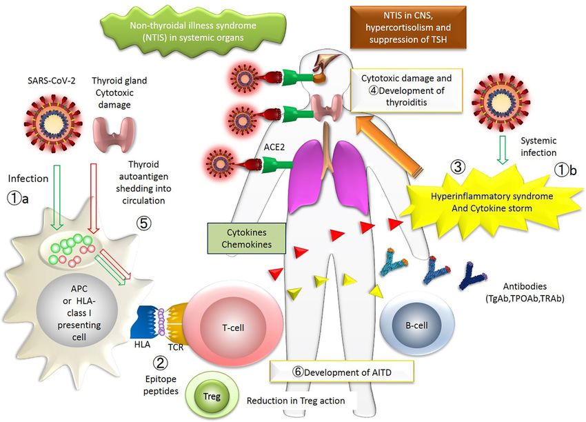
Frontiers in Endocrinology | www.frontiersin.org 5 June 2021 | Volume 12 | Article 708333Inaba and Aizawa COVID and the Thyroid FIGURE 2 | The figure represents the whole picture of the thyroid insults during COVID-19 in the immunological network. The events occurring in patients with thyroid diseases associated with COVID-19 are collectively shown. The numbers approximately correspond to the sequence of events. ①. SARS-CoV-2 infects systemic organs through acquired immunity (①a) and innate immunity (①b). ②. SARS-CoV-2 epitope peptide is presented on the surface of HLA, and T-cell recognizes the epitope peptide. ③. Hyperinflammatory syndrome and cytokine storm occur. ④. Thyroid gland is damaged by immune cells. ⑤. Thyroid autoantigen is shedding into circulation, which is also presented on the surface of HLA. ⑥. Finally, AITD develops as a consequence of new provocation of the disease or activation of previously existing dormant disease. APC, antigen-presenting cells; TCR, T-cell receptor; T-cells include cytotoxic T-cells and effector T-cells; AITD, autoimmune thyroid disease. A putative mechanism for the thyroid insults during COVID-19: an immune-centric view. The numbers approximately correspond to the sequence of events. ①, SARS-CoV-2 infects systemic organs through acquired immunity (①a) and innate immunity (①b). ②, SARS-CoV-2 epitope peptide is presented on the surface of the antigen-presenting (or HLA class I) cells, and T-cell recognizes the epitope peptide. ③, Hyperinflammatory syndrome and cytokine storm occur. ④, Thyroid gland could be a target of the antibodies or T-cells. ⑤,Thyroid autoantigen is shedding into the general circulation, which is also presented on the surface of the HLA-cells. ⑥, Finally, AITD develops as a consequence of the new provocation of the disease or activation of the pre-existing, yet dormant disease. APC, antigen-presenting cells; TCR, T-cell receptor; T-cells include cytotoxic T-cells and effector T-cells; Treg, regulatory T-cell; AITD, autoimmune thyroid disease. activation, triggered by the cytokine storm during SARS-CoV They concluded that the thyroid dysfunction is likely due to infection was likely to be causal for the thyroid damage. The the cytokine storm induced by SARS-Cov-2 with a direct or pathological findings in the three cases of COVID-19 were mediated impact on TSH secretion and deiodinase activity, and essentially the same as those in patients with SARS-CoV, i.e., not likely a destructive thyroiditis (68). apoptosis of follicular cells in the absence of the virus itself in the Of note is that lymphopenia was observed in a case of SARS- thyrocytes (67), suggesting the operation of a similar, cytokine- CoV-2 before the development of PPT, which occurred 4 months mediated thyroid insult in SARS and COVID-19. Recently, after the delivery (28). By the time of the PPT, her lymphocyte Campi et al. found that the majority of patients with COVID- count had returned to normal. Such an alteration in the number 19 had normal thyroid function but that low TSH levels were of lymphocytes in the general circulation was compatible with seen either at admission or during hospitalization in 39% of the idea of the causal link between the SARS-CoV-2 infection patients, associated with low FT3 in half of the cases. and the development of PPT (28). Frontiers in Endocrinology | www.frontiersin.org 6 June 2021 | Volume 12 | Article 708333
Inaba and Aizawa                                                                                                               COVID and the Thyroid

Provocation of Thyroidal Autoimmunity and the                                FUTURE PERSPECTIVES AND
Human Leukocyte Antigen System                                               CONCLUSIONS
Human Leukocyte Antigen (HLA) plays an important role in the
immune system at the interface of antigen presentation by the T-             The case histories reported in the literature indicate that thyroidal
cells and its recognition and antibody production by the B-cells             disorders associated with COVID-19 such as SAT, PT, PPT are usually
(51) (Figure 2). The HLA class I molecules are abundantly                    self-limiting, and GD and HT can be easily treated with thiamazole and
expressed in various cells and induce CD8+T-cell response. On                L-T4, respectively. The beneficial effect of thyroid hormone
the other hand, the expression of HLA class II molecules is                  supplements for patients who exhibit low FT4 and FT3 levels with
limited to antigen-presenting cells and responsible for CD4+ T-              NTIS and/or central hypothyroidism remains to be established.
cell response. Because of polymorphism of HLA genes, divergent                   Management would be more difficult if drug-induced
immune responses toward the same antigen and varied disease                  agranulocytosis were to take place (but fortunately not so far
susceptibility are to be generated.                                          reported), or thyroid storm (84), or myxedema coma were to
    Increased prevalence of certain HLA genotypes in COVID-19                occur (85). As shown here, our understanding of the thyroidal
has been reported in certain populations including Chinese and               manifestation of COVID-19 is far from complete as is the
Italian patients (69–71). The HLA gene and its polymorphisms have            etiologic view of COVID-19 and thyroid insults. Although case
been implicated for many years (over 25 years) in AITD and SAT               reports are definitely important in helping us understand the
(72–74) and now in COVID-19 (75) (Figure 2). Higher incidence of             association, future research, hopefully in a prospective manner
SAT in those with the HLA-B35 or HLA-B67 antigens were                       with longitudinal analyses, is required. This would involve the
reported (72). In 1974, increased frequency of the HL-A8 antigen             histologic and cytological examination of the thyroid gland in a
in the GD patient population was reported (73). To date, Tomer and           large number of patients in order to identify direct evidence
Davies comprehensively reviewed HLA class I and class II                     regarding the nature and cause of thyroid damage with the
association with AITD in wide varieties of ethnics (74).                     COVID-19 virus and the detailed immune response in those
Illustrating the possible association, a Japanese patient with PPT           patients with thyroid dysfunction. Of particular interest is the
and COVID-19 (28) possessed the same HLA alleles predisposing to             need to clarify if thyroid autoimmunity in COVID-19 is an
AITD (HLA-DP5 and HLA-B51) in the Japanese (51, 76).                         innocent-bystander or another culprit in its severity (86).
                                                                                 The strength of this review is its high originality and systematic
Minor Sequelae                                                               description. For the first time, we systematically explained the
Nonspecific Reaction to Severe Illness                                        detailed pathophysiology of the thyroid abnormalities associated
Systemic critical diseases cause NTIS, in which the primary issue is a       with SARS-CoV2 infection, firmly based on the accumulated
derangement in thyroid hormone metabolism. Alteration of the                 evidence in the literature. The limitation of this paper is that
serum thyroid hormone concentration and its metabolites seen in              several review articles dealing with thyroid disorders related to
COVIC-19 may well be added to the long list of causes for NTIS.              COVID-19 have been already reported (30–32).

Angiotensin-Converting Enzyme 2 (ACE2) on Thyroid
and Pituitary Cells                                                          AUTHOR CONTRIBUTIONS
Angiotensin-converting enzyme 2 (ACE2) is the receptor utilized
by SARS-CoV2 creeping into the cells, then initiating replication            HI and TA contributed to conception, design, searching
(77). In many organs including the lung, the gastrointestinal                publications, writing the manuscript. All authors contributed
tract, the liver, the kidney, the skin, the heart, the hematopoietic         to the article and approved the submitted version.
cells, and the spleen, a direct cellular change by the virus was
documented during COVID-19 (78). ACE2 expression has been
known to impair thyroid function and also the function of the                FUNDING
anterior pituitary gland during COVID-19 (77) So far, the
                                                                             This work was partially supported by Takeda Science Foundation.
existence of SARS-CoV-2 in the thyroid gland and pituitary
gland is not clear by examinations of light microscopy,
immunohistochemistry, electron microscopy, and quantitative
                                                                             ACKNOWLEDGMENTS
RT-PCR (79). Furthermore, viral particles of SARS-CoV-2 were
found in the frontal lobe of the brain and brain capillary                   We are greatly indebted to Dr. Terry Davies for manuscript
endothelial cells (80). The SARS-CoV-2 infection has been                    review, technical advice and special insight into thyroid disease.
known to impair olfaction and taste sense by affecting the
central nervous system (81–83). Although clear evidence is
lacking, infection of the thyrocyte, thyrotroph and corticotroph             SUPPLEMENTARY MATERIAL
may result in lowered T3, T4, TSH, ACTH and cortisol. The
dysregulation of the hypothalamic-pituitary-thyroid axis has                 The Supplementary Material for this article can be found online
been considered at least in part responsible for hypothyroidism              at: https://www.frontiersin.org/articles/10.3389/fendo.2021.
in COVID-19 (17, 24, 27) (Figure 2).                                         708333/full#supplementary-material

Frontiers in Endocrinology | www.frontiersin.org                         7                                     June 2021 | Volume 12 | Article 708333
Inaba and Aizawa COVID and the Thyroid
Supplementary Figure 1 | An overview on the postulated mechanism for the and Th2 systems. Tier 4: Patients may present with the symptoms such as fatigue
thyroid insults with COVID-19. Tier 1: The viral infection causes derangement in and lethargy, and a goiter whichmay be painful and is accompaniedwith fever if the
immune system, of which dysregulation of the T-cell system (including regulatory destruction is prominent. Laboratory examination is needed for the correct
T-cell) and resultant cytokine overproduction and storm affect the thyroid gland. Tier diagnosis. There is a general temporal tendency as to the timing between the viral
2: Cellular and humoral immunity in an around the thyroid is provoked or activated: infection and the clinical onset of the thyroid abnormality (See Figure 1). Th1,
the subjects having autoimmune thyroid disorders or those with a particular set of human type 1 helper cell; Th2, human type 2 helper cell; TRAb, anti-TSH receptor
human-leukocyte antigen genotype may be vulnerable. Tier 3: Imbalanced antibody; TgAb, anti-thyroglobulin antibody; TPOAb, antithyroperoxidase antibody;
provocation and/or activation of Th1, Th2 and other subtypes of T/B cells occur, GD, Graves’ disease; SAT, subacute thyroiditis; PT, painless thyroiditis; PPT, post-
leading to the thyroidal stimulation, destruction and suppression, or a combination partum thyroiditis; HT, Hashimoto’s thyroiditis. In this and the following Figures, only
thereof. The thick arrow in the right end implies a mutual stimulation between Th1 Major Sequels of COVID-19 are depicted (see Text).
191 Patients With COVID-19. J Clin Endocrinol Metab (2021) 106(2):e926–
REFERENCES 35. doi: 10.1210/clinem/dgaa813
1. Yang X, Yu Y, Xu J, Shu H, Xia J, Liu H, et al. Clinical Course and Outcomes 18. van Gerwen M, Alsen M, Little C, Barlow J, Naymagon L, Tremblay D, et al.
of Critically Ill Patients With SARS-CoV-2 Pneumonia in Wuhan, China: A Outcomes of Patients With Hypothyroidism and COVID-19: A Retrospective
Single-Centered, Retrospective, Observational Study. Lancet Respir Med Cohort Study. Front Endocrinol (Lausanne) (2020) 11:565. doi: 10.3389/
(2020) 8:475–81. doi: 10.1016/S2213-2600(20)30079-5 fendo.2020.00565
2. Huang C, Wang Y, Li X, Ren L, Zhao J, Hu Y, et al. Clinical Features of 19. Zou R, Wu C, Zhang S, Wang G, Zhang Q, Yu B, et al. Euthyroid Sick
Patients Infected With 2019 Novel Coronavirus in Wuhan, China. Lancet Syndrome in Patients With Covid-19. Front Endocrinol (Lausanne) (2020)
(2020) 395(10223):497–506. doi: 10.1016/S0140-6736(20)30183-5 11:566439. doi: 10.3389/fendo.2020.566439
3. Zhang Y, Xiao M, Zhang S, Xia P, Cao W, Jiang W, et al. Coagulopathy and 20. Gao W, Guo W, Guo Y, Shi M, Dong G, Wang G, et al. Thyroid Hormone
Antiphospholipid Antibodies in Patients With COVID-19. N Engl J Med Concentrations in Severely or Critically Ill Patients With COVID-19.
(2020) 382:e38. doi: 10.1056/NEJMc2007575 J Endocrinol Invest (2021) 44(5):1031–40. doi: 10.1007/s40618-020-01460-w
4. Toscano G, Palmerini F, Ravaglia S, Ruiz L, Invernizzi P, Cuzzoni MG, et al. 21. Chen T, Wu D, Chen H, Yan W, Yang D, Chen G, et al. Clinical
Guillain-Barré Syndrome Associated With SARS-Cov-2. N Engl J Med (2020) Characteristics of 113 Deceased Patients With Coronavirus Disease 2019:
382:2574–6. doi: 10.1056/NEJMc2009191 Retrospective Study. BMJ (2020) 368:m1091. doi: 10.1136/bmj.m1091
5. Lazarian G, Quinquenel A, Bellal M, Siavellis J, Jacquy C, Re D, et al. 22. Chen M, Zhou W, Xu W. Thyroid Function Analysis in 50 Patients With
Autoimmune Haemolytic Anaemia Associated With COVID-19 Infection. COVID-19: A Retrospective Study. Thyroid (2021) 31(1):8–11. doi: 10.1089/
Br J Haematol (2020) 190:29–31. doi: 10.1111/bjh.16794 thy.2020.0363
6. Zulfiqar AA, Lorenzo-Villalba N, Hassler P, Andrès E. Immune 23. Khoo B, Tan T, Clarke SA, Mills EG, Patel B, Modi M, et al. Thyroid Function
Thrombocytopenic Purpura in a Patient With Covid-19. N Engl J Med Before, During and After COVID-19. J Clin Endocrinol Metab (2021) 106(2):
(2020) 382(18):e43. doi: 10.1056/NEJMc2010472 e803–11. doi: 10.1210/clinem/dgaa830
7. Brancatella A, Ricci D, Cappellani D, Viola N, Sgrò D, Santini F, et al. Is 24. Lania A, Sandri MT, Cellini M, Mirani M, Lavezzi E, Gherardo Mazziotti G.
Subacute Thyroiditis an Underestimated Manifestation of SARS-CoV-2 Thyrotoxicosis in Patients With COVID-19: The THYRCOV Study. Eur
Infection? Insights From a Case Series. J Clin Endocrinol Metab (2020) 105: J Endocrinol (2020) 183:381–7. doi: 10.1530/EJE-20-0335
dgaa537. doi: 10.1210/clinem/dgaa537 25. Mateu-Salat M, Urgell E, Chico A. Sars-COV-2 as a Trigger for Autoimmune
8. Asfuroglu Karkan E, Ates I. A Case of Subacute Thyroiditis Associated With Disease: Report of Two Cases of Graves’ Disease After COVID-19.
Covid-19 Infection. J Endocrinol Invest (2020) 43(8):1173–4. doi: 10.1007/ J Endocrinol Invest (2020) 43:1527–8. doi: 10.1007/s40618-020-01366-7
s40618-020-01316-3 26. Harris A, Al Mushref M. Graves’ Thyrotoxicosis Following SARS-CoV-2
9. Ippolito S, Dentali F, Tanda ML. Sars-CoV-2: A Potential Trigger for Infection. AACE Clin Case Rep (2021) 7(1):14–16. doi: 10.1016/j.aace.2020.12.005
Subacute Thyroiditis? Insights Case Rep J Endocrinol Invest (2020) 43 27. Tee LY, Hajanto S, Rosario BH. Covid-19 Complicated by Hashimoto’s
(8):1171–2. doi: 10.1007/s40618-020-01312-7 Thyroiditis. Singapore Med J (2021) 62(5):265. doi: 10.11622/smedj.2020106
10. Brancatella A, Ricci D, Viola N, Sgrò D, Santini F, Latrofa F. Subacute 28. Mizuno S, Inaba H, Kobayashi KI, Kubo K, Ito S, Hirobata T, et al. A Case of
Thyroiditis After Sars-COV-2 Infection. J Clin Endocrinol Metab (2020) 105 Postpartum Thyroiditis Following SARS-CoV-2 Infection. Endocr J (2021) 68
(7):dgaa276. doi: 10.1210/clinem/dgaa276 (3):371–4. doi: 10.1507/endocrj.EJ20-0553
11. Ruggeri RM, Campennì A, Siracusa M, Frazzetto G, Gullo D. Subacute 29. Muller I, Cannavaro D, Dazzi D, Covelli D, Mantovani G, Muscatello A, et al.
Thyroiditis in a Patient Infected With SARS-COV-2: An Endocrine Sars-CoV-2-Related Atypical Thyroiditis. Lancet Diabetes Endocrinol (2020)
Complication Linked to the COVID-19 Pandemic. Hormones (Athens) 8:739–41. doi: 10.1016/S2213-8587(20)30266-7
(2021) 20(1):219–21. doi: 10.1007/s42000-020-00230-w 30. Lundholm MD, Poku C, Emanuele N, Emanuele MA, Lopez N. Sars-CoV-2
12. Campos-Barrera E, Alvarez-Cisneros T, Davalos-Fuentes M. Subacute (Covid-19) and the Endocrine System. J Endocr Soc (2020) 4(11):bvaa144. doi:
Thyroiditis Associated With COVID-19. Case Rep Endocrinol (2020) Sep 10.1210/jendso/bvaa144
28:2020:8891539. doi: 10.1155/2020/8891539 31. Chen W, Tian Y, Li Z, Zhu J, Wei T, Lei J. Potential Interaction Between
13. Mattar SAM, Koh SJQ, Rama Chandran S, Cherng BPZ. Subacute Thyroiditis SARS-CoV-2 and Thyroid: A Review. Endocrinology (2021) 162(2):bqab004.
Associated With COVID-19. BMJ Case Rep (2020) 13(8):e237336. doi: doi: 10.1210/endocr/bqab004
10.1136/bcr-2020-237336 32. Hariyanto TI, Kurniawan A. Thyroid Disease is Associated With Severe
14. San Juan MDJ, Florencio MQV, Joven MH. Subacute Thyroiditis in A Patient Coronavirus Disease 2019 (COVID-19) Infection. Diabetes Metab Syndr
With Coronavirus Disease 2019. AACE Clin Case Rep (2020) 6(6):e361–4. doi: (2020) 14(5):1429–30. doi: 10.1016/j.dsx.2020.07.044
10.4158/ACCR-2020-0524 33. ̇
Inandıklıoğ lu N, Akkoc T. Immune Responses to SARS-CoV, Mers-CoV and
15. Chakraborty U, Ghosh S, Chandra A, Ray AK. Subacute Thyroiditis as a SARS-Cov-2. Adv Exp Med Biol (2020) 1288:5–12. doi: 10.1007/
Presenting Manifestation of COVID-19: A Report of an Exceedingly Rare 5584_2020_549
Clinical Entity. BMJ Case Rep (2020) 13(12):e239953. doi: 10.1136/bcr-2020- 34. Leow MK, Kwek DS, Ng AW, Ong KC, Kaw GJ, Lee LS. Hypocortisolism in
239953 Survivors of Severe Acute Respiratory Syndrome (SARS). Clin Endocrinol
16. Mehmood MA, Bapna M, Arshad M. A Case of Post-COVID-19 Subacute (Oxf.) (2005) 63:197–202. doi: 10.1111/j.1365-2265.2005.02325.x
Thyroiditis. Cureus (2020) 12(12):e12301. doi: 10.7759/cureus.12301 35. Wei L, Sun S, Zhang J, Zhu H, Xu Y, Ma Q, et al. Endocrine Cells of the
17. Lui DTW, Lee CH, Chow WS, Lee ACH, Tam AR, Fong CHY, et al. Thyroid Adenohypophysis in Severe Acute Respiratory Syndrome (SARS). Biochem
Dysfunction in Relation to Immune Profile, Disease Status and Outcome in Cell Biol (2010) 88(4):723–30. doi: 10.1139/O10-022
Frontiers in Endocrinology | www.frontiersin.org 8 June 2021 | Volume 12 | Article 708333Inaba and Aizawa COVID and the Thyroid
36. Wei L, Sun S, Xu CH, Zhang J, Xu Y, Zhu H, et al. Pathology of the Thyroid in 57. Tozzoli R, Barzilai O, Ram M, Villalta D, Bizzaro N, Sherer Y, et al. Infections
Severe Acute Respiratory Syndrome. Hum Pathol (2007) 38:95–102. doi: and Autoimmune Thyroid Diseases: Parallel Detection of Antibodies Against
10.1016/j.humpath.2006.06.011 Pathogens With Proteomic Technology. Autoimmun Rev (2008) 8(2):112–5.
37. Ding Y, He L, Zhang Q, Huang Z, Che X, Hou J, et al. Organ Distribution of doi: 10.1016/j.autrev.2008.07.013
Severe Acute Respiratory Syndrome (SARS) Associated Coronavirus (SARS- 58. Testa A, Castaldi P, Fant V, Fiore GF, Grieco V, De Rosa A, et al. Prevalence of
CoV) in SARS Patients: Implications for Pathogenesis and Virus HCV Antibodies in Autoimmune Thyroid Disease. Eur Rev Med Pharmacol
Transmission Pathways. J Pathol (2004) 203(2):622–30. doi: 10.1002/ Sci (2006) 10(4):183–6.
path.1560 59. McIver B, Gorman CA. Euthyroid Sick Syndrome: An Overview. Thyroid
38. Fliers E, Bianco AC, Langouche L, Boelen A. Thyroid Function in Critically Ill (1997) 7(1):125–32. doi: 10.1089/thy.1997.7.125
Patients. Lancet Diabetes Endocrinol (2015) 3(10):816–25. doi: 10.1016/S2213- 60. Tan T, Khoo B, Mills EG, Phylactou M, Patel B, Eng PC, et al. Association
8587(15)00225-9 Between High Serum Total Cortisol Concentrations and Mortality From
39. Nishihara E, Ohye H, Amino N, Takata K, Arishima T, Kudo T, et al. Clinical COVID-19. Lancet Diabetes Endocrinol (2020) 8(8):659–60. doi: 10.1016/
Characteristics of 852 Patients With Subacute Thyroiditis Before Treatment. S2213-8587(20)30216-3
Intern Med (2008) 47(8):725–9. doi: 10.2169/internalmedicine.47.0740 61. Xiang B, Tao R, Liu X, Zhu X, He M, Ma Z, et al. A Study of Thyroid
40. Pearce EN, Farwell AP, Braverman LE. Thyroiditis. N Engl J Med (2003) Functions in Patients With Cushing’s Syndrome: A Single-Center Experience.
348:2646–55. doi: 10.1056/NEJMra021194 Endocr Connect (2019) 8(8):1176–85. doi: 10.1530/EC-19-0309
41. Desailloud R, Hober D. Viruses and Thyroiditis: An Update. Virol J (2009) 6:5. 62. De Carli M, D’Elios MM, Zancuoghi G, Romagnani S, Del Prete G. Human
doi: 10.1186/1743-422X-6-5 Th1 and Th2 Cells: Functional Properties, Regulation of Development and
42. Izumi Y, Hidaka Y, Tada H, Takano T, Kashiwai T, Tatsumi KI, et al. Simple Role in Autoimmunity. Autoimmunity (1994) 18(4):301–8. doi: 10.3109/
and Practical Parameters for Differentiation Between Destruction-Induced 08916939409009532
Thyrotoxicosis and Graves’ Thyrotoxicosis. Clin Endocrinol (Oxf) (2002) 57 63. Rahimzadeh M, Naderi N. Toward an Understanding of Regulatory T Cells in
(1):51–8. doi: 10.1046/j.1365-2265.2002.01558.x COVID-19: A Systematic Review. J Med Virol (2021) 93(7):4167–81.
43. Bartalena L, Brogioni S, Grasso L, Martino E. Interleukin-6 and the Thyroid. doi: 10.1002/jmv.26891
Eur J Endocrinol (1995) 132(4):386–93. doi: 10.1530/eje.0.1320386 64. Glick AB, Wodzinski A, Fu P, Levine AD, Wald DN. Impairment of
44. Tanaka T, Narazaki M, Kishimoto T. IL-6 in Inflammation, Immunity, and Regulatory T-Cell Function in Autoimmune Thyroid Disease. Thyroid
Disease. Cold Spring Harb Perspect Biol (2014) 6(10):a016295. doi: 10.1101/ (2013) 23(7):871–8. doi: 10.1089/thy.2012.0514
cshperspect.a016295 65. Wong CK, Lam CW, Wu AK, Ip WK, Lee NL, Chan IH, et al. Plasma
45. Han H, Ma Q, Li C, Liu R, Zhao L, Wang W, et al. Profiling Serum Cytokines in Inflammatory Cytokines and Chemokines in Severe Acute Respiratory
COVID-19 Patients Reveals IL-6 and IL-10 Are Disease Severity Predictors. Emerg Syndrome. Clin Exp Immunol (2004) 136:95–103. doi: 10.1111/j.1365-
Microbes Infect (2020) 9(1):1123–30. doi: 10.1080/22221751.2020.1770129 2249.2004.02415.x
46. Davies TF. The Thyroid Immunology of the Postpartum Period. Thyroid 66. Ganesh BB, Bhattacharya P, Gopisetty A, Prabhakar BS. Role of Cytokines in
(1999) 9(7):675–84. doi: 10.1089/thy.1999.9.675 the Pathogenesis and Suppression of Thyroid Autoimmunity. J Interferon
47. Inaba H, Akamizu T. Postpartum Thyroiditis. In: KR Feingold, B Anawalt, A Cytokine Res (2011) 31(10):721–31. doi: 10.1089/jir.2011.0049
Boyce, G Chrousos, WW de Herder, K Dungan, A Grossman, JM Hershman, J 67. Yao XH, Li TY, He ZC, Ping YF, Liu HW, Yu SC, et al. A Pathological Report of
Hofland, G Kaltsas, C Koch, P Kopp, M Korbonits, R McLachlan, JE Morley, Three COVID-19 Cases by Minimal Invasive Autopsies. Zhonghua Bing Li Xue Za
M New, J Purnell, F Singer, CA Stratakis, DL Trence, DP Wilson, editors. Zhi (2020) 49(5):411–7. doi: 10.3760/cma.j.cn112151-20200312-00193
Endotext, vol. 2018. South Dartmouth (MA: MDText.com, Inc (2000). 68. Campi I, Bulgarelli I, Dubini A, Perego GB, Tortorici E, Torlasco C, et al. The
48. Shi X, Li C, Li Y, Guan H, Fan C, Teng Y, et al. Circulating Lymphocyte Spectrum of Thyroid Function Tests During Hospitalization for SARS COV-2
Subsets and Regulatory T Cells in Patients With Postpartum Thyroiditis Infection. Eur J Endocrinol (2021) 184(5):699–709. doi: 10.1530/EJE-20-1391
During the First Postpartum Year. Clin Exp Med (2009) 9(4):263–7. doi: 69. Wang W, Zhang W, Zhang J, He J, Zhu F. Distribution of HLA Allele
10.1007/s10238-009-0046-0 Frequencies in 82 Chinese Individuals With Coronavirus Disease-2019
49. Elefsiniotis IS, Vezali E, Pantazis KD, Saroglou G. Post-Partum Thyroiditis in (Covid-19). HLA (2020) 96(2):194–6. doi: 10.1111/tan.13941
Women With Chronic Viral Hepatitis. J Clin Virol (2008) 41:318–9. doi: 70. Novelli A, Andreani M, Biancolella M, Liberatoscioli L, Passarelli C, Colona
10.1016/j.jcv.2007.12.010 VL, et al. HLA Allele Frequencies and Susceptibility to COVID-19 in a Group
50. Sobue S, Nomura T, Ishikawa T, Ito S, Saso K, Ohara H, et al. Th1/Th2 of 99 Italian Patients. HLA (2020) 96(5):610–4. doi: 10.1111/tan.14047
Cytokine Profiles and Their Relationship to Clinical Features in Patients With 71. Yung YL, Cheng CK, Chan HY, Xia JT, Lau KM, Wong RSM, et al.
Chronic Hepatitis C Virus Infection. J Gastroenterol (2001) 36(8):544–51. doi: Association of HLA-B22 Serotype With SARS-CoV-2 Susceptibility in
10.1007/s005350170057 Hong Kong Chinese Patients. HLA (2021) 97(2):127–32. doi: 10.1111/
51. Inaba H, De Groot LJ, Akamizu T. Thyrotropin Receptor Epitope and Human tan.14135
Leukocyte Antigen in Graves’ Disease. Front Endocrinol (Lausanne) (2016) 72. Ohsako N, Tamai H, Sudo T, Mukuta T, Tanaka H, Kuma K, et al. Clinical
7:120. doi: 10.3389/fendo.2016.00120 Characteristics of Subacute Thyroiditis Classified According to Human
52. Martin A, Davies TF. T Cells and Human Autoimmune Thyroid Disease: Leukocyte Antigen Typing. J Clin Endocrinol Metab (1995) 80(12):3653–6.
Emerging Data Show Lack of Need to Invoke Suppressor T Cell Problems. doi: 10.1210/jcem.80.12.8530615
Thyroid (1992) 2(3):247–61. doi: 10.1089/thy.1992.2.247 73. Grumet FC, Payne RO, Konishi J, Kriss JP. HL-a Antigens as Markers for
53. Fröhlich E, Wahl R. Thyroid Autoimmunity: Role of Anti-Thyroid Antibodies Disease Susceptibility and Autoimmunity in Graves’ Disease. J Clin
in Thyroid and Extra-Thyroidal Diseases. Front Immunol (2017) 9:8:521. doi: Endocrinol Metab (1974) 39(6):1115–9. doi: 10.1210/jcem-39-6-1115
10.3389/fimmu.2017.00521 74. Tomer Y, Davies TF. Searching for the Autoimmune Thyroid Disease
54. Gonzalez-Aguilera B, Betea D, Lutteri L, Cavalier E, Geenen V, Beckers A, Susceptibility Genes: From Gene Mapping to Gene Function. Endocr Rev
et al. Conversion to Graves’ Disease From Hashimoto Thyroiditis: A Study of (2003) 24:694–717. doi: 10.1210/er.2002-0030
24 Patients. Arch Endocrinol Metab (2018) 62(6):609–14. doi: 10.20945/2359- 75. Halpert G, Shoenfeld Y. Sars-CoV-2, the Autoimmune Virus. Autoimmun Rev
3997000000086 (2020) 19(12):102695. doi: 10.1016/j.autrev.2020.102695
55. Lee HJ, Li CW, Hammerstad SS, Stefan M, Tomer Y. Immunogenetics of 76. Ito M, Tanimoto M, Kamura H, Yoneda M, Morishima Y, Yamauchi K, et al.
Autoimmune Thyroid Diseases: A Comprehensive Review. J Autoimmun Association of HLA Antigen and Restriction Fragment Length Polymorphism of T
(2015) 64:82–90. doi: 10.1016/j.jaut.2015.07.009 Cell Receptor Beta-Chain Gene With Graves’ Disease and Hashimoto’s
56. Sultanova A, Cistjakovs M, Gravelsina S, Chapenko S, Roga S, Cunskis E, et al. Thyroiditis. J Clin Endocrinol Metab (1989) 69:100–4. doi: 10.1210/jcem-69-1-100
Association of Active Human Herpesvirus-6 (HHV-6) Infection With 77. Lazartigues E, Qadir MMF, Mauvais-Jarvis F. Endocrine Significance of
Autoimmune Thyroid Gland Diseases. Clin Microbiol Infect (2017) 23(1): SARS-Cov-2’s Reliance on ACE2. Endocrinology (2020) 161(9):bqaa108.
e1–50.e5:50. doi: 10.1016/j.cmi.2016.09.023 doi: 10.1210/endocr/bqaa108
Frontiers in Endocrinology | www.frontiersin.org 9 June 2021 | Volume 12 | Article 708333Inaba and Aizawa COVID and the Thyroid
78. Tabary M, Khanmohammadi S, Araghi F, Dadkhahfar S, Tavangar SM. 84. Pastor S, Molina Á Sr, De Celis E. Thyrotoxic Crisis and COVID-19 Infection:
Pathologic Features of COVID-19: A Concise Review. Pathol Res Pract An Extraordinary Case and Literature Review. Cureus (2020) 12(11):e11305.
(2020) 216(9):153097. doi: 10.1016/j.prp.2020.153097 doi: 10.7759/cureus.11305
79. Bradley BT, Maioli H, Johnston R, Chaudhry I, Fink SL, Xu H, et al. 85. Dixit NM, Truong KP, Rabadia SV, Li D, Srivastava PK, Mosaferi T, et al.
Histopathology and Ultrastructural Findings of Fatal COVID-19 Infections Sudden Cardiac Arrest in a Patient With Myxedema Coma and COVID-19.
in Washington State: A Case Series. Lancet (2020) 396(10247):320–32. doi: J Endocr Soc (2020) 4(10):bvaa130. doi: 10.1210/jendso/bvaa130
10.1016/S0140-6736(20)31305-2 86. Edeas M, Saleh J, Peyssonnaux C. Iron: Innocent Bystander or Vicious Culprit
80. Paniz-Mondolfi A, Bryce C, Grimes Z, Gordon RE, Reidy J, Lednicky J, et al. in COVID-19 Pathogenesis? Int J Infect Dis (2020) 97:303–5. doi: 10.1016/
Central Nervous System Involvement by Severe Acute Respiratory Syndrome j.ijid.2020.05.110
Coronavirus-2 (SARS-CoV-2). J Med Virol (2020) 92(7):699–702. doi:
10.1002/jmv.25915 Conflict of Interest: The authors declare that the research was conducted in the
81. Cooper KW, Brann DH, Farruggia MC, Bhutani S, Pellegrino R, absence of any commercial or financial relationships that could be construed as a
Tsukahara T, et al. Covid-19 and the Chemical Senses: Supporting Players potential conflict of interest.
Take Center Stage. Neuron (2020) 107(2):219–33. doi: 10.1016/j.neuron.
2020.06.032 Copyright © 2021 Inaba and Aizawa. This is an open-access article distributed under
82. Marshall M. Covid’s Toll on Smell and Taste: What Scientists do and Don’t the terms of the Creative Commons Attribution License (CC BY). The use, distribution
Know. Nature (2021) 589(7842):342–3. doi: 10.1038/d41586-021-00055-6 or reproduction in other forums is permitted, provided the original author(s) and the
83. Divani AA, Andalib S, Biller J, Di Napoli M, Moghimi N, Rubinos CA, copyright owner(s) are credited and that the original publication in this journal is
et al. Central Nervous System Manifestations Associated With COVID-19. cited, in accordance with accepted academic practice. No use, distribution or
Curr Neurol Neurosci Rep (2020) 20(12):60. doi: 10.1007/s11910-020-01086-8 reproduction is permitted which does not comply with these terms.
Frontiers in Endocrinology | www.frontiersin.org 10 June 2021 | Volume 12 | Article 708333You can also read