CPG ODN INDUCED ANTIMICROBIAL IMMUNITY IN NEONATAL CHICKS INVOLVES A SUBSTANTIAL SHIFT IN SERUM METABOLIC PROFILES - NATURE
←
→
Page content transcription
If your browser does not render page correctly, please read the page content below
www.nature.com/scientificreports

                OPEN             CpG‑ODN induced antimicrobial
                                 immunity in neonatal chicks
                                 involves a substantial shift in serum
                                 metabolic profiles
                                 Kalhari Bandara Goonewardene1,7, Naama Karu2,6,7, Khawaja Ashfaque Ahmed1,7*,
                                 Shelly Popowich1, Betty Chow‑Lockerbie1, Lisanework E. Ayalew1, Ruwani Karunarathna1,
                                 Thushari Gunawardana1, Mengying Liu1, Suresh K. Tikoo3, Marianna Foldvari4,
                                 Philip Willson5, Rupasri Mandal2, David S. Wishart2 & Susantha Gomis1*

                                 Synthetic CpG-ODNs can promote antimicrobial immunity in neonatal chicks by enriching immune
                                 compartments and activating immune cells. Activated immune cells undergo profound metabolic
                                 changes to meet cellular biosynthesis and energy demands and facilitate the signaling processes. We
                                 hypothesize that CpG-ODNs induced immune activation can change the host’s metabolic demands
                                 in neonatal chicks. Here, we used NMR-based metabolomics to explore the potential of immuno-
                                 metabolic interactions in the orchestration of CpG-ODN-induced antimicrobial immunity. We
                                 administered CpG-ODNs to day-old broiler chicks via intrapulmonary (IPL) and intramuscular (IM)
                                 routes. A negative control group was administered IPL distilled water (DW). In each group (n = 60),
                                 chicks (n = 40) were challenged with a lethal dose of Escherichia coli, two days post-CpG-ODN
                                 administration. CpG-ODN administered chicks had significantly higher survival (P < 0.05), significantly
                                 lower cumulative clinical scores (P < 0.05), and lower bacterial loads (P < 0.05) compared to the DW
                                 control group. In parallel experiments, we compared NMR-based serum metabolomic profiles in
                                 neonatal chicks (n = 20/group, 24 h post-treatment) treated with IM versus IPL CpG-ODNs or distilled
                                 water (DW) control. Serum metabolomics revealed that IM administration of CpG-ODN resulted in
                                 a highly significant and consistent decrease in amino acids, purines, betaine, choline, acetate, and
                                 a slight decrease in glucose. IPL CpG-ODN treatment resulted in a similar decrease in purines and
                                 choline but less extensive decrease in amino acids, a stronger decrease in acetate, and a considerable
                                 increase in 2-hydroxybutyrate, 3-hydroxybutyrate, formic acid and a mild increase in TCA cycle
                                 intermediates (all P < 0.05 after FDR adjustment). These perturbations in pathways associated with
                                 energy production, amino acid metabolism and nucleotide synthesis, most probably reflect increased
                                 uptake of nutrients to the cells, to support cell proliferation triggered by the innate immune response.
                                 Our study revealed for the first time that CpG-ODNs change the metabolomic landscape to establish
                                 antimicrobial immunity in neonatal chicks. The metabolites highlighted in the present study can help
                                 future targeted studies to better understand immunometabolic interactions and pinpoint the key
                                 molecules or pathways contributing to immunity.

                                 Upon microbial entry, pathogens are sensed by host’s innate immune system through several pattern recognition
                                 receptors, predominantly toll-like receptors (TLRs)1–5. These receptors recognize pathogen-associated molecular

                                 1
                                  Department of Veterinary Pathology, Western College of Veterinary Medicine, University of Saskatchewan, 52
                                 Campus Drive, Saskatoon, SK S7N 5B4, Canada. 2Department of Biological Sciences and Computing Science,
                                 University of Alberta, Edmonton, AB T6G 2E9, Canada. 3Vaccinology and Immunotherapy, School of Public Health,
                                 University of Saskatchewan, Saskatoon, SK S7N 5E3, Canada. 4School of Pharmacy, University of Waterloo, 200
                                 University Avenue West, Waterloo, ON N2L 3G1, Canada. 5Canadian Centre for Health and Safety in Agriculture,
                                 University of Saskatchewan, Saskatoon, SK S7N 5E5, Canada. 6Analytical Biosciences and Metabolomics, Division
                                 of Systems Biomedicine and Pharmacology, Leiden Academic Centre for Drug Research, Leiden University,
                                 2300RA Leiden, The Netherlands. 7These authors contributed equally: Kalhari Bandara Goonewardene, Naama
                                 Karu and Khawaja Ashfaque Ahmed. *email: kaa201@mail.usask.ca; susantha.gomis@usask.ca

Scientific Reports |   (2021) 11:9028               | https://doi.org/10.1038/s41598-021-88386-2                                              1

                                                                                                                                         Vol.:(0123456789)
www.nature.com/scientificreports/
patterns or molecules such as lipopeptides, lipoteichoic acid, flagellin, lipopolysaccharides, and unmethylated
CpG motifs containing oligodeoxynucleotides (ODNs). This recognition leads to cell signaling cascades, which
induce the secretion of pro-inflammatory cytokines such as interleukin (IL)-1β, IL-6, tumor necrosis factor as
well as chemokines that attract phagocytic heterophils and macrophages to the site of i nfection6,7. This cascade
ultimately leads to the development of adaptive immunity against the invading p athogens2,8. Chicken TLR21 and
human TLR9 recognize CpG-ODNs containing GTCGTTmotifs, and both have similar intracellular localization,
signaling cascades, and cytokine induction p atterns9–14. CpG-ODNs have great potential as immunotherapeutic
agents and vaccine adjuvants against infections and cancer15–18. Several studies in humans19–21, mice22, cattle and
sheep23, fish24, and c hickens25–27 reported that CpG-ODNs initiate immune responses by activating immune
cells and inducing cytokine secretion16. Our laboratory reported for the first time that standalone CpG-ODN
treatment can protect against bacterial infections in chickens28. We showed that CpG-ODN administration
protects chickens against Escherichia c oli2,26,29,30 and Salmonella typhimurium i nfections27. Other studies have
also demonstrated the antimicrobial function of CpG-ODN against Salmonella enteritidis infection31,32. We
recently demonstrated that CpG-ODN treatment accelerates immune development by enriching immunological
niches in chicks2,33. Furthermore, our recent data established a cause-and-effect relationship by showing that the
levels of CpG-ODN-induced immune enrichment strongly correlate with the levels of protection against E. coli
infection34. Regardless of recent advances, further investigations are needed to understand the mechanisms of
CpG-ODN induced antimicrobial immunity better.
Several recent studies in humans and mice have suggested that energy metabolism significantly regulates
immune cell fate and functions35–37. Macrophages and dendritic cells (the sentinel cells) were shown to have
increased glucose m etabolism38 and increased expression of the glycolytic enzymes, glucose-6-phosphate dehy-
drogenase and hexokinase39. Naïve resting T lymphocytes utilize oxidative phosphorylation for ATP genera-
tion, whereas aerobic glycolysis and glutaminolysis are the main methods of energy generation in activated
T lymphocytes40–43. It was recently reported that the activation of TLR4 by bacterial lipopolysaccharides in
neutrophils increases glucose consumption44. Cells stimulated via pattern recognition receptors (PRR) and
pathogen-associated molecular pattern (PAMP) interactions undergo profound metabolic changes, which is
important not only for the signaling processes but also for biosynthesis and energy production45.
Metabolomics offers an excellent route for exploring the molecular connections between immunity and
metabolism. In particular, nuclear magnetic resonance (NMR) spectroscopy and mass spectrometry (MS) can
be applied to identify metabolites (the end products of biological responses) in biofluids to better understand
disease-induced and immunity induced processes and responses. Despite the potential involvement of the meta-
bolic changes in shaping the immune responses, to the best of our knowledge, no extensive metabolomic profiling
for CpG-ODN-induced immunity has been carried out.
In the present study, we hypothesized that CpG-ODNs potentially regulate metabolic pathways that control
the development of antimicrobial immunity. Antimicrobial immunity, as induced by intramuscular (IM) admin-
istration of CpG-ODN, has been the gold standard to assess challenges against several bacterial p athogens26–28,30.
We recently found that intrapulmonary (IPL) delivery of CpG-ODN also induces antimicrobial immunity in a
dose-dependent manner29,34. Therefore, in this study, we compared NMR-based serum metabolomic profiles in
chickens treated with IM versus IPL CpG-ODNs or distilled water (DW) control.
Results
Immunoprotective efficacy of intramuscular and intrapulmonary delivery of CpG‑ODN com‑
pared to distilled water control group against E. coli septicemia. Chicks that received CpG-ODN
either through the IM or IPL routes or DW controls were challenged with lethal doses (1 × 10⁵ or 1 × 10⁶ CFU) of
a pathogenic strain of E. coli . During the seven days of challenge experiments, chicks that received CpG-ODN
were significantly protected compared to saline controls (Fig. 1). We found that CpG-ODN delivery through the
IM route induced a higher survival against bacterial challenge compared to IPL CpG-ODN delivery. We also
calculated the daily mean CCS for each chick through the seven-day observation period after E. coli challenge.
The birds that received CpG-ODN IM or IPL had significantly lower CCS values (P < 0.001) compared to the
DW controls (Fig. 2a); the lowest CCS values were in birds that received IM CpG-ODN. To measure the bacterial
loads in different groups, we used a semi-quantitative estimate of E. coli isolation on Columbia sheep blood agar
by the quadrant streaking method. In this method, clockwise streaking is done on the agar plate, and bacterial
thinning occurs as streaking goes from quadrant 1 to quadrant 4. Therefore, the isolation of bacterial colonies
in the higher quadrant suggests higher bacterial load in the sample. The number of birds with various bacte-
rial loads is shown in Fig. 2b. These data clearly indicate CpG-ODN treatment led to a substantial reduction in
bacterial loads. When birds in each group were divided into low and high bacterial load categories as mentioned
in the materials and methods section, we found that the CpG-ODN administered birds had a statistically lower
bacterial load in contrast to the DW controls; χ2 = 9.911, P = 0.007 (Fig. 2c). Birds that succumbed to challenge
or were euthanatized had lesions such as pericarditis, airsacculitis or combination of airsacculitis together with
pericarditis or polyserositis.
Effects of CpG‑ODNs on serum metabolites. A total of 40 metabolites per experimental group were
identified and quantified by NMR. These metabolites were further utilised to find differential biomarkers, by
means of univariate and multivariate analysis. First, for quality control measures, unsupervised principle com-
ponent analysis (PCA) was conducted on all samples together, including pooled samples (Fig. S1). Along with
other supporting evidence, this work led to the removal of two outliers from the control group, but not from any
experimental group. To further evaluate the group clustering owing to metabolic differences, discriminant anal-
ysis was applied per CpG-ODN delivery method vs. DW control. Specifically, a partial least squares discrimi-
Scientific Reports | (2021) 11:9028 | https://doi.org/10.1038/s41598-021-88386-2 2
Vol:.(1234567890)
www.nature.com/scientificreports/
Figure 1. Survival percentages of the birds following lethal E. coli infection. Day-old neonatal chicks were
treated with 50 µg of CpG-ODN intramuscularly (IM) or mucosal delivery via intrapulmonary (IPL) route
[6 mg CpG-ODN aerosolized in a closed 0.036 m3 acrylic chamber containing 60 birds for 30 min] or treated
with aerosolized distilled water (DW) as control. On the second-day post-treatment, the birds in each group
(n = 40) were challenged with 1 × 10⁵ CFU or 1 × 10⁶ CFU of E. coli per bird, subcutaneously in the neck. The
mortality was recorded until seven days post-challenge. Birds that received IM CpG-ODN (blue) and IPL CpG-
ODN (red) treatments showed significantly better survival than the DW control (green) group (P < 0.05) over
seven days post challenge.
nant analysis (PLS/DA) model demonstrated a clear separation between IPL CpG-ODN treated chicks and DW
control chicks (Fig. 3a). The predictive accuracy using two latent variables was 0.88, the model fitness capability
R2Y (cum) was 0.75, and the predictive capability Q2 (cum) was 0.57 after 100 cross-validations. The model
significance was further validated via a permutation test with 1000 iterations resulting in P < 0.001. Similarly,
a separate PLS/DA model also showed some degree of separation between IM CpG-ODN treated chicks and
DW control chicks (Fig. 3b). Although the model performance was fine (predictive accuracy = 0.89, R2Y = 0.80,
Q2 = 0.55), the second latent variable contributed little to the model (< 7%), and the model significance was
marginal at P = 0.053 according to the permutation test. The metabolites in the first latent variable of each PLS/
DA model corresponded with the most significant metabolites identified in the Student’s t tests results. These are
detailed in Tables 1 and 2 along with the fold change between treatment and control. They are also described by
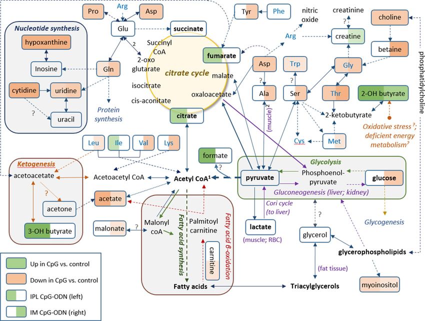
combined box and whisker and dot plots in Fig. 4.
The metabolic changes resulting from the CpG-ODN treatments are further put into biochemical context
and linked to metabolic events as part of the discussion section. Each CpG-ODN treatment was accompanied by
significant changes, compared to DW controls, in about fifteen metabolites (P < 0.05 after adjustment for multiple
comparisons). There was some overlap between the treatments effect, yet unique changes were also recorded
for IPL and for IM vs. DW controls. Treatment with CpG-ODN via IM delivery resulted in more significant
metabolic changes compared to DW controls (eight metabolites with P < 0.001). The most significantly varying
metabolites were typically down-regulated, including of amino acids and derivatives, betaine (decrease of 2.5
fold), choline (decrease of 1.4 fold), glucose (decrease of just 1.15 fold) and nucleotide synthesis intermediates
(decrease of 1.4–1.65 fold). The only significant elevated metabolite in the IM CpG-ODN group was 2-hydroxy-
butyrate (1.35 fold, P = 0.02), which was increased to a lesser degree and significance than in the IPL CpG-ODN
group (1.6 fold, P = 0.006 after FDR correction). Delivery of CpG-ODN via IPL also resulted in a unique 1.45-fold
increase of the ketone body 3-hydroxybutyrate (P = 0.007) and a statistically significant yet quantitatively mod-
erate increase of TCA cycle intermediates, citrate and fumarate (fold change close to 1.2; P < 0.05). Delivery of
CpG-ODN via IPL also showed a more pronounced decrease of acetate than in the IM treatment (1.9 fold change;
P = 0.002), while the decrease of nucleotide synthesis intermediates and of some amino acids was similar to that
of the IM treatment. An additional comparison between the treatments was conducted via one-way ANOVA.
This highlighted differences between IM and IPL delivery methods, mainly in amino acids levels (Table S1).
Pathway enrichment analysis suggested metabolic pathways that were significantly perturbed as a result of
the alterations in metabolites between each CpG-ODN group and DW controls (see supplementary Table S2).
Despite the rather low coverage per pathway reflected in lower numbers of metabolite hits and lower topo-
logical impact, some significant alterations were found in specific pathways involved in energy production and
expenditure. Interestingly, the delivery method of the CpG-ODN affected the significance of the pathways that
were enriched. IM treatment vs. DW controls consistently exhibited higher statistical significance for pathways
involving amino acid metabolism, compared to IPL treatment. On the other hand, IPL was the only delivery
method to alter the TCA cycle when compared to DW controls. IPL treatment also showed higher significance
for pyruvate metabolism and for glycolysis or gluconeogenesis.
Discussion
During an immune response, profound metabolic changes occur to facilitate cell signaling, to enhance cytokine
protein production, to support rapid B-cell and T-cell division and to fulfill increased energy and anabolic
demands45. To the best of our knowledge, the present study is the first report on serum metabolic profiling of
CpG-ODN induced antimicrobial immunity in chickens. We have extensively reported that standalone CpG-
ODN administration via intramuscular (IM)26–28,30 or intrapulmonary (IPL)29,34 routes can induce antimicrobial
immunity against several bacterial pathogens in chickens. Recent metabolomics studies have provided valuable
Scientific Reports | (2021) 11:9028 | https://doi.org/10.1038/s41598-021-88386-2 3
Vol.:(0123456789)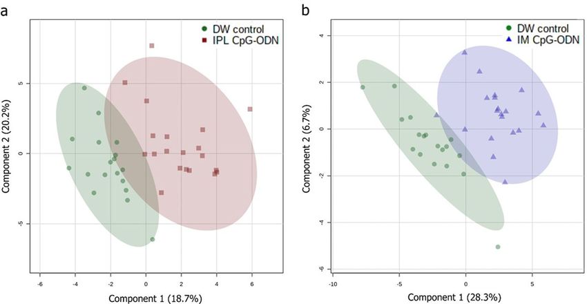
www.nature.com/scientificreports/
Figure 2. Cumulative clinical scores and air sacs bacterial loads in neonatal chicks following E. coli challenge.
Mean cumulative clinical scores (CCS) of each group (a), various scores of bacterial growth isolated (b), and
birds with low and high bacterial loads (c) in neonatal chicks (n = 40/group) following E. coli challenge. (a)
Each data point in line represents the mean CCS value of individual groups on each day, and vertical lines
indicate standard error of the mean. The mean CCS values of the IM CpG-ODN group (blue line) and the
IPL CpG-ODN group (red line) were significantly lower compared to the IPL DW control (green line) group
(P < 0.001). (b) Bar graph shows bacterial scores (0, few, 1 + , 2 + , 3 + & 4 +) of swabs2 taken from air sacs of birds
and cultured on 5% Columbia sheep blood agar by the quadrant streaking method. Higher the bacterial scores
greater are the bacterial loads. Increased bacterial load was observed more frequently in lesions from birds in the
DW control than in the CpG-ODN groups. (c) For the statistical analysis (chi square test) on the bacterial scores
as indicated in Fig. 2b, birds in each group were divided into two categories representing the low bacterial loads
(no growth & few colonies) and high bacterial loads (1 + , 2 + , 3 + & 4 +). Bar graph shows the number of birds
in each group with low and high bacterial loads. The IM CpG-ODN and IPL CpG-ODN administered groups
displayed significantly ( x2 = 9.911, P = 0.007) lower bacterial load compared to the DW control group.
information about the metabolome in various tissues in normal healthy chickens46,47. However, no metabolomic
data has been collected relating to CpG-ODN induced immune responses in chickens. Therefore, the present
study was designed to investigate a potential regulation of metabolic pathways by CpG-ODNs to induce anti-
microbial immunity. Here, we used NMR-based metabolomics to explore the potential of immuno-metabolic
interactions in the orchestration of CpG-ODN-induced antimicrobial immunity. In doing so, we attempted to
identify critical molecules or pathways associated with immunoprotective phenotypes based on their character-
istic serum metabolite profiles. Our findings clearly suggest that immune-metabolic interactions are involved in
CpG-ODN-mediated immunity. In particular, this study revealed a wide array of differential metabolic signatures
in CpG-ODN treated chickens.
In the present study, chickens treated with intramuscular (IM) CpG-ODNs were better protected against
bacterial infection than the intrapulmonary (IPL) group (Figs. 1, 2). This difference between the two CpG-ODN
delivery methods corresponded well with the observed alteration in metabolic profile between the treatment
groups, despite of natural variation in response between birds in the same group (Fig. 4). To better illustrate the
metabolic alterations within their biochemical context, Fig. 5 presents a metabolic pathways map incorporating
the t test results comparing each treatment to control (Tables 1, 2). It is well known that CpG-ODN causes the
immune system’s stimulation, resulting in pro-inflammatory cytokine s ecretion48 and activation of immune cells
such as monocytes, macrophages, and lymphocytes. We recently reported an increased number of mononuclear
Scientific Reports | (2021) 11:9028 | https://doi.org/10.1038/s41598-021-88386-2 4
Vol:.(1234567890)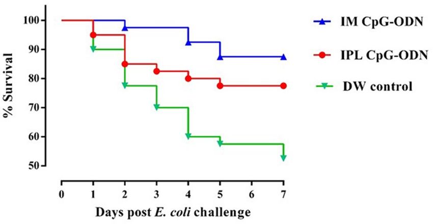
www.nature.com/scientificreports/
Figure 3. Partial Least Squares-Discriminant Analysis (PLS-DA) of serum metabolites in neonatal chicks 24 h
post-treatment. PLS/DA scores plot classifying samples from IPL CpG-ODN treatment vs. DW controls (a), and
IM CpG-ODN vs. DW controls (b), based on measurement of 40 metabolites. DW control, n = 18 (green); IM
CpG, n = 20 (blue); IPL CpG, n = 20 (red).
Metabolite IPL/DW Fold change t test P value FDR—adjusted P value
Choline Down 1.35 1.27E−05 5.08E−04
Acetate Down 1.93 1.24E−04 0.002
Hypoxanthine Down 1.56 3.74E−04 0.005
Proline Down 1.34 6.22E−04 0.006
2-Hydroxybutyrate Up 1.60 7.85E−04 0.006
3-Hydroxybutyrate Up 1.45 0.001 0.007
Cytidine Down 1.56 0.001 0.007
Aspartate Down 1.30 0.002 0.008
Threonine Down 1.37 0.003 0.012
Formate Up 1.49 0.008 0.030
Fumarate Up 1.19 0.010 0.035
Glycine Down 1.19 0.011 0.035
Citrate Up 1.17 0.015 0.047
Uridine Down 1.32 0.018 0.050
Myo-inositol Down 1.39 0.020 0.052
Glutamine Down 1.13 0.037 0.092
Isoleucine Up 1.15 0.041 0.095
Tryptophan Down 1.14 0.043 0.095
Table 1. Selected Student’s t-test results comparing IPL CpG-ODN to DW control. DW controls n = 18; IPL
CpG n = 20. Correction for multiple comparisons was conducted using FDR (n = 40 metabolites). Between-
group direction and fold-change of the mean raw metabolite concentrations are also presented. IPL CpG-ODN
vs. DW control.
cells infiltrating into the lungs of chickens within 24 h of CpG-ODN t reatment2,29,33. Given that immune cells
must grow and divide rapidly upon activation, despite lacking nutrient stores, they require energy-rich resources,
including sugars, amino acids, and fatty acids present in the extracellular e nvironment49. Therefore it is expected
that the levels of these metabolites, during immune activation, would potentially decrease in the s erum50. In
particular, immune activation requires amino acids for protein synthesis, nucleotides for DNA/RNA synthesis,
sugars for energy production and fatty acids for lipid membrane production. As immune cell activation is cer-
tainly metabolically demanding, we assume that the metabolomics changes in serum that we observed in the
present study could be the result of increased enrichment and activation of immunological niches2,29, leading to
enhanced uptake of metabolites by cells during immunological responses. Since blood bathes every organ and
every tissue in the body, it essentially reflects the net metabolic changes resulting from the physiological and
Scientific Reports | (2021) 11:9028 | https://doi.org/10.1038/s41598-021-88386-2 5
Vol.:(0123456789)www.nature.com/scientificreports/
Metabolite IM/DW Fold change t test P value FDR—adjusted P value
Betaine Down 2.46 7.16E−08 2.86E−06
Proline Down 1.51 8.45E−07 1.69E−05
Serine Down 1.35 2.33E−05 3.11E−04
Alanine Down 1.29 8.64E−05 8.64E−04
Choline Down 1.41 1.26E−04 9.28E−04
Glycine Down 1.31 1.86E−04 9.28E−04
Threonine Down 1.49 1.73E−04 9.28E−04
Tyrosine Down 1.30 1.48E−04 9.28E−04
Cytidine Down 1.64 2.31E−04 0.001
Glutamine Down 1.24 4.54E−04 0.002
Hypoxanthine Down 1.51 9.11E−04 0.003
Lysine Down 1.37 0.001 0.004
D-Glucose Down 1.15 0.001 0.004
Acetate Down 1.26 0.007 0.019
Aspartate Down 1.22 0.008 0.020
2-Hydroxybutyrate Up 1.36 0.009 0.021
Uridine Down 1.40 0.009 0.021
Valine Down 1.16 0.017 0.037
Carnitine Down 1.37 0.029 0.057
Methionine Down 1.17 0.032 0.068
Tryptophan Down 1.12 0.037 0.073
Leucine Down 1.19 0.050 0.093
Malonate Down 1.25 0.051 0.093
Creatine Up 1.50 0.068 0.109
Myo-inositol Down 1.29 0.066 0.109
Table 2. Selected Student’s t-test results comparing IM CpG-ODN to DW control. DW controls n = 18;
IM CpG n = 20 (apart from carnitine, where two missing values were removed). Correction for multiple
comparisons was conducted using FDR (n = 40 metabolites). Between-group direction and fold-change of the
mean raw metabolite concentrations are also presented. IM CpG-ODN vs. DW control.
metabolic needs or stresses in different tissues in the animal b ody72. However, our serum metabolomics data do
not distinguish between the possibilities of differential use or synthesis or uptake of metabolites in various tissues
following the CpG-ODN administration. Additional studies and techniques will be required to investigate the
complex issues of differentiating between the use, synthesis, and uptake of substances.
Several studies have reported that amino acids are critical in immune cell proliferation and function51. A
study in mice demonstrated that serine and glycine play an important role in T cell proliferation and f unction50.
In our study, we found a significant decrease in serum levels of essential and also non-essential amino acids.
Altered levels of essential amino acids which are required for protein synthesis, included threonine (35% decrease
in IPL treatment and 50% decrease in IM treatment), glycine (20–30% decrease in both treatments), and lysine
(35% decrease, only in IM treatment). A recent study demonstrated that the metabolism of a single amino acid
(proline) can have a profound effect on the immune responses to pathogens52. This study reported that mito-
chondrial proline catabolism controls innate immunity in Caenorhabditis elegans by regulating reactive oxygen
species (ROS) homeostasis. In both nematodes and birds, antimicrobial ROS produced by m acrophages53 and
neutrophils54 aids in bacterial killing. In our study, we found a substantial reduction in the serum proline levels
that corresponded with increased antimicrobial immunity in both the IPL CpG-ODN and IM CpG-ODN groups.
We hypothesize that the CpG-ODN induced increased leukocytes in various immunological n iches2,33,34, and
enhanced activation of macrophages and heterophils [equivalent to mammalian neutrophils], which probably
led to greater utilization of serum proline. Further studies to test the role of proline utilization and CpG-ODN
induced antimicrobial immunity would be of great interest.
Apart from their role as building blocks in protein synthesis and providing alternative substrates to the TCA
cycle, amino acids are also precursors in nucleotide synthesis and play a vital role in cell proliferation. Specifically,
glutamine is a key amino acid that is further metabolised into purines and downstream nucleotides to generate
DNA and RNA. Increased uptake of glutamine and its precursors aspartate and also proline was observed after
administration of CpG-ODN via IM and also IPL. Within the purine metabolism pathway, the three intermedi-
ates hypoxanthine, cytidine and uridine showed a uniform decrease across the CpG treatments.
In addition to the amino acid changes noted above, we also observed reduced acetate levels in serum upon
the administration of CpG-ODN. The source of acetic acid might be either a catabolism product of ketone
bodies, or metabolism of acetyl-CoA, an essential metabolite of carbohydrate and fatty acid metabolism55. As
mentioned earlier, cell proliferation also increases the demands for fatty acid synthesis, hence acetic acid might
be utilised in this direction. On the other hand, fatty acid oxidation provides energy to the cellular activities
Scientific Reports | (2021) 11:9028 | https://doi.org/10.1038/s41598-021-88386-2 6
Vol:.(1234567890)www.nature.com/scientificreports/
Figure 4. Combined box-and-whisker and dot plots showing the concentrations of selected metabolites in
serum from neonatal chicks 24 h post-treatment. DW control, n = 18 (green); IM CpG-ODN, n = 20 (blue) and
IPL CpG-ODN, n = 20 (red). Values are log-transformed concentrations in μM. One-way ANOVA results are
detailed in supplementary Table S1.
when in need through β-oxidation in mitochondria or peroxisomes. An increased fatty acid uptake in the liver
results in ketogenesis, where fatty acids undergo incomplete o xidation56. Lower serum acetate levels resulting
from CpG-ODN administration might be indicating that immune cells must start dividing and that there is a
need for extra energy. As a result, fatty acids started to undergo oxidation to provide that extra energy. Acetate
also appears to play a signaling role in the immune system. A recent report has shown how acetate promotes
T-cell effector functions57 through epigenetic modifications and that rapidly dividing cells (including both tumor
and immune cells) can use acetate (instead of glucose) as an alternative fuel.
We also found that CpG-ODN administration by both IPL and IM resulted in increased levels of 2-OH
butyrate, which can originate in several amino acids via 2-ketobutyrate. In humans 2-OH butyrate is related to
lipid oxidation, oxidative stress and deficient energy m etabolism58. Interestingly, serum levels of 2-OH butyrate
increased by 60% after the IPL treatment and only by 35% in the IM treatment. Further metabolic differences
were observed between the two administration routes, with a slight decrease in glucose measured only in the
IM group, while a 45% increase in the ketone body 3-OH butyrate was observed only in the IPL-CpG-ODN
group, indicating ketogenesis was being used to provide for higher energy demands. In addition, an increase
Scientific Reports | (2021) 11:9028 | https://doi.org/10.1038/s41598-021-88386-2 7
Vol.:(0123456789)www.nature.com/scientificreports/
Figure 5. Metabolic pathway map. Pathway map incorporating metabolic differences between IPL CpG-ODN
treatment and DW control (left half of each metabolite box) and between IM CpG-ODN treatment and DW
control (right half of each metabolite box). According to the Student’s t test (Table 1, 2), metabolite boxes with
a green background were significantly higher in CpG-ODN vs. DW controls, while metabolite boxes with
a red background were lower in CpG-ODN compared to DW controls, 24 h post treatment. Higher levels
of significance and fold change are reflected by a darker color, and lower levels and trends by a lighter color.
Uncolored part of a box indicates metabolites that did not show any trend of change in concentrations between
CpG-ODN and DW controls. Full arrows represent one-step metabolic conversion, and broken arrows depict
at least two steps between a precursor and its derivative. Essential amino acids are in blue text, and metabolic
events or location are in italic text with different colors1. In animals, acetyl-CoA cannot be used as a substrate
for gluconeogenesis2. The two reactions depend on each other. ? Unestablished in chickens.
in TCA cycle intermediates was recorded only after IPL administration, although the lactate-to-pyruvate ratio
did not differ between the experimental groups. Altogether, these data suggest differences in the extent and rate
of energy metabolism adaptations, potentially due to systemic vs. mucosal targeting by IM and IPL CpG-ODN
administration methods, respectively.
Beyond its function as an energy source, a previous study in cattle reported that beta-hydroxybutyrate (3-OH
butyrate) abrogates the antimicrobial function of neutrophils against E. coli59. Several other studies showed
that 3-hydroxybutyrate upregulates mRNA abundance of proinflammatory cytokines such as IL-1β in mouse
macrophages60, IL-8 in bovine liver61, and several pro-inflammatory cytokines in calf hepatocytes62. The cytokine
mRNA data should be interpreted cautiously, as some cytokines such as IL-1β are highly regulated post-transla-
tionally63. In our previous studies, we found a significant increase in the expression of several proinflammatory
cytokines, including IL-1β and IL-6 following CpG-ODN administration2,33. In the present study, we found an
increased serum level of 3-hydroxybutyrate in the IPL CpG-ODN group, but how this would affect cytokines at
the protein level needs further studies.
Our metabolomic analysis also highlighted two crucial metabolites, choline and betaine. Serum choline levels
were reduced by 35–40% in the CpG-ODN treated birds while betaine levels were significantly lower (2.5-fold)
only in the birds that received intramuscular CpG-ODN treatment. Choline is a precursor in the synthesis of
acetylcholine, phosphorylcholine, and is an important intermediate in phospholipid metabolism. It is well docu-
mented that there is increased consumption of phosphorylcholine under severe oxidative and systemic inflam-
matory conditions64. Choline is oxidized to betaine, and both are linked to the folate-dependent one-carbon
Scientific Reports | (2021) 11:9028 | https://doi.org/10.1038/s41598-021-88386-2 8
Vol:.(1234567890)www.nature.com/scientificreports/
metabolism65. In chickens, choline plays a significant role in improving the humoral and cellular immunity66,
and betaine has been shown to increase lymphocyte infiltration in infected mucosa. Betaine also enhances
phagocytic and nitric oxide production by blood monocytes and h eterophils67. A recent study in cattle reported
that T cell proliferation was linearly enhanced in vitro with increasing doses of c holine68. Reduced choline levels
in our study could also be contributing to the low betaine levels, however betaine is also supplemented via the
feed due to its nutritional importance.
As noted earlier, increased utilization of choline, betaine, and proline by immune cells can enhance the
antimicrobial activity of various immune cells. Additionally, given that choline, betaine, and proline are effec-
tive osmolytes69, their reduced levels in the serum would prevent bacterial proliferation due to a reduced accu-
mulation of osmoprotectants (betaine, choline, and proline), which play an essential role in bacterial growth such
as in the case of E. c oli69 and Staphylococcus a ureus70. It has been reported that betaine provides the best osmotic
protection to E. coli69 growth followed by choline (which is converted to betaine) and p roline69. Interestingly, in
our study, we found that IM CpG-ODN administration dramatically reduced the serum levels of betaine followed
by proline and choline, and provided better protection against E. coli compared to IPL-CpG-ODN, which only
showed significantly reduced serum levels of choline and proline but not betaine. More significant reduction of
bacterial growth supporting osmoprotectants in IM CpG-ODN vs. IPL CpG-ODN correlates well with the rank
order of their protection data (IM-vs. IPL-CpG-ODN).
Overall, the metabolomic data from this study suggest that CpG-ODN-mediated antimicrobial immunity
involves a number of significant metabolic changes in the host that enhance immunity and antagonize microbial
proliferation in the host. CpG-ODN induced metabolomics data generated by the current study provides a unique
and little-appreciated approach to identify regulatory molecules or pathways that give protective immunity to
chickens against bacterial infections. The metabolites highlighted in the present study can help future targeted
studies better understand antimicrobial metabolomic profiles and pinpoint the key molecules or pathways con-
tributing to immunity.
Materials and methods
Housing and maintenance of experimental chickens. This work was carried out in compliance with
the ARRIVE guidelines. The animal study was approved by the Animal Research Ethics Board, University of
Saskatchewan (protocol number 20070008) and adhered to the guidelines of the Canadian Council on Animal
Care. Euthanasia was performed by cervical dislocation following the AVMA guidelines for the euthanasia of
animals. Day-old broiler chickens (Ross 308 strain) were obtained from a commercial hatchery in Saskatchewan.
Groups of chicks were allocated randomly into an animal isolation room at the Animal Care Unit, Western Col-
lege of Veterinary Medicine in Saskatoon, Saskatchewan, and chicks were maintained following the procedure as
described earlier2,33. Briefly, water and commercial broiler starter ration (23% crude protein, 1% calcium, 0.45%
available phosphorous) were provided ad libitum. Air from each room was exhausted through a HEPA filter,
and non-recirculated intake air was provided at a rate of 15–20 air changes/hr. Air pressure differentials and
strict sanitation were maintained in this isolation facility. Broilers were raised at 32 °C for the first 7 days of life
(average weight ~ 180 g); after that, the temperature was decreased by 0.5 °C per day until a room temperature of
27.5 °C was reached. Light (30 lx) was provided for 24 h/d during days 0 to 2 (post-hatch). Darkness was intro-
duced at 3 d post-hatch with 1 h of dark added daily until 4 h of darkness was achieved.
CpG‑ODN delivery. The CpG-ODN [TCGTCGTTGTCGTTTTGTCGTT (2007)] was free of endotoxin
and produced with a phosphorothioate backbone (Operon Biotechnologies, Inc; Huntsville, AL, USA). Syn-
thetic CpG-ODN was diluted in sterile DW and delivered by the IPL route. Briefly, the CpG-ODN solution was
aerosolized as micro-droplets (particle size of 0.5–5 µm) using a Compressor Nebulizer (705–470) unit (AMG
Medical Inc; Montreal, QC, Canada) in a closed 0.036 m3 acrylic chamber containing 60 birds for 30 min (6 mg
CpG-ODN/ chamber) that maintained atmospheric oxygen exchange. The control group of birds (n = 60) were
aerosolized with DW for 30 min in the acrylic chamber using a similar compressor nebulizer. Another group
of birds (n = 60) were administered with CpG-ODN (50 μg/100 μl/bird) by IM injection to the left thigh. The
temperature was maintained at 28–30 °C in the acrylic chamber during the administration of CpG-ODN or DW.
E. coli culture and animal model. In order to confirm the immune protection induced by CpG-ODN
delivery, a parallel E. coli challenge study was performed to the birds. A field isolate of E. coli from a turkey with
septicemia was used as the challenge strain according to our previously established animal m odel29,30. The E. coli
belonged to serogroup O2 was nonhemolytic, serum resistant, aerobactin producing and had K1 capsule with
type I pili28. Aliquots of the bacterial isolate were stored at − 80 °C in brain heart infusion broth (Difco, Detroit,
Mich.) supplemented with 25% (wt/vol) glycerol (VWR Scientific Inc., Montreal, QC, Canada). In order to
challenge the birds, bacteria were cultured on 5% Columbia sheep blood agar for 18–24 h at 37 °C. One colony
of E. coli was added to 100 mL of Luria broth (Difco LB broth, Miller, Becton Dickinson and Company; Sparks,
MD, USA) in a 250 mL Erlenmeyer flask. The culture was grown at 37 °C for 16–18 h, shaking at 150 rpm. This
stationary phase culture contained approximately 1 × 10⁹ colony forming units (CFU) of bacteria per mL, which
was then further diluted into saline to the concentration of bacteria required to challenge birds. The E. coli chal-
lenge dose was confirmed by plating serial dilutions of the diluted culture in duplicate on 5% Columbia sheep
blood agar plates, incubating for 18 h at 37 °C, then counting the number of colonies.
The E. coli challenge study was performed according to the well-established animal model that we docu-
mented earlier26,29. Briefly, on the second-day post-treatment, the birds in each group (n = 40) were challenged
with 1 × 10⁵ CFU (n = 20) or 1 × 10⁶ CFU (n = 20) of bacteria per bird, subcutaneously in the neck. They were
closely monitored three times a day for the most critical period of three days post-challenge and two times a
Scientific Reports | (2021) 11:9028 | https://doi.org/10.1038/s41598-021-88386-2 9
Vol.:(0123456789)www.nature.com/scientificreports/
day thereafter until seven days post-challenge. Each bird was observed for clinical signs, and a daily clinical
score was assigned: 0 = normal; 0.5 = slightly abnormal appearance, slow to move; 1 = depressed, reluctant to
move; 1.5 = reluctant to move, may take a drink and peck some; 2 = unable to stand or reach for food or water;
and 3 = found dead. Birds that received a clinical score of 2 were euthanized by cervical dislocation. At the end
of the trial, each bird was given a cumulative clinical score (CCS) as a sum of daily clinical scores, as previously
described26,29.
When chicks were found dead or euthanized, they were necropsied immediately. All remaining birds were
euthanized on day seven post-challenge. Air sac swabs were obtained from dead or euthanized birds and a
semi-quantitative estimate of E. coli isolation was conducted on 5% Columbia sheep blood agar by the quadrant
streaking method in which bacterial colonies thinning occurs as streaking goes clockwise from quadrant 1 to
quadrant 4. Bacterial growth on these cultures were recorded on a scale from 0 to 4 + , where 0 = no growth;
few = less than 5 colonies; 1 + = growth of bacteria on quadrant 1; 2 + = growth of bacteria on quadrants 1 and 2;
3 + = growth of bacteria on quadrants 1, 2 and 3; and 4 + = growth of bacteria on all quadrants 1–4 as reported
previously71. For the statistical analysis on bacterial loads, birds were divided into two categories in each group. In
category one, birds with low bacterial loads [zero or few (~ 5) bacterial colonies in the blood agar] were included.
Whereas, category two included birds with high bacterial loads [bacterial growth in various quadrants; 1 + , 2 + ,
3 + & 4 + in the blood agar].
Metabolomics analysis of serum. Sample collection. Twenty chicks from each group were euthanized
24 h post-CpG-ODN treatment in the morning between 9 am to 10 a.m. Blood was immediately collected into
serum tubes by severing the necks of the chicks with a sharp pair of scissors. After about 30 min of blood collec-
tion, the clotted blood samples were then centrifuged at 1000g force for 15 min, and serum was separated into
1.5 mL microcentrifuge tubes. Serum samples were stored at − 80 °C, transported on dry ice to The Metabo-
lomics Innovation Centre (TMIC) facility at the University of Alberta in Edmonton, and stored at − 80 °C until
further analysis.
Sample preparation and NMR spectroscopy. Serum samples were thawed on ice and prepared in two batches
according to a randomization template, with the addition of pooled samples for quality control. Plasma and
serum samples contain a significant concentration of large molecular weight proteins and lipoproteins, which
can seriously compromise the quality of 1H-NMR spectra though the generation of intense, broad lines that
interfere with the identification and quantification of lower abundance metabolites. Deproteinization can elimi-
nate these peaks. Deproteinization of the serum samples was done by centrifugation and ultrafiltration using
3-kDa cut-off centrifuge filter units (Microcon YM-3; Sigma-Aldrich, St. Louis, MO), following a previously
reported deproteinization p rocedure72. The deproteinized serum samples (280 μL) were then transferred to a
1.5 mL micro centrifuge tube followed by the addition of 70 μL standard NMR buffer solution (1 mM DSS
(disodium-2, 2-dimethyl-2-silapentane-5-sulphonate), in 10% D2O). These samples (a total volume of 350 μL)
were then transferred to a 3 mm NMR tube for spectral analysis. All 1H-NMR spectra were collected on a Bruker
Avance III Ascend 700 MHz spectrometer with a 5 mm cryo-probe (Bruker Biospin, Rheinstetten, Germany).
1
H-NMR spectra were acquired at 25 °C using the first transient of the noesy-presaturation pulse sequence,
which was chosen for its high degree of quantitative a ccuracy73. Spectra were collected with 128 transients using
a 4 s acquisition time and a 1 s recycle delay.
NMR compound identification and quantification. Before spectral analysis, all free induction decays (FIDs)
were zero-filled to 240 k data points, and a line broadening of 0.5 Hz was applied. The methyl singlet of the added
DSS served as an internal standard for chemical shift referencing (set to 0.00 ppm) and for quantification. All
1
H-NMR spectra were processed and imported into the Chenomx NMR Suite 8.1 software (Edmonton, Canada).
The Chenomx NMR Suite software allows for a quantitative analysis of an NMR spectrum by manually fitting
spectral signatures from an internal database to the spectrum. Specifically, the spectral fitting for metabolite was
done using the standard Chenomx 700 MHz metabolite library. Most of the visible peaks are annotated with
a compound name. Each spectrum was processed and analyzed by at least two experienced NMR spectrosco-
pists to minimize compound misidentification and misquantification. Forty metabolites passed the NMR quality
measures and underwent further statistical analysis.
Data processing and statistical analysis. The MetaboAnalyst 4.0 p ackage74 was used for statistical analysis of the
metabolomics data. Data were log-transformed prior to univariate analysis, and also autoscaled prior to mul-
tivariate analysis. Principal Component Analysis (PCA) was performed for quality-control assessment. Partial
Least Squares-Discriminant Analysis (PLS/DA) was used to classify samples and suggest potential biomark-
ers for treatment effect. Several univariate analysis tests were also employed. In particular, analysis of variance
(ANOVA) was conducted to compare metabolite levels between all three groups, with Tukey’s HSD post-hoc
analysis to indicate significant pairs. Student’s t test was used to compare between two experimental groups in
terms of fold-change analysis and assessment of significance with regard to metabolite differences. In all tests, P
values were further corrected for multiple comparisons by applying the Benjamini–Hochberg method of false
discovery rate (FDR), and considering an FDR-adjusted P value of 0.1 as a threshold for inclusion in tables and
figures. Pathway enrichment analysis was performed in the MetaboAnalyst 4.0 software on log-transformed and
auto scaled data, against the Gallus Gallus pathway database. For each two-group comparison, it consisted of an
ANCOVA test with the use of a relative-betweenness centrality algorithm for pathway topology analysis.
The significance of the observed differences in chick survival and the cumulative clinical score (CCS) were
analyzed using GraphPad Prism 8.0 (GraphPad Software Inc., San Diego, CA) with a significance level of P < 0.05.
Scientific Reports | (2021) 11:9028 | https://doi.org/10.1038/s41598-021-88386-2 10
Vol:.(1234567890)www.nature.com/scientificreports/
The survival data and bacterial scores of both 1 × 10⁵ CFU and 1 × 10⁶ CFU of E. coli challenge were combined for
clarity of analysis and presentation. The level of significance with regard to differences among groups in survival
patterns and median survival times were analyzed using the log-rank test and chi-square statistic. Clinical scores
assigned at each time point were summed up to 7 days post-challenge to generate CCS and thereby daily mean
CCSs were calculated for each group. Two-way ANOVA was performed with Dunnett’s multiple comparison
tests to compare the significant differences in mean CCS. For the statistical analysis on bacterial loads (quadrant
streaking method), birds in each group were divided into two categories (low or high bacterial load). A chi-
square test of independence was performed to examine the relationship between the CpG treatment method and
the ability to recover viable bacteria based on this categorization. The results were interpreted with a statistical
significance of P < 0.05.
Received: 24 July 2020; Accepted: 12 April 2021
References
1. Coffman, R. L., Sher, A. & Seder, R. A. Vaccine adjuvants: Putting innate immunity to work. Immunity 33, 492–503 (2010).
2. Goonewardene, K., Ahmed, K.A., Gunawardana, T. et al. Mucosal delivery of CpG-ODN mimicking bacterial DNA via the
intrapulmonary route induces systemic antimicrobial immune responses in neonatal chicks. Sci. Rep. 10, 5343. https://doi.org/
10.1038/s41598-020-61683-y (2020).
3. Gursel, M. & Gursel, I. Development of CpG ODN based vaccine adjuvant formulations. In Vaccine Design 289–298 (Humana
Press, New York, NY, 2016). https://doi.org/10.1007/978-1-4939-3389-1_20.
4. Gursel, M. & Klinman, D. M. Chapter Use of CpG oligonucleotides as mucosal adjuvants. In Mucosal Immunology 4th edn (eds
Mestecky, J. et al.) 1201–1209 (Academic Press, Cambridge, 2015). https://doi.org/10.1016/B978-0-12-415847-4.00062-8.
5. Krieg, A. M. CpG motifs in bacterial DNA and their immune effects. Annu. Rev. Immunol. 20, 709–760 (2002).
6. Staeheli, P., Puehler, F., Schneider, K., Göbel, T. W. & Kaspers, B. Cytokines of birds: Conserved functions—A largely different
look. J. Interferon Cytokine Res. 21, 993–1010 (2001).
7. Finlay, B. B. & Hancock, R. E. W. Can innate immunity be enhanced to treat microbial infections?. Nat. Rev. Microbiol. 2, 497–504
(2004).
8. Aderem, A. & Ulevitch, R. J. Toll-like receptors in the induction of the innate immune response. Nature 406, 782–787 (2000).
9. Brownlie, R. & Allan, B. Avian toll-like receptors. Cell Tissue Res. 343, 121–130 (2011).
10. Brownlie, R. et al. Chicken TLR21 acts as a functional homologue to mammalian TLR9 in the recognition of CpG oligodeoxynu-
cleotides. Mol. Immunol. 46, 3163–3170 (2009).
11. Hartmann, G. et al. Delineation of a CpG phosphorothioate oligodeoxynucleotide for activating primate immune responses in vitro
and in vivo. J. Immunol. 164, 1617–1624 (2000).
12. Keestra, A. M., de Zoete, M. R., Bouwman, L. I. & van Putten, J. P. M. Chicken TLR21 Is an Innate CpG DNA Receptor Distinct
from Mammalian TLR9. J. Immunol. 185, 460–467 (2010).
13. Krieg, A. M. Toll-like receptor 9 (TLR9) agonists in the treatment of cancer. Oncogene 27, 161–167 (2008).
14. Yeh, D.-W. et al. Toll-like receptor 9 and 21 have different ligand recognition profiles and cooperatively mediate activity of CpG-
oligodeoxynucleotides in zebrafish. PNAS 110, 20711–20716 (2013).
15. Bode, C., Zhao, G., Steinhagen, F., Kinjo, T. & Klinman, D. M. CpG DNA as a vaccine adjuvant. Expert Rev. Vaccines 10, 499–511
(2011).
16. Hanagata, N. CpG oligodeoxynucleotide nanomedicines for the prophylaxis or treatment of cancers, infectious diseases, and
allergies. Int. J. Nanomed. 12, 515–531 (2017).
17. Shirota, H., Tross, D. & Klinman, D. M. CpG oligonucleotides as cancer vaccine adjuvants. Vaccines 3, 390–407 (2015).
18. Zhang, H. & Gao, X.-D. Nanodelivery systems for enhancing the immunostimulatory effect of CpG oligodeoxynucleotides. Mater.
Sci. Eng. C Mater. Biol. Appl. 70, 935–946 (2017).
19. Adamsson, J. et al. Novel immunostimulatory agent based on CpG oligodeoxynucleotide linked to the nontoxic B subunit of
cholera toxin. J. Immunol. 176, 4902–4913 (2006).
20. Weiner, G. J., Liu, H.-M., Wooldridge, J. E., Dahle, C. E. & Krieg, A. M. Immunostimulatory oligodeoxynucleotides containing the
CpG motif are effective as immune adjuvants in tumor antigen immunization. Proc. Natl. Acad. Sci. USA 94, 10833–10837 (1997).
21. Meng, W., Yamazaki, T., Nishida, Y. & Hanagata, N. Nuclease-resistant immunostimulatory phosphodiester CpG oligodeoxynu-
cleotides as human Toll-like receptor 9 agonists. BMC Biotechnol. 11, 88 (2011).
22. Cho, H. C. et al. Cancer immunotherapeutic effects of novel CpG ODN in murine tumor model. Int. Immunopharmacol. 8,
1401–1407 (2008).
23. Nichani, A. K. et al. In vivo immunostimulatory effects of CpG oligodeoxynucleotide in cattle and sheep. Vet. Immunol. Immuno-
pathol. 98, 17–29 (2004).
24. Jørgensen, J. B., Johansen, L.-H., Steiro, K. & Johansen, A. CpG DNA induces protective antiviral immune responses in Atlantic
salmon (Salmo salar L.). J. Virol. 77, 11471–11479 (2003).
25. Dalloul, R. A. et al. In vivo effects of CpG oligodeoxynucleotide on eimeria infection in chickens. Avian Dis. 48, 783–790 (2004).
26. Gomis, S. et al. Protection of neonatal chicks against a lethal challenge of Escherichia coli using DNA containing cytosine-phos-
phodiester-guanine motifs. Avian Dis. 48, 813–822 (2004).
27. Taghavi, A. et al. Protection of neonatal broiler chicks against salmonella typhimurium septicemia by DNA containing CpG motifs.
Avian Dis. 52, 398–406 (2008).
28. Gomis, S. et al. Protection of chickens against Escherichia coli infections by DNA containing CpG motifs. Infect. Immunol. 71,
857–863 (2003).
29. Goonewardene, K. B. et al. Intrapulmonary delivery of CpG-ODN microdroplets provides protection against Escherichia coli
septicemia in neonatal broiler chickens. Avian Dis. 61, 503–511 (2017).
30. Gunawardana, T. et al. Protection of neonatal broiler chickens following in ovo delivery of oligodeoxynucleotides containing CpG
motifs (CpG-ODN) formulated with carbon nanotubes or liposomes. Avian Dis. 59, 31–37 (2014).
31. He, H., Lowry, V. K., Swaggerty, C. L., Ferro, P. J. & Kogut, M. H. In vitro activation of chicken leukocytes and in vivo protection
against Salmonella enteritidis organ invasion and peritoneal S. enteritidis infection-induced mortality in neonatal chickens by
immunostimulatory CpG oligodeoxynucleotide. FEMS Immunol. Med. Microbiol. 43, 81–89 (2005).
32. MacKinnon, K. M. et al. In ovo treatment with CpG oligodeoxynucleotides decreases colonization of Salmonella enteriditis in
broiler chickens. Vet. Immunol. Immunopathol. 127, 371–375 (2009).
33. Gunawardana, T. et al. Synthetic CpG-ODN rapidly enriches immune compartments in neonatal chicks to induce protective
immunity against bacterial infections. Sci. Rep. 9, 341 (2019).
Scientific Reports | (2021) 11:9028 | https://doi.org/10.1038/s41598-021-88386-2 11
Vol.:(0123456789)www.nature.com/scientificreports/
34. Gunawardana, T. et al. CpG-ODN induces a dose-dependent enrichment of immunological niches in the spleen and lungs of
neonatal chicks that correlates with the protective immunity against Escherichia coli. J. Immunol. Res. https://doi.org/10.1155/
2020/2704728 (2020).
35. Cheng, S.-C. et al. mTOR- and HIF-1α-mediated aerobic glycolysis as metabolic basis for trained immunity. Science 345, 1250684
(2014).
36. Gubser, P. M. et al. Rapid effector function of memory CD8+ T cells requires an immediate-early glycolytic switch. Nat. Immunol.
14, 1064–1072 (2013).
37. Pearce, E. L. et al. Enhancing CD8 T cell memory by modulating fatty acid metabolism. Nature 460, 103–107 (2009).
38. Jha, A. K. et al. Network integration of parallel metabolic and transcriptional data reveals metabolic modules that regulate mac-
rophage polarization. Immunity 42, 419–430 (2015).
39. Newsholme, P., Curi, R., Gordon, S. & Newsholme, E. A. Metabolism of glucose, glutamine, long-chain fatty acids and ketone
bodies by murine macrophages. Biochem. J. 239, 121–125 (1986).
40. Donnelly, R. P. & Finlay, D. K. Glucose, glycolysis and lymphocyte responses. Mol. Immunol. 68, 513–519 (2015).
41. Frauwirth, K. A. & Thompson, C. B. Regulation of T lymphocyte metabolism. J. Immunol. 172, 4661–4665 (2004).
42. van Stipdonk, M. J. B. et al. Dynamic programming of C D8+ T lymphocyte responses. Nat. Immunol. 4, 361–365 (2003).
43. Wang, R. et al. The transcription factor Myc controls metabolic reprogramming upon T lymphocyte activation. Immunity 35,
871–882 (2011).
44. Guthrie, L. A., McPhail, L. C., Henson, P. M. & Johnston, R. B. Priming of neutrophils for enhanced release of oxygen metabolites
by bacterial lipopolysaccharide. Evidence for increased activity of the superoxide-producing enzyme. J. Exp. Med. 160, 1656–1671
(1984).
45. Kelly, B. & O’Neill, L. A. Metabolic reprogramming in macrophages and dendritic cells in innate immunity. Cell Res. 25, 771–784
(2015).
46. Jastrebski, S. F., Lamont, S. J. & Schmidt, C. J. Chicken hepatic response to chronic heat stress using integrated transcriptome and
metabolome analysis. PLoS ONE 12, e0181900 (2017).
47. Le Roy, C. I., Mappley, L. J., La Ragione, R. M., Woodward, M. J. & Claus, S. P. NMR-based metabolic characterization of chicken
tissues and biofluids: A model for avian research. Metabolomics 12 (2016).
48. Patel, B. A. et al. Oligodeoxynucleotides containing CpG motifs (CpG-ODN) predominantly induce Th1-type immune response
in neonatal chicks. Dev. Comp. Immunol. 32, 1041–1049 (2008).
49. Ganeshan, K. & Chawla, A. Metabolic regulation of immune responses. Annu. Rev. Immunol. 32, 609–634 (2014).
50. Ma, E. H. et al. Serine is an essential metabolite for effector T cell expansion. Cell Metab. 25, 345–357 (2017).
51. Grohmann, U. et al. Amino-acid sensing and degrading pathways in immune regulation. Cytokine Growth Factor Rev. 35, 37–45
(2017).
52. Tang, H. & Pang, S. Proline Catabolism Modulates Innate Immunity in Caenorhabditis elegans. Cell Rep. 17, 2837–2844 (2016).
53. Abuaita, B. H., Schultz, T. L. & O’Riordan, M. X. Mitochondria-derived vesicles deliver antimicrobial reactive oxygen species to
control phagosome-localized Staphylococcus aureus. Cell Host Microbe 24, 625-636.e5 (2018).
54. Phan, Q. T. et al. Neutrophils use superoxide to control bacterial infection at a distance. PLoS Pathog. 14, e1007157 (2018).
55. Human Metabolome Database: Search Results for metabolite. http://www.hmdb.ca/unearth/q?utf8=%E2%9C%93&query=aceta
te&searcher=metabolites&butt=.
56. Nguyen, P. et al. Liver lipid metabolism. J. Anim. Physiol. Anim. Nutr. 92, 272–283 (2008).
57. Leone, R. D. et al. Glutamine blockade induces divergent metabolic programs to overcome tumor immune evasion. Science 366,
1013–1021 (2019).
58. Gall, W. E. et al. α-Hydroxybutyrate is an early biomarker of insulin resistance and glucose intolerance in a nondiabetic population.
PLoS ONE 5, e10883 (2010).
59. Grinberg, N., Elazar, S., Rosenshine, I. & Shpigel, N. Y. Beta-hydroxybutyrate abrogates formation of bovine neutrophil extracel-
lular traps and bactericidal activity against mammary pathogenic Escherichia coli. Infect. Immun. 76, 2802–2807 (2008).
60. Swartz, T., Mamedova, L. & Bradford, B. Beta-Hydroxybutyrate Alters the mRNA Cytokine Profile from Mouse Macrophages
Challenged with Streptococcus uberis. Kansas Agric. Exp. Stn. Res. Rep. 5 (2019).
61. Zarrin, M., Wellnitz, O., van Dorland, H. A. & Bruckmaier, R. M. Induced hyperketonemia affects the mammary immune response
during lipopolysaccharide challenge in dairy cows. J. Dairy Sci. 97, 330–339 (2014).
62. Shi, X. et al. β-Hydroxybutyrate activates the NF-κB signaling pathway to promote the expression of pro-inflammatory factors in
calf hepatocytes. Cell. Physiol. Biochem. 33, 920–932 (2014).
63. Youm, Y.-H. et al. The ketone metabolite β-hydroxybutyrate blocks NLRP3 inflammasome-mediated inflammatory disease. Nat.
Med. 21, 263–269 (2015).
64. Guleria, A. et al. NMR based serum metabolomics reveals a distinctive signature in patients with Lupus Nephritis. Sci. Rep. 6,
35309 (2016).
65. Ueland, P. M. Choline and betaine in health and disease. J. Inherit. Metab. Dis. 34, 3–15 (2011).
66. Swain, B. K. & Johri, T. S. Effect of supplemental methionine, choline and their combinations on the performance and immune
response of broilers. Br. Poult. Sci. 41, 83–88 (2000).
67. Klasing, K. C., Adler, K. L., Remus, J. C. & Calvert, C. C. Dietary betaine increases intraepithelial lymphocytes in the duodenum
of coccidia-infected chicks and increases functional properties of phagocytes. J. Nutr. 132, 2274–2282 (2002).
68. Garcia, M., Mamedova, L. K., Barton, B. & Bradford, B. J. Choline regulates the function of bovine immune cells and alters the
mRNA abundance of enzymes and receptors involved in its metabolism in vitro. Front. Immunol. 9, 2448 (2018).
69. Cayley, S., Lewis, B. A. & Record, M. T. Origins of the osmoprotective properties of betaine and proline in Escherichia coli K-12.
J. Bacteriol. 174, 1586–1595 (1992).
70. Graham, J. E. & Wilkinson, B. J. Staphylococcus aureus osmoregulation: Roles for choline, glycine betaine, proline, and taurine.
J. Bacteriol. 174, 2711–2716 (1992).
71. Hoeprich, P. D. Infectious diseases: A guide to the understanding and management of infectious processes. (Medical Dept., Harper
& Row, 1972).
72. Psychogios, N. et al. The human serum metabolome. PLOS ONE 6, e16957 (2011).
73. Saude, E. J., Slupsky, C. M. & Sykes, B. D. Optimization of NMR analysis of biological fluids for quantitative accuracy. Metabolomics
2, 113–123 (2006).
74. Chong, J. et al. MetaboAnalyst 4.0: Towards more transparent and integrative metabolomics analysis. Nucleic Acids Res. 46,
W486–W494 (2018).
Acknowledgements
We wish to thank the staff of the Animal Care Unit at the Western College of Veterinary Medicine, University
of Saskatchewan. Financial support was provided by grants from Natural Sciences and Engineering Research
Council of Canada, Western Economic Diversification Canada, Chicken Farmers of Saskatchewan, Canadian
Scientific Reports | (2021) 11:9028 | https://doi.org/10.1038/s41598-021-88386-2 12
Vol:.(1234567890)www.nature.com/scientificreports/
Poultry Research Council, Alberta Livestock and Meat Agency Ltd., Genome Alberta (a division of Genome
Canada) and the Canada Foundation for Innovation.
Author contributions
K.B.G., K.A.A. and S.G. conceived and designed experiments. K.B.G., N.K., K.A.A., S.G., D.S.W., R.M. and P.W.
analyzed data. K.B. G., K.A.A., N.K. and S.G. wrote paper. K.B.G., K.A.A., S.P., B.L., L.E.A., R.K., T.G. and M.L.
performed experiments. M.F., S.K.T. and D.S.W. provided materials and edited manuscript. All authors reviewed
and approved the manuscript.
Competing interests
The authors declare no competing interests.
Additional information
Supplementary Information The online version contains supplementary material available at https://doi.org/
10.1038/s41598-021-88386-2.
Correspondence and requests for materials should be addressed to K.A.A. or S.G.
Reprints and permissions information is available at www.nature.com/reprints.
Publisher’s note Springer Nature remains neutral with regard to jurisdictional claims in published maps and
institutional affiliations.
Open Access This article is licensed under a Creative Commons Attribution 4.0 International
License, which permits use, sharing, adaptation, distribution and reproduction in any medium or
format, as long as you give appropriate credit to the original author(s) and the source, provide a link to the
Creative Commons licence, and indicate if changes were made. The images or other third party material in this
article are included in the article’s Creative Commons licence, unless indicated otherwise in a credit line to the
material. If material is not included in the article’s Creative Commons licence and your intended use is not
permitted by statutory regulation or exceeds the permitted use, you will need to obtain permission directly from
the copyright holder. To view a copy of this licence, visit http://creativecommons.org/licenses/by/4.0/.
© The Author(s) 2021
Scientific Reports | (2021) 11:9028 | https://doi.org/10.1038/s41598-021-88386-2 13
Vol.:(0123456789)You can also read