Original Article Melatonin and tumeric ameliorate aging-induced changes: implication of immunoglobulins, cytokines, DJ-1/NRF2 and apoptosis regulation
←
→
Page content transcription
If your browser does not render page correctly, please read the page content below
Int J Physiol Pathophysiol Pharmacol 2018;10(2):70-82
www.ijppp.org /ISSN:1944-8171/IJPPP0073359

Original Article
Melatonin and tumeric ameliorate aging-induced
changes: implication of immunoglobulins, cytokines,
DJ-1/NRF2 and apoptosis regulation
Ismail Ahmed Ismail1,2, Hanan A El-Bakry3, Safaa S Soliman3
1
 Department of Biology, Faculty of Science, Taibah University, Yanbu Branch, Saudi Arabia; 2Laboratory of Molecu-
lar Cell Biology, Department of Zoology, Faculty of Science, Assiut University, Assiut 71516, Egypt; 3Department of
Zoology, Faculty of Science, Minia University, Egypt
Received January 24, 2018; Accepted April 5, 2018; Epub April 20, 2018; Published April 30, 2018

Abstract: Aging is associated with several biological, physiological, cellular and histological changes. In the present
study, we investigated the effect of aging on different signaling pathways, including antioxidant system, apoptosis
and immune status. Several natural products were used to ameliorate and block aging-related changes. Melatonin
and turmeric have been known to ameliorate and decrease aging-related changes. However, the exact mechanism(s)
of their action is not fully understood. In the present study, we tried to uncover the regulatory mechanism(s) by which
melatonin and turmeric work against aging. We found that aging differentially regulated blood serum immunoglobu-
lins; increased IgA and decreased IgE. Furthermore, all the serum cytokines investigated (TNF-α, IFN-γ, IL-6 and IL-8)
were highly increased by aging. In addition, the antioxidant upstream regulators; DJ-1 and NRF2 were markedly
repressed with aging in thymus tissues. We also found that aging induced apoptosis promoting genes p53 and Bax
mRNA in thymus tissues. Finally, we found clear histological changes in thymus and spleen tissues. Administration
of either melatonin or tumeric clearly ameliorated and blocked to some extinct the effect of aging. Altogether, aging
was associated with downregulation of antioxidant regulators; DJ-1 and NRF2, promoted apoptosis and induced
changes in the immune status. Furthermore, melatonin and tumeric markedly reversed the action of aging through
activating DJ-1/NRF2 signaling pathway and inhibiting p53/Bax apoptotic pathway.

Keywords: Aging, melatonin, tumeric, DJ-1, NRF2

Introduction                                                    endogenous antioxidant activities, which in turn
                                                                exert a powerful antioxidative action [3]. Fur-
Aging is a time-dependent natural physiological                 thermore, melatonin is reported to have benefi-
process; including several biological and physi-                cial effects in rats against aging [4]. In addi-
ological changes. One of the most widely                        tion, curcumin (the active ingredient of turmer-
accepted hypotheses of aging is the increase                    ic) was suggested as a potent antioxidant and
of free radical or oxidative stress [1]. The free-              antiinflammatory candidate having multifacet-
radical theory of aging is based upon the use of                ed therapeutic actions against several age-
antioxidants to diminish the age-related chang-                 related diseases [5].
es and several reports indicated that antioxi-
dants can inhibit and block oxidative damage                    NRF2 is a transcription factor and is considered
to improve the quality of life [1, 2]. There is an              as an antioxidants’ master regulator because it
increasing evidence proposed that various nat-                  regulates the expression of a wide panel of anti-
urally-occurring antioxidants had ameliorative                  oxidant genes [6, 7]. In addition, it is stated that
effect on aging-related changes. Therefore, in                  NRF2 is lined to aging [8, 9]. NRF2 is known as
the current study, the ameliorative effects of                  a downstream target for DJ-1 protein [10].
melatonin and turmeric against aging-induced                    Another study showed that NRF2 could be regu-
changes were investigated and evaluated.                        lated also independent of DJ-1 protein [11].
                                                                DJ-1/PARK7 is initially identified as an onco-
It is stated that, melatonin has its own antioxi-               gene; associated with cancer and male infertil-
dative action in addition to intensifying the                   ity [12]. DJ-1 is reported to be involved in vari-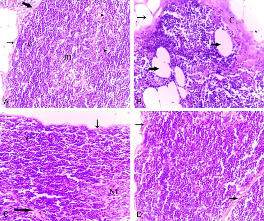
Melatonin and tumeric ameliorate aging-induced changes

ous biological processes; including the regu-               covered with aluminum foil; and kept in a refrig-
lation of ROS levels and induction of apoptosis             erator; fresh solutions were prepared every two
[13]. DJ-1 is therefore, a signaling molecule               days.
responsive to cellular redox state, which exerts
antioxidant functions [14]. Therefore, DJ-1 pro-            Turmeric: The turmeric (Curcuma longa) was
tects cells against apoptosis triggered by                  obtained, in the powder form available com-
increased level of ROS through protecting mito-             mercially, from a local market in El-Minia city,
chondrial integrity by attenuating the transcrip-           Egypt. The powdered turmeric was mixed with
tion of antioxidant genes [15]. Recent studies              laboratory diet at a concentration of 2% w/w.
linked DJ-1 expression fluctuations with aging
[16, 17].                                                   Experimental design

During the present research, the effect of aging            After one week of acclimatization, a group of
in rats (Rattus norvgicus) on the regulation of             young adult rats at 3-4-months of age (n = 6)
selected immunoglobulins (IgA and IgE), aging-              was assigned as normal control adult rats [nor-
related cytokines (TNF-α, IFN-γ, IL-6 and IL-8) in          mal adult (N. adult)]; they were provided with
blood serum and antioxidant master upstream                 normal diet and water ad libitum throughout
signaling molecules such as DJ-1 and NRF2                   the course of the study. Aged rats (16-18
and apoptosis-inducing proteins in the thymus               months-old) were randomly divided into four
tissues as well as the histological changes in              experimental groups.
thymus and spleen tissues were investigated.
Next, the ameliorative effects of two natural               The first group of aged animals was considered
products (melatonin and tumeric) and their                  as normal control aged group (N. aged group; n
mechanisms against aging were investigated.                 = 6) in which animals received laboratory diet,
                                                            and they did not receive any substances or
Materials and methods
                                                            undergo any experimental manipulation.
Animals and housing condition
                                                            The second group of animals [aged+melatonin
Young adult male albino rats (3-4 months),                  (aged+Mel) Group; n = 5] was treated orally
(Rattus norvgicus), weighing from 95-120 g,                 with melatonin (10 mg/kg) five times a week for
and twenty three aged male albino rats (16-18               90 days in the late afternoon (4.00-6.00 pm).
months), weighing from 350-400 g were
obtained from the breeding colony of the                    The third group of rats (n = 6) was used as vehi-
Ministry of Health (Helwan-Egypt). Upon arrival,            cle control for the aged+Mel group, and
the animals were weighed; group housed (three               received 0.5% ethanolic phosphate-buffered
animals per cage) and kept in a well-ventilated             saline by oral intubation in the late afternoon.
animal facility under normal laboratory condi-
tions. They were acclimatized for one week                  The fourth group of rats [aged+turmeric
prior to experimentation. Food and water were               (aged+Tum) Group; n = 6] was fed powdered
available ad libitum throughout the study. All              tumeric mixed with normal laboratory diet at a
procedures were in accordance with institution-             concentration of 2% w/w for 90 consecutive
al guidelines and follow the Guide for Care and             days. The selection of this dose was based on a
Use of Laboratory Animals.                                  previous research study [19].

Chemicals and plant materials                               The design of all experimental groups is sum-
                                                            marized in Table 1. The experiments were con-
Melatonin: Melatonin (N-acetyl-5-methoxytryp-               tinued for 90 days and all animals were weighed
tamine) was purchased from Sigma Chemical                   weekly.
Co. (St. Louis, Mo, USA). It was prepared in eth-
                                                            Blood and tissue sampling
anol due to its instability in non-sterile solu-
tions. Briefly, melatonin was dissolved in 0.5 ml           At the end of the experiment, the animals were
of 100% ethanol and diluted with phosphate-                 decapitated under deep ether anesthesia at
buffered saline (PBS) to a final concentration of           the morning time. Blood samples were collect-
10 mg melatonin/1 ml of 0.5% ethanolic PBS                  ed and allowed to clot at room temperature.
[18]. The bottles of melatonin solution were                After that, they were centrifuged for 30 min at

71                                                   Int J Physiol Pathophysiol Pharmacol 2018;10(2):70-82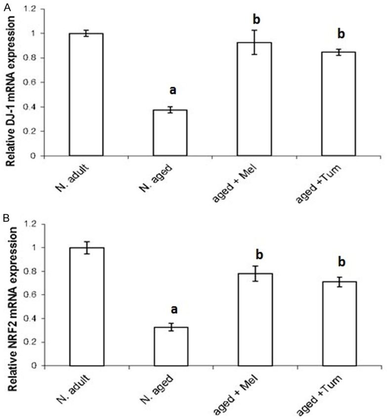
Melatonin and tumeric ameliorate aging-induced changes

Table 1. The scheme of the experimental design
                No. of
Groups                        Dose              Route                         Time                Scarifying time
                 rats
N. adult          6          ……………               ……                            ……                 After 90 days
N. aged           6          ……………               ……                            ……                 After 90 days
Aged+Mel          5       10 mg/kg.b.wt          Oral             Five times a week for 90 days   After 90 days
Vehicle control   6    0.5% ethanolic + PBS      Oral             Five times a week for 90 days   After 90 days
Aged+Tum          6          2% w/w         Mixed with diet                For 90 days            After 90 days
Note: N, normal; Mel, melatonin; Tum, turmeric.

Table 2. Histopathological scoring system of the thymus
         Degree of lympho-         Count of pyknotic Adipose tissue                              IL septae
Score                                                                C/M boundaries C/M ratio
          cytic depletion         nuclei in the cortex  infiltration                            Thickening
(0)            Nil (---)                 None            None (---)       Distinct       > 1:1    Nil (----)
(1)           Mild (+)                     1-3            Mild (+)        Distinct        1:1     Mild (+)
(2)       Moderate (++)                   4-10         Moderate (++) Slightly indistinct < 1:1 Moderate (++)
(3)        Marked (+++)                   > 10         Marked (+++)      Indistinct      < 1:1 Marked (+++)
Note: C/M, cortex to medullary; IL, interlobular.

4000 rpm. Serum aliquots were extracted and                   Immunological studies
stored in microcentrifuge tubes and kept at
-80°C until assayed.                                          Determination of Immunoglobulin-A (IgA) and I
                                                              mmunoglobulin-E (IgE): Total Immunoglobulin
After blood sampling, the thymus and spleen                   (Ig) A and E in blood serum samples were mea-
were immediately removed from each animal.                    sured from all the experimental groups by
Samples of thymus and spleen were fixed in                    Enzyme-Linked Immuosorbent Assay (ELISA)
neutral formalin solution (10%), dehydrated                   kits (CellTrend GmbH, Lukenwalde, Germany;
and embedded in parablast (m.p.54-56;                         Cat No. 52400, 52600, respectively) Then, the
Sigma). Five-micrometer thick sections were                   results were calculated and expressed as nano-
prepared and stained with Haematoxylin and                    grams per milliliter (ng/ml).
Eosin (H&E) using standard procedures for gen-
eral histological architecture. Moreover, sam-                Determination of cytokines Tumor Necrosis
ples of thymus were kept in -80°C until be used               Factor-alfa (TNF-α), Interferon-γ (IFN-γ), inter-
for RNA extraction for real time PCR analyses.                leukin-6 (IL-6), and interleukin-8 (IL-8): The lev-
                                                              els of TNF-α, IFN-γ, IL-6, and IL-8 in the blood
Histological analysis and pathological scoring                serum were determined by enzyme immunoas-
system                                                        say using ELISA kits according to the manufac-
                                                              turer’s recommendations (Koma Biotech Inc;
Sections of thymus and spleen were examined                   Cat. No. K0331196, ScienCell Research
with light microscopy and separately scored                   Laboratories; Cat. No. EK0374, Bioo Scientific;
using a semiquantitative scoring system.                      Cat. No. 2202, and Cusabio Biotech Co; Cat.
                                                              No. CSB-E07273r, respectively). The principal
Thymus: As shown in Table 2, subjective analy-
                                                              method for all the immunoassay was the same
sis of the degree of thymic involution was based
                                                              for all cytokines. The results are expressed as
on various parameters known to change with
                                                              picograms per milliliter (pg/ml).
increasing age. Thymus sections were then
semiquantitatively and qualitatively graded for               Gene expression analyses
the presence of thymic involution using a scale
from 0 to 3 as follows: 0 = absent; 1 = mild; 2 =             The expression levels of the indicated genes
moderate; 3 = severe.                                         were investigated in thymus tissues of rats
                                                              from the all experimental groups using quanti-
Spleen: Sections of spleen were graded for the                tative real-time PCR (RT-PCR). The procedure of
presence of lymphocytic depletion, and a score                analysis was performed as previously described
from 0 to 4 was attributed as: 0 (none), 1 (mini-             [11]. Total RNA was extracted with Qiazol
mal), 2 (mild), 3 (moderate) and 4 (severe).                  (Qiagen, Valencia, CA, USA) according to the
72                                                    Int J Physiol Pathophysiol Pharmacol 2018;10(2):70-82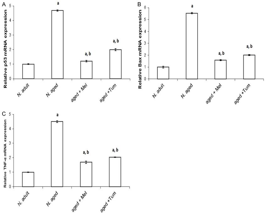
Melatonin and tumeric ameliorate aging-induced changes

                                                                              using 7500 fast (Applied
                                                                              Biosystems, USA). All samples
                                                                              were amplified in triplicate in
                                                                              a 96-well plate using the fol-
                                                                              lowing cycling conditions: 10
                                                                              minutes at 95°C, and 40
                                                                              cycles at 95°C for 15 seconds
                                                                              followed by one minute at
                                                                              60°C.

                                                                              Statistical analysis

                                                                              The data were presented as
                                                                              mean ± standard error. P-
                                                                              value > 0.05 was considered
                                                                              statistically significant (using
                                                                              the SPSS program, version
                                                                              16.0). Statistical investiga-
                                                                              tions were performed using
                                                                              ANOVA (analysis of variance)
                                                                              followed by Post-Hoc test for
                                                                              multiple comparisons. Calcu-
                                                                              lations of real time PCR
                                                                              results were performed by
                                                                              determining the values of
                                                                              Δcycle threshold (ΔCt) using
                                                                              the     endogenous       control
                                                                              (GAPDH) for normalization.
Figure 1. Serum levels of immunoglobulin-A (IgA) (A) and immunoglobulin-E     Then 2-ΔΔCt for each treatment
(IgE) (B) in normal adult rats, normal aged and normal aged treated with      was calculated and statistical
either melatonin or turmeric for 12 weeks. The level of IgA and IgE were
                                                                              analysis for data were per-
investigated in blood serum of rats as indicated in materials and methods
section. Data then were calculated and statistical analyses were performed.   formed as previously descri-
N = normal; Mel = melatonin; Tum = turmeric. A = P < 0.05 vs. normal adult    bed [20].
animals, b = P < 0.05 vs. normal aged rats.
                                                                              Results
manufacturer’s instructions. Five µg of extract-             Attenuation of aging-induced changes in
ed RNA was reverse transcribed into cDNA                     immunoglobulin-A (IgA) and immunoglobulin-E
using the first strand cDNA synthesis kit                    (IgE) by melatonin and Turmeric administration
(Applied Biosystems, Fosters city, CA, USA), and
the resulting cDNA was diluted 10-fold and                   The current data showed the effect of aging on
kept at -20°C. The real time RT-PCR primers                  serum IgA and IgE in rats. Results showed that
were designed by the primer Express 1.5 soft-                aging significantly increased (P < 0.01) serum
ware (Applied Biosystems) as follows: GAPDH                  IgA value as shown in normal aged rats
forward, 5’-ATCTTCTTGTGCAGTGCCAGC-’3 and                     (104.45±0.736) when compared to normal
GAPDH reverse, 5’-GAAGGCAGCCCTGGTAA-                         adult rats (88.1±4.47). Interestingly, melatonin
CC-’3 and Bax forward, 5’-TCATGAAGACAGGGG-
                                                             or turmeric administration to normal aged rats
CCTTT-’3 and Bax reverse, 5’-CTGCAGCTCC-
                                                             for 12 weeks (aged+Mel or aged+Tum groups)
ATGTTGTTGT-’3 and NRF2 forward, 5’-TG-
                                                             resulted in a significant decrease (P < 0.01) in
TCAGCTACTCCCAGGTTG-’3 and NRF2 reverse,
5’-ATCAGGGGTGGTGAAGACTG-’3 and DJ-1 for-                     serum IgA levels when compared with normal
ward, 5’-GGAGCAGAGGAGATGGAGAC-’3 and                         aged rats and restored them to their normal
DJ-1 reverse, 5’-TCACCAAAGCCGACTCAGAT-’3                     level (Figure 1A). On the other hand, the serum
and TNF-α forward, 5’-CGTCGTAGCAAACCACC-                     IgE level was significantly decreased (P < 0.01)
AAG-’3 and TNF-α reverse, 5’-GAGGCTGACTTTC-                  in the normal aged group when compared to
TCCTGGT-’3 and P53 forward, 5’-CTCCTCTCC-                    normal adult rats. However, treatment of aged
CCAGCAAAAGA-’3 and P53 reverse, 5’-GTAG-                     rats with melatonin or turmeric for 12 weeks
ACTGGCCCTTCTTGGT-’3. QPCR was carried out                    (aged+Mel and aged+Tum groups) markedly
73                                                  Int J Physiol Pathophysiol Pharmacol 2018;10(2):70-82
Melatonin and tumeric ameliorate aging-induced changes
Figure 2. Serum levels of Tumor Necrosis Factor-alpha (TNF-α) (A), interferon gamma (IFN-γ) (B), interleukon 6 (IL-6)
(C), and interleukon 8 (IL-8) (D) in normal adult rats, normal aged, and normal aged treated with either melatonin or
turmeric for 12 weeks as indicated in methods section. N = normal; Mel = melatonin; Tum = turmeric. A = P < 0.01
vs. normal adult animals, b = P < 0.01 vs. normal aged rats.
increased (P < 0.01) the serum IgE and restored Modulatory effects of melatonin and turmeric
its low level as in normal adult rats (Figure 1B). on aging-induced changes in apoptosis-related
genes
Attenuation of aging-induced increases in
serum cytokines by melatonin and turmeric To determine the age-related changes in gene
expression of DJ-1, NRF2, p53, Bax and TNF-α
The modulatory effect of melatonin and turmer- in thymus, gene expression analyses for the
ic on the changes induced by aging in serum indicated genes were investigated using quan-
cytokines; Tumor Necrosis Factor-alpha (TNF- titative real time PCR (Figures 3, 4). The mRNA
α), Interferon-gamma (IFN-γ), Interleukin-6 (IL- levels of DJ-1 (Figure 3A) and NRF2 (Figure 3B)
6) and Interleukin-8 (IL-8) were evaluated. The genes were highly expressed in thymus tissues
present data showed that, the levels of Tumor of normal adult rats. On aging, the DJ-1 and
Necrosis Factor-alpha (TNF-α), Interferon-gam- NRF2 mRNA level were significantly repressed
ma (IFN-γ), Interleukin-6 (IL-6) and Interleukin-8 (P < 0.01) when compared with the normal
(IL-8) were significantly increased (P < 0.01) in adult rats. Interestingly, treatment the aged
serum of normal aged rats when compared rats with melatonin or turmeric for 12 weeks
with those of normal adult rats (Figure 2). In (aged+Mel or aged+Tum) increased markedly
contrast, melatonin or turmeric administration the mRNA expression levels of both DJ-1 and
to normal aged rats for 12 weeks (aged+Mel or NRF2 (Figure 3A, 3B).
aged+Tum groups, respectively) resulted in a
significant decrease (P < 0.01) in their levels On the other hand, basal mRNA level of p53
toward normal adult levels, but their values (Figure 4A), Bax (Figure 4B) and TNF-α (Figure
were still significantly higher than those of nor- 4C) were low expressed in thymus tissues of
mal adult rats (Figure 2). normal adult rats. The p53, Bax and TNF-α
74 Int J Physiol Pathophysiol Pharmacol 2018;10(2):70-82Melatonin and tumeric ameliorate aging-induced changes
tive tissue capsule. Each lobe
is partly divided by connective
tissue septa into incomplete
lobules. The thymic lobules
comprise a darkly stained
peripheral cortex and a lightly
stained central medulla. The
cortex consists mainly of
small and densely packed
lymphocytes (Figure 5A). The
medulla consists of a large
number of reticulo-epithelial
cells (Figure 5A), in addition
to a few fully mature lympho-
cytes, macrophages and
occasional Hassall’s corpu-
scles.
Thymus of aged rats showed
marked thymic involution
compared with those ob-
tained from the normal adult
rats. The capsule and inter
lobular septa became slightly
thickened. The cortex showed
obvious lymphocytic deple-
tion with the appearance of
Figure 3. Age-related changes in mRNA expression levels of DJ-1 and NRF2 numerous shrunken, necrotic
in thymus tissue. DJ-1 (A) and NRF2 (B) mRNA expression levels in thymus as well as apoptotic lympho-
tissues of normal adult rats, normal aged and normal aged treated with���� ei-
��� cytes (Figure 5B). The cortical
ther melatonin or turmeric for 12 weeks were investigated using real time reticuloepithelial cells were
quantitative PCR. Data were then statistically analyzed as indicated in meth-
clearly identified due to the
ods section. N = normal; Mel = melatonin; Tum = turmeric. A = P < 0.05 vs.
normal adult animals. B = P < 0.05 vs. normal aged rats. reduction in the numbers of
cortical lymphocytes (Figure
5B). Such alterations in the
mRNA expression levels were significantly ele- cortical cellularity resulted in a great loss of
vated (P < 0.01) in thymus tissues of normal demarcation between cortex and medulla. In
aged rats (N. aged group), compared with the addition, increased numbers of adipocytes infil-
normal adult control rats (N. adult group). The tered the capsular and septal areas, and
enhanced expressions of p53, Bax and TNF-α extended into the cortex (Figure 5B). In the
by aging were significantly (P < 0.01), and then medulla, the epithelial component appeared
diminished in aged animals treated with either more prominent due to the decrease in the den-
melatonin or turmeric for 12 weeks (aged+Mel sity of medullary lymphocytes. A variety of epi-
or aged+Tum groups) (Figure 4A-C respective- thelial cells were arranged in the cortex or in
ly). However, their values after treatment with the medulla as prominent tubular structures
either melatonin or turmeric didn’t reach their lined by cuboidal epithelium.
basal levels in control normal adult rats (Figure
4). Melatonin or turmeric administration resulted
in obvious alterations in the cellular density and
Aging induced cellular morphological changes cellular compositions of the thymus of normal
in rat thymus and the ameliorative effect of aged rats. However, the histological appear-
melatonin and turmeric ance of the aged thymus under these various
treatment conditions was similar. Specifically,
In normal control adult rats, the thymus gland melatonin or turmeric administration to normal
is a bi-lobed organ covered with a thin connec- aged rats for 12 weeks (aged+Mel or aged+Tum
75 Int J Physiol Pathophysiol Pharmacol 2018;10(2):70-82Melatonin and tumeric ameliorate aging-induced changes
Figure 4. Age-related changes in p53, Bax and TNF-α
mRNA expression levels. The expression levels of
p53 (A), Bax (B) and TNF-α (C) mRNA in thymus tis-
sue of normal adult rats, normal aged and normal
aged treated with either melatonin or turmeric were
investigated using real time quantitative PCR and
statistical significance were then analyzed. N = nor-
mal; Mel = melatonin; Tum = turmeric. A = P < 0.05
vs. normal adult; b = P < 0.05 vs. normal aged rats.
with densely backed lymphocytes
and light stained medulla (m)
with lymphocytes and macro-
phages (short arrows) as well as
epithelial cells (irregular arrows).
Note the thin capsule (thin arrow)
and the thin interlobular septum
(thick arrow). (B) Thymus tissues
of normal aged rats showing the
lymphocytes depletion of the
cortex. Note the thickened cap-
sule (C) and increased numbers
of adipocytes infiltered the cap-
sular (thin arrow) and extended
into the cortex (thick arrows).
(C) Thymus of a normal aged rat
treated with melatonin showing
increased lymphocytic population
in the cortical (C) and medullary
(M) regions. Note the thin cap-
sule (thin arrow) and interlobular
septum (thick arrow), as well as
the absence of adipocytes. (D)
Thymus of a normal aged rat
treated with turmeric showing the
histological improvement in the
Figure 5. Photomicrographs of thymus tissues of normal adult rat (A), nor- thymic tissue. Note: the cortical
mal aged (B), normal aged treated with either melatonin (C) or turmeric (D). (C) and medullary (M) regions.
(A) Thymus tissues of normal adult rats showing a dark stained cortex (c) (H&E, 400X).
76 Int J Physiol Pathophysiol Pharmacol 2018;10(2):70-82Melatonin and tumeric ameliorate aging-induced changes
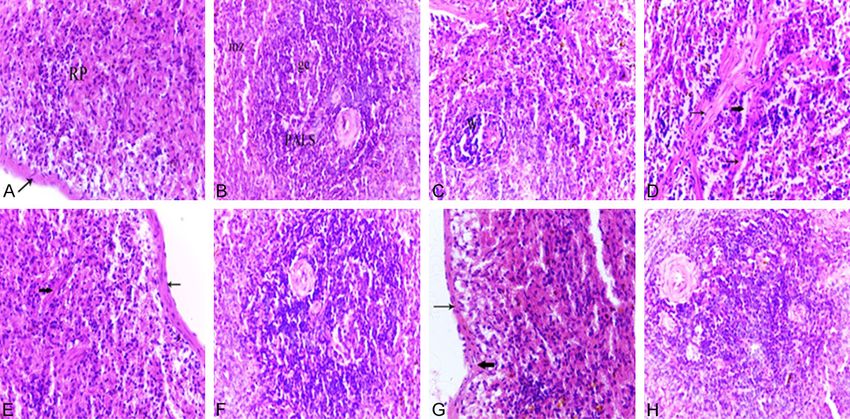
Figure 6. Photomicrographs of spleen tissues of a normal adult rat (A, B), normal aged (C, D), normal aged treated
with either melatonin (E, F) or turmeric (G, H). (A) Spleen of an adult rat showing the splenic red pulp (RP). Note
the less-thickened capsule (arrow). (B) Spleen of an adult rat showing the PALS and marginal zones (mz). Note the
presence of follicles containing prominent germinal centers (gc) with tangible body macrophages which appeared
with cytoplasmic engulfed apoptotic debris. (C) Spleen of a normal aged rat revealed the decreased cellularity of
both white pulp and red pulp areas. Note the lymphocytic depletion of the white pulp (W) and the marginal zone is
hardly defined. (D) Spleen of a normal aged rat showing the lymphocytic depletion of the white pulp (W) with shrink-
age and apoptotic cells. Note the tangible body macrophages with cytoplasmic engulfed apoptotic bodies. (H&E,
400X). (E) Spleen of a normal aged rat treated with melatonin showing the increased lymphocytic population of the
splenic red pulp. Note the less-thickened capsule (thin arrow) and trabeculae (thick arrow). (F) Spleen of a normal
aged rat treated with melatonin showing a marked improvement where the lymphocytic population of the white pulp
was increased. (G) Spleen of a normal aged rat treated with turmeric showing the increased lymphocytic population
of the splenic red pulp. Note the less-thickened capsule (thin arrow) and trabeculae (thick arrow) and the decrease
number of pigments. (H) Spleen of a normal aged rat treated with turmeric showing apparent increase in the lym-
phocyte population of white pulp. (H&E, 400X).
groups, respectively) produced a marked (Figure 6A, 6B); trabeculae of smooth muscles
improvement in aged thymic tissue. The density and fibroelastic tissue were also seen in the
of the cortical lymphocytes was apparently splenic parenchyma. The red pulp was consist-
increased, and the number of necrotic thymo- ed of a meshwork of splenic cords, which con-
cytes seemed to be decreased compared to tained various types of cells, and venous sinus-
that seen in the thymus of normal aged rats es (Figure 6A). The white pulp was composed
(Figure 5C, 5D). Also, the thickened interlobular of periarteriolar lymphoid sheath (PALS), lym-
septa as well as the thickened capsule phoid follicles, and prominent marginal zone
appeared thinner than those observed in the located between the white and red pulp (Figure
thymus of normal aged rats (Figure 5C, 5D). 6B). The follicle sometimes had germinal cen-
Moreover, it has been noticed that no adipo- ters which occasionally contained apoptotic
cytes were found in all examined thymus sec- cells and tingible body macrophages engulfing
tions of these groups. apoptotic bodies (Figure 6B). Furthermore, a
variety of pigments were detected in the spleen
Aging induced cellular morphological changes of control rats. Particularly, deposits were seen
in rat spleen and the ameliorative effect of throughout the cytoplasm of macrophages in
melatonin and turmeric the red pulp, and sometimes in the white pulp
as well.
The spleen of adult rats showed normal histo-
logical architecture composed of two distinct The histological architecture of spleen sections
compartments, the red pulp and the white pulp from normal aged rats showed striking altera-
77 Int J Physiol Pathophysiol Pharmacol 2018;10(2):70-82Melatonin and tumeric ameliorate aging-induced changes
Table 3. Histopathological scores of the thymus appeared clearly decreased compared to
Groups N. adult N. aged Aged+Mel Aged+Tum that observed in the spleen of normal aged
Scores 0.3.±0.16 2.5±0.06a 0.8±0.44b 0.5±0.28b rats (Figure 6E and 6G).
The histopathological alterations were graded according to
parameters previously described in the Materials and Methods
Histopathological scores of thymus and
section. Data represent means ± SEM. N = normal; Mel = mela- spleen
tonin; Tum = turmeric. a = P < 0.01 vs. normal adult animals. b
= P < 0.01 vs. normal aged rats. The thymus and spleen histopathological
scores were higher (P < 0.01) in normal aged
rats than in normal adult rats (Tables 3, 4). In
Table 4. Histopathological scores of the spleen contrast, melatonin or turmeric administra-
Groups N. adult N. aged Aged+Mel Aged+Tum tion to normal aged rats for 12 weeks
Scores 0.0±0.0 3.5±0.28a 0.6±0.33b 1±0.57b (aged+Mel or aged+Tum groups, respectively)
The histopathological alterations were graded as described in caused a significant decrease (P < 0.01) in
the Materialsand Methods section. Data represent means ± the histopathological scores, when com-
SEM. N = normal; Mel = melatonin; Tum = turmeric. a = P < 0.01
vs. normal adult animals. b = P < 0.01 vs. normal aged rats. pared to aged rats (Tables 3, 4). However,
there were no significant differences in the
thymic or splenic histopathology scores
tions compared to those of adult animals. between aged+Mel or aged+Tum groups and
Specifically, all spleen sections of aged rats the normal adult animals (Tables 3, 4).
revealed an obvious decrease in the cellularity
of both white pulp and red pulp areas com- Discussion
pared with normal adult rats. The white pulp
areas showed lymphocytic depletion with the Ample data suggested that melatonin is impli-
appearance of numerous shrunken and necrot- cated in aging regulation [21-23]. It could be
ic lymphocytes (Figure 6C). The PALS of the observed that melatonin declined with age, and
white pulp showed marginal zones with dilated this reduction in the level of melatonin is an
marginal sinusoids. Additionally, many apoptot- important indicator for the increased oxidative
ic cells and tangible body macrophages with stress by aging [21]. Furthermore, turmeric is
cytoplasmic-engulfed apoptotic bodies were reported also to be involved in aging regulation
seen in the white pulp. Pigment-laden macro- [5, 24]. In the present research, the effect of
phages were commonly observed in the red aging on selected immunoglobulins (IgA and
pulp and white pulp (Figure 6C and 6D). Also, IgE), some cytokines (TNF-α, IFN-γ, IL-6 and
pronounced dilatation and congestion of splen- IL-8), antioxidant markers (NRF2 and DJ-1), and
ic blood vessels and sinuses were found (Figure apoptotic markers (p53 and Bax) in addition to
6D), accompanied by an increase in hemosid- the histological changes accompanied with
erin pigments (hemosiderosis). The capsule aging were investigated in rats. In addition, the
and trabeculae appeared more thickened com- ameliorative effect of melatonin and turmeric
pared to those of adult animals (Figure 6D). against aging was investigated.
Melatonin or turmeric administration for 12 In the present work, IgA was significantly
weeks exerted prominent ameliorative effects increased in serum of normal aged rats with a
on the histological alterations induced by aging concurrent decrease in IgE level when com-
in the spleen tissue. In specific, the white pulp pared to that of normal adult rats. These find-
became easily defined with increased lympho- ings are in agreement with those reported that
cytic population (Figure 6F and 6H). The mar- aging resulted in elevation in the levels of
ginal zone became well-differentiated (Figure serum IgA and IgG [25]. Total serum IgE levels
6F and 6H). The splenic cords of the red pulp increased progressively from the age of 1
exhibited an obvious increase in the cellular month to the age of 5 months and then declined
population, specifically lymphocytes (Figure 6E after the age of one-year [26]. Such observa-
and 6G). The hemosiderin and pigment-laden tions were also reported that serum IgE levels
macrophages were reduced all over the splenic and antigen-specific IgE production both
tissue (Figure 6E-H). Furthermore, the thick- decreased with age [27]. Currently, we found
ness of the trabeculae as well as the capsule that administration of both melatonin and tur-
78 Int J Physiol Pathophysiol Pharmacol 2018;10(2):70-82Melatonin and tumeric ameliorate aging-induced changes
meric decreased IgA and increased IgE serum the finding that the expression of the proapop-
level in aged rats, and this is consistent with totic Bax protein was increased with aging [39].
the findings that melatonin and IgA were cor- Consistent with our data, it could be found that
relatively expressed [28]. Also, consistently melatonin reversed the effect of aging on p53
with the current results, it is stated that melato- and Bax expression [40]. Consistently, other
nin administration inhibited the serum IgE lev- studies showed that turmeric treatment
els in the model mice [29]. Meanwhile, we decreased p53 and Bax expression levels [41,
found that curcumin uncovered aging-induced 42].
immunoglobulin A and E changes. On the other
hand, it could be also found that curcumin ele- The thymus gland is a primary lymphoid organ
vated immunoglobulin-A in rats [30] and inhib- responsible for the differentiation and matura-
ited IgE in Guinea pigs [31]. Also, another study tion of T lymphocytes and is termed the “the
showed that curcumin had no effect on the immunological clock of aging” [43]. The thymus
immune status [32]. Melatonin is a potent ther- gradually decreases its capacity to generate
apeutic candidate regulating immune function immunocompetent T cells and becomes mini-
in aged individuals [22]. mally functional gradually with aging; under-
goes dramatic alterations in its size, morpholo-
In the present study, the levels of Tumor Ne- gy and cell composition, a process termed
crosis Factor-alpha (TNF-α), Interferon-gamma “age-associated thymic involution” [44]. The
(IFN-γ), Interleukin-6 (IL-6) and Interleukin-8 (IL- present observations showed that, thymus of
8) were significantly increased in serum of nor- aged rats showed a marked thymic involution
mal aged rats compared to that recorded in compared to normal adult rats. The data pre-
normal adult rats. These findings were support- sented herein confirm the earlier reports which
ed by the findings that, aging is associated with stated that thymus of old rats showed decre-
increased levels of pro-inflammatory cytokines ased cortical lymphocytes with apparent of epi-
such as TNF-α, IL-6, IL-1β [33]. Moreover, our thelial cells, increased fibrovascular tissues,
findings indicated that melatonin significantly loss of cortical and medullary boundaries, aug-
decreased TNF-α, IFN-γ, IL-6 and IL-8 when mented levels of apoptosis [44, 45]. The
compared to normal aged rats without melato- appearance of numerous shrunken, necrotic as
nin supplementation. In this respect, melatonin well as apoptotic lymphocytes in the thymus of
was found to modulate the inflammatory status aged rats is associated with a concurrent
of through aging [34]. Similarly, turmeric admin- increase in the level of TNF-α in serum of nor-
istration to normal aged rats decreased TNF-α, mal aged rats. Furthermore, the present data
IFN-γ, IL-6 and IL-8 when compared to normal also revealed an increased number of adipo-
aged rats. These anti-inflammatory effects of cytes infiltered the capsular and septal areas,
turmeric were supported by finding that, cur- and extended into the cortex of thymus of aged
cumin counteracted the pro-inflammatory sta- rats. These results supported data from other
tus [5]. studies, which demonstrated that thymus is
the major immune organ that is largely replaced
Regarding the effect of aging on the antioxidant with fat during aging [46].
master regulator NRF2 and its upstream signal-
ing DJ-1 protein; current data showed that both It is well established that the pineal gland
DJ-1 and NRF2 mRNA levels were significantly through its principal hormone melatonin affects
declined with aging. In this respect, it is stated lymphoid tissue size, structures and function
that DJ-1 is implicated in oxidative stress and [47]. In the present work, administration of mel-
aging regulation [35, 36]. Furthermore, NRF2 is atonin to normal aged rats produced a marked
suggested as a potential therapeutic candidate improvement in aged thymic tissue such as
against oxidative stress and aging [6, 8]. increased the density of the cortical lympho-
Consistent with the present data, it is stated cytes, decreased the number of necrotic thy-
that decline of NRF2 caused age-related fea- mocytes and absence of adipocytes when com-
tures [9]. The tumor suppressor p53 was cor- pared to that seen in the thymus of normal
relatively expressed with aging [37, 38]. aged rats. These results are consistent with the
Consistently, here we found that p53 mRNA finding that, melatonin prevented age-related
expression level increased with aging in thymus thymic involution through regulation of thymo-
tissues. Also, current data is consistent with cyte apoptosis [48]. The adipose tissue is also
79 Int J Physiol Pathophysiol Pharmacol 2018;10(2):70-82Melatonin and tumeric ameliorate aging-induced changes
an important target for the action of melatonin. Conclusively, the present study showed that
In this issue, it was demonstrated that melato- aging is implicated with changes of several sig-
nin reverted age-related changes caused by naling cascades such as immunoglobulins, cyo-
adiposity [49]. This is the first report to the kines, and antioxidants- and apoptosis-related
available knowledge, showing that turmeric has proteins. These changes could be reversed
beneficial effects on thymus and splenic tis- using melatonin and tumeric administration.
sues abnormalities in aged rats. Similar to mel- These data suggest melatonin and tumeric as
atonin treatment, turmeric administration to potent anti-aging candidates through activating
normal aged rats for 12 weeks produced a the antioxidant upsetream regulators; DJ-1 and
marked improvement in aged thymic tissue. In NRF2 and inhibiting apoptosis-inducing pro-
addition, curcumin may suppress preadipocyte teins p53 and Bax.
differentiation and thus reduce the number of
adipocytes and fat content of adipose tissue Disclosure of conflict of interest
[50]. Thus, the current data support the sug-
gestion that aging is a chronic inflammatory None.
process with a shift towards a proinflammatory
Address correspondence to: Ismail Ahmed Ismail,
cytokine profile in tissues. This suggestion is
Department of Biology, Faculty of Science, Taibah
further confirmed by current immunological
University, Yanbu Branch, Saudi Arabia; Laboratory
results in which the levels of pro-inflammatory
of Molecular Cell Biology, Department of Zoology,
cytokines (TNF-α, IFN-γ, IL-6, IL-8) were signifi-
Faculty of Science, Assiut University, Assiut 71516,
cantly increased in serum of normal aged rats
Egypt. E-mail: ismailahmed@aun.edu.eg; ismai-
compared to that recorded in serum of normal
l75eg@yahoo.com
adult rats.
Histopathological examination showed a loss References
of the cellularity of both the white and red pulps
[1] Harman D. Free radical involvement in aging.
in the spleen of aged rats compared to those Drugs Aging 1993; 3: 60-80.
from controls. These alterations were associ- [2] Harman D. Free radical theory of aging: an up-
ated with the appearance of numerous shrunk- date. Ann N Y Acad Sci 2006; 1067: 10-21.
en and necrotic lymphocytes and congestion of [3] Reiter RJ, Tan DX, Osuna C and Gitto E. Actions
the splenic sinusoids. The present findings are of melatonin in the reduction of oxidative
consistent with numerous studies that confirm stress. J Biomed Sci 2000; 7: 444-458.
the occurrence of condition of immunosuppres- [4] Mauriz JL, Molpeceres V, García-Mediavilla MV,
sion in aging [45, 51]. In this context, normal González P, Barrio JP, González-Gallego J. Mel-
aging has been shown to induce noticeable atonin prevents oxidative stress and changes
alterations in the splenic architecture including in antioxidant enzyme expression and activity
in the liver of aging rats. J Pineal Res 2007; 42:
the presence of thickly dyed karyopyknosis and
222-230.
many apoptotic cells as well as congested [5] Sikora E, Scapagnini G and Barbagallo M. Cur-
splenic sinus and increased macrophages. cumin, inflammation, ageing and age-related
Similar results were obtained by different diseases. Immun Ageing 2010; 7: 1.
authors who observed that with the develop- [6] Petri S, Korner S and Kiaei M. Nrf2/ARE signal-
ment of aging, there is a substantial reduction ing pathway: key mediator in oxidative stress
in lymphocytes of splenic white pulp and huge and potential therapeutic target in ALS. Neurol
increasing macrophages [45, 51]. Furthermore, Res Int 2012; 2012: 878030.
in the present study, splenic sections of aged [7] Jung KA, Kwak MK. The Nrf2 system as a po-
rats treated with melatonin showed a marked tential target for the development of indirect
improvement in the histological picture, where antioxidants. Molecules 2010; 15: 7266-
7291.
the white pulp became easily defined with
[8] Chapple SJ, Siow RC, Mann GE. Crosstalk be-
increased lymphocytic population. The margin- tween Nrf2 and the proteasome: therapeutic
al zone became well-differentiated. The hemo- potential of Nrf2 inducers in vascular disease
siderin pigments were reduced in the red pulp and aging. Int J Biochem Cell Biol 2012; 44:
compared to that seen in the spleen of normal 1315-1320.
aged rats. These results are in agreement with [9] Suh JH, Shenvi SV, Dixon BM, Liu H, Jaiswal AK,
the finding that melatonin has a potential role Liu RM and Hagen TM. Decline in transcrip-
in maintaining the function and activity of tional activity of Nrf2 causes age-related loss
splenic tissue in old rats [45]. of glutathione synthesis, which is reversible
80 Int J Physiol Pathophysiol Pharmacol 2018;10(2):70-82Melatonin and tumeric ameliorate aging-induced changes
with lipoic acid. Proc Natl Acad Sci U S A 2004; nin, immune function and aging. Immun Age-
101: 3381-3386. ing 2005; 2: 17.
[10] Gan LI, Johnson DA and Johnson JA. Keap1- [23] Turek FW, Zee P and Van Reeth O. Melatonin
Nrf2 activation in the presence and absence and aging. Adv Exp Med Biol 1999; 460: 435-
of DJ-1. Eur J Neurosci 2010; 31: 967-977. 440.
[11] Ismail IA, Abdel Shakor AB and Hong SH. DJ-1 [24] Anand P, Sundaram C, Jhurani S, Kunnumak-
protects breast cancer cells against 2’-Benzoy- kara AB and Aggarwal BB. Curcumin and can-
loxycinnamaldehyde-induced oxidative stress cer: an “old-ageâ€disease with an
independent of Nrf2. J Cell Physiol 2015; 230: “age-old†solution. Cancer Lett 2008;
2262-9. 267: 133-164.
[12] Nagakubo D, Taira T, Kitaura H, Ikeda M, Tamai [25] Fagnoni FF, Vescovini R, Passeri G, Bologna G,
K, Iguchi-Ariga SM, Ariga H. DJ-1, a novel onco- Pedrazzoni M, Lavagetto G, Casti A, Franceschi
gene which transforms mouse NIH3T3 cells in C, Passeri M and Sansoni P. Shortage of circu-
cooperation with ras. Biochem Biophys Res lating naive CD8+ T cells provides new insights
Commun 1997; 231: 509-513. on immunodeficiency in aging. Blood 2000;
[13] Andres-Mateos E, Perier C, Zhang L, Blanchard- 95: 2860-2868.
Fillion B, Greco TM, Thomas B, Ko HS, Sasaki [26] Pauwels R, Bazin H, Platteau B and Van der
M, Ischiropoulos H and Przedborski S. DJ-1 Straeten M. The effect of age on IgE produc-
gene deletion reveals that DJ-1 is an atypical tion in rats. Immunology 1979; 36: 145.
peroxiredoxin-like peroxidase. Proc Natl Acad [27] Stoy PJ, Roitman-Johnson B, Walsh G, Gleich
Sci U S A 2007; 104: 14807-14812. GJ, Mendell N, Yunis E and Blumenthal MN. Ag-
[14] Zhong N and Xu J. Synergistic activation of the ing and serum immunoglobulin E levels, imme-
human MnSOD promoter by DJ-1 and PGC-1al- diate skin tests, and RAST. J Allergy Clin Immu-
pha: regulation by SUMOylation and oxidation. nol 1981; 68: 421-426.
Hum Mol Genet 2008; 17: 3357-67. [28] Park SJ and Tokura H. Bright light exposure
[15] Milani P, Ambrosi G, Gammoh O, Blandini F during the daytime affects circadian rhythms
and Cereda C. SOD1 and DJ-1 converge at of urinary melatonin and salivary immunoglob-
Nrf2 pathway: a clue for antioxidant therapeu- ulin A. Chronobiol Int 1999; 16: 359-71.
tic potential in neurodegeneration. Oxid Med [29] Park G, Lee SH, Oh DS and Kim YU. Melatonin
Cell Longev 2013; 2013: 836760. inhibits neuronal dysfunction-associated with
[16] Bohm MR, Melkonyan H and Thanos S. Life- neuroinflammation by atopic psychological
time expression of the proteins peroxiredoxin, stress in NC/Nga atopic-like mouse models. J
beta-synuclein, PARK7/DJ-1, and stathmin in Pineal Res 2017; 63.
the primary visual and primary somatosensory [30] Okazaki Y, Han Y, Kayahara M, Watanabe T, Ar-
cortices in rats. Front Neuroanat 2015; 9: 16. ishige H and Kato N. Consumption of curcumin
[17] Kahle PJ, Waak J and Gasser T. DJ-1 and pre- elevates fecal immunoglobulin A, an index of
vention of oxidative stress in Parkinson’s dis- intestinal immune function, in rats fed a high-
ease and other age-related disorders. Free fat diet. J Nutr Sci Vitaminol (Tokyo) 2010; 56:
Radic Biol Med 2009; 47: 1354-61. 68-71.
[18] Hafez AM. Antigenotixic activity of melatonin [31] Thakare VN, Osama MM and Naik SR. Thera-
and selenium against genetic damage induced peutic potential of curcumin in experimentally
by paraquat. Australian Journal of Basic and induced allergic rhinitis in guinea pigs. Int Im-
Applied Sciences 2009; 3: 2130-2143. munopharmacol 2013; 17: 18-25.
[19] Sadeek EA and El-Razek FH. The chemo-pro- [32] Ilsley SE, Miller HM and Kamel C. Effects of di-
tective effect of turmeric, chili, cloves and car- etary quillaja saponin and curcumin on the
damom on correcting iron overload-induced performance and immune status of weaned
liver injury, oxidative stress and serum lipid piglets. J Anim Sci 2005; 83: 82-8.
profile in rat models. Journal of American Sci- [33] Bruunsgaard H, Pedersen M and Pedersen BK.
ence 2010; 6: 7. Aging and proinflammatory cytokines. Curr
[20] Livak KJ and Schmittgen TD. Analysis of rela- Opin Hematol 2001; 8: 131-136.
tive gene expression data using real-time [34] Puig Á, Rancan L, Paredes SD, Carrasco A, Es-
quantitative PCR and the 2(-Delta Delta C(T)) cames G, Vara E, Tresguerres JA. Melatonin
Method. Methods 2001; 25: 402-8. decreases the expression of inflammation and
[21] Reiter RJ, Craft CM, Johnson JE Jr, King TS, apoptosis markers in the lung of a senescence-
Richardson BA, Vaughan GM and Vaughan accelerated mice model. Exp Gerontol 2016;
MK. Age-associated reduction in nocturnal pi- 75: 1-7.
neal melatonin levels in female rats. Endocri- [35] Bonilha VL, Bell BA, Rayborn ME, Samuels IS,
nology 1981; 109: 1295-1297. King A, Hollyfield JG, Xie C and Cai H. Absence
[22] Srinivasan V, Maestroni GJ, Cardinali DP, Es- of DJ-1 causes age-related retinal abnormali-
quifino AI, Perumal SR and Miller SC. Melato- ties in association with increased oxidative
81 Int J Physiol Pathophysiol Pharmacol 2018;10(2):70-82Melatonin and tumeric ameliorate aging-induced changes
stress. Free Radic Biol Med 2017; 104: 226- [43] Boren E and Gershwin ME. Inflamm-aging: au-
237. toimmunity, and the immune-risk phenotype.
[36] Meulener MC, Xu K, Thomson L, Ischiropoulos Autoimmunity reviews 2004; 3: 401-406.
H, Bonini NM. Mutational analysis of DJ-1 in [44] Taub DD and Longo DL. Insights into thymic ag-
Drosophila implicates functional inactivation ing and regeneration. Immunol Rev 2005;
by oxidative damage and aging. Proc Natl Acad 205: 72-93.
Sci U S A 2006; 103: 12517-12522. [45] El-Sokkary GH, Reiier RJ and Abdel-Ghaffar SK.
[37] Biteau B and Jasper H. It’s all about balance: Melatonin supplementation restores cellular
p53 and aging. Aging (Albany NY) 2009; 1: proliferation and DNA synthesis in the splenic
884. and thymic lymphocytes of old rats. Neuroen-
[38] Vigneron A and Vousden KH. p53, ROS and se- docrinol Lett 2003; 24: 215-223.
nescence in the control of aging. Aging (Albany [46] Yang H, Youm YH, Vandanmagsar B, Rood J,
NY) 2010; 2: 471. Kumar KG, Butler AA and Dixit VD. Obesity ac-
[39] Kapasi AA and Singhal PC. Aging splenocyte celerates thymic aging. Blood 2009; 114:
and thymocyte apoptosis is associated with 3803-3812.
enhanced expression of p53, bax, and cas- [47] Nelson RJ and Demas GE. Role of melatonin in
pase-3. Mol Cell Biol Res Commun 1999; 1: mediating seasonal energetic and immuno-
78-81. logic adaptations. Brain Res Bull 1997; 44:
[40] Caballero B, Vega-Naredo I, Sierra V, Huidobro- 423-430.
Fernández C, Soria-Valles C, De Gonzalo-Calvo [48] Provinciali M, Di Stefano G, Bulian D, Tibaldi A
D, Tolivia D, Pallás M, Camins A, Rodríguez- and Fabris N. Effect of melatonin and pineal
Colunga MJ, Coto-Montes A. Melatonin alters grafting on thymocyte apoptosis in aging mice.
cell death processes in response to age-relat- Mech Ageing Dev 1996; 90: 1-19.
ed oxidative stress in the brain of senescence- [49] Grad BR and Rozencwaig R. The role of melato-
accelerated mice. J Pineal Res 2009; 46: 106- nin and serotonin in aging: update. Psychoneu-
14. roendocrinology 1993; 18: 283-295.
[41] Banerjee B, Chakraborty S, Ghosh D, Raha S, [50] Aggarwal BB. Targeting inflammation-induced
Sen PC and Jana K. Benzo(a)pyrene induced obesity and metabolic diseases by curcumin
p53 mediated male germ cell apoptosis: Syn- and other nutraceuticals. Annu Rev Nutr 2010;
ergistic protective effects of curcumin and res- 30: 173-199.
veratrol. Front Pharmacol 2016; 7: 245. [51] Kumar R and Burns EA. Age-related decline in
[42] Zhang Y, Yan Y, Cao Y, Yang Y, Zhao Q, Jing R, immunity: implications for vaccine responsive-
Hu J and Bao J. Potential therapeutic and pro- ness. Expert Rev Vaccines 2008; 7: 467-479.
tective effect of curcumin against stroke in the
male albino stroke-induced model rats. Life Sci
2017.
82 Int J Physiol Pathophysiol Pharmacol 2018;10(2):70-82You can also read