DL-3-n-butylphthalide enhances synaptic plasticity in mouse model of brain impairments
←
→
Page content transcription
If your browser does not render page correctly, please read the page content below
RESEARCH ARTICLE
DL-3-n-butylphthalide enhances synaptic plasticity in mouse
model of brain impairments
Qian Ding1, Qian Yu2, Lei Tao2, Yifei Guo3, Juan Zhao1 and Jun Yu3*
1
Department of Anesthesiology, Xi’an International Medical Center Hospital, Northwest University, Xi’an,
Shaanxi, China; 2Department of Anesthesiology, The Second Affiliated Hospital of Air Force Medical University,
Xi’an, Shaanxi, China; 3Clinical Experimental Center, Xi’an International Medical Center Hospital, Northwest
University, Xi’an, Shaanxi, China
Abstract
Synaptic impairment results in cognitive dysfunction of Alzheimer’s disease (AD). As a plant extract,
it is found that DL-3-n-butylphthalide (L-NBP) rescues abnormal cognitive behaviors in AD animals.
However, the regulatory effects of L-NBP on synaptic plasticity remains unclear. APP/PS1 mice at
12 months old received oral L-NBP treatment for 12 weeks. A water maze test assessed cognitive perfor-
mances. In vitro patch-clamp recordings and in vivo field potential recordings were performed to evaluate
synaptic plasticity. The protein expression of AMPA receptor subunits (GluR1 and GluR2) and NMDA
receptor subunits (NR1, NR2A, and NR2B) was examined by Western blot. In addition, glutaminase
activity and glutamate level in the hippocampus were measured by colorimetry to evaluate presynaptic
glutamate release. L-NBP treatment could significantly improve learning and memory ability, upregulate
NR2A and NR2B protein expressions, increase glutaminase activity and glutamate level in the hippocam-
pus, and attenuate synaptic impairment transmission in the AD mice. L-NPB plays a beneficial role in AD
mice by regulating NMDA receptor subunits’ expression and regulating presynaptic glutamate release.
Keywords: synaptic plasticity; L-3-n-butylphthalide; glutamate; Alzheimer’s disease; learning and memory ability
Received: 19 November 2021; Revised: 30 November 2021; Accepted: 2 December 2021; Published: 05 January 2022
L
earning and memory impairment is one of the typi- mouse model of AD (11). L-NBP was also reported to pre-
cal symptoms of Alzheimer’s disease (AD). Synaptic vent synaptic plasticity impairment in a rat model with cere-
function underlies the mechanism of memory for- bral ischemia (12). Previous studies found that a derivative of
mation and storage (1), and synaptic impairment was sug- L-NBP, potassium 2-(1-hydroxypentyl)-benzoate, could en-
gested to be the basis of memory dysfunction in AD (2). AD hance the LTP of APP/PS1 mice (13). Furthermore, L-NBP
patients showed loss of synapses in the hippocampus (3). could rescue hippocampal synaptic loss in aged APP/PS1
Synaptic changes were also identified in AD mice, such as mice (14). However, it is not clear whether L-NBP modulates
APP/PS1 mice and Tg2576. These changes included synap- synaptic plasticity. Herein, we sought to determine the effects
tic loss, decreased spine density, and long-term potentiation of L-NBP on synaptic plasticity of APP/PS1 mice and inves-
(LTP) impairments (4). On the other hand, chemicals that tigate the possible underlying mechanisms.
were shown to attenuate learning and memory deficits in ex-
perimental AD animals could also prevent the impairment Methods
of synaptic function (5–7). Thus, targeting synaptic dysfunc-
tion was considered as a potential intervention in AD (8, 9). Animals and drug treatment
DL-3-n-butylphthalide (L-NBP), as a seed extract obtained In our study, 200 male APPswe/PSEN1ΔE9 (APP/PS1)
from Apium graveolens Linn, was found to improve mice mice (Jackson Laboratory, Bar Harbor, ME, USA) and
spatial learning and memory (10) and in a triple transgenic their littermates of wild-type (WT) were used totally.
STEMedicine 2022. © 2022 Qian Ding et al. This is an Open Access article distributed under the terms of the Creative Commons Attribution 4.0 International License (http://creativecommons. 1
org/licenses/by/4.0/), allowing third parties to copy and redistribute the material in any medium or format and to remix, transform, and build upon the material for any purpose,
even commercially, provided the original work is properly cited and states its license. Citation: STEMedicine 2022, 3(1): e113 - http://dx.doi.org/10.37175/stemedicine.v3.i1.113Qian Ding et al.
Mice were aged 12 months and weighted at about 40 g at We placed the mice in the water from a start position in
the beginning of the experiment. They were divided into training trials and allowed 60 sec to navigate to escape to
four groups: 1) WT (vehicle treatment); 2) WT+L-NBP the platform. If the platform were not found in 60 sec,
(L-NBP treatment); 3) Tg (APP/PS1, vehicle treatment); 4) the experimenter would guide the mouse to the platform.
Tg+L-NBP (APP/PS1, L-NBP treatment). Mice in the Four start positions were used, with one for each quad-
L-NBP (Shijiazhuang Group NBP Pharmaceuticals, rant, and the start position was randomly chosen from
China, dissolved in vegetable oil, 3 mg/mL) treatment the four start positions in each training trial. The training
groups took L-NBP orally at a 15 mg/kg gavage dose, lasted for 5 days, and each mouse received six trials per
while mice in control groups received an equal amount of day. A probe test proceeded on the 6th day. First, each
vegetable oil. L-NBP or vegetable oil was administrated mouse was placed in the water opposite the target quad-
5 days per week for 3 months. At the end of drug treat- rant (where the platform was previously located). After
ment, mice were at the age of 15 months. Animal handles removing the platform from the tank, they could swim
were performed following the Guide of the Ethics Com- freely in the water for 1 min. Time in the target quadrant
mittee of Xi’an International Medical Center Hospital. was recorded. Animal tracking was analyzed by commer-
cial software (JiLiang Software Technology, Shanghai,
Water maze test China).
A 120-cm diameter white tank with a 10-cm diameter
transparent escape platform was named the water maze. In vivo field potential recordings
We added white food coloring to the water. The platform After being anesthetized by urethane (1.5 g/kg), the
was invisible to the mice, which was 0.5 cm below the mouse’s head was fixed in a stereotaxic apparatus. Fol-
water surface. The tank was considered to be segmented lowing surgical procedures, a stimulating electrode was
into four quadrants, and the platform was fixed in the inserted and located at the Schaffer collaterals, and a re-
center of one quadrant during training trials (Fig. 1a). cording electrode was inserted into the stratum radiatum.
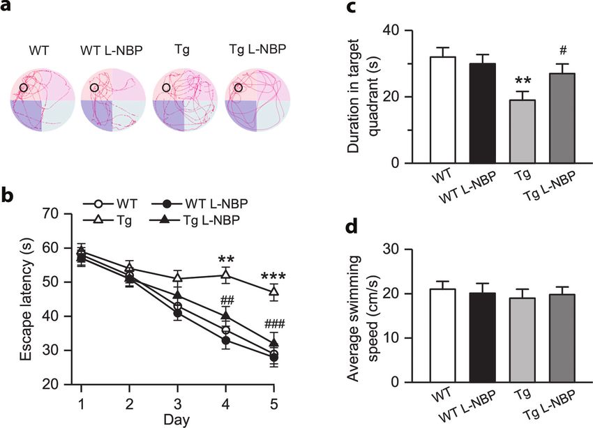
Fig. 1. L-NBP treatment ameliorates spatial learning and memory deficits in APP/PS1 mice. (a) The Morris water maze test
assessed mice (n = 11 mice per group) for spatial learning and memory. Sample swim traces are shown. (b) Longer escape
latency to reach the platform during training and (c) shorter duration time in the target quadrant during the probe trial
were in the Tg group than in the WT group. L-NBP treatment ameliorated this effect, shortened the escape latency, and
prolonged the duration in the target quadrant. (d) Average swimming speeds had no significant difference among the four
groups. WT group: **P < 0.01 and ***P < 0.001; Tg group: #P < 0.05, ##P < 0.01, and ###P < 0.001. Vertical bars represent
one standard error.
Citation: STEMedicine 2022, 3(1): e113 - http://dx.doi.org/10.37175/stemedicine.v3.i1.113 2Synaptic plasticity in mouse model of brain impairments
The input–output curves were plotted by stepping stimula- glutaminase activity and glutamate level were measured by
tion intensity from 0 to 1.5 mA. Two paired stimuli evoked colorimetry using commercial kits (A124 and A074, respec-
paired-pulse facilitation (PPF) at varying inter-pulse in- tively, Jiancheng Bioengineering Institute, Wuhan, China).
tervals (20–300 ms). After testing for baseline, LTP was
evoked by three sets of high-frequency stimulation (HFS, Statistics
10 trains of 20 stimuli, 200 Hz, inter-train interval 2 sec) Data were shown as mean ± standard error of mean
with an interset interval of 5 min. (SEM). Two-way analysis of variance (ANOVA) followed
by a post hoc Bonferroni test was used to analyze statisti-
Patch-clamp recordings cal differences. P-value less than 0.05 was considered as a
At the end of the experiments, the brain of the sacrificed statistical difference.
mouse was quickly removed. The hippocampus was iso-
lated, split into 400-µm thick horizontal sections, incu- Results
bated in oxygenated artificial cerebrospinal fluid at 22°C
for 1 h, and then transferred to the recording chamber. L-NBP ameliorated cognitive deficits
The whole-cell recording was performed on the CA1 py- As shown in Fig. 1, on the 4th and 5th day of training,
ramidal neurons, visualized by a differential interference Tg mice had obviously longer latency to reach the plat-
contrast (DIC) microscope (Nomarski). The borosilicate form than WT mice. However, Tg mice treatment with
glass electrodes (5–7 MΩ) filled with intracellular solu- L-NBP could remarkably reduce the escape latency
tion. Stimulating pulses in 0.1 Hz were used to evoke (Fig. 1a and b). In the probe test, the durations in the
excitatory postsynaptic currents (EPSCs). At +40 mV target quadrant among four groups were significantly dif-
holding potential, NMDA-EPSCs were recorded with the ferent (P < 0.01). It indicated that WT mice spent con-
presence of picrotoxin (GABA antagonist, 50 μM) and siderably more time in the target quadrant than Tg mice
NBQX (AMPA antagonist, 10 μM). At -60 mV, AM- (P < 0.01), while Tg mice treatment with L-NBP could
PA-EPSCs were recorded with picrotoxin (50 μM) and dramatically increase the time in the target quadrant
±APV (NMDA antagonist, 100 μM). (P < 0.05) (Fig. 1c). However, there was no significant dif-
ference in the average swimming speeds of the four groups
Western-blotting (P > 0.05) (Fig. 1d). It meant that L-NBP did not influ-
After the experiments, the protein of the hippocampal ence motor behavior in the mice.
tissues was extracted by using radioimmunoprecipita-
tion buffer (Beyotime Biotechnology). Sodium dodecyl L-NBP treatment ameliorated the impairment of LTP in the
sulfate–polyacrylamide gel electrophoresis separated hippocampal CA1 area
protein samples (30 µg for each lane) and then trans- The input/output curves indicated that the fEPSP slope
ferred pieces to a polyvinylidene fluoride membrane. evoked by high-intensity currents was lower in Tg mice
The membranes were blocked by bovine serum albumin than WT mice (Fig. 2a). We found that HFS could evoke
(5%) for 1 h and incubated with the indicated primary LTP in all groups (Fig. 2b). However, the amplitude of
antibodies at 4°C overnight. The primary antibodies LTP in Tg mice was significantly smaller compared with
were anti-NR1 (#5704S, 1:800, Cell Signaling Technol- WT mice (P < 0.001, Fig. 2b–d). In addition, the LTP am-
ogy, Danvers, MA, USA), anti-NR2A (#4206S, 1:700, plitude in Tg+L-NBP mice was substantially larger than
Cell Signaling Technology), anti-NR2B (#4207S, 1:700, in Tg mice (P < 0.01, Fig. 2b–d).
Cell Signaling Technology), anti-GluR1 (ab183897,
1:800, Abcam, Cambridge, MA, USA), anti-GluR2 L-NBP treatment restored NMDA receptor-mediated EPSCs
(ab206293, 1:800, Abcam), and anti-β-actin (sc-8432, A one-way ANOVA was separately performed to assess
1:900, Santa Cruz, Dallas, TX, USA). Horse radish whether amplitudes of AMPA receptor or NMDA-medi-
peroxidase-conjugated secondary antibodies were fur- ated EPSCs. As shown in Fig. 3, amplitudes of AMPA
ther used to incubate the membranes, following visual- or AMPA receptor-mediated EPSCs, as well as the
ized by Chemiluminescent Substrate (Thermo Fisher, NMDA/AMPA ratio, were remarkably larger in WT
Waltham, MA). Results were analyzed using the Image mice than Tg mice (P < 0.001, P < 0.05 and P < 0.05,
J software. respectively) (Fig. 3a–c). L-NBP treatment robustly
boosted NMDA-EPSCs amplitude (P < 0.01, Fig. 3a, b)
Measurement of glutamate level and glutaminase activity and NMDA/AMPA ratio (P < 0.05) (Fig. 3c) in Tg mice.
The homogenates of the hippocampal tissues were cen- However, it had no significant influence on the AM-
trifuged at 4°C for 10 min (at 8,000 rpm for glutaminase PA-EPSCs amplitude (P > 0.05, Fig. 3a, 3b). It might
activity measurement and at 2,500 rpm for glutamate level result in an increased NMDA/AMPA ratio in Tg mice
measurement), and the supernatants were collected. The (P < 0.05, Fig. 3c).
Citation: STEMedicine 2022, 3(1): e113 - http://dx.doi.org/10.37175/stemedicine.v3.i1.113 3Qian Ding et al.
Fig. 2. L-NBP treatment ameliorates the impairment of long-term potentiation (LTP) in the hippocampal CA1 area of APP/
PS1 mice. (a) Input/output curves showed the field excitatory postsynaptic potential (fEPSP) slopes as a function of stimu-
lus current intensities in the four groups (n = 14 mice per group). (b) The fEPSP slope assessed the magnitude of LTP after
high-frequency stimulation (HFS) (n = 14 mice per group). The arrow indicates the onset of HFS. (c) Sample waveforms of
fEPSPs before and after HFS. (d) The fEPSP slope at 60 min was less increased in the Tg group than in the WT group. This
suppression of LTP was reversed by L-NBP treatment. WT group: ***P < 0.001; Tg group: ##P < 0.01. Vertical bars represent
one standard error.
L-NBP treatment restored protein expressions of NR2A and and glutamate level in Tg mice (P < 0.05 and P < 0.05,
NR2B receptors respectively) (Fig. 5a–e).
Compared to WT mice, NR2A, NR2B, and GluR1 pro-
tein expressions were significantly decreased in the hip- Discussion
pocampus of Tg mice (P < 0.01, P < 0.01 and P < 0.001, The present study reveals that L-NBP improved cogni-
respectively) (Fig. 4a, b). Conversely, L-NBP treatment in tive performances in AD mice, which is consistent with
Tg mice significantly increased NR2A and NR2B protein previous findings that L-NBP could enhance learning
expressions (P < 0.05 and P < 0.01, respectively) but failed and memory through Morris water maze test in various
to affect the expression of GluR1 (P > 0.05, Fig. 4a, b). experimental AD models, including Aβ intracerebroven-
tricularly infused rats, double transgenic APP/PS1 mice,
L-NBP treatment restored presynaptic function and transmitter and triple transgenic 3xTg-AD mice (10, 11, 15). These
synthesis and release results demonstrated a beneficial effect of L-NBP in AD
The peak PPF ratio (at 40 ms inter-pulse interval) of models. Furthermore, considering that synaptic plasticity
Tg mice was remarkably smaller than in WT mice (P < is known as the base of learning and memory, the impact
0.05, Fig. 5a–c). Compared with WT mice, the Tg mice of L-NBP on synaptic plasticity was explored in the cur-
also showed significantly lower glutaminase activity and rent study.
glutamate level (P < 0.05 and P < 0.01, respectively) LTP was known to correlate with learning and memory,
(Fig. 5d, e). L-NBP treatment reversed the impairment and it is well accepted as an electrophysiological measure
of PPF (P < 0.05) and increased the glutaminase activity of synaptic plasticity (16). A previous study revealed an
Citation: STEMedicine 2022, 3(1): e113 - http://dx.doi.org/10.37175/stemedicine.v3.i1.113 4Synaptic plasticity in mouse model of brain impairments Fig. 3. L-NBP treatment restores NMDA receptor-mediated synaptic transmission in APP/PS1 mice. (a) Excitatory synaptic transmission was assessed by recording AMPA and NMDA receptor-mediated excitatory postsynaptic currents (EPSCs) in the hippocampal CA1 area (n = 12 mice per group). Sample waveforms of evoked EPSCs are shown. (b) Amplitudes of AMPA and NMDA receptor-mediated EPSCs were considerably lower in the Tg group compared with the WT group. The decreased ampli- tude of NMDA receptor-mediated EPSCs and (c) the ratio of NMDA receptor-mediated EPSCs to AMPA receptor-mediated EPSCs in the Tg group were considerably increased by L-NBP treatment. WT group: *P < 0.05 and ***P < 0.001; Tg group: # P < 0.05 and ##P < 0.01. Vertical bars represent one standard error. Fig. 4. L-NBP treatment restores the expression of NR2A and NR2B receptors in the APP/PS1 mice. (a) Sample images of Western blot of AMPA receptor subtypes (GluR1 and GluR2) and NMDA receptor subtypes (NR1, NR2A, and NR2B) during the four groups (n = 12 mice per group). (b) NR2A, NR2B, and GluR1 were significantly lower in the Tg group than in the WT group. The decreased levels of NR2A and NR2B were reversed by L-NBP treatment. Quantified results were normalized to levels of β-actin. Values were expressed as percentages compared to the WT group. WT group: **P < 0.01 and ***P < 0.001; Tg group: # P < 0.05 and ##P < 0.01. Vertical bars represent one standard error. Citation: STEMedicine 2022, 3(1): e113 - http://dx.doi.org/10.37175/stemedicine.v3.i1.113 5
Qian Ding et al. Fig. 5. L-NBP treatment restores presynaptic transmission and transmitter synthesis and releases in APP/PS1 mice. (a) Paired- pulse facilitation (PPF) was recorded at varying interpulse intervals in the hippocampal CA1 area (n = 14 mice per group). (b) Sample waveforms of fEPSPs evoked by paired pulses. (c) PPF ratio (fEPSP2/fEPSP1) at 40 ms of the interpulse interval, (d) glutaminase activity, and (e) glutamate level of the Tg group, which was significantly lower than in the WT group, were improved by L-NBP treatment. For the glutaminase activity and glutamate level assays, data were collected from 11 mice in each group. WT group: *P < 0.05 and **P < 0.01; Tg group: #P < 0.05. Vertical bars represent one standard error. impairment of LTP in APP/PS1 mice that were more than but not AMPA-EPSCs amplitude, the effect of L-NBP 8 months old (17). Consistent with this report, our results in NMDA/AMPA ratio might be via restoring NMDA of in vivo LTP recording showed that impaired LTP was receptor-mediated synaptic transmission. found in APP/PS1 mice aged 15 months. Furthermore, It is well documented that the signaling properties of L-NBP was found to attenuate LTP impairment in other these receptors would be influenced by the subunit com- animal models with cognitive impairment, including position of NMDA and AMPA receptors (19). NMDA chronic cerebral ischemia and diabetes with cognitive dys- receptors are heteromers composed of subunits: NR1, NR2 function (12, 18). Similarly, L-NBP could also attenuate (A to D), and NR3 (A and B), and those consisting of LTP impairment in the AD mice and cognitive impair- NR1 subunit and NR2A or NR2B subunit are identified ment through restoring synaptic plasticity. as the primary type of functional NMDA receptors in the NMDA and AMPA receptors are vital components brain (22, 23). AMPA receptors are heteromers composed underlying the LTP mechanism and are crucial for con- of combinations of subunits GluR1–GluR4, and those trolling synaptic plasticity. These two receptors are coex- containing subunits (GluR1 and GluR2) are involved in pressed in the synapse, and the ratio of currents through synaptic transmission in the brain (24). Postmortem stud- the two types of channels (NMDA/AMPA ratio) is rel- ies have found reductions of NR2A/NR2B mRNA levels atively fixed (19). Therefore, the balance of the NMDA/ of the hippocampus and reduced GluR1 protein levels in AMPA ratio is crucial for the formation of synaptic plas- the dentate gyrus in AD patients (25, 26). Decreased levels ticity (20). A previous study observed a reduction of the of NR2A and NR2B were also observed in APP/PS1 mice NMDA/AMPA ratio in the AD mice (21). In our study, (27). Another study found that APP/PS1 mice showed the mice also had a significantly lower NMDA/AMPA reduced mRNA expression of NR2B and GluR1 when ratio than their WT littermates. Treatment with L-NBP they developed cognitive dysfunction (28). In accordance was found to rescue the decrease of the NMDA/AMPA with these results, NR2A, NR2B, and GluR1 protein ex- ratio in our study. As our results showed that L-NBP pressions were significantly decreased. Treatment with recovered the decrease in NMDA-EPSCs amplitude L-NBP could restore the NR2A and NR2B expressions Citation: STEMedicine 2022, 3(1): e113 - http://dx.doi.org/10.37175/stemedicine.v3.i1.113 6
Synaptic plasticity in mouse model of brain impairments
but not GluR1. These results might support that the effect Conflict of interest and funding
of L-NBP on LTP was via repairing NMDA receptor- The authors have nothing conflict of interest to declare.
mediated synaptic transmission. The impact of L-NBP This study was supported by the National Natural Science
in upregulating NR2B was previously reported in an ani- Foundation of China (81671195).
mal model of diabetes with cognitive dysfunction (18). In
accordance with that study, our result showed a striking References
effect of L-NBP on NR2B level, which almost returned 1. Takeuchi T, Duszkiewicz AJ, Morris RGM. The synaptic plas-
to normal level after L-NBP treatment. Previous evidence ticity and memory hypothesis: encoding, storage and persistence.
suggested that selective reduction in NR2B participated Philos Trans R Soc B Biol Sci 2014; 369(1633): 20130288. doi:
in the cognitive impairments in AD animal models and 10.1098/rstb.2013.0288
2. Shankar GM, Walsh DM. Alzheimer’s disease: synaptic dys-
patients (27). These results indicated that the impact of
function and Aβ. Mol Neurodegener 2009; 4(1): 48. doi:
L-NBP in advancing learning and memory should be 10.1186/1750-1326-4-48
partially via upregulating NR2B level. 3. Scheff SW, Price DA, Schmitt FA, DeKosky ST, Mufson EJ.
Furthermore, we examined the effect of L-NBP on Synaptic alterations in CA1 in mild Alzheimer disease and mild
presynaptic functions, which is another critical compo- cognitive impairment. Neurology 2007; 68(18): 1501–8. doi:
nent of synaptic plasticity. PPF is a short-term synaptic 10.1212/01.wnl.0000260698.46517.8f
4. Pozueta J, Lefort R, Shelanski ML. Synaptic changes in
plasticity that is evoked by two closely spaced stimuli.
Alzheimer’s disease and its models. Neuroscience 2013; 251:
The mechanism underlying PPF involves increased pre- 51–65. doi: 10.1016/j.neuroscience.2012.05.050
synaptic Ca2+ concentration and neurotransmitter release 5. Cavanagh C, Wong TP. Preventing synaptic deficits in Alzheimer’s
(29). Thus, PPF is commonly used to measure presyn- disease by inhibiting tumor necrosis factor alpha signaling.
aptic functions. Previous studies found that the PPF IBRO Rep 2018; 4: 18–21. doi: 10.1016/j.ibror.2018.01.003
ratio was reduced in the mice at 15 months (17). Con- 6. Sheng C, Xu P, Zhou K, Deng D, Zhang C, Wang Z. Icariin
Attenuates Synaptic and Cognitive Deficits in an A β1-42-
sistently, we demonstrated that the peak PPF ratio was
Induced Rat Model of Alzheimer’s disease. BioMed Res Int
significantly reduced, which indicated an impaired pre- 2017; 2017: 12. doi: 10.1155/2017/7464872
synaptic function in the hippocampus of this AD model. 7. Xu ZP, Li L, Bao J, Wang ZH, Zeng J, Liu EJ, et al. Magnesium
In addition, glutamate levels decreased in AD patients’ protects cognitive functions and synaptic plasticity in strepto-
brains (30, 31). zotocin-induced sporadic Alzheimer’s model. PLoS One 2014;
Meanwhile, postmortem studies found a reduced level 9(9):e108645. doi: 10.1371/journal.pone.0108645
8. Bereczki E, Branca RM, Francis PT, Pereira JB, Baek J-H,
of glutaminase and loss of glutaminase-positive neurons
Hortobágyi T, et al. Synaptic markers of cognitive decline in
in AD brains (32, 33), which might explain the decrease neurodegenerative diseases: a proteomic approach. Brain 2018;
of glutamate levels. Glutamate level was also reduced 141(2): 582–95. doi: 10.1093/brain/awx352
in the hippocampus (34). Consistent with these reports, 9. Anand R, Gill KD, Mahdi AA. Therapeutics of Alzheimer’s
we found that the glutaminase activity and glutamate disease: past, present and future. Neuropharmacology 2014; 76:
levels were dramatically reduced in the mice, indicating 27–50. doi: 10.1016/j.neuropharm.2013.07.004
10. Peng Y, Hu Y, Xu S, Li P, Li J, Lu L, et al. L-3-n-butylphthalide
a reduced presynaptic glutamate release in this model.
reduces tau phosphorylation and improves cognitive deficits in
L-NBP treatment reversed the impairment in PPF and AβPP/PS1-Alzheimer’s transgenic mice. J Alzheimers Dis 2012;
increased the glutaminase activity and glutamate level 29: 379–91. doi: 10.3233/JAD-2011-111577
in the mice in our study. It indicated that L-NBP could 11. Peng Y, Sun J, Hon S, Nylander AN, Xia W, Feng Y, et al. L-3-
restore presynaptic function by restoring glutamate-me- n-butylphthalide improves cognitive impairment and reduces
diated synaptic transmission, which might underlie the amyloid-beta in a transgenic model of Alzheimer’s disease.
J Neurosci 2010; 30(24): 8180–9. doi: 10.1523/JNEUROSCI.
role of L-NBP in alleviating learning and memory defi-
0340-10.2010
cits in APP/PS1 mice. 12. Xu J, Wang Y, Li N, Xu L, Yang H, Yang Z. l-3-n-butylphtha-
lide improves cognitive deficits in rats with chronic cerebral isch-
Conclusion emia. Neuropharmacology 2012; 62(7): 2424–9. doi: 10.1016/j.
In summary, this study revealed that L-NBP treatment neuropharm.2012.02.014
13. Li PP, Wang WP, Liu ZH, Xu SF, Lu WW, Wang L, et al.
could enhance the protein levels of NR2A and NR2B,
Potassium 2-(1-hydroxypentyl)-benzoate promotes long-term
glutaminase activity, and glutamate levels and attenu- potentiation in Abeta1-42-injected rats and APP/PS1 transgenic
ate impairments of synaptic transmission, which might mice. Acta Pharmacol Sin 2014; 35(7): 869–78. doi: 10.1038/
underlie the effect of L-NBP in improving learning and aps.2014.29
memory in APP/PS1 mice. 14. Zhang Y, Huang L-J, Shi S, Xu S-F, Wang X-L, Peng Y. L-3-
n-butylphthalide rescues hippocampal synaptic failure and
attenuates neuropathology in aged APP/PS1 mouse model of
Acknowledgment Alzheimer’s disease. CNS Neurosci Ther 2016; 22(12): 979–87.
None. doi: 10.1111/cns.12594
Citation: STEMedicine 2022, 3(1): e113 - http://dx.doi.org/10.37175/stemedicine.v3.i1.113 7Qian Ding et al.
15. Peng Y, Xing C, Xu S, Lemere CA, Chen G, Liu B, et al. L-3- and entorhinal cortex in Alzheimer’s disease. J Neurol Sci 2002;
n-butylphthalide improves cognitive impairment induced by 200(1): 11–18. doi: 10.1016/S0022-510X(02)00087-4
intracerebroventricular infusion of amyloid-beta peptide in 26. Wakabayashi K, Narisawa-Saito M, Iwakura Y, Arai T, Ikeda
rats. Eur J Pharmacol 2009; 621(1–3): 38–45. doi: 10.1016/j. K, Takahashi H, et al. Phenotypic down-regulation of glutamate
ejphar.2009.08.036 receptor subunit GluR1 in Alzheimer’s disease☆. Neurobiol
16. Song S, Wang X, Sava V, Weeber EJ, Sanchez-Ramos J. In vivo Aging 1999; 20(3): 287–95.
administration of granulocyte colony-stimulating factor restores 27. Huang H-J, Liang K-C, Ke H-C, Chang Y-Y, Hsieh-Li HM.
long-term depression in hippocampal slices prepared from Long-term social isolation exacerbates the impairment of spa-
transgenic APP/PS1 mice. J Neurosci Res 2014; 92(8): 975–80. tial working memory in APP/PS1 transgenic mice. Brain Res
doi: 10.1002/jnr.23378 2011; 1371: 150–60. doi: 10.1016/j.brainres.2010.11.043
17. Gengler S, Hamilton A, Holscher C. Synaptic plasticity in the 28. Dickey CA, Loring JF, Montgomery J, Gordon MN, Eastman
hippocampus of a APP/PS1 mouse model of Alzheimer’s dis- PS, Morgan D. Selectively reduced expression of synaptic plas-
ease is impaired in old but not young mice. PLoS One 2010; ticity-related genes in amyloid precursor protein + presenilin-1
5(3):e9764. doi: 10.1371/journal.pone.0009764 transgenic mice. J Neurosci 2003; 23(12): 5219–26.
18. Li J, Zhang S, Zhang L, Wang R, Wang M. Effects of L-3-n- 29. Zucker RS, Regehr WG. Short-term synaptic plasticity. Annu
butylphthalide on cognitive dysfunction and NR2B expression Rev Physiol 2002; 64(1): 355–405. doi: 10.1146/annurev.
in hippocampus of streptozotocin (STZ)-induced diabetic physiol.64.092501.114547
rats. Cell Biochem Biophys 2015; 71(1): 315–22. doi: 10.1007/ 30. Fayed N, Modrego PJ, Rojas-Salinas G, Aguilar K. Brain gluta-
s12013-014-0200-5 mate levels are decreased in Alzheimer’s disease: a magnetic res-
19. Rao VR, Finkbeiner S. NMDA and AMPA receptors: old onance spectroscopy study. Am J Alzheimers Dis Other Demen
channels, new tricks. Trends Neurosci 2007; 30(6): 284–91. doi: 2011; 26(6): 450–6. doi: 10.1177/1533317511421780
10.1016/j.tins.2007.03.012 31. Antuono PG, Jones JL, Wang Y, Li SJ. Decreased glutamate
20. Luscher C, Malenka RC. NMDA receptor-dependent long-term + glutamine in Alzheimer’s disease detected in vivo with (1)
potentiation and long-term depression (LTP/LTD). Cold Spring H-MRS at 0.5 T. Neurology 2001; 56(6): 737–42. doi: 10.1212/
Harb Perspect Biol 2012; 4(6): a005710. doi: 10.1101/cshper- WNL.56.6.737
spect.a005710 32. Burbaeva G, Boksha IS, Tereshkina EB, Savushkina OK,
21. Tozzi A, Sclip A, Tantucci M, de Iure A, Ghiglieri V, Costa C, Prokhorova TA, Vorobyeva EA. Glutamate and GABA-
et al. Region- and age-dependent reductions of hippocampal metabolizing enzymes in postmortem cerebellum in Alzheimer’s
long-term potentiation and NMDA to AMPA ratio in a genetic disease: phosphate-activated glutaminase and glutamic acid
model of Alzheimer’s disease. Neurobiol Aging 2015; 36(1): decarboxylase. Cerebellum 2014; 13(5): 607–15. doi: 10.1007/
123–33. doi: 10.1016/j.neurobiolaging.2014.07.002 s12311-014-0573-4
22. Belayev L, Lu Y, Bazan NG. Chapter 35 – brain ischemia and 33. Akiyama H, McGeer PL, Itagaki S, McGeer EG, Kaneko T.
reperfusion: cellular and molecular mechanisms in stroke injury Loss of glutaminase-positive cortical neurons in Alzheimer’s
A2 – Brady, Scott T. In: Siegel GJ, Albers RW, Price DL, eds. disease. Neurochem Res 1989; 14(4): 353–8. doi: 10.1007/
Basic neurochemistry (Eighth Edition). New York: Academic BF01000038
Press; 2012, pp. 621–42. 34. González-Domínguez R, García-Barrera T, Vitorica J, Gómez-
23. Miller SL, Yeh HH. Chapter 3 – neurotransmitters and neurotrans- Ariza JL. Region-specific metabolic alterations in the brain of
mission in the developing and adult nervous system A2 – Conn, P. the APP/PS1 transgenic mice of Alzheimer’s disease. Biochim
Michael. In: Michael P, ed. Conn’s translational neuroscience. San Biophys Acta 2014; 1842(12, Part A): 2395–402. doi: 10.1016/j.
Diego, CA: Academic Press; 2017, pp. 49–84. bbadis.2014.09.014
24. Banerjee A, Borgmann-Winter KE, Ray R, Hahn CG.
Chapter 8 – the PSD: a microdomain for converging molecu-
lar abnormalities in schizophrenia. In: Abel T, Nickl-Jockschat *Jun Yu
T, eds. The neurobiology of schizophrenia. San Diego, CA: Clinical Experimental Center
Academic Press; 2016, pp. 125–47. Xi’an International Medical Center Hospital
25. Bi H, Sze C-I. N-methyl-d-aspartate receptor subunit NR2A and Northwest University, Xi’an, Shaanxi, China
NR2B messenger RNA levels are altered in the hippocampus Email: pclamper@nwu.edu.cn
Citation: STEMedicine 2022, 3(1): e113 - http://dx.doi.org/10.37175/stemedicine.v3.i1.113 8Synaptic plasticity in mouse model of brain impairments Citation: STEMedicine 2022, 3(1): e113 - http://dx.doi.org/10.37175/stemedicine.v3.i1.113 9
You can also read