DRUG REPURPOSING FOR CORONAVIRUS (SARS COV 2) BASED ON GENE CO EXPRESSION NETWORK ANALYSIS - NATURE
←
→
Page content transcription
If your browser does not render page correctly, please read the page content below
www.nature.com/scientificreports
OPEN Drug repurposing for coronavirus
(SARS‑CoV‑2) based on gene
co‑expression network analysis
Habib MotieGhader1,2*, Esmaeil Safavi1,3, Ali Rezapour4, Fatemeh Firouzi Amoodizaj1 &
Roya asl Iranifam1
Severe acute respiratory syndrome (SARS) is a highly contagious viral respiratory illness. This illness
is spurred on by a coronavirus known as SARS-associated coronavirus (SARS-CoV). SARS was first
detected in Asia in late February 2003. The genome of this virus is very similar to the SARS-CoV-2.
Therefore, the study of SARS-CoV disease and the identification of effective drugs to treat this disease
can be new clues for the treatment of SARS-Cov-2. This study aimed to discover novel potential drugs
for SARS-CoV disease in order to treating SARS-Cov-2 disease based on a novel systems biology
approach. To this end, gene co-expression network analysis was applied. First, the gene co-expression
network was reconstructed for 1441 genes, and then two gene modules were discovered as significant
modules. Next, a list of miRNAs and transcription factors that target gene co-expression modules’
genes were gathered from the valid databases, and two sub-networks formed of transcription factors
and miRNAs were established. Afterward, the list of the drugs targeting obtained sub-networks’ genes
was retrieved from the DGIDb database, and two drug-gene and drug-TF interaction networks were
reconstructed. Finally, after conducting different network analyses, we proposed five drugs, including
FLUOROURACIL, CISPLATIN, SIROLIMUS, CYCLOPHOSPHAMIDE, and METHYLDOPA, as candidate
drugs for SARS-CoV-2 coronavirus treatment. Moreover, ten miRNAs including miR-193b, miR-192,
miR-215, miR-34a, miR-16, miR-16, miR-92a, miR-30a, miR-7, and miR-26b were found to be significant
miRNAs in treating SARS-CoV-2 coronavirus.
Coronaviruses infect both animals and humans, causing intestinal and respiratory infections1,2. Severe acute
respiratory syndrome (SARS) is a coronavirus-associated respiratory disease that was originally discovered in
China in February 20033. Ten years after SARS coronavirus, Middle East respiratory syndrome (MERS) corona-
virus was broke out in Middle Eastern countries2,4,5. SARS-CoV and MERS uses angiotensin-converting enzyme
2 (ACE2) and dipeptidyl peptidase 4(DPP4) as a receptor, r espectively2. Additionally, in the autumn of 2019,
the coronavirus SARS-CoV-2 broke out in the Chinese city of W uhan4,6. Given that, there is a high similarity
between SARS-CoV and SARS-CoV-2, the spread speed of SARS-CoV-2 is faster than SARS-CoV4. SARS-CoV-2
like SARS-CoV utilize the host cell ACE2 r eceptor4. ACE2 is a membrane receptor on the surface of many cell
types and tissues including the lungs, heart, blood vessels, kidney, liver, and gastrointestinal2,4. SARS-CoV, SARS-
CoV-2, and MERS all have similar genetic characteristics, and SARS-CoV-2 is very similar to SARS-CoV7. Due
to the high genetic similarity of SARS-CoV and SARS-CoV-2, the results of the study on SARS-CoV can be a
clue to the treatment of SARS-CoV-2. Due to the disease’s significance and high death rate, early detection and
treatment are essential. The medicinal drugs used to treat coronavirus infections are only intended to be used
temporarily. Besides, clinical trials on medications and vaccines that are efficient in curing diseases take quite a
long time. Furthermore, handling SARS viruses in vivo is often challenging and risky. However, the knowledge
obtained by sequencing their genes, proteins, or RNA is simple and easy to manage through artificial intelligence
8
. Additionally, miRNA-mRNA data sources have progressed considerably as prospective techniques for gaining
a better understanding of potential SARS-CoV therapies, allowing network science and computational systems
biology to become feasible9. According to these data, many scientists seek to identify the involved host genes
and proteins in diseases to find a new therapy.
1
Department of Basic Sciences, Biotechnology Research Center, Tabriz Branch, Islamic Azad University, Tabriz,
Iran. 2Department of Biology, Tabriz Branch, Islamic Azad University, Tabriz, Iran. 3Department of Basic Sciences,
Faculty of Veterinary Medicine, Tabriz Branch, Islamic Azad University, Tabriz, Iran. 4Department of Animal Science,
Faculty of Agriculture, Tabriz Branch, Islamic Azad University, Tabriz, Iran. *email: habib_moti@ut.ac.ir
Scientific Reports | (2021) 11:21872 | https://doi.org/10.1038/s41598-021-01410-3 1
Vol.:(0123456789)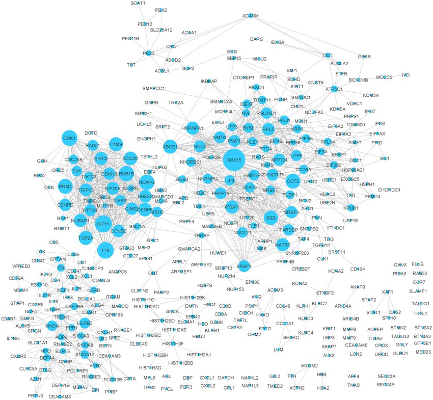
www.nature.com/scientificreports/
Recent research has identified a set of antiviral genes, such as ISG15, IFIH1, MX1, OAS1-3, IRF7, IRF9, and
STAT1 expressed by host cells, which could be used as a new therapeutic target against coronavirus due to their
response to viral i nfection10–18. In addition to therapeutic gene targets, miRNAs can also be used to suppress the
viral genome due to their ability to regulate gene expression. MiRNAs are small non-coding RNA molecules that
prevent mRNAs from being t ranslated19,20. Therefore, miRNA-based therapy could be proposed for SARS-CoV
treatment21,22. On the other hand, the role of pathologic processes in miRNAs, such as inflammatory responses
and viral infection, has been recently verified21,23,24.
Drug repurposing (DR) is a strategy for identifying new therapeutic uses for approved or investigational
drugs25,26. This approach is also referred to as drug reprofiling, drug re‑tasking, drug repositioning, drug thera-
peutic and drug recyclining26. It is an efficient approach for the development or discovery of drug molecules with
new therapeutic i ndications25. Generally, the process of drug repurposing consists of three s teps26: 1. identification
of a candidate molecule for a particular indication. 2. mechanistic evaluation of the drug effect in preclinical
models. 3. evaluation of efficacy in phase clinical trials II. Of these three steps, step 1 is crucial, and it is here that
modern approaches to hypothesising may be most u seful26. These systematic approaches can be divided into
experimental and computational approaches . Some of the computational approaches are: molecular docking,
26
signature machiing, genetic association, network mapping, retrospective clinical analysis and novel data sources26.
As well as, among the experimental methods, Binding assays to identify relevant target interactions and Phenotypic
screening approaches can be mentioned26.
Recently, different articles based on network approaches have been published for drug repurposing27.
SAveRUNNER28 is a network-based drug repurposing algorithm, which predicts drug-disease assosiations using
network-based similarity measure. This algorithm provided as a freely available R-code29. SAveRUNNER is also
been used as a drug repurposing tool for amyotrophic lateral sclerosis (ALS) d isease30. Pasquale and c olleaqes31,
examined three different network-based approaches and identified 399 repurposable drugs for COVID-19 using
SAveRUNNER algorithm. Another algorithm based on artificial intelligence, network diffusion, and network
proximity introduced as a drug repurposing method for SARS-CoV-2 d isease32. We also recently introduced a
protein–protein interaction network approach in order to propose candidate drugs to treatment of SARS-CoV-2
disease21.
Tasnimul and colleague, recently introduced a network-based method for identifying and repurposing drugs
for the treatment of SARS-CoV-2 disease33. In this method, differentially expressed genes between Idiopathic
pulmonary fibrosis (IPF) and SARS-CoV-2 samples were compared and finally, some IPF drugs were proposed
as candidate drugs to treat SARS-CoV-2 disease. Yadi et al.34 proposed a novel network-based drug repurpos-
ing methodology based on human interactome and protein–protein interaction networks. This method quntify
the interplay between the drug targets and HCoV–host interactome in the human protein–protein interaction
network. In this study, 16 potential drugs was introduced to treat SARS-CoV-2 disease. In another study, Hangyu
and colleaque35 developed a machine-learning -based method to predict virus-host interactions at both organ-
ism and protein levels for SARS-Cov-2 disease. In this method, a multi-layer virus-host interaction network was
constructed. CoVex36, an interactive online platform for SARS-CoV-2, introduced by Sepideh and colleaque. This
platform, integrates human protein–protein interactions, virus-human protein interactions and drug-target inter-
actions. Zhihao and c olleaque37 constructed a autophagy interaction network based on competitive endogenous
RNA(ceRNA) in SARS-CoV-2 infection. In this study, hsa-miR-4772–5p, hsa-miR- 192–5p, hsa-miR-652–3p,
hsa-miR- 192–5p, hsa-miR-340–3p, CCR2 and TP53INP2 introduced as potential biomarkers in predicting
changes in mild SARS-CoV-2 infection. In comparison to the mentioned network-based methods, in our study,
the gene co-expression network is used. In addition to the gene co-expression network, regulatory interactions
including miRNA-Gene, TF-Gene, and TF-miRNA have also been used in our study. Moreover, drug-gene and
drug-TF interaction networks have been studied and investigated in this study. As well as, some of the genes that
regulate more miRNAs, are also introduced as effective miRNAs in SARS-Cov disease. Changes in the expression
of these genes can affect the expression of target miRNAs.
The current research aimed to discover the genes and miRNAs involved in SARS-CoV disease and repurpose
candidate drugs for this diease in order to treating SARS-CoV-2 coronavirus based on a co-expression network
analysis. In this regard, this study used a co-expression network analysis to identify potential drugs for the treat-
ment of SARS-CoV. The methodology we used in this study is an entirely novel method based on gene module
identification.
The technique first entails identifying a list of genes (human genes) expressed differentially in healthy and
SARSCoV-infected samples. After obtaining differentially expressed genes between healthy and SARSCoV-
infected samples, the co-expression network is reconstructed in STRING online tool38. Then, two significant gene
modules are discovered from the gene co-expression network. Afterward, a list of miRNAs and transcription
factor genes that have a regulatory impact on modules’ genes are collected from a valid database (TRRUST v 239
and miRWalk v240), and different network analyses are done on these biomolecules. Finally, the list of drugs that
target modules’ genes are gathered from the (DGIdb)41 database, and then two drug-gene interaction networks
are reconstructed. The workflow diagram of this study is demonstrated in Fig. 1. As shown in this figure, the
method’s output is some candidate drugs for the treatment of SARS coronavirus. In this study, FLUOROURACIL,
CISPLATIN, SIROLIMUS, CYCLOPHOSPHAMIDE, and METHYLDOPA are the key drugs reported for treating
SARS-CoV-2 coronavirus. Moreover, hsa-miR-193b, hsa-miR-192, hsa-miR-215, hsa-miR-34a, hsa-miR-16, hsa-
miR-16, hsa-miR-92a, hsa-miR-30a, hsa-miR-7, and hsa-miR-26b are candidate miRNAs, which are significant
in the treatment of SARS-CoV-2 disease.
Scientific Reports | (2021) 11:21872 | https://doi.org/10.1038/s41598-021-01410-3 2
Vol:.(1234567890)
www.nature.com/scientificreports/

                                  Figure 1.  The overall workflow of the proposed method. In this method, a network-based approach is applied
                                  to drug repurposing for coronavirus disease treatment. (a) At first, a transcriptome profile for healthy (control)
                                  and SARS-CoV-infected samples were taken from the GEO database with the accession number GSE1739. (b)
                                  Then, after identifying differentially expressed genes in the control and disease groups, the gene co-expression
                                  network is reconstructed, and two significant gene modules are discovered from the co-expression network.
                                  (c) Next, for every gene module, the TF-miRNA-TG network is reconstructed independently. The information
                                  of TFs-miRNAs, TFs-TGs, and miRNAs-TGs regulations are taken from the TransmiR42, TRRUST39, and
                                  miRWalk40 databases, respectively. (d) Afterward, Drug-gene and Drug-TF networks are reconstructed for
                                  TF-miRNA-TG networks independently. (e) Finally, 19 drugs are proposed as candidate drugs for coronavirus
                                  treatment.

                                  Result
                                  Gene co‑expression network analysis and gene modulation.. First 1441 differentially expressed
                                  genes between normal and SARS infected groups with p-values less than 0.05 were assumed as primary genes.
                                  Then, using this primary gene list, the gene co-expression network was reconstructed in the STRING database.
                                  In this network 1050 genes out of 1441 primary genes were disconnected. Therefore, these disconnected genes
                                  were removed from the network and a network with 391 genes was obtained. Figure 2 shows the co-expression
                                  network for these genes. Supplementary file S6 contains more information on the topological characteristics of
                                  this network.

Scientific Reports |   (2021) 11:21872 |               https://doi.org/10.1038/s41598-021-01410-3                                                      3

                                                                                                                                               Vol.:(0123456789)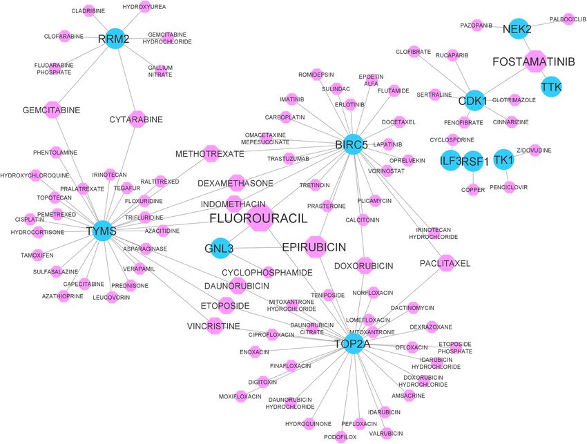
www.nature.com/scientificreports/
Figure 2. Gene co-expression network for the primary genes (disconnected genes were removed from the
network). The size of the nodes indicates its degree. There are 391 nodes and 1273 edges in this network.
DHX15 is the highest degree node in this network.
Module name Gene names
TOP2A (24), TTK (24), NUSAP1(24), UBE2C(24), CENPF(24), KIF20A(24), CDK1(24), CDC20(24), BIRC5(24),
Module A TRIP13(23), NCAPG(24), GINS2(23), KIF11(24), CENPE(22), BUB1B(24), CCNB2(24), CDC25A(17), TACC3(17),
RAD51(20), TYMS(22), CDKN3(22), RRM2(24), TK1(19), NEK2(19), PTTG1(22)
SRSF1(16), SYNCRIP(14), HNRNPU(16), DHX15(29), SSB(19), MATR3(17), GNL3(13), NUDT21(16), U2AF2(17),
HNRNPF(17), SRSF7(15), SRSF3(17), KHDRBS1(14), FUBP1(13), PRPF19(14), BYSL(12), ILF3(17), NLE1(12),
Module B
UTP6(12), RSL24D1(12), MRTO4(13), DIEXF(12), PTBP1(17), UTP3(12), PWP1(12), PNO1(12), KRR1(12),
NMD3(12), HNRNPA1(17), TARDBP(17)
Table 1. Gene names of modules A and B. Numbers inside the parentheses represent the genes degree.
After analyzing the network in Cytoscape s oftware43, generally, 391 nodes and 1273 edges were observed in
the reconstructed co-expression network. After clustering the gene co-expression network, we discovered two
significant modules (Module A and Module B) in the co-expression network. The list of genes for these two
modules is reported in Table 1.
Scientific Reports | (2021) 11:21872 | https://doi.org/10.1038/s41598-021-01410-3 4
Vol:.(1234567890)www.nature.com/scientificreports/
Module name TF Number of TGs Number of target miRNAs
TF-miRNA-TG_A MYC 3 91
TP53 5 89
MYC 1 91
NFKB1 1 67
RELA 1 59
STAT3 1 53
MYCN 1 52
SP1 4 38
ESR1 1 30
TF-miRNA-TG_B E2F3 1 20
KLF4 1 18
CTNNB1 1 16
DNMT1 1 15
NANOG 1 14
TP73 2 12
LEF1 1 12
MYB 1 10
FOXO3 1 9
Table 2. High degree nodes of the TFs in the TF-miRNA-TG_A and B sub-networks.
Transcription factors, miRNAs, and target genes interaction network. At first two TF-miRNA-
TG sub-networks named TF-miRNA-TG_A and TF-miRNA-TG_B were reconstructed for gene modules A and
B, respectively. As demonstrated in previous section, two significant gene modules were considered for more
analysis. To do this, the data of TFs-TGs, TFs-miRNAs, and miRNAs-TGs regulatory interactions were retrieved
from the TRRUST, TransmiR, and miRWalk databases, respectively. These two sub-networks are shown in Fig-
ure S1 (See supplementary file S6). The information for TFs and TGs Interactions are activation, repression, and
unknown. In addition, regulatory interactions information for TFs and TGs are: activation, activation(feedback),
regulation, regulation(feedback), autoregulatory negative feedback, loop(feedback), repression, repression(feedback),
auto-regulatory feedback circuit, and activation (negative regulatory loop). This information for TF-miRNA-TG_A
and TF-miRNA-TG_B sub-networks (see supplementary file S7) are reported in supplementary file S2. Moreo-
ver, the topological properties for these sub-networks was reported in supplementary file S6.
In order to analyze the TFs, which regulate more Genes and miRNAs in the TF-miRNA-TG_A and TF-
miRNA-TG_B networks, the high degree TF nodes were selected and reported as significant TFs. To do this,
among TF nodes in the TF-miRNA-TG_A and TF-miRNA-TG_B sub-networks, the TFs with a degree of 10 and
higher were selected (Table 2). The number of TGs and target miRNAs for every TF are listed in Table 2 as well.
In addition to the TFs, a list of miRNAs that regulate more genes in the TF-miRNA-TG_A and TF-miRNA-
TG_B networks were selected and reported as key miRNAs. To this end, for every TF-miRNA-TG network,
five miRNAs with high degrees were assumed. These miRNAs for TF-miRNA-TG_A were hsa-miR-193b, hsa-
miR-192, hsa-miR-215, hsa-miR-34a, and hsa-miR-16. For TF-miRNA-TG_B network, the selected miRNAs
were hsa-miR-16, hsa-miR-92a, hsa-miR-30a, hsa-miR-7, and hsa-miR-26b. Among these miRNAs, hsa-miR-16
regulates more genes in both subnetworks. The list of these miRNAs, along with their degree and target genes,
are reported in Table 3.
Enrichment analysis of genes. Gene ontology was performed for the module A and B gene lists, sepa-
rately. The results for module A gene list show that they significantly enriched in mitotic cell cycle process, nuclear
division, and mitotic nuclear division biological processes. Moreover, the gene list of module B significantly
enriched in RNA processing, mRNA metabolic process, and RNA metabolic process biological processes. Then, a
pathway enrichment analysis was done for modules A and B gene lists separately. The results showed that the
module A and B genes, significantly enriched in Resolution of Sister Chromatid Cohesion and mRNA Splicing—
Major Pathway pathways, respectively. More details of the GO and pathway enrichment analyses are reported in
supplementary file S3.
Enrichment analysis of miRNAs. In order to check miRNA family for the TF-miRNA-TG_A and TF-
miRNA-TG_B sub-networks, at first the list of miRNAs are imported into the TAM online tool. Then, the
obtained result is reported in supplementary file S4. As reported in this file, both sub-networks significantly
enriched in the let-7 and mir-17 families.
Drug‑Gene interaction network. After gathering drug-gene interactions for TF-miRNA-TG_A and TF-
miRNA-TG_B genes, the drug-gene interaction network was reconstructed and is demonstrated in Fig. 3. As
shown in this network, some drugs have a high degree, which means that these drugs target and regulate more
Scientific Reports | (2021) 11:21872 | https://doi.org/10.1038/s41598-021-01410-3 5
Vol.:(0123456789)www.nature.com/scientificreports/
miRNA Degree Target genes
TF-miRNA-TG_A
RRM2, RAD51, CDC25A, CDK1, CDC20, NCAPG, KIF11, TYMS, UBE2C, TACC3, GINS2, TRIP13,
hsa-miR-193b 13
BUB1B
hsa-miR-192 10 RAD51, CDC25A, CDC20, CENPF, CDKN3, KIF20A, TTK, TRIP13, BUB1B, CENPE
hsa-miR-215 9 RAD51, CDC20, CENPF, CDKN3, KIF20A, TTK, TRIP13, BUB1B, CENPE
hsa-miR-34a 7 RRM2, CDC25A, BIRC5, CDC20, NCAPG, KIF11, TYMS, hsa-miR-34a
hsa-miR-16 7 CDC25A, BIRC5, CDK1, CDC20, NCAPG, UBE2C, CENPF
TF-miRNA-TG_B
hsa-miR-16 8 HNRNPF, HNRNPA1, SRSF1, NLE1, NMD3, DIEXF, UTP3, BYSL
hsa-miR-92a 7 HNRNPF, PTBP1, PNO1, NLE1, NMD3, KHDRBS1, U2AF2
hsa-miR-30a 6 HNRNPA1, HRNPU, SRSF7, UTP6, PTBP1, PRPF19
hsa-miR-7 4 HNRNPU, MATR3, SRSF1, hsa-miR-7, ILF3
hsa-miR-26b 4 MATR3, SYNCRIP, NMD3, GNL3
Table 3. High degree nodes of the miRNAs in the TF-miRNA-TG_A and B sub-networks.
Figure 3. The drug-gene interaction network for TF-miRNA-TG_A and TF-miRNA-TG_B sub-networks. Blue
circles show TGs, and pink octagons show drugs. The size of the octagon nodes indicates its degree. The high
degree drug is FLUOROURACIL with 4 target genes.
genes. Therefore, high-degree drugs were selected and reported as significant, as they regulate more genes in
module_A and module_B. In this regard, the drugs with a degree of 3 or higher were selected and are reported
in Table 4. As reported, FLUOROURACIL, EPIRUBICIN, and FOSTAMATINIB are effective drugs and targeted
4, 3, and 3 genes, respectively.
Scientific Reports | (2021) 11:21872 | https://doi.org/10.1038/s41598-021-01410-3 6
Vol:.(1234567890)www.nature.com/scientificreports/
Drug Degree Target genes
FLUOROURACIL 4 BIRC5, GNL3, TYMS, TOP2A
EPIRUBICIN 3 BIRC5, GNL3, TOP2A
FOSTAMATINIB 3 NEK2, CDK1, TTK
Table 4. High degree drugs in the drug-gene network. Of all the drugs, only those with degree 3 or higher
have been reported.
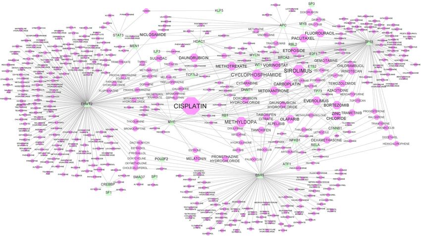
Figure 4. Drug-TF interaction network for TF-miRNA-TG_A and TF-miRNA-TG_B sub-networks. Green
triangle shapes show genes, and pink octagon shapes show drugs. The size of the octagon nodes indicates its
degree. The high degree drug is CISPLATIN with 11 target genes.
In addition to the Drug-gene interaction network, the drug-TF network was reconstructed to discover the
drugs that target the transcription factors of the TF-miRNA-TG_A and TF-miRNA-TG_B sub-networks. This
network is shown in Fig. 4. In this network, the drugs with a degree of 4 and higher were selected and reported
as significant drugs. More details of the effective drugs are reported in Table 5. As the table shows, CISPLATIN,
SIROLIMUS, and CYCLOPHOSPHAMIDE drugs targeted 11, 6, and 5 TFS, respectively, and the others targeted
4 TFs. The complete information of Drug-Gene and Drug-TF interactions are provided in supplementary file
S5. Additionally, supplemental file S6 has detailed information on the network topological characteristics of the
Drug-Gene and Drug-TF networks.
Recently, the potential effect of 6710 drugs as SARS-CoV-2 inhibitors is tested in vitro and in vivo44. The
results of this report show that some of the drugs proposed in this article (see Tables 4 and 5) have an inhibitory
effect on SARS-CoV-2. According to this report, OLAPARIB, NICLOSAMID and METHOTREXATE have a very
weak, weak, and strong effects on SARS-CoV-2, respectively. As well as, DAUNORUBICIN and BORTEZOMIB
have a cytotoxic effect on this disease.
GSEA and candidate drugs validation. To evaluate the proposed drugs, the Connectivity Map(CMAP)
analysis was utilized. To do this, from the Drug-Gene and Drug-TF networks, the drugs with a degree of 4 or
above were selected and imported to Enrichr CMAP database. Then, the impact of these drugs on target genes
was evaluated. Enrichr CMAP database contains CMAP-up and CMAP-down datasets. In Tables 3 and 4, from
among the 19 repurposed drugs for SARS-CoV disease, only nine drugs, including SIROLIMUS, METHYL-
DOPA, VORINOSTAT, PACLITAXEL, DAUNORUBICIN, METHOTREXATE, NICLOSAMIDE, and ETOPO-
SIDE, were validated by CMAP analysis. Table 6 shows the validated drugs together with the corresponding
unregulated or downregulated target genes.
Scientific Reports | (2021) 11:21872 | https://doi.org/10.1038/s41598-021-01410-3 7
Vol.:(0123456789)www.nature.com/scientificreports/
Drug Degree Target TFs
CISPLATIN 11 BRCA2, DNMT1, E2F1, EHMT2, ESR1, MYC, MYCN, RB1, TP53, TP73
SIROLIMUS 6 APC, RB1, RBL2, TCF7L2, TP53, WT1
CYCLOPHOSPHAMIDE 5 BRCA2, CTNNB1, EHMT2, MYCN, TP53
METHYLDOPA 5 EHMT2, ESR1, HDAC1, TP53, TP73
VORINOSTAT 4 HDAC1, MYC, RB1, TP53
OLAPARIB 4 BRCA2, ESR1, MYC, TP53
MITOXANTRONE 4 DNMT1, EHMT2, NFKB1, TP53
FLUOROURACIL 4 APC, E2F1, MYB, TP53
EVEROLIMUS 4 BRCA2, CTNNB1, ESR1, RB1
PACLITAXEL 4 BRCA2, E2F1, MYB, TP53
DAUNORUBICIN 4 EHMT2, HDAC1, TP53, WT1
ZINC CHLORIDE 4 ESR1, HDAC1, TP53, TP73
METHOTREXATE 4 E2F1, EHMT2, RB1, TP53
CARBOPLATIN 4 BRCA2, ETS2, TP53, TP73
BORTEZOMIB 4 E2F1, NFKB1, RB1, TP53
NICLOSAMIDE 4 APC, EHMT2, STAT3, TP53
ETOPOSIDE 4 BRCA2, E2F1, MYCN, TP53
Table 5. High degree drugs in the Drug-TF network. Of all the drugs, only those with degree 4 or higher have
been reported.
Drug name Gene names (↓:Downregulated and ↑: upregulated)
SIROLIMUS TYMS (↑), NLE1 (↑), CDC25A (↑), BYSL (↓), UTP3 (↓), GNL3 (↓), MRTO4 (↓), UTP6 (↓), KRR1 (↓), CDC25A (↓)
METHYLDOPA NMD3 (↓)
VORINOSTAT CCNB2 (↓)
PACLITAXEL KHDRBS1 (↑)
DAUNORUBICIN BUB1B (↓), CENPE (↓), NMD3 (↓)
UBE2C (↓), CDC20 (↓), BUB1B (↓), KIF20A (↓), PTTG1 (↓), CDKN3 (↓), KIF11 (↓), TACC3 (↓), TOP2A (↓), TTK
METHOTREXATE
(↓), CENPF (↓), CCNB2 (↓)
NICLOSAMIDE HNRNPA1 (↑) GINS2 (↓), BYSL (↓), RRM2 (↓)
UBE2C (↓), CDC20 (↓), KIF20A (↓), NCAPG (↓), PTTG1 (↓), PNO1 (↓), NEK2 (↓), CENPF (↓), TOP2A (↓), KIF11
ETOPOSIDE
(↓), CDKN3 (↓), TACC3 (↓), TTK (↓)
Table 6. The validated candidate drugs by CMAP analysis.
Discussion
In this article, a network-based approach was applied to discover therapeutic drugs for SARS-CoV-2 disease. Due
to the high genetic similarity of SARS-CoV and SARS-CoV-2, the results of the study on SARS-CoV can be a clue
to the treatment of SARS-CoV-2. To this end, at first, differentially expressed significant genes (P-value < 0.05) in
healthy and SARS-CoV infected samples were selected, then the gene co-expression network was reconstructed
in STRING database for the filtered genes.
After reconstructing the gene co-expression network, we discovered two significant gene modules using
ClusterViz plugin in Cytoscape software. These two obtained gene co-expression modules (Module A and Mod-
ule B) contained 25 and 30 genes, respectively. In the next step, a list of TFs and miRNAs which regulate these
module’s genes were gathered from the TRRUST and miRWalk2.0 databases, respectively. Moreover, the regula-
tion information of the TFs and miRNAs were obtained from the TransmiR database. After collecting the TFs,
miRNAs, and TGs regulation information, the two sub-networks of TF-miRNA-TG_A and TF-miRNA-TG_B
were reconstructed. TF-miRNA-TG_A contains 347 miRNAs, 25 TGs, and 57 TFs, and TF-miRNA-TG_B contains
116 miRNAs, 25 TGs, and 4 TFs.
To analyze the biological processes and pathways in which Module A and Module B are involved, Gene Ontol-
ogy (GO) and pathway enrichment analyses were done using a DAVID tool. The GO enrichment analysis showed
that the genes in Module A significantly enriched in mitotic cell cycle process, nuclear division, and mitotic nuclear
division biological processes. In Module B, the genes significantly enriched in RNA processing, mRNA metabolic
process, and RNA metabolic process terms. The Reactome pathway enrichment analysis showed that modules A
and B enriched in Resolution of Sister Chromatid Cohesion and mRNA Splicing—Major Pathway terms, respec-
tively (see supplementary file S3). To analyze the TF-miRNA-TG_A and TF-miRNA-TG_B sub-network’s miR-
NAs, the TAM online tool was utilized. Using this tool, we identified miRNAs families for these sub-networks.
Scientific Reports | (2021) 11:21872 | https://doi.org/10.1038/s41598-021-01410-3 8
Vol:.(1234567890)www.nature.com/scientificreports/
The results showed that the miRNAs of both sub-networks significantly enriched in let-7 and mir-17 families.
Some other significant miRNAs families are reported in the supplementary file S4.
Given that the transcription factors have considerable roles in the gene expression process, the TFs of both
TF-miRNA-TG_A and TF-miRNA-TG_B sub-networks were studied and evaluated. To this end, from any TF-
miRNA-TG sub-networks, the TFs with a degree of 10 and above were selected and reported. The high-degree
TFs were MYC, TP53, NFKB1, RELA, STAT3, MYCN, SP1, ESR1, E2F3, KLF4, CTNNB1, DNMT1, NANOG,
TP73, LEF1, MYB, and FOXO3. These 17 transcription factor genes can be evaluated in further studies on SARS-
CoV-2 coronavirus disease.
Hub miRNAs in TF-miRNA-TG_A and TF-miRNA-TG_B sub-networks are essential and crucial, as they
regulate more genes of the subnetworks at a post-translational regulation level and can impact biological pro-
cesses. Therefore, for each TF-miRNA-TG subnetwork, five high degree miRNAs were selected and reported as
significant miRNAs. Based on the findings, hsa-miR-193b, hsa-miR-192, hsa-miR-215, hsa-miR-34a, and hsa-
miR-16 were significant miRNAs for TF-miRNA-TG_A subnetwork, and hsa-miR-16, hsa-miR-92a, hsa-miR-30a,
hsa-miR-7, and hsa-miR-26b were found to be significant miRNAs for TF-miRNA-TG_B subnetwork as well.
Among these miRNAs, hsa-miR-16 regulates more genes in both subnetworks. According to the literature, some
of these miRNAs have been studied in SARS coronavirus.
Kyung Hee Choi et al.45 found that has-miR-193 is a dual-strand tumor suppressor and a novel therapeutic
target for lung cancer. In another study, Huajun Hu et al.46 reported that this miRNA is a tumor suppressor in
Non-small cell lung cancer (NSCLC). Liang Sun et al.47 concluded that hsa-miR-193b regulates the RAB22A
oncogene, inhibits breast cancer growth, and may have significant implications for cancer therapy. In addition,
this miRNA regulates breast cancer cell migration and vasculogenic mimicry by DDAH148. Moreover, it could
function as a tumor-suppressive miRNA in breast cancer49 and inhibits breast cancer m etastasis50.
51
Martyna Filipska et al. found that hsa-miR-192-5p has a functional role in squamous cell lung cancer cells.
In Peng Zou et al.52 reported that this miRNA suppresses the progression of lung cancer bone metastasis by tar-
geting TRIM44. Moreover, this miRNA has been introduced as a prognostic marker for NSCLC participants53.
Importantly, hsa-miR-192 induces Cisplatin-resistance, inhibits cell apoptosis in lung c ancer54 and the prolif-
eration, migration, and invasion of osteosarcoma cells, and promotes a poptosis55. Xiaopan et al.56 reported that
hsa-miR-215 suppresses proliferation and migration of non-small cell lung cancer cells(NSCLC). This miRNA is
downregulated in NSCLC tissues and may play a key role in the development of NSCLC. The lower expression
of has-miR-215 in NSCLC is negatively associated with lymphatic metastasis and TNM s taging57. This miRNA
targets ZEB2 in human non-small cell lung cancer and functions as a tumor s uppressor58. Ariana Centa et al.59
expressed that has-miR-34a-5p is identified as the regulator of mRNA targets involved in endothelial, inflam-
matory signaling pathways, and viral diseases. Furthermore, in the present study, the expression of this miRNA
was significantly down-regulated in the COVID-19 patients compared to the Controls. Also, Martin Hart et al.60,
in their systems biology, analysis identified miR-34a as strongly associated with pathogenesis. In another study,
Rieko Aida et al.61 reported that apigenin might induce apoptosis by down-regulating SNAI1 through miR-
34a-5p up-regulation in A549 cells. Woo Ryung Kim et al.62 reported that hsa-miR-16-5p is commonly bound to
SARS-CoV, MERS-CoV, and SARS-CoV-2. In Zofia Wicik et al.63 showed that this miRNA could regulate ACE2
networks. Moreover, this miRNA can link the pathogenesis of HIV-1 and m alaria64–66. Similarly, Jianghong Wei
et al.67 found that overexpression of miR-16 inhibited the growth and metastasis of the DMS-53 lung cancer cells.
Alireza Paniri et al.68 reported that hsa-miR-26b-5p strongly targets ACE2 and have an important effect on SARS
coronavirus. Like has-miR-16-5p, has-mir-26b-5p can regulate ACE2 networks as w ell63. Moreover, this paper
reported that has-miR-26b-5p may plays a significant role in the pathogenesis of HF in COVID-19 patients. The
effect of this miRNA in SARS coronavirus was studied by Laura Teodori and her c olleagues69. Moreover, Yang
Gao et al.70 reported that this miRNA plays an important role in tumor suppression in lung cancer. According
to M Xia et al., this miRNA could suppress lung cancer cells’ proliferation, migration, and invasion. Min Jiang
et al.71 reported that has-miR-92a family could be ideal biomarkers for cancer diagnosis and prognosis. Also,
our study revealed that the expression of has-mir-92a was upregulated in lung squamous cell carcinoma (LUSC).
Besides, this miRNA could promote growth, metastasis, and chemoresistance in NSCLC cells72. This miRNA was
thus introduced as a plasma biomarker for small cell lung c ancer73. Jianhua Gong et al.74 revealed that the has-
miR-92a up-regulation could significantly induce proliferation and inhibit apoptosis of lung cancer cells. Jianjie
Zhu et al.75 revealed that the upregulation of has-mir-30-5p in lung cancer cell lines inhibited cell proliferation
in vitro and in vivo. This miRNA suppresses lung cancer progression by targeting S IRT176. Also, the lack of its
expression promotes the growth of lung cancer cells by targeting MEF2D. Moreover, Xiaowei Quan et al.77 and
Ruixue Tang et al.78 revealed that miR-30a-5p expression is downregulated in NSCLC. In addition, the increase in
miR-30a-5p level could enhance Bax protein level and decrease Bcl-2 protein level77. In the field of pharmaceuti-
cal research, Xiaojie Xu et al.79 reported that miR-30a-5p enhances paclitaxel sensitivity in non-small cell lung
cancer through targeting BCL-2 expression. Haiping Xiao et al.80 believed that has-miR-7-5p suppresses tumor
metastasis of NSCLC by targeting NOVA2. Plus, Kenneth L undstrom81 revealed that Rotavirus (RV) miR-7 can
inhibit rotavirus replication by targeting the RV nonstructural protein 5. In another study, it was found that
has-miR-7 could repress fibrogenesis of lung fibroblasts induced by TGF-β182. In addition, Xiaofei Zhang et al.83
reported that the overexpressed CDR1as functions as an oncogene to promote the tumor progression via miR-7
in non-small-cell lung cancer.
From the perspective of pharmacological studies, our finding shows that 470 drugs target TF and non-TF
genes in both TF-miRNA-TG_A and TF-miRNA-TG_B subnetworks. Of 470 drugs, 62 drugs target both TF
and non-TF genes, 95 drugs target non-TF genes, and 436 drugs target TF genes. From among the 470 obtained
drugs, the drugs which target more genes were selected and discussed. In the drug-gene network, the drugs with
a degree of 3 or above were selected and reported as potential and effective drugs for treating patients infected
with SARS coronavirus. These drugs, including FLUOROURACIL, EPIRUBICIN, and FOSTAMATINIB, target
Scientific Reports | (2021) 11:21872 | https://doi.org/10.1038/s41598-021-01410-3 9
Vol.:(0123456789)www.nature.com/scientificreports/
at least three genes in the drug-gene network. Also, the drugs with a degree of 4 or above were selected and
reported for the drug-TF network. These high-degree drugs were CISPLATIN, SIROLIMUS, CYCLOPHOSPHA-
MIDE, METHYLDOPA, VORINOSTAT, OLAPARIB, MITOXANTRONE, FLUOROURACIL, EVEROLIMUS,
PACLITAXEL, DAUNORUBICIN, ZINC CHLORIDE, METHOTREXATE, CARBOPLATIN, BORTEZOMIB,
NICLOSAMIDE, and ETOPOSIDE. Of the reported drugs, CISPLATIN targets 11 Transcription factor genes and
may have a crucial impact on SARS coronavirus disease. We found that some of these drugs have been studied
in SARS-CoV and SARS-CoV-2 coronaviruses, and others can be assumed as candidate drugs for SARS-CoV-2
coronavirus disease therapeutic.
Shamim I. Ahmad, in his recent study, revealed that FLUOROURACIL, in combination with deoxyribose and
deoxyribonucleosides, can be a therapeutic option for SARS c oronavirus84. EPIRUBICIN, VAPREOTIDA, and
SAQUINAVIR have been proposed as key drugs in SARS coronavirus t reatment85. Also, Strich et al.86 introduced
the FOSTAMATINIB as a potential therapeutic for COVID-19. Moreover, FOSTAMATINIB has the potential
to treat serious outcomes of coronavirus COVID-19, including acute lung injury (ALI) and acute respiratory
distress syndrome (ARDS)87,88. In addition, several studies evaluated and showed the impact of FOSTAMATINIB
on SARS coronavirus89–92. The mTOR signaling plays a crucial role in MERS-CoV infection93. In this regard, Yadi
Zhou et al.34 observed that the SIROLIMUS is an inhibitor of mTOR with both antifungal and antineoplastic
properties. In addition, this drug has been presented as a viral protein expression b locker94. Swaroop Revannasid-
daiaha et al.95 showed that CYCLOPHOSPHAMIDE had a potential role in mitigation of acute respiratory distress
syndrome among patients with SARS-CoV-2. Moreover, Brocato et al.96 , Othenin-Girard et al.97, Corso et al.98,
Schaecher et al.99, and Revannasiddaiah et al.95 evaluated and showed the impact of CYCLOPHOSPHAMIDE on
SARS coronavirus disease with different approaches.
Al-Rashedi et al.100 noted that the OLAPARIB is a potential drug for treating patients infected with SARS-
COV-2. MITOXANTRONE has also been introduced as potential inhibitors of SARS-CoV-2 Mpro101. Safavi et al.102
showed that the METHOTREXATE silence the immune activation in patients with COVID-19. Also, Sujoy Khan
et al.103 proposed this drug as a potential drug for treating patients infected with COVID-19. Additionally, this
drug has a protective effect on SARS-CoV-2 infection via downregulating A CE2104. In this study, another drug
that we have reported as a potential drug for SARS coronavirus was NICLOSAMIDE. This drug has previously
been reported as an antiviral agent against COVID-19105,106. Other studies have reported the potential of this
drug in treating patients infected with COVID-19107,108. Different studies have been undertaken on the effect of
ETOPOSIDE on SARS coronavirus disease109–112. In our previous research based on protein–protein-network
analysis, we proposed PACLITAXEL, CARBOPLATIN, BORTEZOMIB, VORINOSTAT, and DAUNORUBICIN
as potential drugs for SARS-CoV-2 coronavirus t reatment21. In this study, PACLITAXEL was introduced as the
most potent therapeutic candidate drug. In previous research, rare studies examined the effect of this drug on
SARS-CoV-2 disease.
In conclusion, based on our results, these 19 drugs can be assumed as candidate therapeutic drugs for SARS-
CoV-2 coronavirus. Moreover, along with some other drugs, nine miRNAs were proposed as candidate miR-
NAs, which may play an important role in treating SARS-CoV-2 disease. These candidate miRNAs include
hsa-miR-193b, hsa-miR-192, hsa-miR-215, hsa-miR-34a,hsa-miR-16, hsa-miR-92a, hsa-miR-30a, hsa-miR-7, and
hsa-miR-26b.
Conclusion
In this study, focusing on the gene expression profile of SARS-CoV samples, an attempt was made to identify
effective drugs for the treatment of this disease with a gene co-expression network-based approach. Given
that the genomes of SARS-CoV and SARS-CoV-2 are very similar, it is expected that the drugs introduced to
treat SARS-CoV coronavirus would also be effective in treating SARS-CoV-2 disease. Current research aimed
to discover novel potential drugs for SARS-CoV disease in order to treating SARS-CoV-2 coronavirus based
on a co-expression network analysis. To this end, at first, significant DEGs in normal and SARS-CoV infected
samples were selected and then the gene co-expression network was reconstructed and two gene modules were
discovered as significant modules. Then, two significant gene modules were discovered from the reconstructed
co-expression network. Next, for the obtained modules, two sub-networks named TF-miRNA-TG_A and TF-
miRNA-TG_B were drawn. Afterward, the list of the drugs targeting TF-miRNA-TG_A and TF-miRNA-TG_B
sub-networks’ genes was extracted, and two drug-gene and drug-TF interaction networks were drawn. Eventually,
five drugs including FLUOROURACIL, CISPLATIN, SIROLIMUS, CYCLOPHOSPHAMIDE, and METHYLDOPA
are proposed as poteintial drugs for SARS-CoV-2 coronavirus treatment. As well as, ten miRNAs including
miR-193b, miR-192, miR-215, miR-34a, miR-16, miR-16, miR-92a, miR-30a, miR-7, and miR-26b were found to
be significant miRNAs in treating SARS-CoV-2 coronavirus.
Methods
Dataset and preprocessing. The gene expression data used in this work were downloaded from the
NCBI Gene Expression Omnibus (GEO) database with the accession number GSE1739. This data contains gene
expression profiles of normal and Severe Acute Respiratory Syndrome (SARS) infected patients’ blood samples.
To assign probes to gene IDs, the annotation file published by Affymetrix was used.
In this article, a network-based approach was applied to discover therapeutic drugs for SARS-CoV-2 disease.
To this end, at first, differentially expressed significant genes (p_value < 0.05) in healthy and SARS-CoV infected
samples were selected, and then the gene co-expression network was reconstructed in STRING database for the
filtered genes.
Scientific Reports | (2021) 11:21872 | https://doi.org/10.1038/s41598-021-01410-3 10
Vol:.(1234567890)www.nature.com/scientificreports/
Network reconstruction and module extraction. At first, differentially expressed genes were extracted
for the normal and SARS infected groups. In order to calculate the differentially expressed genes, adjusted p_
value was calculated using Benjamini & Hochberg false discovery rate method. Then, 1441 genes with p-values
less than 0.05 were assumed as primary genes, which were then used in network reconstruction. The list of
mentioned primary genes is reported in Supplementary file S1. Afterward, the gene co-expression network is
reconstructed by primary genes in STRING database. In this web tool, the minimum required interaction score
parameter is adjusted to 0.04.
In order to analyze the reconstructed co-expression network, the Cytoscape s oftware43 version 3.8.2 was used.
ClusterViz113 plugin was used to identify gene modules (highly interconnected regions) in the co-expression
network. ClusterViz is a Cytoscpae plugin, which discovers modules in a biological network using various
clustering algorithms. This plugin contains three commonly used clustering algorithms, including FAG-EC,
EAGLE, and MCODE.
In this research, we have applied all of the algorithms and the results did not have significant difference, so
we have decided to select one of them. Therefore, the MCODE (Molecular Complex Detection) algorithm was
used to find the gene co-expression modules. MCODE is a graph theoretic clustering algorithm for discovering
strongly connected regions in a given network113. This algorithm selects the seed nodes and expand them based
on the density of the cluster and density of the local n eighborhood113. The MCODE algorithm was performed
with the following parameters: Degree threshold = 2, NodeScore Threshold = 0.2, K-Core Threshold = 2, and
Maxdepth = 100.
Transcription factors, miRNAs, and target genes interaction network. To investigate the effect
of Transcription Factors (TF) and microRNAs (miRNA) on target genes (TG), the TF-miRNA-TG sub-networks
were reconstructed for gene modules.
Transcription factors (TF) are proteins that regulate the rate of transcription of genetic information
from DNA to messenger RNA114. miRNAs are small non-coding RNAs that function in RNA silencing and
post-transcriptional gene regulation23,115. Both TFs and miRNAs regulate gene expression116. To get regulatory
interactions information of TFs-TGs, the TRRUST online database was utilized. This database contains 8,444
and 6,552 TF-target regulatory relationships of 800 human TFs and 828 mouse TFs, respectively. In addition to
TFs-TGs regulatory information, TFs-miRNAs regulatory interactions are essential. This information is obtained
from the TransmiR V 242 database. TransmiR v2.0 incorporates 3,730 TF-miRNA regulatory interactions, covering
623 TFs and 785 miRNAs for 19 organisms. In this study, the information of miRNAs-TGs regulatory interactions
was retrieved from the miRWalk 2.0 database. This database contains both validated and predicted interactions.
In this study, only experimentally validated miRNAs–TGs interactions were considered.
In order to analyze the reconstructed co-expression network, the Cytoscape software was used. After analyzing
the network, generally, 391 nodes and 1273 edges were observed in the reconstructed co-expression network.
ClusterViz113 plugin was used to identify gene modules (highly interconnected regions) in the co-expression net-
work. ClusterViz is a Cytoscpae plugin, which discovers modules in a biological network using various clustering
algorithms. This plugin contains three commonly used clustering algorithms, including FAG-EC, EAGLE, and
MCODE. The MCODE (Molecular Complex Detection) algorithm was used to find the gene co-expression mod-
ules in this study. The MCODE algorithm was performed with the following parameters: Degree threshold = 2,
NodeScore Threshold = 0.2, K-Core Threshold = 2, and Maxdepth = 100. After clustering the gene co-expression
network, we discovered two significant modules (Module A and Module B) in the co-expression network.
Enrichment analysis. The Database for Annotation, Visualization, and Integrated Discovery (DAVID )
v6.8117,118 was used for the enrichment analysis of the genes. The gene ontology and pathway enrichment analysis
were done for obtained gene modules.
To identify miRNAs family, the TAM online tool119 was applied. To do this, all the miRNAs in TF-miRNA-TG
sub-networks were imported to the TAM tool separately, and then significant miRNA families were identified.
Drug‑Gene interaction network. To identify the drugs that target TF-miRNA-TG_A and TF-miRNA-
TG_B TF and non-TF genes, the Drug Gene Interaction Database (DGIdb)41 was used. This database retrieves
drug-gene interaction information from 24 other related databases. In this study, to identify drug-genes interac-
tion, only approved drugs were used.
GSEA and candidate drugs validation. In order to evaluate the proposed drugs for SARS-CoV-2 dis-
ease, the GSEA was performed by querying the Enrichr d atabase120. To this end, the Enrichr database was uti-
lized to perform the Connectivity Map(CMAP) analysis121.
Received: 14 August 2021; Accepted: 28 October 2021
References
1. Masters, P. & Perlman, S. (Lippincott Williams & Wilkins, 2013).
2. Cui, J., Li, F. & Shi, Z.-L. Origin and evolution of pathogenic coronaviruses. Nat. Rev. Microbiol. 17, 181–192 (2019).
3. McBride, R. & Fielding, B. C. The role of severe acute respiratory syndrome (SARS)-coronavirus accessory proteins in virus
pathogenesis. Viruses 4, 2902–2923 (2012).
Scientific Reports | (2021) 11:21872 | https://doi.org/10.1038/s41598-021-01410-3 11
Vol.:(0123456789)www.nature.com/scientificreports/
4. Rabaan, A. A. et al. SARS-CoV-2, SARS-CoV, and MERS-COV: a comparative overview. Infez Med 28, 174–184 (2020).
5. Zaki, A. M., Van Boheemen, S., Bestebroer, T. M., Osterhaus, A. D. & Fouchier, R. A. Isolation of a novel coronavirus from a
man with pneumonia in Saudi Arabia. N. Engl. J. Med. 367, 1814–1820 (2012).
6. El-Hachem, N. et al. Integrative transcriptome analyses empower the anti-COVID-19 drug arsenal. Iscience 23, 101697 (2020).
7. Zhu, N. et al. A novel coronavirus from patients with pneumonia in China. New England J. Med. 2, 1056 (2020).
8. MotieGhader, H., Gharaghani, S., Masoudi-Sobhanzadeh, Y. & Masoudi-Nejad, A. Sequential and mixed genetic algorithm and
learning automata (SGALA, MGALA) for feature selection in QSAR. Iran. J. Pharm. Res. IJPR 16, 533 (2017).
9. Wu, Q., Coumoul, X., Grandjean, P., Barouki, R. & Audouze, K. Endocrine disrupting chemicals and COVID-19 relationships:
A computational systems biology approach. Environ. Int. 12, 106232 (2020).
10. Wei, J. et al. Genome-wide CRISPR screen reveals host genes that regulate SARS-CoV-2 infection. Biorxiv (2020).
11. Chakraborty, C. et al. Consider TLR5 for new therapeutic development against COVID-19. J. Med. Virol. 2, 1069 (2020).
12. Sharma, G., Hanania, N. A. & Shim, Y. M. The aging immune system and its relationship to the development of chronic obstruc-
tive pulmonary disease. Proc. Am. Thorac. Soc. 6, 573–580 (2009).
13. Saha, A. et al. Tocilizumab: a therapeutic option for the treatment of cytokine storm syndrome in COVID-19. Arch. Med. Res.
51, 595–597 (2020).
14. Bhattacharya, M. et al. Development of epitope-based peptide vaccine against novel coronavirus 2019 (SARS-COV-2): Immu-
noinformatics approach. J. Med. Virol. 92, 618–631 (2020).
15. Blanco-Melo, D. et al. Imbalanced host response to SARS-CoV-2 drives development of COVID-19. Cell 181, 1036–1045. e1039
(2020).
16. Zhou, Z. et al. Overly exuberant innate immune response to SARS-CoV-2 infection. (2020).
17. Xiong, Y. et al. Transcriptomic characteristics of bronchoalveolar lavage fluid and peripheral blood mononuclear cells in COVID-
19 patients. Emerging microbes & infections 9, 761–770 (2020).
18. Prasad, K. et al. Targeting hub genes and pathways of innate immune response in COVID-19: a network biology perspective.
Int. J. Biol. Macromol. 163, 1–8 (2020).
19. Zakeri, N. S. S., Pashazadeh, S. & MotieGhader, H. Gene biomarker discovery at different stages of Alzheimer using gene co-
expression network approach. Sci. Rep. 10, 1–13 (2020).
20. Adhami, M., MotieGhader, H., Haghdoost, A. A., Afshar, R. M. & Sadeghi, B. Gene co-expression network approach for predict-
ing prognostic microRNA biomarkers in different subtypes of breast cancer. Genomics 112, 135–143 (2020).
21. Adhami, M., Sadeghi, B., Rezapour, A., Haghdoost, A. A. & MotieGhader, H. Repurposing novel therapeutic candidate drugs
for coronavirus disease-19 based on protein-protein interaction network analysis. BMC Biotechnol. 21, 1–11 (2021).
22. Sardar, R., Satish, D., Birla, S. & Gupta, D. Comparative analyses of SAR-CoV2 genomes from different geographical locations and
other coronavirus family genomes reveals unique features potentially consequential to host-virus interaction and pathogenesis.
BioRxiv (2020).
23. Motieghader, H., Kouhsar, M., Najafi, A., Sadeghi, B. & Masoudi-Nejad, A. mRNA–miRNA bipartite network reconstruction
to predict prognostic module biomarkers in colorectal cancer stage differentiation. Mol. BioSyst. 13, 2168–2180 (2017).
24. MotieGhader, H., Masoudi-Sobhanzadeh, Y., Ashtiani, S. H. & Masoudi-Nejad, A. mRNA and microRNA selection for breast
cancer molecular subtype stratification using meta-heuristic based algorithms. Genomics 112, 3207–3217 (2020).
25. Rudrapal, M., Khairnar, J. & Jadhav, G. Drug repurposing (DR): an emerging approach in drug discovery. Drug Repurposing
Hypothesis Mol. Asp. Ther. Appl (2020).
26. Pushpakom, S. et al. Drug repurposing: progress, challenges and recommendations. Nat. Rev. Drug Discovery 18, 41–58 (2019).
27. Conte, F. et al. A paradigm shift in medicine: A comprehensive review of network-based approaches. Biochimica et Biophysica
Acta (BBA)-Gene Regulatory Mechanisms 1863, 194416 (2020).
28. Fiscon, G., Conte, F., Farina, L. & Paci, P. SAveRUNNER: a network-based algorithm for drug repurposing and its application
to COVID-19. PLoS Comput. Biol. 17, e1008686 (2021).
29. Fiscon, G. & Paci, P. SAveRUNNER: an R-based tool for drug repurposing. BMC Bioinf. 22, 1–10 (2021).
30. Fiscon, G., Conte, F., Amadio, S., Volonté, C. & Paci, P. Drug repurposing: a network-based approach to amyotrophic lateral
sclerosis. Neurotherapeutics, 1–14 (2021).
31. Sibilio, P. et al.. In silico drug repurposing in COVID-19: a network-based analysis. Biomed. Pharmacother. 142, 111954 (2021).
32. Gysi, D.M. et al.. Network medicine framework for identifying drug-repurposing opportunities for COVID-19. Proc. Natl. Acad.
Sci. 118 (2021).
33. Taz, T. A. et al. Network-based identification genetic effect of SARS-CoV-2 infections to Idiopathic pulmonary fibrosis (IPF)
patients. Brief. Bioinf. 22, 1254–1266 (2021).
34. Zhou, Y. et al. Network-based drug repurposing for novel coronavirus 2019-nCoV/SARS-CoV-2. Cell Discov. 6, 1–18 (2020).
35. Du, H., Chen, F., Liu, H. & Hong, P. Network-based virus-host interaction prediction with application to SARS-CoV-2. Patterns
2, 100242 (2021).
36. Sadegh, S. et al. Exploring the SARS-CoV-2 virus-host-drug interactome for drug repurposing. Nat. Commun. 11, 1–9 (2020).
37. Chen, Z. et al. Construction of an autophagy interaction network based on competitive endogenous RNA reveals the key path-
ways and central genes of SARS-CoV-2 infection in vivo. Microb. Pathogenesis, 105051 (2021).
38. Jensen, L. J. et al. STRING 8—a global view on proteins and their functional interactions in 630 organisms. Nucleic Acids Res.
37, D412–D416 (2009).
39. Han, H. et al. TRRUST v2: an expanded reference database of human and mouse transcriptional regulatory interactions. Nucleic
Acids Res. 46, D380–D386 (2018).
40. Dweep, H. & Gretz, N. miRWalk2.0: a comprehensive atlas of microRNA-target interactions. Nat. Methods 12, 697–697 (2015).
41. Freshour, S.L. et al.. Integration of the Drug–Gene Interaction Database (DGIdb 4.0) with open crowdsource efforts. Nucleic
Acids Res. 49, D1144-D1151 (2021).
42. Wang, J., Lu, M., Qiu, C. & Cui, Q. TransmiR: a transcription factor–microRNA regulation database. Nucleic Acids Res. 38,
D119–D122 (2010).
43. Shannon, P. et al. Cytoscape: a software environment for integrated models of biomolecular interaction networks. Genome Res.
13, 2498–2504 (2003).
44. Patten, J. et al.. Multidose evaluation of 6,710 drug repurposing library identifies potent SARS-CoV-2 infection inhibitors In Vitro
and In Vivo. bioRxiv (2021).
45. Choi, K.H., Shin, C.H., Lee, W.J., Ji, H. & Kim, H.H. Dual-strand tumor suppressor miR-193b-3p and-5p inhibit malignant
phenotypes of lung cancer by suppressing their common targets. Biosci. Rep. 39 (2019).
46. Hu, H., Li, S., Liu, J. & Ni, B. MicroRNA-193b modulates proliferation, migration, and invasion of non-small cell lung cancer
cells. Acta Biochim Biophys. Sin 44, 424–430 (2012).
47. Sun, L. et al. Regulation of RAB22A by mir-193b inhibits breast cancer growth and metastasis mediated by exosomes. Int. J.
Oncol. 53, 2705–2714 (2018).
48. Hulin, J.-A. et al. MiR-193b regulates breast cancer cell migration and vasculogenic mimicry by targeting dimethylarginine
dimethylaminohydrolase 1. Sci. Rep. 7, 1–15 (2017).
49. Hu, S. et al. CD44v6 targeted by miR-193b-5p in the coding region modulates the migration and invasion of breast cancer cells.
J. Cancer 11, 260 (2020).
Scientific Reports | (2021) 11:21872 | https://doi.org/10.1038/s41598-021-01410-3 12
Vol:.(1234567890)www.nature.com/scientificreports/
50. Hashemi, Z. S., Moghadam, M. F., Farokhimanesh, S., Rajabibazl, M. & Sadroddiny, E. Inhibition of breast cancer metastasis by
co-transfection of miR-31/193b-mimics. Iran. J. Basic Med. Sci. 21, 427 (2018).
51. Filipska, M. et al. MiR-192 and miR-662 enhance chemoresistance and invasiveness of squamous cell lung carcinoma. Lung
Cancer 118, 111–118 (2018).
52. Zou, P. et al. miR-192-5p suppresses the progression of lung cancer bone metastasis by targeting TRIM44. Sci. Rep. 9, 1–9 (2019).
53. Wang, T., Li, W., Li, H. & Li, W. Dysregulation of exosomal miR-192 and miR-194 expression in lung adenocarcinoma patients.
Saudi J. Biol. Sci. 28, 1561–1568 (2021).
54. Zhang, F. et al.. MiR-192 confers cisplatin resistance by targeting Bim in lung cancer. Zhongguo fei ai za zhi 17 (2014).
55. Shang, G. et al. MicroRNA-192 inhibits the proliferation, migration and invasion of osteosarcoma cells and promotes apoptosis
by targeting matrix metalloproteinase-11. Oncol. Lett. 15, 7265–7272 (2018).
56. Cai, X. et al. miR-215 suppresses proliferation and migration of non-small cell lung cancer cells. Oncol. Lett. 13, 2349–2353
(2017).
57. Yao, Y., Shen, H., Zhou, Y., Yang, Z. & Hu, T. MicroRNA-215 suppresses the proliferation, migration and invasion of non-small
cell lung carcinoma cells through the downregulation of matrix metalloproteinase-16 expression. Exp. Ther. Med. 15, 3239–3246
(2018).
58. Hou, Y. et al. miR-215 functions as a tumor suppressor and directly targets ZEB2 in human non-small cell lung cancer. Oncol.
Lett. 10, 1985–1992 (2015).
59. Centa, A. et al. Deregulated miRNA expression is associated with endothelial dysfunction in post-mortem lung biopsies of
COVID-19 patients. Am. J. Physiol. Lung Cell. Mol. Physiol. 320, L405–L412 (2021).
60. Hart, M. et al. Identification of miR-34a-target interactions by a combined network based and experimental approach. Oncotarget
7, 34288 (2016).
61. Aida, R. et al. miR-34a-5p might have an important role for inducing apoptosis by down-regulation of SNAI1 in apigenin-treated
lung cancer cells. Mol. Biol. Rep. 48, 2291–2297 (2021).
62. Kim, W. R. et al. Expression Analyses of MicroRNAs in Hamster Lung Tissues Infected by SARS-CoV-2. Mol. Cells 43, 953
(2020).
63. Wicik, Z. et al. ACE2 interaction networks in COVID-19: a physiological framework for prediction of outcome in patients with
cardiovascular risk factors. J. Clin. Med. 9, 3743 (2020).
64. Mariconti, M. et al. Correction to: Role of microRNAs in host defense against Echinococcus granulosus infection: a preliminary
assessment. Immunol. Res. 67, 98–98 (2019).
65. Biswas, S., Haleyurgirisetty, M., Lee, S., Hewlett, I. & Devadas, K. Development and validation of plasma miRNA biomarker
signature panel for the detection of early HIV-1 infection. EBioMedicine 43, 307–316 (2019).
66. Ketprasit, N. et al. The characterization of extracellular vesicles-derived microRNAs in Thai malaria patients. Malar. J. 19, 1–14
(2020).
67. Wei, J. et al. MicroRNA-16 inhibits the proliferation and metastasis of human lung cancer cells by modulating the expression
of YAP1. J. BU ON. Off. J. Balkan Union Oncol. 25, 862–868 (2020).
68. Paniri, A., Hosseini, M.M., Moballegh-Eslam, M. & Akhavan-Niaki, H. Comprehensive in silico identification of impacts of
ACE2 SNPs on COVID-19 susceptibility in different populations. Gene Rep. 22, 100979 (2021).
69. Teodori, L. et al.. MicroRNAs bioinformatics analyses identifying HDAC pathway as a putative target for existing anti‐COVID‐19
therapeutics. Front. Pharmacol. 11 (2020).
70. Gao, Y. & Yang, F. MiR-26b regulates invasion and migration of lung cancer cells through targeting hENT1 depending on RhoA/
ROCK-1 pathway. Zhong nan da xue xue bao. Yi xue ban Journal of Central South University. Medical Sciences 42, 755–761 (2017).
71. Jiang, M., Li, X., Quan, X., Li, X. & Zhou, B. MiR-92a family: a novel diagnostic biomarker and potential therapeutic target in
human cancers. Front. Mol. Biosci. 6, 98 (2019).
72. Ren, P., Gong, F., Zhang, Y., Jiang, J. & Zhang, H. MicroRNA-92a promotes growth, metastasis, and chemoresistance in non-
small cell lung cancer cells by targeting PTEN. Tumor. Biol. 37, 3215–3225 (2016).
73. Yu, Y. et al. Plasma miR-92a-2 as a biomarker for small cell lung cancer. Cancer Biomark. 18, 319–327 (2017).
74. Gong, J. et al. The relationship between miR-17-5p, miR-92a, and let-7b expression with non-small cell lung cancer targeted
drug resistance. J. Buon 22, 454–461 (2017).
75. Zhu, J. et al. CD73/NT5E is a target of miR-30a-5p and plays an important role in the pathogenesis of non-small cell lung cancer.
Mol. Cancer 16, 1–15 (2017).
76. Guan, Y., Rao, Z. & Chen, C. miR-30a suppresses lung cancer progression by targeting SIRT1. Oncotarget 9, 4924 (2018).
77. Quan, X., Li, X., Yin, Z., Ren, Y. & Zhou, B. p53/miR-30a-5p/SOX4 feedback loop mediates cellular proliferation, apoptosis, and
migration of non-small-cell lung cancer. J. Cell. Physiol. 234, 22884–22895 (2019).
78. Tang, R. et al. Downregulation of MiR-30a is associated with poor prognosis in lung cancer. Med. Sci. Monit. Int. Med. J. Exp.
Clin. Res. 21, 2514 (2015).
79. Xu, X. et al. miR-30a-5p enhances paclitaxel sensitivity in non-small cell lung cancer through targeting BCL-2 expression. J.
Mol. Med. 95, 861–871 (2017).
80. Xiao, H. MiR-7-5p suppresses tumor metastasis of non-small cell lung cancer by targeting NOVA2. Cell. Mol. Biol. Lett. 24, 1–13
(2019).
81. Lundstrom, K. (Multidisciplinary Digital Publishing Institute, 2020).
82. Yao, W. et al. The CDR1as/miR-7/TGFBR2 axis modulates EMT in silica-induced pulmonary fibrosis. Toxicol. Sci. 166, 465–478
(2018).
83. Zhang, X., Yang, D. & Wei, Y. Overexpressed CDR1as functions as an oncogene to promote the tumor progression via miR-7 in
non-small-cell lung cancer. Onco. Targets. Ther. 11, 3979 (2018).
84. Ahmad, S.I. 5-Fluorouracil in combination with deoxyribonucleosides and deoxyribose as possible therapeutic options for the
Coronavirus, COVID-19 infection. Med. Hypotheses 142, 109754 (2020).
85. Khan, M.A. et al.. Comparative molecular investigation of the potential inhibitors against SARS-CoV-2 main protease: a molecu-
lar docking study. J. Biomol. Struct. Dyn. 1–7 (2020).
86. Strich, J. R. et al. Fostamatinib inhibits neutrophils extracellular traps induced by COVID-19 patient plasma: a potential thera-
peutic. J. Infect. Dis. 223, 981–984 (2021).
87. Tabassum, N., Zhang, H. & Stebbing, J. Repurposing fostamatinib to combat SARS-CoV-2-induced acute lung injury. Cell Rep.
Med. 1, 100145 (2020).
88. Malimova, M. et al. A high content screen for mucin-1-reducing compounds identifies fostamatinib as a candidate for rapid
repurposing for acute lung injury during the COVID-19 pandemic. bioRxiv (2020).
89. Apostolidis, S.A. et al. Signaling through FcγRIIA and the C5a-C5aR pathway mediates platelet hyperactivation in COVID-19.
bioRxiv (2021).
90. Liu, D.-Y. et al. Drug repurposing for COVID-19 treatment by integrating network pharmacology and transcriptomics. Phar-
maceutics 13, 545 (2021).
91. Hoepel, W. et al. High titers and low fucosylation of early human anti–SARS-CoV-2 IgG promote inflammation by alveolar
macrophages. Sci. Transl. Med. 13 (2021).
Scientific Reports | (2021) 11:21872 | https://doi.org/10.1038/s41598-021-01410-3 13
Vol.:(0123456789)You can also read