Alcohol pretreatment of stools effect on culturomics - Nature
←
→
Page content transcription
If your browser does not render page correctly, please read the page content below
www.nature.com/scientificreports

            OPEN           Alcohol pretreatment of stools
                           effect on culturomics
                           Pamela Afouda1,2, Marie Hocquart1,2, Thi-Phuong-Thao Pham1,2, Edmond Kuete1,2,
                           Issa Isaac Ngom1,2, Niokhor Dione1,2, Camille Valles1,2, Sara Bellali1,2, Jean-Christophe Lagier1,2,
                           Grégory Dubourg1,2 & Didier Raoult1,2*
                           Recent studies have used ethanol stool disinfection as a mean of promoting valuable species’ cultivation
                           in bacteriotherapy trials for Clostridium difficile infections (CDI) treatment with a particular focus on
                           sporulating bacteria. Moreover, the culturomic approach has considerably enriched the repertoire
                           of cultivable organisms in the human gut in recent years. This study aimed to apply this culturomic
                           approach on fecal donor samples treated with ethanol disinfection to evidence potential beneficial
                           microbes that could be used in bacteriotherapy trials for the treatment of CDI. Thereby, a total of 254
                           bacterial species were identified, 9 of which were novel. Of these, 242 have never been included in
                           clinical trials for the treatment of CDIs, representing potential new candidates for bacteriotherapy
                           trials. While non-sporulating species were nevertheless more affected by the ethanol pretreatment
                           than sporulating species, the ethanol disinfection technique did not specifically select bacteria able
                           to sporulate, as suggested by previous studies. Furthermore, some bacteria previously considered as
                           potential candidates for bacteriotherapy have been lost after ethanol treatment. This study, while
                           enriching the bacterial repertoire of the human intestine, would nevertheless require determining the
                           exact contribution of each of species composing the bacterial consortia intended to be administered for
                           CDI treatment.

                           Clostridium difficile infection (CDI) represents a public health problem worldwide as it is associated with signif-
                           icant morbidity and mortality1–4. This infection, due to the establishment of toxigenic C. difficile in the human
                           digestive tract is a consequence of intestinal microbiota imbalance5 due to antibiotic intake. Until recently, the
                           administration of antimicrobial agents was the treatment of choice for this type of infection and therefore exposed
                           patients to the risk of recurrence of CDI6. The modification of the gut microbiota during antibiotic treatment
                           induces an increase in the production of succinate. Indeed, Clostridium difficile can use this succinate produced
                           by converting it into butyrate, thus promoting its colonization of the host’s intestine7. Otherwise, alternative
                           treatments to antibiotics are increasingly being used, such as the use of monoclonal antibodies against toxins
                           produced by Clostridium difficile, vaccination against Clostridium difficile infection, transplantation of non-toxic
                           strains of Clostridium difficile, but also the use of transplants of healthy faecal microbiota from healthy sub-
                           jects8–10. Fecal microbiota transplantation for the treatment of CDI recurrence has been shown to be effective in
                           recent years11–14. However, its non-standardization and its unattractive character15 have led to the emergence of
                           studies on bacteriotherapy, which consists of using non-toxic, bacterial cocktails, sporulating or not, isolated from
                           the feces of fecal transplant donors to treat or prevent CDI recurrence9,16–18. Several mixtures of bacterial strains
                           (previously known or new species) have already been proposed, mainly species belonging to phyla Firmicutes
                           and Bacteroidetes, with an interesting efficacy in the majority of patients treated16–20. Otherwise, the treatment of
                           clinical samples or mixed cultures with ethanol has been described as very effective for the isolation of sporulated
                           bacteria18,21. A recent study used disinfection of donor stools with ethanol21 before bacterial selection to eliminate
                           vegetative forms, resulting in the identification by metagenomics of very few organisms (i.e., 34 genera of bacte-
                           ria)18. However, metagenomics does not allow distinguishing alive from dead bacteria, nor does it provide biolog-
                           ical material for the bacteriotherapy approach. As part of the culturomic approach that has substantially increased
                           the bacterial diversity associated with human in recent years22,23, we propose herein to apply this technique to
                           fecal stool transplant donors. This would allow to assess (i) which microbes are remaining after such disinfection
                           and (ii) to obtain biological material for those that could be included as part of a bacteriotherapy strategy.
                               This work therefore consists of an exhaustive analysis of the fresh intestinal microbiota of 8 fecal transplant
                           donors and 3 samples of stool infusions from fecal transplant donors after pretreatment with ethanol using the

                           1
                            Aix Marseille Université, IRD, AP-HM, MEPHI, Marseille, France. 2IHU Méditerranée Infection, Marseille, France.
                           *email: didier.raoult@gmail.com

Scientific Reports |   (2020) 10:5190 | https://doi.org/10.1038/s41598-020-62068-x                                                             1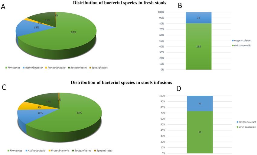
www.nature.com/scientificreports/ www.nature.com/scientificreports
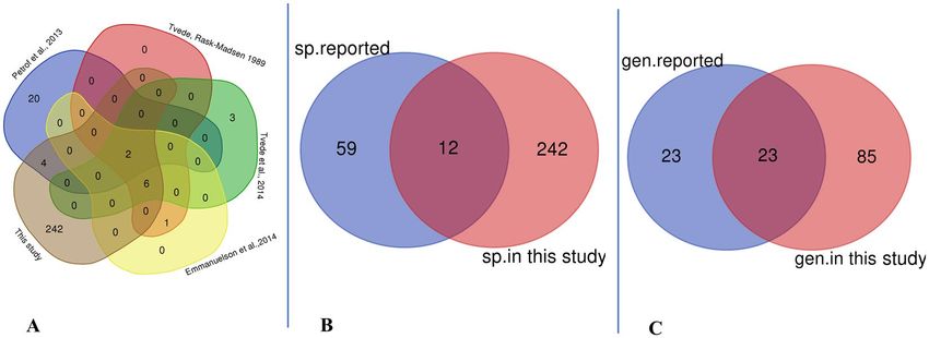
Figure 1. (A) Classification in phylum of the 196 bacteria species of the 8 fresh stools of fecal transplant donors
pretreated with ethanol. (B) Oxygen tolerance of the 196 bacteria species of the 8 fresh stools of fecal transplant
donors pretreated with ethanol. (C) Classification in phylum of the 135 bacteria species of the 3 fecal infusions
of fecal transplant donors pretreated with ethanol. (D) Oxygen tolerance of the 135 bacteria species of the 3 fecal
infusions of fecal transplant donors pretreated with ethanol.
culturomic approach. The objective is to evaluate the panel of bacterial species isolated from these stool samples
that have not been reported in previous studies and would have a potential therapeutic effect on Clostridium
difficile infections (CDI).
Results
Distribution of bacterial species. The 16 enrichment conditions (Supplementary Table 1) and 6 differ-
ent directs cultures used allowed us to test a total of 38,016 bacterial colonies by MALDI-TOF MS among the 8
fresh stool specimens. As a result, 196 bacterial species were identified, of which 99 were known in the human
gut (50%), 13 in humans but not in the gut (7%), 12 unknown from human being (6%), while 63 were new
species previously discovered as part of other culturomics studies (32%)23 and 9 were new species discovered
in this study (5%) (Appendix 1). The classification in phylum shows a predominance of Firmicutes (67%), fol-
lowed by Actinobacteria (15%), Bacteroidetes (15%) and small portions of Proteobacteria (2%) and Synergistetes
(1%) (Fig. 1A). These species are mostly anaerobes (158/196 = 80.61%) (Fig. 1B). Concerning the 3 fecal infu-
sions, a total of 16,500 colonies were tested by MALDI-TOF MS, representing 135 different species, 81 of which
are already known in the human gut (60%), 12 in the human, but not in gut (9%), 8 were non-Human (6%),
34 were culturomics new species (25%), but no other new species was discovered. The same profile of phylum
distribution is observed in the fecal infusions. Firmicutes represent 63%, Bacteroidetes 17%, Actinobacteria
11% followed by small portions of Proteobacteria (8%) and Synergistetes (1%) (Fig. 1C). The majority were also
anaerobes (73.33%) (Fig. 1D). A total of 254 bacterial species were isolated from 11 stools samples disinfected
with ethanol, mainly anaerobes (194/254 = 73.83%) (Appendix 1). These species are predominated by the fam-
ilies Clostridiaceae (37/254 = 14.57%), Ruminococcaceae (17/254 = 6.69%), Bacteroidaceae (16/254 = 6.30%),
Bacillaceae (15/254 = 5.91%) and Lachnospiraceae (13/254 = 5.12%) (Appendix 1). The richness of bacterial spe-
cies obtained in stools is very variable from one stool to another (from 35 to 68 species for fresh stool and 33 to
83 for fecal infusions) (Appendix 2).
Overall, for each fresh stool, the proportion of new species previously known in culture and those added in
this study represents a little more than one third (72/196 species = 36.73%) of the total proportion of bacterial
species obtained (Appendix 1), and those found in fecal infusions represent 1/4 (34/135 species; 25.18%) of these
total species (Appendix 1). For the 11 samples combined, the proportion of new species previously isolated in cul-
turomics and those discovered following ethanol disinfection, represent 32.67% (83/254) of the total proportion
of bacterial species (Appendix 1).
Impact of ethanol disinfection. The 18 usual culturomics conditions were carried out in parallel with
those following ethanol disinfection on the same fecal samples. To assess the impact of ethanol disinfection, we
compared the culture data obtained before and after ethanol disinfection of the same stool sample. Ethanol disin-
fection applied to the 8 fresh samples allows 60 species that were absent under the 18 standard cultivation condi-
tions to be cultivated, the same figure being 49 species for the 3 fecal infusions (Fig. 2, Appendix 1). Considering
Scientific Reports | (2020) 10:5190 | https://doi.org/10.1038/s41598-020-62068-x 2
www.nature.com/scientificreports/                                                               www.nature.com/scientificreports

                           Figure 2. Venn diagram of the bacterial species obtained before and after ethanol disinfection: FS bf.
                           OH = bacterial species obtained in the 8 fresh stools before ethanol disinfection; FI bf. OH = bacterial species
                           obtained in the 3 fecal infusions before ethanol disinfection; FS af. OH = bacterial species obtained in the 8
                           fresh stools after ethanol disinfection; FI af. OH = bacterial species obtained in the 3 fecal infusions after ethanol
                           disinfection.

                           Figure 3. Graphical representation of the mean impact of ethanol disinfection toward several bacterial genera.
                           The mean impact was assessed by summing the number of samples for which the genus was gained and
                           subtracting the number of samples for which it was lost. Each loss corresponds to the sum of the number of
                           species belonging to this genus present before disinfection, but absent after disinfection. Each gain corresponds
                           to the sum of the number of species belonging to this genus isolated absent before disinfection, but present after
                           disinfection.

                           all bacterial species isolated in the 11 samples, 68 bacterial species were unique to ethanol disinfection, while 98
                           and 329 different species are acquired and lost at least once, respectively (Appendix 2). In detail, ethanol disinfec-
                           tion has eliminated bacteria such as Phascolarctobacterium faecium (Supplementary Table 2) and Barnesiella intes-
                           tinihominis (Appendix 2), but also several species of the genera Alistipes, Bacteroides, Dialister, Bifidobacterium
                           for which the mean differential frequency of the different species are respectively −3.81, −3.52, −3.50 and −3.33
                           (Fig. 3, Table 1). Species belonging to the genera Bacillus, Clostridium, Blautia, Lactobacillus and Prevotella seem
                           to be less affected by this bacterial elimination caused by ethanol disinfection (Fig. 3, Table 1). All species gained
                           and lost in each stool with ethanol disinfection are illustrated in Appendix 2. More particularly, at the family level,
                           we observed after ethanol disinfection an enrichment in Ruminococcaceae, Bacteroidaceae and Lachnospiraceae,
                           whose rates decreased respectively from 2.70%, 5.16% and 4.18% before disinfection to 6.69%, 6.30% and 5.12%
                           after disinfection (Appendix 1).

                           Impact on spore forming species.             When all samples are combined, the number of species gained at least
                           once with ethanol disinfection was 98, 26 of which (26.53%) were sporulating species. Among the 329 species lost
                           at least once with ethanol disinfection, 10.94% (36 species) were sporulating species (Appendix 2, Fig. 4). In addi-
                           tion, only 32.65% of the species found in both ethanol and ethanol-free conditions are sporulating (16 species).

Scientific Reports |   (2020) 10:5190 | https://doi.org/10.1038/s41598-020-62068-x                                                              3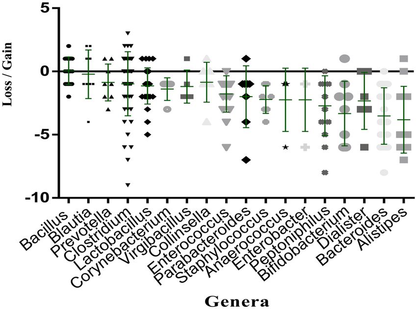
www.nature.com/scientificreports/                                                                    www.nature.com/scientificreports

                                              Number of      Number of     Average of    Standard
                            Genera            species lost   total species total species deviation
                            Bacillus          5              19             0.055       1.05
                            Blautia           4              9             −0.22        1.92
                            Prevotella        5              8             −0.87        1,45
                            Clostridium       21             47            −0.97        2,53
                            Lactobacillus     20             27            −1.14        1.43
                            Corynebacterium   5              5             −1.40        0.89
                            Virgibacillus     4              5             −1.75        0.50
                            Collinsella       4              7             −0.85        1.57
                            Enterococcus      17             18            −1.77        1.43
                            Parabacteroides   7              8             −2.00        2.44
                            Staphylococcus    9              9             −2.22        1.09
                            Anaerococcus      4              4             −2.25        2.50
                            Enterobacter      4              4             −2.25        2.50
                            Peptoniphilus     13             14            −2.71        2.36
                            Bifidobacterium   8              9             −3.33        2.54
                            Dialister         4              6             −2.33        2.25
                            Bacteroides       20             21            −3.52        2.22
                            Alistipes         9              11            −3.81        2.63

                           Table 1. Bacterial genera for which at least 4 different bacterial species are lost under the ethanol conditions.

                           Figure 4. Graphical representation of the mean impact of ethanol disinfection on sporulated and non-
                           sporulated species. Each point represents a species that has been classified as sporulated or non-sporulated. We
                           have assessed the mean impact of ethanol disinfection for each species by summing the number of samples for
                           which the species was gained and subtracting the number of samples for which it was lost. A gain corresponds
                           to a species absent before disinfection, but recovered after disinfection, while a loss corresponds to a species
                           present before disinfection, but absent after disinfection. Error bars are shown in green; p-value = 0.0003 with
                           Mann-Whitney test.

                           While these data suggest that ethanol incubation does not select only spore-forming species, non-spore-forming
                           species are nevertheless more affected by ethanol pre-treatment than spore-forming species(p = 0.0003) (Fig. 4).

                           Impact of the culturomics strategy on the cultivation of potential bacterial species of interest
                           for bacteriotherapy. Of the 71 species previously reported in bacteriotherapy trials16–20, 12 (17%) were
                           recovered in this study (Fig. 5A,B). The species are as follows: Bacteroides ovatus, Bacteroides thetaiotaomi-
                           cron, Bacteroides vulgatus, Bifidobacterium adolescentis, Bifidobacterium longum, Clostridium bifermentans,
                           Clostridium innocuum, Clostridium ramosum, Collinsella aerofaciens, Enterococcus fecalis, Escherichia coli and
                           Parabacteroides distasonis. In addition, 242 bacterial species isolated in this study were not considered in bacte-
                           riotherapy trials (Fig. 5B, Appendix 3). These are mainly strict anaerobes (185/242 = 76.45%) and predominated
                           by phyla Firmicutes (158/242), Bacteroidetes (33/242) and Actinobacteria (31/242) and with a low proportion of

Scientific Reports |   (2020) 10:5190 | https://doi.org/10.1038/s41598-020-62068-x                                                             4
www.nature.com/scientificreports/ www.nature.com/scientificreports
Figure 5. Venn Diagram comparing bacterial strains known previously in bacteriotherapy with those obtained
in this study; (A) Separate comparison of all bacterial species previously known against those of this study;
two bacterial species are shared between all studies. These are: Escherichia coli and Bacteroides ovatus. (B)
Grouped comparison of all bacterial species previously known in bacteriotherapy against those of this study;
12 bacterial species are shared between these two groups; these are: Clostridium ramosum, Enterococcus fecalis,
Clostridium bifermentans, Escherichia coli, Collinsella aerofaciens, Clostridium innocuum, Bifidobacterium
longum, Bacteroides thetaiotaomicron, Bacteroides vulgatus, Bacteroides ovatus, Parabacteroides distasonis
and Bifidobacterium adolescentis. (C) Grouped comparison of all previously known bacterial genera in
bacteriotherapy against those of this study; 23 bacterial genera are shared between these two groups. These are
genera Bifidobacterium, Streptococcus, Dorea, Terrisporobacter, Turicibacter, Ruminiclostridium, Enterococcus,
Bacteroides, Parabacteroides, Anaerostipes, Hungatella, Clostridium, Anaerotruncus, Anaerofustis, Flavonifractor,
Ruminococcus, Escherichia, Intestinibacter, Oscillibacter, Eubacterium, Collinsella, Lactobacillus and Blautia.
Proteobacteria and Synergistetes (Appendix 3). Of the 59 bacterial species absent from this study, 6 were actually
collected from samples prior to ethanol disinfection (i.e., as part of the 18 standard culture conditions), suggesting
that they did not survive the disinfection procedure (i.e., LactobacilIus rhamnosus, Faecalibacterium prausnitzii,
Acidaminococcus intestinalis, Dorea longicatena, Streptococcus mitis and Lactobacillus paracasei) (Appendix 1,
Appendix 3).
Succinate production. As high levels of succinate within the gut microbiota could promote CDI7, we
assessed the ability from bacteria isolated as part of this study to produce succinate. Considering the species
cultured following ethanol disinfection or not, we obtained information for 158/427 species. Of these, 112 were
succinate-producing bacteria and 46 were non-succinate-producing bacteria (Appendix 2). However, no signif-
icant difference was observed in the impact of ethanol disinfection on succinate-producing species compared to
species unable to produce succinate (Mann-Whitney test; p = 0.7693) (Fig. 6).
New species. The new genera and species found in this study were all found for the first time in fresh stools
and were named as follows: Massiliimalia timonensis (=CSUR P3753 = CCUG 7163), Lactobacillus timonen-
sis (=CSUR P3825 = CCUG 70711), Ethanolibacter massiliensis (=CSUR P5640 = CECT 9563), Prevotella
merdae (=CSURP4119 = CECT9566), Ruminococcus merdae (=CSUR P4123), Clostridium cacamassiliense
(=CSUR P5205), Dialister massiliensis (=CSURP5638), Neochristensenella massiliensis (=CSURP4260) and
Pseudoruminocossus massiliensis (=CSURP3876). Of these, only Massiliimalia timonensis was found in half of the
fresh stool samples. The others were found either in 3 (Ruminococcus merdae), 2 (Lactobacillus timonensis), or in
one stool sample at a time (Ethanolibacter massiliensis, Prevotella merdae, Clostridium cacamassiliense, Dialister
massiliensis, Neochristensenella massiliensis, Pseudoruminococcus massiliensis) (Appendix 2). None of these spe-
cies were found in the 3 fecal infusions.
Discussion
Herein, we carried out a complete culture analysis of 11 fecal samples after ethanol treatment using a culturomics
approach that will then be used as bacteriotherapy, particularly for patients with Clostridium difficile infections
(CDI). As a result, we cultured a total of 254 species mostly anaerobic, of which 68 bacterial species (containing 9
new bacterial taxa) were obtained only after treatment with ethanol (Appendix 1).
As disinfection technique has already been reported in a bacteriotherapy trial for the treatment of Clostridium
difficile infections18, in particular for the selection of sporulated bacteria21, the primary aim of this study was
to identify potential bacterial species that could be used for bacteriotherapy trials through the culturomics
approach22,23. Secondly, this work could be used to assess the potential relevance of this culture condition to
cultural studies.
Focusing on the contribution of the culturomics strategy to the recapture of bacteria of interest in bacteriother-
apy, we found that, compared to previous studies, 23 bacterial genera are shared between the two groups, with 85
bacterial genera representing at least 242 species being reported only in this study (Fig. 5A–C, Appendix 3). This
large difference can be explained on the one hand, by the size of our sampling, which is much more important than
in the previous studies, and, on the other hand, by our culturomics strategy, which targets the cultivation of a large
Scientific Reports | (2020) 10:5190 | https://doi.org/10.1038/s41598-020-62068-x 5www.nature.com/scientificreports/ www.nature.com/scientificreports
Figure 6. Graphical representation of the mean impact of ethanol disinfection on succinate producers and non-
succinate producers. We have assessed the mean impact of ethanol disinfection for each species by summing
the number of samples for which the species was gained and by subtracting the number of samples for which
it was lost. A gain corresponds to a species absent before disinfection, but recovered after disinfection, while a
loss corresponds to a species present before disinfection but absent after disinfection. Error bars are shown in
green; p-value = 0.7693 with Mann-Whitney test. Error bars are shown in green; p-value = 0.7693 with Mann-
Whitney test.
number of fastidious species. On the other hand, the genera Coprobacillus, Lachnoclostridium, Acidaminococcus,
Peptostreptococcus, Faecalibacterium, Tyzzerella, Coprococcus and Holdemanella, previously identified as candi-
dates16–20 and found under culturomics conditions prior ethanol disinfection, were all eliminated after ethanol
disinfection protocol. Similarly, the LactobacilIus rhamnosus, Faecalibacterium prausnitzii, Acidaminococcus intes-
tinalis, Dorea longicatena, Streptococcus mitis and Lactobacillus paracasei species do not appear to have survived
this disinfection protocol. These findings could appear counterintuitive as Lactobacillus species are frequently
included in probiotic formulations for preventing CDI relapses, while Faecalibacterium prausnitzii has been used
in a bacteriotherapy trial aiming to eradicate CDI in two patients16–20. As these microbes are often administered
in combination24, the exact contribution of each of its species to the treatment or prevention of CDI remains
undetermined and requires further studies.
When focusing on bacterial taxa dramatically affected by ethanol disinfection, species such as
Phascolarctobacterium faecium and Barnesiella intestinihominis (Appendix 2), but also some species of the genera
Alistipes, Bacteroides and Bifidobacterium were strongly eliminated (Table 1, Fig. 3). Apart from the Bacteroides
and Bifidobacterium genera previously reported in bacteriotherapy for the treatment of Clostridium difficile infec-
tions, the others have not been found in any bacteriotherapy studies16–20, although it has been reported that many
of these anaerobes are described as essential for human intestinal homeostasis25–29. These results may suggest
that the majority of these ethanol-eliminated bacterial species would not be fully essential for the treatment of
Clostridium difficile infections. Among the genera least affected by ethanol disinfection in this study, Blautia,
Clostridium and Lactobacillus were identified as bacteriotherapy candidates for the treatment of Clostridium dif-
ficile infections16–20. Indeed, ethanol stool disinfection would therefore select the majority of bacterial genera
sufficient to restore the diversity of the gut microbiota in the treatment of Clostridium difficile infections.
Interestingly, ethanol disinfection has enriched the proportion of species belonging to the Ruminococcaceae
and Lachnospiraceae (Appendix 1, Appendix 4). These two bacterial families have been suggested to be predictive
of favorable outcome following FMT for treating Clostridium difficile infections30. Our list of bacteria obtained
with ethanol disinfection is therefore quite consistent and contains probable candidates for CDI bacteriotherapy
trials.
Strikingly, sporulated bacterial species only represent 26.53% of all the species gained by ethanol disinfection
(Appendix 2), and the majority of species gained at least 3 times are new non-spore forming culturomics species23
(Supplementary Table 3). This highlights the fact that ethanol disinfection is ultimately an effective approach to
recover fastidious and minority species that have not been found under standard culturomics conditions22,23 for
the same samples and is therefore suitable for the exploration of the human gut microbiota. However, our data do
not support the idea that only sporulating species can survive this procedure (Fig. 4), as previously suggested18,21,
even though they have been less affected by ethanol disinfection than non-sporulating species.
Furthermore, some anaerobic bacteria reported in this study produce31–34 or consume 28,35 succinate
(Appendix 2). This production of succinate by intestinal bacteria modulates glucose metabolism in the healthy
host by inducing activation of intestinal gluconeogenesis36. However, in the host suffering from CDI, Clostridium
difficile can exploit the succinate produced by converting it into butyrate to multiply and exert an increased
Scientific Reports | (2020) 10:5190 | https://doi.org/10.1038/s41598-020-62068-x 6www.nature.com/scientificreports/ www.nature.com/scientificreports
pathogenic effect7. In our study, ethanol disinfection has no particular effect on the succinate producing species
as they were eliminated by ethanol incubation as much as non-succinate producing species (Fig. 6).
Finally, considering the gains and losses of stool bacterial species in each stool after ethanol treatment, we
noted that 98 minority bacterial species are gained at least once, versus 329 majority species lost at least once
(Appendix 2). This suggests a complementarity between standard culturomics conditions and ethanol disinfec-
tion conditions for the isolation of high numbers of microorganisms from stool samples. Indeed, ethanol stool
disinfection may therefore be an additional condition to be added to our laboratory culture strategy for future
studies, to explore and increase the microbial flora not yet cultivated in order to enrich the bacterial repertoire of
rare bacterial species. While the alcohol disinfection was empirically designed to select the sporulated bacteria,
another point of consideration could be to explore the impact of heat shock on fecal specimen from donors to
search for new bacteriotherapy trials candidates.
Conclusion
In conclusion, we demonstrated here that ethanol disinfection associated with the culturomic approach could be a
promising approach to explore the diversity of the human gut microbiota by selecting bacterial species of interest,
which can be potentially usable in bacteriotherapy. High-scale culture approach applied to 11 samples allowed
us to isolate 242 species that have never been reported in previous bacteriotherapy trials and that could be the
subject of further studies in the treatment of Clostridium difficile infections.
Material and Methods
Samples information. The material consists of 11 samples of healthy subjects, 8 of which represent fresh
stools from fecal transplant donors and 3 were samples of fecal infusion obtained from frozen stools (80 °C).
These 11 samples were collected from 9 different fecal donors: the 8 fresh stools represent 8 different donors,
fecal infusions 1 and 2 were collected from donors of fresh stools 1 and 4, and fecal infusion 3 was obtained from
the ninth donor (Supplementary Table 4). Each fecal donor gave informed and signed consent. The study was
approved by the ethics committee of the Institut Hospitalo-Universitaire-Méditerranée Infection under agree-
ment number 2016–011 and all the methods were performed in accordance with relevant and regulations.
Standard microbiological procedures. According to the French Recommendations of the National
Agency for the Safety of Medicines (ANSM)37, the stool have been qualified before being used for fecal transplan-
tation. This qualification procedure includes the search for pathogens and for transferable resistance mechanisms
from the stool and blood of donor. Any positive result to a pathogen or resistance mechanisms precludes the use
of this donor’s stool (Supplementary Table 5).
Preparation of fecal infusion. Fecal infusion is a stool donation prepared to be transplanted to a receiver,
while fecal transplant is the action of transplanting the fecal infusion to a receiver. Between September 2016 and
December 2017, 3 different fecal infusions were prepared according to the procedure previously reported38 and
from 3 different frozen fecal transplant donor stools. Donors were, pre-selected from questionnaires and medical
tests according to the ANSM37 (Supplementary Data). Briefly, for the preparation of each fecal infusion, each
donor’s stool is thawed at room temperature for 4 hours. In a pitcher, 500 mL of saline is added to the stool. The
mixture is then mixed for 5 minutes and passed through a sieve having a pore size of 1 mm in diameter. The fecal
infusion is collected in 10 mL syringes and then kept under anaerobic conditions (ie in a plastic bag + GENbag
Anaer systems (bioMérieux)). Each fecal infusion represents one sample and one donor.
Process. Manipulations of the 11 samples used (8 fresh stools samples and 3 fecal infusions samples) were
performed under microbiological hood and anaerobic chamber. Each stool was disinfected separately with eth-
anol according to a previous study18 in order to eliminate vegetative forms as much as possible to promote the
growth of bacteria resistant to alcohol18 and capable of sporulating18,21. To this end, about 10 g of each stool was
®
homogenized in 10 mL of saline solution (NaCL 0.9%, Versylene Fresenius, Sevres, France). Then, the mixture
is filtered through a sieve with a pore size of 1 mm in diameter and the supernatant is recovered. In falcon tubes,
10 mL of 100% ethanol is added to 10 mL of supernatant containing the bacterial cells and the spores. This mix-
ture is then incubated at room temperature under anaerobic conditions for 1 hour. Thereafter, the mixture is
centrifuged for 2 minutes at 5000 rpm to remove ethanol (which in this case is the supernatant). The pellet was
washed twice with saline solution by centrifugation to remove any trace of the remaining ethanol before proceed-
ing to microbial culturomics.
Microbial culturomics. After ethanol disinfection of the 11 samples, we performed, the culture on 6 differ-
ent solid culture media and, on 16 different pre-enrichment conditions (Supplementary Table 1), which will then
be subcultured on sheep blood-enriched Columbia agar (COS) medium (bioMérieux, Marcy l’Etoile, France). A
total of 22 culture conditions were thereby used in this study. Briefly, the direct culture after ethanol treatment
was carried out in anaerobic chamber on these 6 different types of culture media: Reinforced clostridial agar
™
(HiMedia Laboratories Pvt Lt, India), Wilkins Chalgren agar (Becton, Dickinson company, Le Pont-de-Claix,
France), Brain-heart infusion agar (Becton, Dickinson company, Le Pont-de-Claix, France), deMan, Rogosa and
Sharpe agar (Sigma-Aldrich, Saint-Louis, USA)16, 5% sheep blood-enriched columbia agar (COS) (bioMérieux,
Marcy l’Etoile, France), Yeast Extract-Casein Hydrolysate-Fatty Acids (YCFA) agar, according to the composi-
tion previously described39, supplemented with 0.002 g/ml each of glucose, maltose, cellobiose and 0.1% sodium
taurocholate40. In parallel, the stools were pre-incubated in blood culture bottles supplemented or not with 5%
of the rumen, 5% of blood or both under aerobic and anaerobic conditions at 37 °C and then at 28 °C, under
16 selected culturomics conditions22,23. These blood cultures were seeded every 3 days on Columbia agar with
5% sheep blood (bioMérieux, Marcy l’Etoile, France) under aerobic and anaerobic conditions for 30 days22,23
Scientific Reports | (2020) 10:5190 | https://doi.org/10.1038/s41598-020-62068-x 7www.nature.com/scientificreports/ www.nature.com/scientificreports
(Supplementary Table 1). All the morphologically different colonies obtained in direct culture and preincubation
were subcultured on COS and bacterial identification was performed after 24–72 hours of incubation. The subcul-
tures were identified using MALDI-TOF mass spectrometry with a Microflex LT spectrometer (Bruker Daltonics,
Leipzig, Germany) as previously described41. When identification was not possible by MALDI-TOF, 16S rRNA
gene sequencing was performed on unidentified colonies.
16S rRNA gene sequencing. DNA extraction was performed using the EZ1 DNA Tissue Kit and BioRobot
EZ1 Advanced XL (Qiagen, Courtaboeuf, France). DNA extracts were used for 16S rRNA amplification using
the fD1 and rP2 primers (Eurogentec, Angers, France). Amplicon sequencing was performed using the Big Dye
Terminator v1.1 Cycle Sequencing Kit and an ABI Prism 3130xl Genetic Analyzer capillary sequencer (Applied
®
Biosystems), as previously described42. The obtained 16S rRNA sequences were compared with those available in
GenBank (http://www.ncbi.nlm.nih.gov/genbank). Identification at the species level was defined by a 16S rRNA
gene sequence similarity ≥98.65% with the sequence of the prototype strain of a species with standing in nomen-
clature. When this percentage of identity was lower than the generally accepted thresholds of 98.65% or 95%, the
strain studied was considered a putative new species or genus, respectively43,44.
Succinate production and sporulation. The “Google” search engine was used to search for data on the
production or non-production of succinate and sporulation of our isolated bacterial species in this study using
the keywords “name of bacteria” followed by “succinate production” or “spore”. For species for which full descrip-
tions or work on succinate and spore production were available, searches were carried out using “PubMed” and
“Google scholar” databases, but also using “List of Prokaryotic names with Standing in Nomenclature” (http://
www.bacterio.net/). Concerning the different new species of “culturomics” found in this study, we used our lab-
oratory data (published or not).
Venn diagrams. Venn diagrams comparing the bacterial species obtained in this study with those previously
reported in bacteriotherapy were produced online at: http://bioinformatics.psb.ugent.be/webtools/Venn.
Statistical test. Plots and statistical analyses were performed using the GraphPad Prism software (version
6.01; GraphPad Software, Inc., www.graphpad.com) for Figs. 3, 4 and 6 based on the number of bacterial taxa
gained and lost in the 11 stools samples (Appendix 2).
Data availability
Additional data on the bacterial species isolated in this study are presented in Appendix 1 to 4. Supplementary
Tables and Appendices legends are available in “Supplementary Data”.
Received: 10 April 2019; Accepted: 2 March 2020;
Published: xx xx xxxx
References
1. Pépin, J. et al. Clostridium difficile-associated diarrhea in a region of Quebec from 1991 to 2003: a changing pattern of disease
severity. CMAJ Can. Med. Assoc. J. J. Assoc. Medicale Can. 171, 466–472 (2004).
2. McDonald, L. C. et al. An epidemic, toxin gene-variant strain of Clostridium difficile. N. Engl. J. Med. 353, 2433–2441 (2005).
3. Loo, V. G. et al. A predominantly clonal multi-institutional outbreak of Clostridium difficile-associated diarrhea with high morbidity
and mortality. N. Engl. J. Med. 353, 2442–2449 (2005).
4. Lessa, F. C. et al. Burden of Clostridium difficile infection in the United States. N. Engl. J. Med. 372, 825–834 (2015).
5. Khoruts, A., Dicksved, J., Jansson, J. K. & Sadowsky, M. J. Changes in the composition of the human fecal microbiome after
bacteriotherapy for recurrent Clostridium difficile-associated diarrhea. J. Clin. Gastroenterol. 44, 354–360 (2010).
6. Lewis, B. B. et al. Loss of Microbiota-Mediated Colonization Resistance to Clostridium difficile Infection With Oral Vancomycin
Compared With Metronidazole. J. Infect. Dis. 212, 1656–1665 (2015).
7. Ferreyra, J. A. et al. Gut microbiota-produced succinate promotes C. difficile infection after antibiotic treatment or motility
disturbance. Cell Host Microbe 16, 770–777 (2014).
8. Gens, K. D., Elshaboury, R. H. & Holt, J. S. Fecal microbiota transplantation and emerging treatments for Clostridium difficile
infection. J. Pharm. Pract. 26, 498–505 (2013).
9. Gerding, D. N. et al. Administration of spores of nontoxigenic Clostridium difficile strain M3 for prevention of recurrent C. difficile
infection: a randomized clinical trial. JAMA 313, 1719–1727 (2015).
10. Dieterle, M. G., Rao, K. & Young, V. B. Novel therapies and preventative strategies for primary and recurrent Clostridium difficile
infections. Ann. N. Y. Acad. Sci. 1435, 110–138 (2019).
11. Hamilton, M. J., Weingarden, A. R., Sadowsky, M. J. & Khoruts, A. Standardized frozen preparation for transplantation of fecal
microbiota for recurrent Clostridium difficile infection. Am. J. Gastroenterol. 107, 761–767 (2012).
12. van Nood, E. et al. Duodenal infusion of donor feces for recurrent Clostridium difficile. N. Engl. J. Med. 368, 407–415 (2013).
13. Brandt, L. J. American Journal of Gastroenterology Lecture: Intestinal microbiota and the role of fecal microbiota transplant (FMT)
in treatment of C. difficile infection. Am. J. Gastroenterol. 108, 177–185 (2013).
14. Goldenberg, S. D. et al. Comparison of Different Strategies for Providing Fecal Microbiota Transplantation to Treat Patients with
Recurrent Clostridium difficile Infection in Two English Hospitals: A Review. Infect. Dis. Ther. 7, 71–86 (2018).
15. Zipursky, J. S., Sidorsky, T. I., Freedman, C. A., Sidorsky, M. N. & Kirkland, K. B. Patient attitudes toward the use of fecal microbiota
transplantation in the treatment of recurrent Clostridium difficile infection. Clin. Infect. Dis. Off. Publ. Infect. Dis. Soc. Am. 55,
1652–1658 (2012).
16. Petrof, E. O. et al. Stool substitute transplant therapy for the eradication of Clostridium difficile infection: ‘RePOOPulating’ the gut.
Microbiome 1, 3 (2013).
17. Tvede, M., Tinggaard, M. & Helms, M. Rectal bacteriotherapy for recurrent Clostridium difficile-associated diarrhoea: results from
a case series of 55 patients in Denmark 2000–2012. Clin. Microbiol. Infect. Off. Publ. Eur. Soc. Clin. Microbiol. Infect. Dis. 21, 48–53
(2015).
18. Khanna, S. et al. A Novel Microbiome Therapeutic Increases Gut Microbial Diversity and Prevents Recurrent Clostridium difficile
Infection. J. Infect. Dis. 214, 173–181 (2016).
Scientific Reports | (2020) 10:5190 | https://doi.org/10.1038/s41598-020-62068-x 8www.nature.com/scientificreports/ www.nature.com/scientificreports
19. Tvede, M. & Rask-Madsen, J. Bacteriotherapy for chronic relapsing Clostridium difficile diarrhoea in six patients. Lancet Lond. Engl.
1, 1156–1160 (1989).
20. Emanuelsson, F., Claesson, B. E. B., Ljungström, L., Tvede, M. & Ung, K.-A. Faecal microbiota transplantation and bacteriotherapy
for recurrent Clostridium difficile infection: a retrospective evaluation of 31 patients. Scand. J. Infect. Dis. 46, 89–97 (2014).
21. Koransky, J. R., Allen, S. D. & Dowell, V. R. Use of ethanol for selective isolation of sporeforming microorganisms. Appl. Environ.
Microbiol. 35, 762–765 (1978).
22. Lagier, J.-C. et al. Microbial culturomics: paradigm shift in the human gut microbiome study. Clin. Microbiol. Infect. Off. Publ. Eur.
Soc. Clin. Microbiol. Infect. Dis. 18, 1185–1193 (2012).
23. Lagier, J.-C. et al. Culture of previously uncultured members of the human gut microbiota by culturomics. Nat. Microbiol. 1, 16203
(2016).
24. Barker, A. K. et al. A randomized controlled trial of probiotics for Clostridium difficile infection in adults (PICO). J. Antimicrob.
Chemother. 72, 3177–3180 (2017).
25. Tojo, R. et al. Intestinal microbiota in health and disease: role of bifidobacteria in gut homeostasis. World J. Gastroenterol. 20,
15163–15176 (2014).
26. Ruiz, L., Delgado, S., Ruas-Madiedo, P., Sánchez, B. & Margolles, A. Bifidobacteria and Their Molecular Communication with the
Immune System. Front. Microbiol. 8, 2345 (2017).
27. Routy, B. et al. The gut microbiota influences anticancer immunosurveillance and general health. Nat. Rev. Clin. Oncol. 15, 382–396
(2018).
28. Wu, F. et al. Phascolarctobacterium faecium abundant colonization in human gastrointestinal tract. Exp. Ther. Med. 14, 3122–3126
(2017).
29. Ishaq, H. M. et al. Molecular Alteration Analysis of Human Gut Microbial Composition in Graves’ disease Patients. Int. J. Biol. Sci.
14, 1558–1570 (2018).
30. Wilson, B. C., Vatanen, T., Cutfield, W. S. & O’Sullivan, J. M. The Super-Donor Phenomenon in Fecal Microbiota Transplantation.
Front. Cell. Infect. Microbiol. 9 (2019).
31. Van der Meulen, R., Adriany, T., Verbrugghe, K. & De Vuyst, L. Kinetic analysis of bifidobacterial metabolism reveals a minor role
for succinic acid in the regeneration of NAD+ through its growth-associated production. Appl. Environ. Microbiol. 72, 5204–5210
(2006).
32. Fischbach, M. A. & Sonnenburg, J. L. Eating for two: how metabolism establishes interspecies interactions in the gut. Cell Host
Microbe 10, 336–347 (2011).
33. Rios-Covian, D. et al. Interactions between Bifidobacterium and Bacteroides Species in Cofermentations Are Affected by Carbon
Sources, Including Exopolysaccharides Produced by Bifidobacteria. Appl. Env. Microbiol. 79, 7518–7524 (2013).
34. Morotomi, M., Nagai, F., Sakon, H. & Tanaka, R. Dialister succinatiphilus sp. nov. and Barnesiella intestinihominis sp. nov., isolated
from human faeces. Int. J. Syst. Evol. Microbiol. 58, 2716–2720 (2008).
35. Del Dot, T., Osawa, R. & Stackebrandt, E. Phascolarctobacterium faecium gen. nov, spec. nov., a Novel Taxon of the Sporomusa
Group of Bacteria. Syst. Appl. Microbiol. 16, 380–384 (1993).
36. De Vadder, F. et al. Microbiota-Produced Succinate Improves Glucose Homeostasis via Intestinal Gluconeogenesis. Cell Metab. 24,
151–157 (2016).
37. La transplantation de microbiote fécal et son encadrement dans les essais cliniques - Point d’Information - ANSM: Agence nationale
de sécurité du médicament et des produits de santé. https://ansm.sante.fr/S-informer/Points-d-information-Points-d-information/
La-transplantation-de-microbiote-fecal-et-son-encadrement-dans-les-essais-cliniques-Point-d-Information2.
38. Lagier, J.-C. et al. Dramatic reduction in Clostridium difficile ribotype 027-associated mortality with early fecal transplantation by
the nasogastric route: a preliminary report. Eur. J. Clin. Microbiol. Infect. Dis. Off. Publ. Eur. Soc. Clin. Microbiol. 34, 1597–1601
(2015).
39. Duncan, S. H., Hold, G. L., Harmsen, H. J. M., Stewart, C. S. & Flint, H. J. Growth requirements and fermentation products of
Fusobacterium prausnitzii, and a proposal to reclassify it as Faecalibacterium prausnitzii gen. nov., comb. nov. Int. J. Syst. Evol.
Microbiol. 52, 2141–2146 (2002).
40. Browne, H. P. et al. Culturing of ‘unculturable’ human microbiota reveals novel taxa and extensive sporulation. Nature 533, 543–546
(2016).
41. Seng, P. et al. Ongoing Revolution in Bacteriology: Routine Identification of Bacteria by Matrix-Assisted Laser Desorption
Ionization Time-of-Flight Mass Spectrometry. Clin. Infect. Dis. 49, 543–551 (2009).
42. Dubourg, G. et al. The gut microbiota of a patient with resistant tuberculosis is more comprehensively studied by culturomics than
by metagenomics. Eur. J. Clin. Microbiol. Infect. Dis. Off. Publ. Eur. Soc. Clin. Microbiol. 32, 637–645 (2013).
43. Stackebrandt, E. & Ebers, J. Taxonomic parameters revisited: Tarnished gold standards. Microbiol. Today 8, 6–9 (2006).
44. Kim, M., Oh, H.-S., Park, S.-C. & Chun, J. Towards a taxonomic coherence between average nucleotide identity and 16S rRNA gene
sequence similarity for species demarcation of prokaryotes. Int. J. Syst. Evol. Microbiol. 64, 346–351 (2014).
Acknowledgements
The authors thank the sequencing platform of the IHU Mediterranee-Infection for the 16S rRNA gene sequencing
of some strains of this study. This study was supported by the “Méditerranée Infection” foundation and by the
French Government under the « Investissements d’avenir » (Investments for the Future) program managed by the
Agence Nationale de la Recherche (ANR, fr: National Agency for Research), (reference: Méditerranée Infection
10-IAHU-03). This work was also supported by Région Provence-Alpes-Côte d’Azur and European funding
FEDER PRIMI.
Author contributions
D.R. conceived and designed the experiments. P.A., M.H., T.P.T.P., E.K., I.I.N., N.D., C.V. and S.B. performed the
experiments; P.A. and G.D analyzed the data; P.A., G.D., J.C.L. and D.R. wrote the manuscript. All authors read
and approved the final manuscript.
Competing interests
The authors declare no competing interests.
Additional information
Supplementary information is available for this paper at https://doi.org/10.1038/s41598-020-62068-x.
Correspondence and requests for materials should be addressed to D.R.
Reprints and permissions information is available at www.nature.com/reprints.
Scientific Reports | (2020) 10:5190 | https://doi.org/10.1038/s41598-020-62068-x 9www.nature.com/scientificreports/ www.nature.com/scientificreports
Publisher’s note Springer Nature remains neutral with regard to jurisdictional claims in published maps and
institutional affiliations.
Open Access This article is licensed under a Creative Commons Attribution 4.0 International
License, which permits use, sharing, adaptation, distribution and reproduction in any medium or
format, as long as you give appropriate credit to the original author(s) and the source, provide a link to the Cre-
ative Commons license, and indicate if changes were made. The images or other third party material in this
article are included in the article’s Creative Commons license, unless indicated otherwise in a credit line to the
material. If material is not included in the article’s Creative Commons license and your intended use is not per-
mitted by statutory regulation or exceeds the permitted use, you will need to obtain permission directly from the
copyright holder. To view a copy of this license, visit http://creativecommons.org/licenses/by/4.0/.
© The Author(s) 2020
Scientific Reports | (2020) 10:5190 | https://doi.org/10.1038/s41598-020-62068-x 10You can also read