Floral Morphology and Embryology of - Acer oblongum
←
→
Page content transcription
If your browser does not render page correctly, please read the page content below
©Verlag Ferdinand Berger & Söhne Ges.m.b.H., Horn, Austria, download unter www.biologiezentrum.at

              Floral Morphology and Embryology of
                       Acer oblongum
                                            By
                                 Ishwari KHUSHALANI
            Department of Botany, Birla College, Pilani (Rajasthan), India
                                   With 66 Figures
                                Received May 30, 1963
                                      Contents
   1. Introduction
   2. Materials and methods
   3. Observations
      3. 1. External morphology
      3. 2. Floral anatomy
      3. 3. Organography and organogeny
      3. 4. Microsporogenesis and male gametophyte
      3. 5. Megasporogenesis and female gametophyte
      3. 6. Endosperm
      3. 7. Pericarp and seed
   4. Discussion
   5. Summary
   6. Literature cited
                                1. Introduction
        BENTHAM    & HOOKER 1862: 409 included Acer in Sapindaceae while
   some taxonomists have separated it from Sapindaceae and included under
   the family Aceraceae (LAWRENCE 1958, HTJTCHINSON 1959, COPELAND 1957,
   PORTER 1959). Embryology of Acer is very little understood. MOTTIER
   1893 worked out the development of embryo sac in Acer rubrum. However,
   some work has been done on the floral anatomy of the family. SAUNDERS
   1939, briefly described the floral anatomy of Acer pseudoplatanus and
   A. negundo. HALL 1951 has reviewed the earlier literature on Aceraceae
   and given a detailed account of the floral anatomy of Acer. On the basis
   of the floral anatomy HALL 1951 has divided nine species of Acer into
   six anatomical types, forming a single series, from the most primitive
   to the most highly evolved. HALL 1954 has described the variability in
   the floral anatomy of Acer negundo. HALL 1959 while studying the inflores-
   cence in the Aceraceae confirmed the view that simpler inflorescence types
   have been derived phylogenetically from more complex ones through loss
   of branches and the shortening of internodes. Aceraceae provides an
   excellent material for such a study since they include a wide variety of
   inflorescence types within a group of closely related species.

                                                                         18*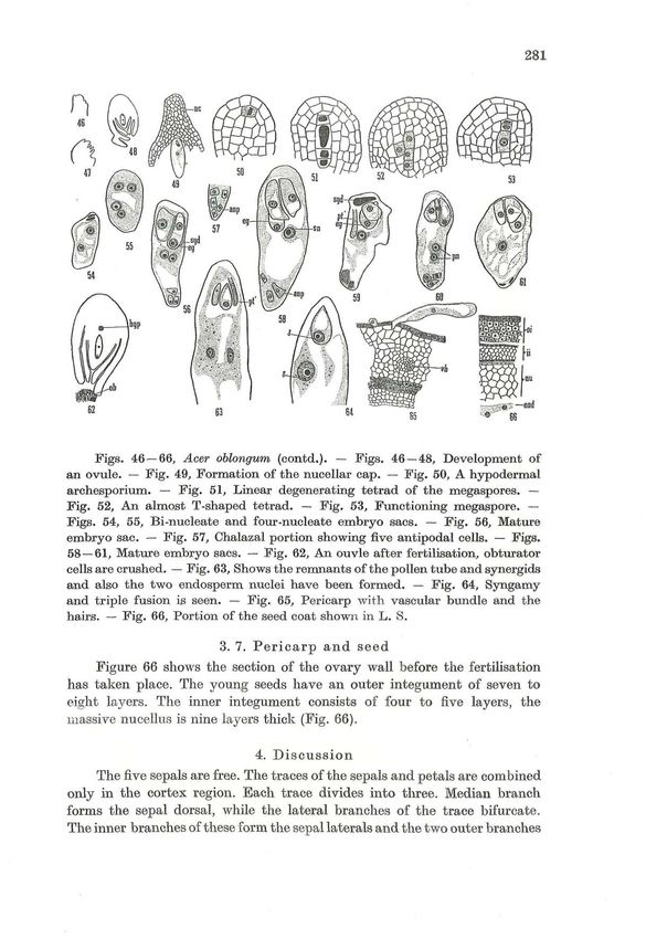
©Verlag Ferdinand Berger & Söhne Ges.m.b.H., Horn, Austria, download unter www.biologiezentrum.at

        276

                               2. Materials and methods
            Acer oblongum was collected from Ootacamund and fixed in formalin-
       acetic-alcohol. Dehydration and imbedding were followed according to
       the usual alcohol xylol paraffin method. Sections were cut at 6—15 \x.
       For embryological purpose staining was done with combinations of safranin
       and fast green, safranin and light green and Heidenhains iron-alum haemato-
       xylin. For the floral anatomy the combination erythrosine and crystal
       violet gave better results.

                                     3. Observations
                              3. 1. External morphology
            The small pedicellate flowers borne on panicles, are hypogynous and
       aetinomorphic having five imbricate sepals and five free petals. A very
       distinctly lobed disc is present within the petals. The stamens are in two
       whorls, the outer whorl consists of five and the inner one of four stamens.
       The number of stamens varies from eight to nine but in all the cases, the
       antipetalous stames are suppressed. The ovary is superior, bicarpellary
       and compressed at right angles to the septum. There are two distinct and
       divergent styles, each having a terminal stigma.

                                  3. 2. Floral anatomy
             The floral anatomy described here is that of the functionally bisexual
       flower with five sepals, five petals, nine stamens occuring in two whorls
       of five and four respectively and a bicarpellary ovary (Fig. 1) *).
             The pedicel consists of a siphonostele in the centre (Fig. 2). In the
       receptacular region the stele bulges out to give five traces which constitute
       the fused sepal and petal supply. Three out of these five traces are given
       off slightly at lower level (Figs. 3 and 4). Each of these traces is resolved
       into three branches (Figs. 5 & 6), the middle one of which forms the midrib
       of the sepal, while the lateral two branches once again bifurcate (Fig. 6).
       The inner branches of these enter the sepal, thus forming the two marginals
       of the same sepal while the outer ones move out tangentially. Two such
       branches of the adjacent sides unite and later on each of these fused bundles
       enters the petal (Fig. 7). The gaps created by the fused sepal and petal

           *) Abbreviations in figures 1 — 66: anp = antipodal cells, cb = carpel-
      lary wall bundles,, cd = carpel dorsal, cr = carpel, cv = carpel ventral, di = disc,
      ds = disc supply, eg = egg, end = endosperm, hyp = hypostase, ii = inner
      integument, lo = locules, nc = nucellar cap, nu = nucellus, ob = obturator,
      oi = outer integument, ot = ovular trace, pe = petal, pn = polar nuclei,
      pt — petal trace, pt' = pollen tube, se = sepal, sm = stamen, sn = secondary
      nucleus, spe = sepal lateral, spm = sepal dorsal, spt = sepal petal trace,
      st = staminal trace, syd = synergids, vb = vascular bundle.
©Verlag Ferdinand Berger & Söhne Ges.m.b.H., Horn, Austria, download unter www.biologiezentrum.at
                                                                                    277

   are closed soon. At the higher level each petal has only one central bundle
   which does not divide further while each sepal is supplied with three.
        The central stele, at a slightly higher level, gives out eight traces
   which constitute the supply of disc, stamens and ovary wall (Fig. 8).
   Small irregular branches are given off from these traces towards the outer
   side for the disc (Figs. 9, 10). At a slightly higher level, the staminal traces
   are separated from the two dorsals and several carpellary wall traces to
   which they were fused at the lower level (Fig. 10). All the staminal traces
   arise at the same level.
        In the centre now are left two bundles which are fused ventrals of the
   two carpels (Figs. 8—11). At this level the lower portions of the locules
   are marked off (Fig. 12). Before supplying the ovules each of the fused
   ventral divides (Fig. 13) into two and these become inversely oriented
   (Fig. 14). After giving off the traces to the ovules these ventral bundles
   move to the septal radii where they fuse with the carpellary wall bundles
   (Fig. 15). At the terminus of the ovular region the ventral bundles travel
   along with the two dorsals through the stylar region (Figs. 16—18). At a
   higher level one dorsal and the two ventrals of one carpel fuse together.
   Thus, in the tip region each stigmatic lobe has a single bundle.

                      3.3. Organography and Organogeny
         Unicellular hairs are present on some of the floral parts. The cell
   destined to develop into a hair gets differentiated from the rest of the cells
   (Fig. 19) by a large nucleus and its large size. Later on, it becomes papillate
   (Fig. 20), increases in length, and becomes vacuolated (Fig. 21). Along with
   the unicellular hairs, stalked multicellular glands are also present on the
   outer side of the ovary wall.
         The precursor of a gland becomes differentiated from the rest of the
   cells with a prominent nucleus and protrudes out slightly (Fig. 22). It
   divides transversely forming an outer and an inner cell (Fig. 23), the inner
   one being in continuation with the epidermis. The outer cell divides first
   transversely and later in various planes to form the stalk and head of the
   gland (Figs. 24, 25).
         In this plant two types of ovaries are found, one with stunted growth
   having papillate hairs on the stigma and another which is with a normal
   growth, the latter being more common. Though the ovay may be stunted,
   the embryo sacs in the ovules of these ovaries are quite normal and even
   fertilization has been found to have taken place. The funicular obturator
   is present and its cells are elongated and vacuolated.
         There are two anatropus, superimposed ovules in each locule of the
   normal ovary. The funicular obturator is present and its cells are elongated
   and vacuolated.
         The floral organs develop in the sequence, sepals, petals, disc, stamens
   and carpels (Figs. 26 — 28).
©Verlag Ferdinand Berger & Söhne Ges.m.b.H., Horn, Austria, download unter www.biologiezentrum.at
278
3. 4. Microsporogenesis and male gametophyte
The young anther of the flower is four-lobed (Fig. 29). The primary
partietal layer cut off by the archesporium divides to form four layers
of cells beneath the epidermis. The cells of epidermis become tangentially
elongated and their cytoplasm is gathered on one side. The subepidermal
layer develops into the endothecium, the cells of which become radially
elongated and develop the characteristic fibrous thickenings when uni-
nucleate pollen grains are formed in the microsporangium (Fig. 30). Just
beneath the endothecium are two middle layers, which get crushed and
absorbed. No trace of them is found when the pollen grains are uninucleate
(Fig. 30).
The innermost wall layer is the tapetum. Its cells are uninucleate at
first but they become two-three-nucleate at the time of the meiotic divisions
in the microspore mother cells. Some of the tapetal nuclei are very large
and contain a varying number of nucleoli. The nuclei often fuse with each
other. The tapetum is of glandular type (Fig. 31). When the uninucleate
pollen grains are distinguished in the anther, the tapetal cells show signs of
degeneration.
The primary sporogenous cells function directly as the spore mother
cells (Fig. 32).
The mother cells undergo usual meiotic divisions in a simultaneous
manner, forming isobilateral and tetrahedral tetrads, the latter being more
predominant. The divisions are not synchronous in the four lobes of the
anther. Quadripartition takes place by centripetal furrowing (Figs. 33—38).
Later the microspores acquire their own walls. The young microspore
has a dense cytoplasm and a centrally placed large nucleus (Fig. 39). The
development of male gametophyte is in a normal way. Generally the pollen
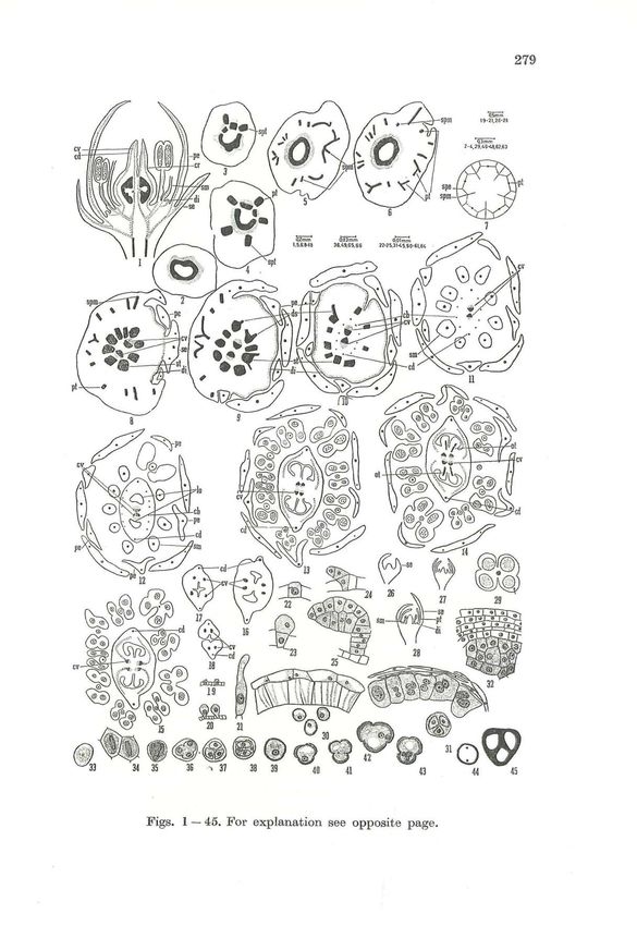
E x p l a n a t i o n of figures
Figs. 1 — 45, Acer oblongum. — Fig. 1, L. S. of the flower. — Fig. 7, A
diagrammatic representation. Figs. 2 — 7 and 8 —18., Serial transection of the
flower from pedicel onwards. (Sepals and petals avoided in Fig. 15). — Fig.
19 — 21, Development of the hairs, which are found on the inner side of the
ovary wall. — Figs. 22 — 25, Development of the gland. — Figs. 26 — 28, Devel-
opment of floral organs. — Fig. 29, T. S. of young anther. — Fig. 30, Fibrous
thickenings in the endothecium. — Fig. 31, Multinucleate, multinucleolate
and vacuolated tapetal cells are seen, along with the degenerating middle
layers. — Fig. 32, It is an enlargement of the portion marked by arrow in
Fig. 29. It shows the wall layers and the sporogenous tissue. — Figs. 33 — 36,
Meiotic divisions of the microspore mother cell. — Figs. 37 — 38, Tetrahedral
and isobilateral tetrads. — Fig. 39 — 40, Uninucleate pollen grains. — Fig. 41,
Division of the nucleus in the microspore. — Fig. 42, Bicelled pollen grain. —
Fig. 43, Three-celled pollen grain. — Figs. 44—45, Degeneration taking place
after the first and the second division in the microspore mother cell.©Verlag Ferdinand Berger & Söhne Ges.m.b.H., Horn, Austria, download unter www.biologiezentrum.at
279
33 34 35 36 •* 37 38 39 40 41 44 45
Figs. 1 — 45. For explanation see opposite page.©Verlag Ferdinand Berger & Söhne Ges.m.b.H., Horn, Austria, download unter www.biologiezentrum.at
280
grains are shed at two-celled stage but in one case, a three-celled pollen
grain was observed (Fig. 43).
Pollen grains are tricolporate. Sterility of pollen grains is very high.
Some of the dyads and tetrads were also degenerating (Figs. 44—45).
3. 5. Megasporogenesis and female gametophyte
The ovules are bitegmic, crassinucellate and anatropous. The ovular
protuberance is erect at the earlier stages (Fig. 46). Soon it curves (Fig. 47),
and at the eight-nucleate embryo sac stage, it becomes anatropous (Fig. 48).
The inner integument develops when the hypodermal archesporium is
distinguished in the young nucellus. The outer one also develops soon. The
micropyle is formed by the inner integument. A nucellar cap is present
which is eight to ten cell layers in thickness at the mature embryo sac
stage (Fig. 49).
The single hypodermal archesporial cell divides periclinally to form
a primary parietal cell and a primary sporogenous cell (Fig. 50). The
primary parietal cell undergoes further periclinal divisions while the primary
sporogenous cell functions directly as the megaspore mother cell.
The megaspore mother cell increases in size and undergoes the usual
meiotic divisions to form the tetrad. In most of the cases linear tetrads are
formed but in one case an almost T-shaped tetrad was observed (Fig. 52).
In one case of the linear tetrad all the spores had degenerated (Fig. 51).
The chalazal megaspore is usually functional. In Fig. 53, two degenerating
spores at the micropylar end were observed. The development of the
embryo sac conforms to the Polygonum-type (Figs. 54—56). The mature
embryo sac has a narrow chalazal end and a broad micropylar end. The
synergids are hooked and the egg is flask-shaped. The three antipodal
cells may degenerate soon. More than three antipodal cells have also been
observed (Fig. 57). The embryo sac contains numerous starch grains
(Fig. 63). The two polar nuclei meet slightly above the centre (Fig. 58).
In the ovaries with stunted growth, the embryo sacs were of a normal
type. Even a pollen tube has been observed lying in the embryo sac in
one case (Fig. 59). Figures 60 and 61 show mature embryo sacs. The funi-
cular obturator is seen crushed in the later stages. A well marked hypostase
is present (Fig. 62).
The entry of the pollen tube is through the micropyle. The remnants
of the pollen tube have been observed (Fig. 63). In one case, one male
gamete is seen fusing with the egg and another with the secondary nucleus
(Fig. 63).
3. 6. Endosperm
The endosperm is of the Nuclear type (Fig. 65). The development
of the embryo is arrested for a long time. The seeds from the fruits which
were naturally shed showed only undivided zygotes with man3^ endosperm
nuclei.©Verlag Ferdinand Berger & Söhne Ges.m.b.H., Horn, Austria, download unter www.biologiezentrum.at
281
Figs. 46 — 66, Acer oblongum (contd.). — Figs. 46 — 48, Development of
an ovule. — Fig. 49, Formation of the nucellar cap. — Fig. 50, A hypodermal
archesporium. — Fig. 51, Linear degenerating tetrad of the megaspores. —
Fig. 52, An almost T-shaped tetrad. — Fig. 53, Functioning megaspore. —
Figs. 54, 55, Bi-nucleate and four-nucleate embryo sacs. — Fig. 56, Mature
embryo sac. — Fig. 57, Chalazal portion showing five antipodal cells. — Figs.
58 — 61, Mature embryo sacs. — Fig. 62, An ouvle after fertilisation, obturator
cells are crushed. — Fig. 63, Shows the remnants of the pollen tube and synergids
and also the two endosperm nuclei have been formed. — Fig. 64, Syngamy
and triple fusion is seen. — Fig. 65, Pericarp with vascular bundle and the
hairs. — Fig. 66, Portion of the seed coat shown in L. S.
3. 7. Pericarp and seed
Figure 66 shows the section of the ovary wall before the fertilisation
has taken place. The young seeds have an outer integument of seven to
eight layers. The inner integument consists of four to five layers, the
massive nucellus is nine layers thick (Fig. 66).
4. Discussion
Thefivesepals are free. The traces of the sepals and petals are combined
only in the cortex region. Each trace divides into three. Median branch
forms the sepal dorsal, while the lateral branches of the trace bifurcate.
The inner branches of these form the sepal laterals and the two outer branches©Verlag Ferdinand Berger & Söhne Ges.m.b.H., Horn, Austria, download unter www.biologiezentrum.at
282
of the two adjacent traces fuse and form a petal trace. This phenomenon
raises a question as to whether it is brought about by shifting of the sepal
laterals to petal trace or vice-versa ? Or it is the internode between the
lateral sepal traces and petal traces reduced thus bringing together the
two whorls of the traces and ultimately bringing about the fusion ? Earlier,
PUEI 1942 and 1954 having observed a similar condition in Moringaceae
and Cucurbitaceae has propounded the view that it is a case of adnation
where bundles arising very closely to each other or in the same sector
often remain united for shorter or longer distances.
The petal trace in Acer oblongum does not divide in the petal even
at the higher level. The disc receives the supply from the staminal traces.
It will not be proper on the part of the author to draw any conclusions
regarding the nature of the disc only on the basis of one species.
The stamens are in two whorls, with five stamens in the outer whorl
and four in the inner. Since the vascular bundles to all the stamens diverge
out at the same level from the central stele, then it may be assumed that
the original two whorls of the traces of stamens have been compressed
into one whorl due to the lack of the space. Reduction in the number of
the stamens has involved the antipetalous whorl in Acer oblongum.
The ovary is bicarpellary and syncarpous. The syncarpy affects the
vascular system also. The single dorsal bundle was distinguished by its
prominent size among the carpellary wall branches given off by the staminal
traces, but the secondary marginals were not observed.
The ovary is bicarpellary and ovules are borne on the dorsal radii.
The placental strands are formed by the ventrals of the same carpels and
they lie opposite the dorsal bundles. Hence the placentation is axile in
view of PUBI'S 1952 interpretation.
ALMSTEDT 1933 has proposed a phylogenetic series in Aceraceae on
the basis of inflorescence. Acer oblongum is the most primitive type according
to this series, the flowers being in loose panicles. Similarly the primitiveness
of Acer oblongum is substantiated by basic vascularisation of the flower
being similar to the 1st type of HALL'S 1951 anatomical series. According
to him the presence of disc is an additional factor in support of the primitive-
ness of the family Aceraceae. Considering all these aspects, Acer oblongum
seems to be quite primitive in nature.
PAX 1885 suggested that Acer has been evolved from ancestors that
were pentamerous in all whorls. Evidence in support of this interpretation
was found in the androeceum of the flowers. The author supports PAYER
1857 who found evidence in the ontogeny of A. tatarium which has a
complete pentamerous antisepalous whorl of which two or three stamens
have been lost phylogenetically.
MOTHER 1893 while studying the development of the embryo sac in
Acer rubrum states that "The mother cell in all probability arises from©Verlag Ferdinand Berger & Söhne Ges.m.b.H., Horn, Austria, download unter www.biologiezentrum.at
283
a single hypodermal cell, but as growth proceeds it becomes more deeply
situated in the nucellus by the multiplication of epidermal cells by tan-
gential or periclinal division." The above statement indicates that the mother
cell is deeply situated due the repeated division of the epidermis. While
describing the mode of the division of the megaspore mother cell in Acer
nibrum MOTTIER 1893 states "this cell which has now elongated considerably
divides by a wall at right angles to its long axis. The upper cell divides
again in a similar manner so that there are three cells resulting from the
mother cell. The lower one of these three now enlarges gradually absorbing
the two upper, its large nucleus soon divides".
A T-shaped tetrad was seen in A. oblongum. Five antipodal cells were
observed in one case only. MOTTIEK, 1893 in A. rubrum reported only three
antipodal cells and in one case he observed three antipodal cells lying
close to each other without any walls.
5. Summary
Theflowersof Acer oblongum are pentamerous. Sepal and petal supply
arise conjointly. The disc is supplied by the staminal traces. The stamens
are in two whorls but their traces are given off at the same level. The
dorsals and the carpellary wall bundles are fused with the staminal traces
at an earlier stage. The placentation is axile.
The tapetum is secretory, the division of the microspore mother cells
is simultaneous and the pollen grains are shed mostly at the two-celled
stage. The ovules are bitegmic, crassinucellate and anatropous. Micropyle
is formed by the inner integument. The development of the embryo sac
conforms to Polygonum-type. The triple fusion has been observed. The
endosperm is of the Nuclear type. Acer oblongum is considered to be the
primitive species of the genus.
The author wishes to express her sincere thanks to Prof. B. N. MULAY
for his valuable suggestions and interest in the work.
6. L i t e r a t u r e cited
ALMSTEDT M. F. 1933. An anatomical study of inflorescence of certain species
of Acer. — Thesis Cornell University.
BENTHAM G. & HOOKER J. D. 1862. Genera Plantarum, 1 (1). London.
COPELAND H. F. 1957. Forecast of a system of the dicotyledons. — Madrono,
14: 1-9.
HALL B. A. 1951. The floral anatomy of the genus Acer. — Amer. J. Bot. 38:
793-799.
— 1954. Variability in the floral anatomy of Acer negundo.
— 1959. Inflorescences in the Aceraceae. — IX. International bot. Congr.
Proc. 2, 2a.
HTJTCHINSON J. 1959. The families of flowering plants, I. Dicotyledons. Oxford.
LAWRENCE G. H. M. 1958. Taxonomy of vascular plants. New York.
MOTHER D. M. 1893. Development of the embryo sac in Acer rubrum. —
Bot. Gaz. 18: 375-377.©Verlag Ferdinand Berger & Söhne Ges.m.b.H., Horn, Austria, download unter www.biologiezentrum.at
284
*) PAX F. 1885. Monographie der Gattung Acer. — Bot. Jb. 6: 287 — 374.
*) PAYER J. B. 1857. Traite d' organogenie comparöe de la fleur. Paris.
PORTER C. L. 1959. Taxonomy of flowering plants. San Francisco and London.
PUKI V. 1942. Studies in floral anatomy, II. Floral anatomy of the Moringaceae,
with reference to gynaecium constitution. — Proc. Nat. Inst. Sei.
India, 8: 71-88.
— 1952. Placentation in Angiosperms. — Bot. Rev. 18: 603 — 651.
— 1954. Studies in floral anatomy, VII. On the placentation in the Cucur -
bitaceae. — Phytomorphology 4: 278 — 299.
SAUNDERS K. 1939. Floral Morphology. A New outlook. Cambridge.
*) Not seen in original.You can also read