Gain- of- function mutations in ALPK1 cause an NF- κB- mediated autoinflammatory disease: functional assessment, clinical phenotyping and disease ...
←
→
Page content transcription
If your browser does not render page correctly, please read the page content below
Autoinflammatory disorders
Ann Rheum Dis: first published as 10.1136/annrheumdis-2022-222629 on 22 July 2022. Downloaded from http://ard.bmj.com/ on September 19, 2022 by guest. Protected by copyright.
CLINICAL SCIENCE
Gain-of-function mutations in ALPK1 cause an NF-
κB-mediated autoinflammatory disease: functional
assessment, clinical phenotyping and disease course
of patients with ROSAH syndrome
Christina Torres Kozycki ,1,2 Shilpa Kodati,3 Laryssa Huryn,3 Hongying Wang,1
Blake M Warner ,4 Priyam Jani,4 Dima Hammoud,5 Mones S Abu-Asab,6
Yingyos Jittayasothorn,3 Mary J Mattapallil,3 Wanxia Li Tsai,7 Ehsan Ullah,8 Ping Zhou,9
Xiaoying Tian,9 Ariane Soldatos,10 Niki Moutsopoulos,4 Marie Kao-Hsieh,4
Theo Heller,11 Edward W Cowen,12 Chyi-Chia Richard Lee,13 Camilo Toro,14,15
Shelley Kalsi,16 Zohreh Khavandgar,4 Alan Baer,4 Margaret Beach,4 Debra Long Priel,17
Michele Nehrebecky,1 Sofia Rosenzweig,1 Tina Romeo,1 Natalie Deuitch,1,18
Laurie Brenchley,4 Eileen Pelayo,4 Wadih Zein,3 Nida Sen,3 Alexander H Yang,11
Gary Farley,19 David A Sweetser,20,21 Lauren Briere,20 Janine Yang,22
Fabiano de Oliveira Poswar,23,24 Ida Schwartz,23,24 Tamires Silva Alves,23
Perrine Dusser,25 Isabelle Koné-Paut ,26 Isabelle Touitou,27 Salah Mohamed Titah,28
Petrus Martin van Hagen,29 Rogier T A van Wijck,30 Peter J van der Spek,30
Hiromi Yano,31 Andreas Benneche,32 Ellen M Apalset,33 Ragnhild Wivestad Jansson,34
Rachel R Caspi,35 Douglas Byron Kuhns,17 Massimo Gadina,7 Hidetoshi Takada,36
Hiroaki Ida,37 Ryuta Nishikomori,38 Elena Verrecchia,39,40 Eugenio Sangiorgi,41
Raffaele Manna,39 Brian P Brooks,8 Lucia Sobrin,22 Robert Hufnagel,8 David Beck,42
Feng Shao,9 Amanda K Ombrello,1 Ivona Aksentijevich,1 Daniel L Kastner ,1
Undiagnosed Diseases Network
Handling editor Josef S
Smolen
ABSTRACT
► Additional supplemental WHAT IS ALREADY KNOWN ON THIS TOPIC
material is published online
Objectives To test the hypothesis that ROSAH (retinal
only. To view, please visit the dystrophy, optic nerve oedema, splenomegaly, anhidrosis ⇒ The p.Thr237Met variant in ALPK1 has been
journal online (http://d x.doi. and headache) syndrome, caused by dominant mutation associated with a dominantly inherited form of
org/1 0.1136/annrheumdis- in ALPK1, is an autoinflammatory disease. progressive blindness. ALPK1’s role in human
2022-2 22629). physiology and immune regulation is still under
Methods This cohort study systematically evaluated
27 patients with ROSAH syndrome for inflammatory investigation but the protein is known to act as
For numbered affiliations see
end of article. features and investigated the effect of ALPK1 mutations a sensor for bacterial sugars.
on immune signalling. Clinical, immunologic and
Correspondence to radiographical examinations were performed, and
Dr Christina Torres Kozycki; inflammation and improved quality of life, anti-IL-6
10 patients were empirically initiated on anticytokine
c hristina.kozycki@n ih.gov (tocilizumab) was the only anticytokine therapy that
Dr Daniel L Kastner; therapy and monitored. Exome sequencing was used to
improved intraocular inflammation (two of two patients).
d an.kastner@nih.gov identify a new pathogenic variant. Cytokine profiling,
Patients’ primary samples and in vitro assays with
transcriptomics, immunoblotting and knock-in mice were
Received 8 April 2022 mutated ALPK1 constructs showed immune activation
used to assess the impact of ALPK1 mutations on protein
Accepted 6 June 2022 with increased NF-κB signalling, STAT1 phosphorylation
function and immune signalling. and interferon gene expression signature. Knock-in mice
Results The majority of the cohort carried the with the Alpk1 T237M mutation exhibited subclinical
p.Thr237Met mutation but we also identified a new inflammation.
© Author(s) (or their ROSAH-associated mutation, p.Tyr254Cys.
employer(s)) 2022. Re-use Clinical features not conventionally attributed to
Nearly all patients exhibited at least one feature inflammation were also common in the cohort and
permitted under CC BY.
Published by BMJ. consistent with inflammation including recurrent fever, included short dental roots, enamel defects and
headaches with meningeal enhancement and premature decreased salivary flow.
To cite: Kozycki CT, basal ganglia/brainstem mineralisation on MRI,
Kodati S, Huryn L, et al. Conclusion ROSAH syndrome is an autoinflammatory
Ann Rheum Dis Epub ahead deforming arthritis and AA amyloidosis. However, there disease caused by gain-of-function mutations in
of print: [please include Day was significant phenotypic variation, even within families ALPK1 and some features of disease are amenable to
Month Year]. doi:10.1136/ and some adults lacked functional visual deficits. While immunomodulatory therapy.
annrheumdis-2022-222629 anti-TNF and anti-IL-1 therapies suppressed systemic
Kozycki CT, et al. Ann Rheum Dis 2022;0:1–12. doi:10.1136/annrheumdis-2022-222629 1
Autoinflammatory disorders
Ann Rheum Dis: first published as 10.1136/annrheumdis-2022-222629 on 22 July 2022. Downloaded from http://ard.bmj.com/ on September 19, 2022 by guest. Protected by copyright.
have an inflammatory signature. However, systematic analysis of
WHAT THIS STUDY ADDS inflammatory features in humans or mice harbouring activating
⇒ This is the first study to demonstrate that retinal dystrophy, mutations in ALPK1 has not been performed.
optic nerve oedema, splenomegaly, anhidrosis and headache Given ALPK1’s role as an innate immune sensor, we hypothe-
(ROSAH) syndrome is an autoinflammatory disease caused sised that ROSAH syndrome is an autoinflammatory disease. To
by gain-of-function mutations in ALPK1 and it identifies a test this hypothesis, we characterised a large cohort of molecu-
second ALPK1 mutation associated with human disease. The larly diagnosed patients and analysed the effect of ALPK1 patho-
study also establishes that ROSAH syndrome can present genic variants on protein function and immune signalling.
with a range of systemic features including recurrent fever,
uveitis, deforming arthritis, AA amyloidosis, meningeal METHODS
enhancement and premature mineralisation of the basal This cohort study included 27 patients with ROSAH syndrome
ganglia, substantia nigra and red nuclei on MRI and many from 8 countries. Twenty patients from 12 unrelated families
manifestations of disease are amenable to modulation with were confirmed to carry the T237M variant. Six individuals who
anticytokine therapy. were a first-degree relative of a proband and had at least two of
three features of optic nerve oedema or advanced retinal degen-
eration, anhidrosis and splenomegaly secondary to red pulp
HOW THIS STUDY MIGHT AFFECT RESEARCH, PRACTICE, OR congestion were also included in the cohort. One patient with
POLICY the ROSAH syndrome phenotype lacked the T237M variant
⇒ This study introduces ROSAH syndrome as a new but was found to carry a previously unreported heterozygous
autoinflammatory disease and emphasises the need for missense variant, ALPK1 p.Tyr254Cys (figure 1A,B).
broader awareness of the disease to facilitate early diagnosis Between September 2019 and April 2022, information on
so that patients can be evaluated for immunomodulatory demographics, clinical manifestations, laboratory parameters
treatment before they suffer irreversible damage from chronic and disease course were compiled through interviews of patients
inflammation. It also lays the foundation for future studies to or first-degree relatives and review of medical records. During
investigate the specific impact of IL-6 inhibition on disease this same period, 11 of these patients were also evaluated at
course given its success in reducing intraocular inflammation the National Institutes of Health (NIH) Clinical Centre using
for two patients. clinical, radiographic, ultrasonographic and functional examina-
tions. Biological specimens were collected for functional anal-
yses and patients were empirically given immunomodulatory
therapies including adalimumab, anakinra, canakinumab and
INTRODUCTION tocilizumab when clinically appropriate and acceptable to the
A heterozygous missense variant p.Thr237Met (T237M) in patient.
the alpha kinase 1 gene (ALPK1) has been shown to cause a Additional details are provided in the supplement and include
syndrome termed retinal dystrophy, optic nerve oedema, sple- methods for identifying the previously unreported Y254C
nomegaly, anhidrosis and headache (ROSAH), denoting the variant and methods for measurement of soluble biomarkers,
features of ROSAH.1 Nevertheless, ALPK1’s role in human gene-expression studies, luciferase assay and development of a
biology is still under investigation, and little has been reported knock-in mouse model.
about the mechanism through which the ALPK1 mutation causes
ROSAH syndrome.
RESULTS
The initial paper describing families with ROSAH focused on
Patient population
the ophthalmologic manifestations of the disease and proposed
Twenty-seven patients with ROSAH syndrome were included in
that, like many other forms of heritable retinal degeneration,
this cohort (online supplemental table 1 and figure 1).
ROSAH syndrome may be a ciliopathy.2 In support of this
hypothesis, the authors showed that ALPK1 localises to the
ciliary basal body in retinal pigment epithelial cells, and primary Identification of Y254C variant in an individual with ROSAH
cilia formation is dysfunctional in primary patient cells.1 syndrome
However, there is increasing evidence that ALPK1 plays a role Exome sequencing in patient F13.1 led to identification of
in innate immune activation.3–8 ALPK1 has been shown to act a novel heterozygous missense substitution NM_025144.4:
as an intracellular sensor for metabolites produced by a variety c.761A>G; p.Tyr254Cys (Y254C). The patient is of European
of bacteria including Helicobacter pylori, Shigella flexneri and ancestry, and she is the only clinically affected member in her
Burkholderia cenocepacia. Specifically, the N-terminal domain family. The variant was absent in her mother, and no samples
of ALPK1 binds bacterial sugars, including ADP-beta-D -manno- were available from her deceased father or first-degree relatives.
heptose (ADP- heptose). On activation, the kinase domain of The variant occurs in the ligand binding domain, is predicted
ALPK1 phosphorylates TRAF- interacting protein with fork to be damaging to protein function by multiple in silico algo-
head-associated domain, leading to enhanced NF-κB signalling.3 rithms, including Varsome, PolyPhen-2, SIFT and CADD (24.3)
Wild-type mice injected with subcutaneous ADP-heptose demon- and is absent in the population database gnomAD.12 The Tyr254
strated massive neutrophil recruitment and increased produc- residue is evolutionarily conserved in vertebrates (figure 1C).
tion of NF-κB- induced cytokines and chemokines, whereas
these responses were compromised in ALPK1 knock-out mice.3 Clinical features of ROSAH syndrome
ALPK1 has also been linked to inflammatory conditions in ROSAH patients presented with variable clinical features;
humans. Single- nucleotide polymorphisms in ALPK1 have however, penetrance was complete in all identified family
been associated with an increased risk of gout, while rare vari- members. Prominent clinical characteristics are summarised
ants have been identified in patients with recurrent periodic in figure 1D, and representative images are provided in
fevers.9–11 These data suggested that patients with ROSAH may figure 2A–G.
2 Kozycki CT, et al. Ann Rheum Dis 2022;0:1–12. doi:10.1136/annrheumdis-2022-222629
Autoinflammatory disorders
Ann Rheum Dis: first published as 10.1136/annrheumdis-2022-222629 on 22 July 2022. Downloaded from http://ard.bmj.com/ on September 19, 2022 by guest. Protected by copyright.
Figure 1 Heterozygous missense mutations of ALPK1 in the cohort and overview of clinical manifestations observed in patients with ROSAH
syndrome. (A) Electropherogram of the previously unreported Y254C mutation for patients F13.1. (B) Domain structure of ALPK1 protein, indicating
the location of the observed ROSAH-associated mutations (T237M and Y254C). (C) Schematic showing cross-species conservation of ALPK1 in the
regions flanking the T237M and Y254C mutations. Sequences were obtained from Uniprot and multiple sequence alignments were created on Clustal
Omega. (D) Bar chart indicating the prevalence of clinical manifestations reported in our ROSAH syndrome cohort. Patient F2.4 has cerebral palsy and
was unable to provide any information about subjective clinical features. Blue shading indicates yes, and grey shading indicates no. Splenomegaly
as determined by ultrasounds and arthritis as demonstrated by X-ray. Arthritis was present in all individuals evaluated by X-ray. ROSAH, retinal
dystrophy, optic nerve oedema, splenomegaly, anhidrosis and headache.
For all families in this cohort, the genetic testing of the Arthralgia was common (77% (20/26)), with patients
proband was prompted by findings on ophthalmologic exam- reporting involvement of the hands, wrists, elbows, spine, knees,
ination. Ocular manifestations included optic nerve elevation, ankles and feet. Nine adults had deforming joint disease that was
uveitis, retinal vasculitis and retinal degeneration (online supple- grossly appreciable on clinical examination or as erosive changes
mental table 1). For most patients, the initial ophthalmological visible on X-ray (figure 2B). In some patients, joint disease was
examination was prompted by subjective visual changes and, in the first clinical manifestation. Patient F12.1 had prominent
addition to bilateral optic disc elevation, patients were noted to knee and ankle arthritis by age 4 years, and, after developing
have intraocular inflammation. However, patient F9.1 had no debilitating arthritis at age 7, patient F13.1 was evaluated for
subjective visual symptoms, but bilateral optic disc elevation was systemic juvenile idiopathic arthritis (sJIA).
observed on a routine health screening examination at age 32. Gastrointestinal symptoms were reported in 14 patients and
It is also notable that the onset and course of ophthalmolog- ranged from episodic abdominal pain, gastro-oesophageal reflux
ical disease varied considerably even within families, and several disease (GERD) and dysphagia to constipation and ileus. On
adults (patients F3.2, F5.2, F9.1, F11.2) lacked subjective visual endoscopy performed for dysphagia, patient F2.2 was found to
deficits. The mean age at which patients initially recognised have linear oesophageal furrows, gastric erythema and duodenal
subjective visual deficits was 14.9 years of age (online supple- erosions consistent with ongoing inflammation (figure 2C).
mental figure 2). As an example of the variability in ocular disease, Similar erythema of the gastric mucosa was noted by endoscopy
patient F11.3 began experiencing problems with her vision at in patients F5.2 and F12.1. Most patients evaluated by abdom-
age 15 and was legally blind by age 21, while her 27-year-old inal ultrasound (72% (13/18)) had splenomegaly, and seven
brother (patient F11.2) remains without any significant visual patients underwent splenectomy for abdominal discomfort
impairment and her 51-year-old mother (patient F11.1) only or cytopenias. Splenic tissue was notable for red pulp expan-
has decreased night vision. Additionally, patient F5.3 developed sion (figure 2D, online supplemental table 2). Five patients
retinal detachment leading to left eye blindness by the age of had hepatomegaly. Transabdominal ultrasound with Doppler
7, while his 42-year-old father (patient F5.2) remains without imaging, transient liver elastography and abdominal MRI were
any visual deficit. Optic disc elevation was nearly universal in not consistent with a diagnosis of portal hypertension. Patient
this cohort and was often dramatic but was remarkably subtle in F4.1 was found to have microalbuminuria and AA amyloid
some patients (figure 2A). present on a fat pad biopsy.
Nearly all patients exhibited at least one inflammatory feature Cognitive deficits were rare (4% (1/27)) and only present
which included recurrent fever, malaise, episodic abdominal in patient F2.4 who has cerebral palsy after preterm delivery
pain, headaches, transient cytopenias and uveitis with retinal that was complicated by severe intraventricular haemorrhage.
vasculitis. Most patients experienced episodic malaise and many However, recurrent headaches were experienced by many
patients experienced non-infectious low-grade fevers. The fever patients (50%, (13/26)) and abnormalities on brain MRIs were
episodes lasted less than 24 hours before resolving spontaneously. common. We reviewed brain MRIs for eight adults who had
Kozycki CT, et al. Ann Rheum Dis 2022;0:1–12. doi:10.1136/annrheumdis-2022-222629 3
Autoinflammatory disorders
Ann Rheum Dis: first published as 10.1136/annrheumdis-2022-222629 on 22 July 2022. Downloaded from http://ard.bmj.com/ on September 19, 2022 by guest. Protected by copyright.
Figure 2 Clinical manifestations associated with ROSAH syndrome. (A) Optic disc elevation. Fundus photographs demonstrating flagrant optic
disc oedema (black arrow) in patient F9.2 (left) and more subtle changes in patient F9.3 (right). (B) Inflammatory arthritis with erosive changes
in patient F3.3 (top left) and patient F13.1 (top right). X-ray demonstrating advanced diffuse changes of inflammatory arthritis involving wrist,
metacarpophalangeal and interphalangeal joints with evolving joint deformities for patient F13.1 (bottom). (C) Gastrointestinal inflammation. Patient
F2.2’s endoscopy for dysphagia revealed oesophageal linear furrows (left, arrows) and erythematous duodenal mucosa (right, arrow). (D) Massive
splenomegaly. Splenic histology showing red pulp expansion with mild histiocytic hyperplasia from patient F2.2 (resected at age 13, 26×15×6 cm,
weighing 1320 grams) (left). Abdominal MRI from patient F1.1 at age 13 demonstrating hepatosplenomegaly with spleen craniocaudal diameter
of 22.5 cm and liver craniocaudal diameter of 18.6 cm (right) in the coronal plane (normal range for age: spleen 8–12 cm, liver 8.5–14 cm).27
(E) Premature basal ganglia and brainstem mineralisation. Susceptibility weighted imaging from brain MRIs showing decreased signal intensity
consistent with premature mineralisation of the globus pallidi (small black arrow), substantia nigra (open white arrows) and red nuclei (red arrows).
The mineralisation worsens with age eventually involving the caudate nuclei and putamina (white arrow heads) (top row: patient F1.1, bottom row:
patient F7.1). (F) Dental caries. Sjögren’s disease-like pattern of dental caries in patient F4.1. (G) Short dental roots. Three-dimensional rendering and
two-dimensional slice of the maxillary central incisor from F5.3 (left) and an age matched healthy control (right). The crown length is similar between
the teeth (green line to blue line). However, the root length is one third in F5.3 (blue line to red line). ROSAH, retinal dystrophy, optic nerve oedema,
splenomegaly, anhidrosis and headache.
imagining performed as part of evaluation for headaches or for figure 3c). Two patients had small optic nerves, likely secondary
deep-phenotyping of the cohort. None of the of the patients to atrophy, and one subject had chronic retinal detachment.
endorsed extrapyramidal symptoms such as parkinsonism or Dental abnormalities were prevalent. Most patients (85%
involuntary movements, although, on clinical examination, (23/27)) had multiple dental caries (figure 2F), 5 adults were
patient F7.1 had subtle cog wheel rigidity elicited only with edentulous, and all seven patients who underwent dental exam-
reinforcement manoeuvres. However, seven of the patients had ination at the NIH had some degree of enamel defects with
premature mineralisation/calcification of the globus pallidi, the severity increasing with age. Younger individuals had mild
red nuclei and substantia nigra, worsening with age, eventually defects in the form of enamel pitting and grooves. Older indi-
involving the rest of the basal ganglia (figure 2E). White matter viduals had severe enamel defects including loss of enamel from
abnormalities have also been reported and patient F10.1 initially the tooth surface. Radiographic examination revealed presence
received a diagnosis of multiple sclerosis after she presented with of short and stunted roots in five of the seven patients (online
loss of colour vision and was reported to have multiple lesions supplemental figure 4a-d). Notably, two of the youngest individ-
on MRI. Focal areas of hyperintensity were noted on fluid- uals (both 14 years old) had the shortest roots with root length
attenuated inversion recovery (FLAIR) in the subcortical white being less than half of the crown length (figure 2G). Enlarge-
matter of three patients (online supplemental figure 3a). Among ment of the pulp cavity leading to short dental roots in a pattern
the subjects who received post contrast FLAIR imaging (n=8), known as taurodontism was noted in four of seven individuals,
four showed foci of meningeal enhancement (online supple- with mandibular second molars being the most affected.13
mental figure 3b), suggesting the possibility of ongoing central Because patients with ROSAH syndrome had a pattern of
nervous system (CNS) inflammation. Additionally, MRI of the dental caries that was reminiscent of Sjögren’s Disease (SjD),
orbits showed disc oedema and/or thickening/enhancement four patients underwent formal evaluation for SjD (online
of the posterior aspects of the globes in 7 out of 10 subjects, supplemental table 3).14 15 Patients had evidence of hyposaliva-
suggesting retinal/choroidal inflammation (online supplemental tion with decreased mean unstimulated and stimulated salivary
4 Kozycki CT, et al. Ann Rheum Dis 2022;0:1–12. doi:10.1136/annrheumdis-2022-222629
Autoinflammatory disorders
Ann Rheum Dis: first published as 10.1136/annrheumdis-2022-222629 on 22 July 2022. Downloaded from http://ard.bmj.com/ on September 19, 2022 by guest. Protected by copyright.
Figure 3 ROSAH patient salivary glands demonstrate salivary hypofunction, altered echoarchitecture and histopathological evidence of
inflammation, atrophy and fibrosis. (A) Whole unstimulated saliva flow and total stimulated saliva flow (TSSF, collected while stimulating with 2%
citric acid every 30 s) were measured in 4 patients with ROSAH and compared with patients with Sjögren’s disease (SjD) and healthy volunteers
(HV). Like SjD, unstimulated and stimulated salivary flow rates were reduced in patients with ROSAH as compared with HV. Statistical significance
was only reached for TSSF. (B) The parotid (PAR) and submandibular (SMG) salivary gland ultrasound (SGUS) of ROSAH patients exhibited abnormal
echogenicity and homogeneity compared with HV. The most striking finding were isolated (50% total surface
area) round, hypoechoic lesions which ranged in size from 1.5 mm to 6.5 mm (average of 3–3.5 mm; red arrows). These differ from hypoechoic lesions
seen in SjD in shape, size, and distribution and are most likely attributable to pockets of trapped saliva (ie, sialectasias). (C) Labial minor salivary
glands (LSG) were inspected using light microscopy. HV LSG are typified by mixed seromucous and mucous acinar cells, and associated ducts, with
minimal atrophy or fibrosis and only minimal scattered, typically plasmacytic, inflammation. Alternately, SjD LSG exhibit overall architectural distortion
with decreased proportions of seromucous >mucous acinar cells and increased proportion of immune infiltrates (eg, periductal focal lymphocytic
sialadenitis (dashed ellipsis) with enhanced diffuse non-sialadenitis). Additional features included: atrophy (eg, decreased lobular size, decreased
acinar size), fibrosis (eg, increased interlobular and intralobular collage deposition), adipocyte infiltration (‘fatty infiltration’; black arrow). SMG from
patients with ROSAH syndrome exhibit features similar to SjD including periductal focal lymphocytic sialadentis (two of three cases; dashed elipsis),
decreased seromucous acinar cells (three of three), prominent increased periductal fibrosis (three of three) and atrophy and increased fatty infiltration
(three of three). Additional features included perivascular inflammation, and damage to ducts with mucous extravasation reaction was observed in
two cases. ROSAH, retinal dystrophy, optic nerve oedema, splenomegaly, anhidrosis and headache.
flow (figure 3A). The cause of the decreased stimulated salivary Immune system dysregulation
flow in ROSAH syndrome is unclear but ultrasonic imaging of C reactive protein (CRP) levels were highly variable in untreated
the parotid and submandibular salivary glands was notable for patients and significant elevations occurred without change in
round, hypoechoic lesions that may represent pockets of trapped systemic symptoms and resolved without intervention (figure 4A).
saliva (figure 3B). Histopathologic analysis of ROSAH labial For patients with a spleen, episodes of CRP elevation were typically
salivary glands revealed focal inflammation (foci of lymphocytic associated with transient cytopenias (online supplemental figure
infiltrate), glandular atrophy with adipocytic replacement and 5a). Seven patients experienced transient neutropenia. Eight indi-
focal mild fibrosis (figure 3C, online supplemental figure 4e). viduals underwent bone marrow biopsy for evaluation of cytope-
nias and there was no evidence of hypocellularity. While cytopenias
However, subjective eye dryness was not prominent, and features
appeared to improve after splenectomy (online supplemental figure
of dry eye disease were not appreciated on slit lamp exam.
5b), elevated CRPs were observed after splenectomy in patients F2.2,
However, of the 3 patients that had fluorescein ocular staining
F11.3 and F12.1 at 101 mg/L, 65 mg/L and 217 mg/L, respectively.
and Schirmer testing, one individual had an ocular staining score Lymphocyte phenotyping by flow cytometry was performed for 12
consistent with SjD and one individual had Schirmer testing patients (online supplemental figure 6). Lymphopenia was present in
consistent with SjD. 41% of the patients (5 of 12) without clear predilection for a specific
Dysfunctional production of sweat and breast milk were lineage. There was no evidence of increased susceptibility to docu-
also common features among patients with ROSAH syndrome. mented bacterial infections.
Hypohidrosis or anhidrosis was a nearly universal, present from Among immune cells, ALPK1 transcription is highest in neutro-
birth. In this cohort, four parous women were unable to lactate phils (online supplemental figure 7a).16 17 Therefore, neutrophil func-
after a total of eight live births. tion was assessed using assays to evaluate phagosome formation and
Kozycki CT, et al. Ann Rheum Dis 2022;0:1–12. doi:10.1136/annrheumdis-2022-222629 5
Autoinflammatory disorders
Ann Rheum Dis: first published as 10.1136/annrheumdis-2022-222629 on 22 July 2022. Downloaded from http://ard.bmj.com/ on September 19, 2022 by guest. Protected by copyright.
Figure 4 Inflammatory signature in untreated patients with ROSAH syndrome. (A) CRP, cytokine and chemokine levels in serum (n=7) and plasma
(n=5) of untreated patients with ROSAH syndrome. Grey zone indicates normal range. (B) Top 10 activated canonical pathways predicted based on
differentially expressed genes from whole blood RNA of untreated adults with ROSAH syndrome (n=4) based on Ingenuity Pathway Analysis. Bars
denote the different pathways based on Z-scores. CRP, C reactive protein; GM-CSF: granulocyte-macrophage colony-stimulating factor; ROSAH, retinal
dystrophy, optic nerve oedema, splenomegaly, anhidrosis and headache.
oxidative burst (online supplemental figure 7b). Based on ingestion of autoinflammation, most ROSAH syndrome patients lacked
of Staphylococcus aureus labelled with pH-sensitive dye, neutrophils high-titre autoantibodies (online supplemental table 4). Specifically,
from one untreated patient (F2.4) demonstrated an early increase in relevant to the poor dentition observed in patients with ROSAH
phagocytosis as compared with neutrophils from her affected rela- syndrome, patients had normal IgA levels and lacked anti-SSA and
tives on cytokine inhibitors and two healthy controls. No difference anti-SSB antibodies.
in phagocytic activity was appreciated in monocytes (online supple-
mental figure 7c). Dihydrorhodamine flow cytometric assay did not
detect any abnormalities in NADPH oxidase activity before or after Inflammatory signature
stimulation with phorbol myristate acetate (PMA). Untreated patients had recurrent elevations of CRP as well as
Pretreatment immunoglobulin levels were normal in most patients proinflammatory cytokines and chemokines (figure 4A). Plasma
(online supplemental figure 8), and consistent with other diseases TNF levels were persistently elevated in untreated patients and
6 Kozycki CT, et al. Ann Rheum Dis 2022;0:1–12. doi:10.1136/annrheumdis-2022-222629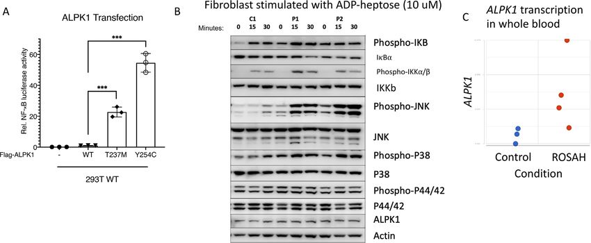
Autoinflammatory disorders
Ann Rheum Dis: first published as 10.1136/annrheumdis-2022-222629 on 22 July 2022. Downloaded from http://ard.bmj.com/ on September 19, 2022 by guest. Protected by copyright.
IL-6, CCL2 (MCP-1), soluble IL-2 receptor alpha and IL-10 protein. Additionally, the previously unreported Y254C variant
were also frequently elevated (online supplemental figure 9a). showed a significantly higher NF-κB activity than the mutant
Patients with ROSAH syndrome also demonstrated elevations of T237M plasmid (figure 5A).
additional cytokines and chemokines including plasma CXCL10 To explore the effect of the ALPK1 mutation ex vivo, we
(interferon gamma- induced protein 10 (IP- 10)) and serum used T237M patient- derived fibroblasts from two unre-
CXCL1 (GRO-alpha (previously known as neutrophil-activating lated patients to study the activity of the canonical NF-κB
protein 3)) (online supplemental figure 9b- c). However, no pathway in response to stimulation with the ALPK1 agonist
elevations in intracellular IFN-gamma, TNF-alpha or IL-4 were ADP-heptose (the patient with the Y254C mutation declined
observed after stimulation of peripheral blood cells from two skin biopsy). As compared with healthy controls, stimulated
patients (online supplemental table 5). patients’ cells showed increased phosphorylation of IκBα,
Analysis of cerebral spinal fluid (CSF) was suggestive of CNS IKKα/β, and MAP kinases p38 and JNK, which are hallmarks
inflammation (online supplemental table 6). CSF neopterin, of the activated canonical NF-κB pathway (figure 5B). Addi-
produced by immune cells after interferon stimulation and tionally, we observed higher mRNA expression of ALPK1 and
shown to correlate with CSF interferon-alpha titres in Aicardi- increased expression of NF-κB regulated genes in RNAseq data
Goutières syndrome (AGS), was measured in patients F2.2 and from patients with ROSAH syndrome (figure 5C and online
F3.4 and found to be elevated.18–20 CSF cytology was performed supplemental figure 10).
on patient F10.1’s sample and was notable for numerous cells
consistent with activated monocytes and lymphocytes. Patient
F2.2 had a banked sample available for CSF cytokine analysis ROSAH syndrome mutations are associated with
and the results were notable for elevation of IL-13 and soluble increased signal transducer and activator of transcription
IL-2 receptor alpha (online supplemental table 7). phosphorylation and expression of interferon-regulated
RNA extracted from whole blood for four untreated adults genes
with ROSAH had a distinct transcriptomic signature as compared Based on the presence of premature CNS basal ganglia miner-
with healthy controls (figure 4B). Many of the differentially alisation reminiscent of that seen in classic type-I interferonop-
expressed genes are involved in innate immune signalling path- athies including AGS, we were interested in assessing signal
ways. Production of nitric oxide and reactive oxygen species in transducer and activator of transcription (STAT1) phosphory-
macrophages, fMLP signalling in neutrophils, integrin signalling lation and expression of interferon-regulated genes.21 Immuno-
and Fcγ receptor-mediated phagocytosis were among the top blotting for phospho-STAT1 in patient fibroblasts and in 293 T
upregulated canonical pathways. cells transfected with WT and mutant constructs revealed that
ROSAH-associated mutations result in constitutive STAT1 acti-
ROSAH syndrome mutations are gain of function and result in vation (figure 6A,B). Unstimulated monocytes isolated from a
enhanced NF-κB signalling pre-treatment ROSAH patient demonstrated increased STAT1
We assessed the activity of mutant ALPK1 using an NF-κB phosphorylation as compared with cells isolated from a healthy
luciferase assay. Transiently transfected mutant proteins had control and monocytes from a ROSAH patient treated with a
increased constitutive NF-κB activity relative to the wild-type TNF inhibitor (figure 6C).
Figure 5 Gain-of-function mutations in ALPK1 are associated with enhanced NF-κB activation in transfected cells and fibroblasts from patients
with ROSAH syndrome. (A) 293 T cells were transiently cotransfected with an NF-B–responsive luciferase reporter gene and Flag-ALPK1 (wild-type
or disease-associated mutant [T237M or Y254C]). Luciferase assay of NF-κB activation is shown as mean±SD. From three technical replicates (two-
tailed unpaired Student’s t-test, ***pAutoinflammatory disorders
Ann Rheum Dis: first published as 10.1136/annrheumdis-2022-222629 on 22 July 2022. Downloaded from http://ard.bmj.com/ on September 19, 2022 by guest. Protected by copyright.
Figure 6 ALPK1 mutations affect STAT1 phosphorylation, plasma levels of interferon-induced cytokines and transcription of interferon-regulated
genes. (A, B) 293 T cells transiently transfected with ALPK1 variants (A) and ROSAH patient derived fibroblasts (B) were stimulated with ADP-heptose
(5 uM) and whole cell lysates from both experiments were subjected to Western blotting for indicated proteins. Constitutive STAT1 phosphorylation
(pSTAT) was observed in both transfected cells and patient fibroblasts. (-) reflects transfection with empty vector. (C) CD14-labelled monocytes from
an untreated ROSAH patient (F2.4, middle of panel) showed constitutively phosphorylated STAT1 (pSTAT1) as compared with healthy control (top)
and ROSAH patient treated with TNF-inhibitor (F2.2, bottom of panel). (D) Plasma CXCL10 (interferon-inducible protein 10 (IP-10)) as measured in
patients F1.1, F2.2, F2.3, F2.4 and F7.1. Grey shaded area represents the mean plus or minus 2 SD from 114 healthy controls. (E) Heat map showing
increased expression of interferon-regulated genes (type I: top (GO:0060337) and type II: bottom (GO:0034341)) in four untreated patients with
ROSAH syndrome as compared with three healthy controls. Upregulated genes are shown in red and down-regulated genes in blue. ROSAH, retinal
dystrophy, optic nerve oedema, splenomegaly, anhidrosis and headache; STAT1, signal transducer and activator of transcription.
We also observed elevated levels of CXCL10 in the peripheral clinical feature of ROSAH syndrome. Patients F3.4 and F5.3
whole blood samples (n=5 patients) and increased expression of lacked subjective symptoms, and patient F13.1 denied subjec-
interferon-regulated genes (n=4 patients) (figure 6D,E). tive benefit but discontinued anti-IL-1 therapy (anakinra) after
less than 1 week secondary to intolerable injection site reac-
Mouse model tions. Anti-TNF therapy (adalimumab) led to improvement
Consistent with the observations in patients with ROSAH in fatigue, headache, or arthralgia for four of four patients in
syndrome, knock-in mice with the Alpk1 T237M mutation had whom these features were present. Additionally, three of these
elevated serum levels of CXCL1, CXCL10 and CCL2 (online patients were noted to have normalised CRPs (figure 7A) and a
supplemental figure 11a). At 16 weeks, mice did not have an decline in inflammatory cytokines (figure 7B) while on therapy.
increase in spleen size or weight (online supplemental figure Six patients were treated with anti-IL-1 therapy (anakinra or
11b) and mice did not exhibit visual decline (online supple- canakinumab) and reported some improvement in subjective
mental figure 11c, d) or evidence of retinal degeneration due to symptoms; however, serum CRP levels were not consistently
Alpk1 mutation at up to 12 months of age (online supplemental suppressed.
figure 11e). However, we cannot exclude the possibility that Whole blood RNA sequencing was performed on paired pre-
retinal abnormalities could manifest in mice at a later age. and post-treatment samples from patients F1.1 and F2.2. Prior
to the initiation of treatment, patients with ROSAH syndrome
Response to therapy demonstrated increased expression of many genes linked to
Ten patients have been treated with anti- cytokine therapy inflammation (figure 7C, online supplemental table 8). After
(online supplemental table 1), and seven patients with systemic initiation of anti-TNF therapy, both patients had transcriptome
symptoms reported subjective improvement in at least one changes consistent with decreased inflammation. Patients F2.1
8 Kozycki CT, et al. Ann Rheum Dis 2022;0:1–12. doi:10.1136/annrheumdis-2022-222629Autoinflammatory disorders
Ann Rheum Dis: first published as 10.1136/annrheumdis-2022-222629 on 22 July 2022. Downloaded from http://ard.bmj.com/ on September 19, 2022 by guest. Protected by copyright.
Figure 7 Response to anticytokine therapy. (A) Pretreatment and post-treatment CRPs for patients initiated on anticytokine therapy (n=6). Shaded
zone represents normal range. (B) Pretreatment and post-treatment cytokines from patients F1.1 (plasma), F2.2 (serum), F2.3 (serum). Shaded area
indicates time on anti-cytokine therapy. Specific therapies are as indicated in the figures. (C) Heatmap showing differentially expressed inflammatory
response genes (GO: 0006954) in whole blood of pre-treatment (n=4) and post-adalimumab (n=2) patients with ROSAH syndrome. Patient F2.2 had
post-treatment samples collected on three separate visits. Upregulated genes are shown in red, and downregulated genes in blue. Complete list of
genes in online supplemental table 9. (D) Fluorescein angiography from patient F2.3 demonstrating retinal vasculitis (dotted white arrows) and disc
leakage (solid red arrow) that improved after initiation of tocilizumab. (E) Optical coherence tomography from patient F2.3 demonstrating cystoid
macular oedema (white arrows) that improved after initiation of tocilizumab. CRP, C reactive protein; ROSAH, retinal dystrophy, optic nerve oedema,
splenomegaly, anhidrosis and headache.
and F3.3 are already blind and have declined treatment with DISCUSSION
anticytokine therapy. Although the initial report of ROSAH syndrome emphasised the
Our ability to determine the impact of therapy on ocular disease visual manifestations associated with the disease, our work addi-
was limited because most patients in this cohort already exhib- tionally establishes ROSAH syndrome as a disease of systemic
ited advanced retinal disease at the time of evaluation. However, inflammation caused by gain-of-function mutations in the innate
two patients (F2.3 and F5.3) had substantially decreased intra- immune receptor ALPK1. This conclusion is supported by both
ocular inflammation after starting the IL-6 receptor antagonist, our in vitro work as well as our systematic analysis of inflam-
tocilizumab. Patient F2.3 had almost complete resolution of matory features in the largest cohort of patients reported to
her cystoid macular oedema after 3 months of treatment with date. These findings have important implications for both basic
tocilizumab and this was maintained at 9 months on tocilizumab
science and clinical practice.
and fluorescein angiography showed significant improvement in
Our discovery of a second ROSAH- associated mutation
retinal vascular leakage (figure 7D,E). After 5 months of tocili-
occurring in the ligand-binding domain of ALPK1 emphasises
zumab treatment, patient F5.3 had decreased retinal vascular
the importance of this domain in protein activation and provides
leakage and this improvement was maintained during 14 months
a solid foundation for establishing the pathogenicity of missense
of treatment. Patients F1.1 and F4.1 had continued visual decline
with progressive constriction of visual fields despite adalimumab mutations affecting the region. While the exact impact of these
and canakinumab monotherapies, respectively. Subsequently, mutations on protein structure and function remains to be eluci-
patient F4.1 was switched from canakinumab to sarilumab, dated, there is clearly a strong phenotypic overlap between
the only subcutaneously administered IL- 6 receptor antag- patients with the recurrent p.T237M variant and the patient
onist locally available to the patient but within 1 week of the with the p.Y254C mutation. Additionally, our in vitro work
first 150 mg subcutaneous dose, developed grade 4 neutropenia demonstrates that both mutations are associated with increased
(absolute neutrophil countAutoinflammatory disorders
Ann Rheum Dis: first published as 10.1136/annrheumdis-2022-222629 on 22 July 2022. Downloaded from http://ard.bmj.com/ on September 19, 2022 by guest. Protected by copyright.
The findings from this large, international cohort study a ciliopathy, this nosology may change as there are an increasing
provide valuable insights on the clinical spectrum of disease number of proteins that were initially labelled as ‘ciliary’ but
associated with mutations in ALPK1 and highlight the possi- have now also been observed at the innate immune synapse.25 26
bility that patients with ROSAH syndrome may be currently The prevalence of clinical manifestations is this cohort was
unrecognised in cohorts of more common inflammatory disor- likely biased by the fact that all diagnostic genetic testing in the
ders. We have found that ROSAH syndrome can present with cohort was prompted by ophthalmological examination findings,
periodic fevers, malaise, headaches, uveitis, deforming joint and the prevalence of specific clinical features is likely to change
disease, abdominal pain, premature CNS mineralisation and as more patients without prominent ocular manifestations are
focal meningeal enhancement on brain MRI and it has mimicked screened for mutations in ALPK1. Additionally, the prevalence of
diseases including sJIA, sarcoidosis, neuro-Behçet’s disease, SjD disease features for deceased patients was limited to what could
and multiple sclerosis. We also found that untreated patients be recalled by their surviving children.
with ROSAH syndrome had frequent elevations of CRP and In conclusion, we have demonstrated that ROSAH syndrome
proinflammatory plasma cytokines including TNF and IL- 6. is an autoinflammatory disease that can manifest with a spec-
Additionally, we have seen that diagnosis may be aided by the trum of inflammatory features including recurrent fever, uveitis,
presence of ocular involvement, splenomegaly, decreased or deforming arthritis and cyclical cytopenias. For patients with
inability to sweat or multiple dental caries, but none of these advanced retinal degeneration, TNF inhibitors and IL-1 inhibi-
features is universal. While advanced retinal degeneration was tors can be considered for treatment of non-ocular disease mani-
common among adults in our cohort, three adults lacked signifi- festations including fevers, headaches, and arthritis. However,
cant visual impairment but suffered from other systemic inflam- for patients with active intraocular inflammation, our findings
matory manifestations of the disease.
indicate that tocilizumab may be the preferred treatment and
Patients’ clinical improvement on anticytokine therapies also
future studies should be pursued to determine if this result is
highlights the role of immune activation in disease pathogenesis
reproducible in additional patients. Continued study of ALPK1
and emphasises the importance of referring patients with ROSAH
function and ROSAH syndrome may also provide valuable
syndrome for multidisciplinary evaluation and care. While all
insights for more common disorders of inflammation, such as
index patients in this cohort had routine ophthalmology care at
gout or periodic fever, aphthous stomatitis, pharyngitis, cervical
the time of initial contact with the NIH, most patients did not
adenitis (PFAPA), where endogenous ligands may play a role as
have a regular provider experienced in management of systemic
damage-associated molecular patterns.
inflammation. Yet thorough examination revealed that many
patients had elevations of serum CRP and non-ophthalmological
Author affiliations
indications to consider systemic immunomodulatory treatment 1
Inflammatory Disease Section, National Human Genome Research Institute,
including recurrent headaches, disabling episodes of fatigue, Bethesda, Maryland, USA
arthritis, abdominal pain, and AA amyloidosis. Most patients 2
National Institute of Allergy and Infectious Diseases, Bethesda, Maryland, USA
3
who received anti-TNF or anti-IL1 therapy reported subjective National Eye Institute, Bethesda, Maryland, USA
4
improvement in systemic symptoms. While these therapies may National Institute of Dental and Craniofacial Research, Bethesda, Maryland, USA
5
Radiology and Imaging Sciences, National Institutes of Health Clinical Center,
be appropriate for treating non-ocular inflammatory manifesta- Bethesda, Maryland, USA
tion, there is no evidence that they are efficacious for treating 6
Section of Histopathology, National Eye Institute, Bethesda, Maryland, USA
intraocular inflammation or that they can influence progres- 7
National Institute of Arthritis and Musculoskeletal and Skin Diseases, Bethesda,
sive vision loss. Thus, additional prospective studies are needed Maryland, USA
8
to determine optimal treatment for this disease, but providers Ophthalmic Genetics & Visual Function Branch, National Eye Institute, Bethesda,
Maryland, USA
should consider alternative therapies in patients with active, 9
National Institute of Biological Sciences Beijing, Beijing, China
vision-threating intraocular inflammation. 10
National Institute of Neurological Disorders and Stroke, Bethesda, Maryland, USA
The IL- 6 inhibitor tocilizumab has shown very promising 11
National Institute of Diabetes and Digestive and Kidney Diseases, Bethesda,
results in patients F2.3 and F5.3. Both patients had intraocular Maryland, USA
12
inflammation that was unresponsive to TNF and IL-1 inhibition Dermatology Branch, NIH, National Institute of Arthritis and Musculoskeletal and
Skin Diseases, Bethesda, Maryland, USA
but showed dramatic improvement on tocilizumab, and we are 13
National Cancer Institute, Bethesda, Maryland, USA
actively seeking to determine if these results can be replicated 14
Undiagnosed Diseases Program, Bethesda, Maryland, USA
in additional patients. It should also be noted that patients with 15
National Human Genome Research Institute, Bethesda, Maryland, USA
ROSAH syndrome have an interferon gene expression signature, 16
National Heart Lung and Blood Institute, Bethesda, Maryland, USA
17
premature basal ganglia mineralisation and elevated CNS neop- Neutrophil Monitoring Laboratory, Applied/Developmental Research Directorate,
terin that suggest the disease may be an interferonopathy, which Frederick National Laboratory for Cancer Research, Frederick, Maryland, USA
18
Oncogenesis and Development Section, National Human Genome Research
might indicate that patients would benefit from treatment with Institute, Bethesda, Maryland, USA
a JAK-inhibitor.21 22 19
Drs. Gilbert and Farley, OD, PC, Colonial Heights, Virginia, USA
Deep-phenotyping of this cohort illustrates the potential for 20
Massachusetts General Hospital Center for Genomic Medicine, Boston,
monogenic diseases to advance our understanding on ALPK1’s Massachusetts, USA
21
role in human biology. Several clinical features of ROSAH Division of Medical Genetics & Metabolism, Department of Pediatrics,
Massachusetts General Hospital, Boston, Massachusetts, USA
syndrome, including short dental roots as well as the aberrant 22
Massachusetts Eye and Ear, Boston, Massachusetts, USA
production of sweat, breast milk and saliva are not classically 23
Hospital de Clínicas de Porto Alegre, Porto Alegre, Brazil
associated with inflammation but may reflect ALPK1’s role in 24
Post Graduate Program in Genetics and Molecular Biology, Universidade Federal do
ciliary functioning. Indeed, primary cilia are present in the dental Rio Grande do Sul, Porto Alegre, Brazil
25
epithelium and mesenchyme at various stages of tooth develop- Service de Rhumatologie Pédiatrique, Centre de Référence des Maladies Auto-
Inflammatoires de l’enfant, Hôpital Bicêtre, AP HP, Université Paris Sud, Bicetre,
ment, and the clinical and radiographic features of teeth in this
France
cohort were very similar to past reports of dental anomalies in 26
Service de Rhumatologie Pédiatrique, Centre de Référence des Maladies Auto-
ciliopathy disorders.23 24 While, to date, no monogenic diseases Inflammatoires et de l’amylose inflammatoire CEREMAIA, Hôpital Bicêtre, AP HP,
have been categorised as both an autoinflammatory disease and Université Paris Saclay, Bicetre, France
10 Kozycki CT, et al. Ann Rheum Dis 2022;0:1–12. doi:10.1136/annrheumdis-2022-222629Autoinflammatory disorders
Ann Rheum Dis: first published as 10.1136/annrheumdis-2022-222629 on 22 July 2022. Downloaded from http://ard.bmj.com/ on September 19, 2022 by guest. Protected by copyright.
27
CeRéMAIA, CHU Montpellier, INSERM, University of Montpellier, Montpellier, Ron Scott, Judy Schaechter, Timothy Schedl, Kelly Schoch, Daryl A Scott, Vandana
France Shashi, Jimann Shin, Edwin K Silverman, Janet S Sinsheimer, Kathy Sisco, Edward C
28
Hôpital Fondation Adolphe de Rothschild, Paris, France Smith, Kevin S Smith, Emily Solem, Lilianna Solnica-Krezel, Ben Solomon, Rebecca C
29
Depts Internal Medicine and Immunology, Erasmus MC, Rotterdam, The Spillmann, Joan M Stoler, Jennifer A Sullivan, Kathleen Sullivan, Angela Sun, Shirley
Netherlands Sutton, David A Sweetser, Virginia Sybert, Holly K Tabor, Amelia L M. Tan, Queenie
30
Pathology & Clinical Bioinformatics, Erasmus MC, Rotterdam, The Netherlands K-G Tan, Mustafa Tekin, Fred Telischi, Willa Thorson, Cynthia J Tifft, Camilo Toro,
31
Iizuka Hospital, Iizuka, Japan Alyssa A Tran, Brianna M Tucker, Tiina K Urv, Adeline Vanderver, Matt Velinder, Dave
32
Department of Medical Genetics, Haukeland University Hospital, Bergen, Norway Viskochil, Tiphanie P Vogel, Colleen E Wahl, Stephanie Wallace, Nicole M Walley,
33
Bergen Group of Epidemiology and Biomarkers in Rheumatic Disease, Department Melissa Walker, Jennifer Wambach, Jijun Wan, Lee-kai Wang, Michael F Wangler,
of Rheumatology, Haukeland University Hospital, Bergen, Norway Patricia A Ward, Daniel Wegner, Monika Weisz-Hubshman, Mark Wener, Tara Wenger,
34
Department of Ophthalmology, Haukeland University Hospital, Bergen, Norway Katherine Wesseling Perry, Monte Westerfield, Matthew T Wheeler, Jordan Whitlock,
35
Laboratory of Immunology, National Eye Institute, NIH, Bethesda, Maryland, USA Lynne A Wolfe, Kim Worley, Changrui Xiao, Shinya Yamamoto, John Yang, Diane B
36
Department of Child Health, University of Tsukuba Faculty of Medicine, Tsukuba, Zastrow, Zhe Zhang, Chunli Zhao, Stephan Zuchner, Hugo Bellen, Rachel Mahoney.
Japan
37
Division of Respirology, Neurology, and Rheumatology, Department of Medicine, Contributors CTK, SK, LH, BMW, PJ, DH, MSA-A, EU, AS, NM, MK-H, TH, EWC,
Kurume University School of Medicine, Kurume, Japan C-CRL, CT, SK, ZK, ABa, MB, MN, SR, TR, ND, LB, EP, WZ, NS, AHY, GF, DAS, LB, JY,
38
Department of Pediatrics and Child Health, Kurume University School of Medicine, FdOP, IS, TSA, PD, IK-P, IT, SMT, PMvH, RTAvW, PJvdS, HY, ABe, EMA, RWJ, HT, HI, RN,
Kurume, Japan EV, ES, RM, BPB, LS, RH, DB, AKO, IA, DLK were involved in identification, diagnosis
39
Department of Internal Medicine, Periodic Fevers Research Center, Università and/or clinical characterisation of people with ROSAH syndrome. CTK, HW, YJ, MJM,
Cattolica del Sacro Cuore, Roma, Italy WLT, PZ, XT, DLP, RRC, DBK, MG, FS performed laboratory assays and/or developed
40
Dipartimento di scienze dell’invecchiamento, neurologiche, ortopediche e della and evaluated the mouse model. All authors reviewed the manuscript, and approved
testa-collo, Fondazione Policlinico Universitario Agostino Gemelli IRCCS, Roma, Italy the final version for submission. CTK acts as guarantor for this work.
41
Istitute of Genomic di Medicine, Universita Cattolica del Sacro Cuore, Roma, Italy Funding This work was supported by the Divisions of Intramural Research of the
42
NYU, New York, New York, USA National Human Genome Research Institute, the National Institute of Allergy and
Acknowledgements We thank Drs. David Kleiner, Bibi Bielekova, Sabina Desar Infectious Diseases, the National Eye Institute, and the National Institute for Dental
and Billel Gasmi as well as Ms. Silke Williams for assistance with processing samples and Craniofacial Research, as well as the NIH Clinical Centre, the NIH Common
and reviewing sample histology. We thank Drs. Charles Hesdorffer, Charles Bolan, Fund, through the Office of Strategic Coordination/Office of the NIH Direction
Rebecca Clark, Bernadette Redd, Amisha Barochia, and Danica Novacic as well as (U01HG007690 (DAS, LCB)) and the Hill Family Fund for the Diagnosis and
Ms. Emily Place for providing clinical care to this cohort. We thank Dr. JaeJin Chae Management of Rare and Undiagnosed Diseases at Mass General Hospital.
for his support in establishing the mouse colony. We thank the staff at the NIH Disclaimer The content is solely the responsibility of the authors and does not
Intramural Sequencing Centre for performing RNA sequencing and Dr. Hiro Oda and necessarily represent the official views of the National Institutes of Health.
Mr. Kyle Amende for their support in processing and transferring the data. We thank
Ms. Daniela Ospina Cardona for collecting patient samples and the healthy control Competing interests FS is a cofounder and stockholder of Pyrotech therapeutics,
volunteers who participated in this study. Finally, we express our greatest gratitude a company that aims to develop agonist/inhibitor drugs for ALPK1. RM has received
to the affected patients and their families that have entrusted us with their care and honorary fees for lectures from SHIRE-TAKEDA- SANOFI- NOVARTIS-SOBI and Nida
dedicated their time and tissues to advancing our understanding of this rare disease. Sen is employed by Janssen.
Collaborators Maria T Acosta, Margaret Adam, David R Adams, Justin Alvey, Patient and public involvement Patients and/or the public were involved in the
Laura Amendola, Ashley Andrews, Euan A Ashley, Mahshid S Azamian, Carlos design, or conduct, or reporting, or dissemination plans of this research. Refer to the
A Bacino, Guney Bademci, Ashok Balasubramanyam, Dustin Baldridge, Jim Bale, Methods section for further details.
Michael Bamshad, Deborah Barbouth, Pinar Bayrak-Toydemir, Anita Beck, Alan H Patient consent for publication Not applicable.
Beggs, Edward Behrens, Gill Bejerano, Jimmy Bennet, Beverly Berg-Rood, Jonathan
A Bernstein, Gerard T Berry, Anna Bican, Stephanie Bivona, Elizabeth Blue, John Ethics approval This study involves human participants and was approved
Bohnsack, Devon Bonner, Lorenzo Botto, Brenna Boyd, Lauren C Briere, Elly Brokamp, by Institutional Review Board of the NIH: protocols 94-HG-0105, 14-EI-0064,
Gabrielle Brown, Elizabeth A Burke, Lindsay C Burrage, Manish J Butte, Peter Byers, 15-D-0051, 94-D-0094, 15-HG-0130. Mass General Brigham IRB protocol #
William E Byrd, John Carey, Olveen Carrasquillo, Thomas Cassini, Ta Chen Peter 2015P001514. Participants gave informed consent to participate in the study before
Chang, Sirisak Chanprasert, Hsiao-Tuan Chao, Gary D Clark, Terra R Coakley, Laurel taking part.
A Cobban, Joy D Cogan, Matthew Coggins, F Sessions Cole, Heather A Colley, Provenance and peer review Not commissioned; externally peer reviewed.
Cynthia M Cooper, Heidi Cope, William J Craigen, Andrew B Crouse, Michael
Data availability statement Data are available on reasonable request.
Cunningham, Precilla D’Souza, Hongzheng Dai, Surendra Dasari, Joie Davis, Jyoti G
Dayal, Matthew Deardorff, Esteban C Dell’ Angelica, Katrina Dipple, Daniel Doherty, Supplemental material This content has been supplied by the author(s).
Naghmeh Dorrani, Argenia L Doss, Emilie D Douine, Laura Duncan, Dawn Earl, It has not been vetted by BMJ Publishing Group Limited (BMJ) and may not
David J Eckstein, Lisa T Emrick, Christine M Eng, Cecilia Esteves, Marni Falk, Liliana have been peer-reviewed. Any opinions or recommendations discussed are
Fernandez, Elizabeth L Fieg, Paul G Fisher, Brent L Fogel, Irman Forghani, William A solely those of the author(s) and are not endorsed by BMJ. BMJ disclaims all
Gahl, Ian Glass, Bernadette Gochuico, Rena A Godfrey, Katie Golden-Grant, Madison liability and responsibility arising from any reliance placed on the content.
P Goldrich, Alana Grajewski, Irma Gutierrez, Don Hadley, Sihoun Hahn, Rizwan Where the content includes any translated material, BMJ does not warrant the
Hamid, Kelly Hassey, Nichole Hayes, Frances High, Anne Hing, Fuki M Hisama, Ingrid accuracy and reliability of the translations (including but not limited to local
A Holm, Jason Hom, Martha Horike-Pyne, Alden Huang, Yong Huang, Wendy Introne, regulations, clinical guidelines, terminology, drug names and drug dosages), and
Rosario Isasi, Kosuke Izumi, Fariha Jamal, Gail P Jarvik, Jeffrey Jarvik, Suman Jayadev, is not responsible for any error and/or omissions arising from translation and
Orpa Jean-Marie, Vaidehi Jobanputra, Lefkothea Karaviti, Jennifer Kennedy, Shamika adaptation or otherwise.
Ketkar, Dana Kiley, Gonench Kilich, Shilpa N Kobren, Isaac S Kohane, Jennefer N
Open access This is an open access article distributed in accordance with the
Kohler, Deborah Krakow, Donna M Krasnewich, Elijah Kravets, Susan Korrick, Mary
Koziura, Seema R Lalani, Byron Lam, Christina Lam, Grace L LaMoure, Brendan C Creative Commons Attribution 4.0 Unported (CC BY 4.0) license, which permits
Lanpher, Ian R Lanza, Kimberly LeBlanc, Brendan H LeeRoyLevitt, Richard A Lewis, others to copy, redistribute, remix, transform and build upon this work for any
Pengfei Liu, Xue Zhong Liu, Nicola Longo, Sandra K Loo, Joseph Loscalzo, Richard purpose, provided the original work is properly cited, a link to the licence is given,
L Maas, Ellen F Macnamara, Calum A MacRae, Valerie V Maduro, Bryan C Mak, and indication of whether changes were made. See: https://creativecommons.org/
May Christine V Malicdan, Laura A Mamounas, Teri A Manolio, Rong Mao, Kenneth licenses/by/4.0/.
Maravilla, Ronit Marom, Gabor Marth, Beth A Martin, Martin G Martin, Julian A
ORCID iDs
Martínez-Agosto, Shruti Marwaha, Jacob McCauley, Allyn McConkie-Rosell, Alexa
Christina Torres Kozycki http://orcid.org/0000-0001-5405-4694
T McCray, Elisabeth McGee, Heather Mefford, J Lawrence Merritt, Matthew Might,
Blake M Warner http://orcid.org/0000-0002-4961-018X
Ghayda Mirzaa, Eva Morava, Paolo M Moretti, Mariko Nakano-Okuno, Stan F Nelson,
Isabelle Koné-Paut http://orcid.org/0000-0001-8939-5763
John H Newman, Sarah K Nicholas, Deborah Nickerson, Shirley Nieves-Rodriguez,
Daniel L Kastner http://orcid.org/0000-0001-7188-4550
Donna Novacic, Devin Oglesbee, James P Orengo, Laura Pace, Stephen Pak, J Carl
Pallais, Christina GS Palmer, Jeanette C Papp, Neil H Parker, John A PhillipsIII, Jennifer
E Posey, Lorraine Potocki, Barbara N Pusey, Aaron Quinlan, Wendy Raskind, Archana REFERENCES
N Raja, Deepak A Rao, Anna Raper, Genecee Renteria, Chloe M Reuter, Lynette Rives, 1 Williams LB, Javed A, Sabri A, et al. ALPK1 missense pathogenic variant in five families
Amy K Robertson, Lance H Rodan, Jill A Rosenfeld, Natalie Rosenwasser, Francis leads to ROSAH syndrome, an ocular multisystem autosomal dominant disorder.
Rossignol, Maura Ruzhnikov, Ralph Sacco, Jacinda B Sampson, Mario Saporta, C Genet Med 2019;21:2103–15.
Kozycki CT, et al. Ann Rheum Dis 2022;0:1–12. doi:10.1136/annrheumdis-2022-222629 11You can also read