High-Efficient Production of Adipose-Derived Stem Cell (ADSC) Secretome Through Maturation Process and Its Non-scarring Wound Healing Applications
←
→
Page content transcription
If your browser does not render page correctly, please read the page content below
ORIGINAL RESEARCH
                                                                                                                                               published: 16 June 2021
                                                                                                                                       doi: 10.3389/fbioe.2021.681501

                                            High-Efficient Production of
                                            Adipose-Derived Stem Cell (ADSC)
                                            Secretome Through Maturation
                                            Process and Its Non-scarring Wound
                                            Healing Applications
                                            Young-Hyeon An 1,2 , Dae Hyun Kim 3 , Eun Jung Lee 3 , Dabin Lee 4 , Mihn Jeong Park 5 ,
                                            Junghyeon Ko 1 , Dong Wook Kim 4 , Jiwan Koh 3 , Hyun Sook Hong 6 , Youngsook Son 6,7 ,
                                            Je-Yoel Cho 4 , Ji-Ung Park 8 , Sun-Dong Kim 3 and Nathaniel S. Hwang 1,2,5*
                                            1
                                              School of Chemical and Biological Engineering, Institute of Chemical Processes, Seoul National University, Seoul,
                                            South Korea, 2 BioMax/N-Bio Institute, Seoul National University, Seoul, South Korea, 3 Senior Science & Life, Inc., Seoul,
                                            South Korea, 4 Department of Biochemistry, BK21 PLUS Program for Creative Veterinary Science Research and Research
                                            Institute for Veterinary Science, College of Veterinary Medicine, Seoul National University, Seoul, South Korea,
                                            5
                                              Interdisciplinary Program in Bioengineering, Seoul National University, Seoul, South Korea, 6 Department of Biomedical
                                            Science and Technology, Kyung Hee University, Seoul, South Korea, 7 Department of Genetic Biotechnology and Graduate
                            Edited by:      School of Biotechnology, Kyung Hee University, Yongin, South Korea, 8 Department of Plastic and Reconstructive Surgery,
                         Lia Rimondini,     Seoul National University Boramae Hospital, Seoul National University College of Medicine, Seoul, South Korea
  University of Eastern Piedmont, Italy
                       Reviewed by:
                         Philipp Seib,
                                            Recently, the stem cell-derived secretome, which is the set of proteins expressed by
            University of Strathclyde,      stem cells and secreted into the extracellular space, has been demonstrated as a
                     United Kingdom
                                            critical contributor for tissue repair. In this study, we have produced two sets of high
                      Agata Przekora,
  Medical University of Lublin, Poland      concentration secretomes from adipose-derived mesenchymal stem cells (ADSCs) that
                  *Correspondence:          contain bovine serum or free of exogenous molecules. Through proteomic analysis,
                  Nathaniel S. Hwang        we elucidated that proteins related to extracellular matrix organization and growth
                  nshwang@snu.ac.kr
                                            factor-related proteins are highly secreted by ADSCs. Additionally, the application of
                    Specialty section:      ADSC secretome to full skin defect showed accelerated wound closure, enhanced
         This article was submitted to      angiogenic response, and complete regeneration of epithelial gaps. Furthermore, the
                          Biomaterials,
                a section of the journal
                                            ADSC secretome was capable of reducing scar formation. Finally, we show high-dose
       Frontiers in Bioengineering and      injection of ADSC secretome via intraperitoneal or transdermal delivery demonstrated no
                        Biotechnology
                                            detectable pathological conditions in various tissues/organs, which supports the notion
           Received: 16 March 2021
                                            that ADSC secretome can be safely utilized for tissue repair and regeneration.
            Accepted: 04 May 2021
           Published: 16 June 2021          Keywords: stem cells, secretome, proteomic analysis, skin regeneration, tissue repair
                             Citation:
     An Y-H, Kim DH, Lee EJ, Lee D,
      Park MJ, Ko J, Kim DW, Koh J,        INTRODUCTION
 Hong HS, Son Y, Cho J-Y, Park J-U,
      Kim S-D and Hwang NS (2021)          One of the promising techniques to treat severe non-healing wound is stem cell-based therapy,
             High-Efficient Production     which reconstructs the wound healing process through trophic, paracrine, and immunomodulatory
of Adipose-Derived Stem Cell (ADSC)
                                           properties (Del Papa et al., 2015; Marfia et al., 2015). However, direct transplantation of stem cells is
      Secretome Through Maturation
Process and Its Non-scarring Wound
                                           constrained by their low survival rate in vivo environment and potential contribution to teratoma
                Healing Applications.      formation (Lindeman and Visvader, 2010; Tu, 2010). To this end, the paracrine activity of the stem
Front. Bioeng. Biotechnol. 9:681501.       cells has attracted attention, suggesting that the use of the secreted molecules of stem cells would
     doi: 10.3389/fbioe.2021.681501        provide a therapeutic impact (Salgado et al., 2010; Lee et al., 2014; Robert et al., 2019). Secretome,

Frontiers in Bioengineering and Biotechnology | www.frontiersin.org                1                                              June 2021 | Volume 9 | Article 681501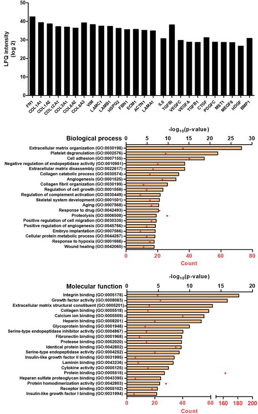
An et al.                                                                                                 The ADSCs Secretome for Wound Healing

also known as conditioned medium, is the complex set of the                Preparation of SC and Serum-Free
secreted molecules from stem cells, which includes extracellular           Secretome
vesicles and soluble fractions, such as cytokines and growth
                                                                           The clinically graded secretome of ADSCs was produced with
factors (Lee et al., 2010). Among the several sources of stem
                                                                           GMP-compliant manufacturing (Senior Lifescience, Co., Ltd.).
cells, enormous attention has been focusing on the adipose
                                                                           ADSCs within passage 5 were cultured in 75 cm2 flasks
tissue-derived stem cells (ADSCs) due to the easy isolating
                                                                           using alpha-modified minimum essential medium (α-MEM),
procedure, liposuction, with a higher harvest rate than bone
                                                                           which was the absence of both phenol red and antibiotics.
marrow (Hodgkinson et al., 2020). The ADSC-secretome has
                                                                           The medium was changed once ADSCs reached about 100%
been proven to be useful not only for regenerating non-healing
                                                                           confluence, and the culture medium was continuously matured
cutaneous and corneal wounds, cardiovascular disease, etc., but
                                                                           for 7 days to obtain both the SC and the SF without medium
also to be utilized in a cosmeceutical application (Damous et al.,
                                                                           change. The secretome of each patient was collected and mixed
2018; Lombardi et al., 2019).
                                                                           homogeneously to minimize the donor variability.
    Skin wound healing is an orchestrated and highly regulated
process, which includes inflammation, proliferation, matrix
formation, and remodeling (Gurtner et al., 2008). When
                                                                           Enzyme-Linked Immunosorbent Assay
                                                                           The level of both TGF-β1 and VEGF was quantified using
the process is disrupted, chronic non-healing wound occurs,
                                                                           enzyme-linked immunosorbent assay (ELISA) within the SF and
resulting in not only significantly lowering the quality of life of
                                                                           SC, respectively. In the case of SF, the aqueous phase was dried
the patient but also causing social costs due to the high prevalence
                                                                           out at room temperature using an evaporator for adjusting the
rate (Jarbrink et al., 2016; Chan et al., 2017). In particular, the
                                                                           protein levels similar to those of SC.
wound healing process is delayed or failed in severe conditions,
such as diabetic ulcers and severe burns. Despite the increase
of interest in secretome-based skin wound therapy, most studies            Sample Preparation and Labeling for
are performed using serum-free secretome (SF) since the serum-             Proteomic Analysis
containing secretome (SC) is susceptible to be contaminated                Only the SF was evaluated by proteomic analysis. A total of
(Shin et al., 2019); at the same time, the complicated procedure           240 µg protein of SF was subjected to filter-aided sample
hinders the analysis of the SC (Weng et al., 2016). However,               preparation (FASP) digestion (Sielaff et al., 2017). Proteins were
in the case of SF, the protein or cytokine level was highly                trypsin digested at 37◦ C for 16 h, 3% trifluoroacetic acid was
reduced compared with SC; thus, a strategy is actually required            added into elute to stop the reaction, and the C18 tip column
to increase the amount of secreted molecules in the absence                was used to remove salts. A total of 2 µg/5 µl of the digested
of serum proteins.                                                         peptide was used to analyze using liquid chromatography-mass
    For these reasons, in this study, we produced a concentrated           spectrometry (LC-MS/MS).
SF by culturing the ADSCs with the maturation process and
subsequently evaporating it. It differs from previous methods              LC-MS/MS Analysis
that collect the secretome (or conditioned medium) at the time             Spectra raw data were acquired on an Orbitrap Fusion Lumos
when the cell confluency is about 80–90% (Lombardi et al., 2019).          (Thermo Fisher Scientific, San Jose, CA) with EASY-nLC 1200
In addition, we adjusted the level of both transforming growth             (Thermo Fisher Scientific, San Jose, CA). An autosampler was
factor-beta 1 (TGF-β1) and vascular endothelial growth factor              used to load 5 µl aliquots of the peptide solutions into an EASY
(VEGF) in SF, which are representative growth factors for the              column, Acclaim PepMapTM 100 of i.d. 75 µm, length 2 cm,
wound healing process, to those of SC. Finally, the comparative            and particle size of 3 µm (Thermo Fisher Scientific, San Jose,
study of the in vivo wound healing effects between SF and SC was           CA). The trapped peptides were then separated on an EASY-
carried out in mice dorsal skin wound model.                               Spray Column, C18 analytic-column of i.d. 75 µm and length
                                                                           500 mm and 2 µm particle size (100 Å from Thermo Scientific).
                                                                           The mobile phases were composed of 100% water (A) and 100%
MATERIALS AND METHODS                                                      acetonitrile (B), and each contained 0.1% formic acid. Liquid
                                                                           chromatography with 2 h gradient at a flow rate of 250 nl/min
Adipose Tissue-Derived Stem Cells                                          was used. During the chromatographic separation, the Orbitrap
Isolation and Culture                                                      Fusion Lumos was operated in a data-dependent acquisition
Human adipose tissue-derived stem cells (ADSCs) were collected             mode. Survey full scans were acquired on the mass range 400–
from five patients (age 32–60) with the liposuction process, which         1,600 m/z, maximum injection time of 100 ms, automatic gain
was implemented with consent from the patients and approval                control target 2 × 10⁵ ions with a resolution of 120,000, and
of the Boramae Medical Center (IRB No, 20160113/16-2016-                   analyzed using the Orbitrap. MS/MS precursors were selected
3/021). After minced into small pieces, the adipose tissues were           from top n intense ions in 3 s between survey scans, which
digested using 250 U/ml type I collagenase for 1 h, followed by            were fragmented by 37.5% higher-energy collisional dissociation.
neutralization with DMEM containing 10% fetal bovine serum                 MS/MS was acquired on a maximum injection time of 54 ms,
(FBS). The digested mixture was filtered through a cell strainer of        automatic gain control 5 × 10⁴ ions with a resolution of
40 µm. The suspension of cells was centrifuged, and the obtained           30,000, and analyzed using the Orbitrap. Previously fragmented
cells were cultured in DMEM with 10% FBS.                                  precursors were excluded for 30 s.

Frontiers in Bioengineering and Biotechnology | www.frontiersin.org    2                                      June 2021 | Volume 9 | Article 681501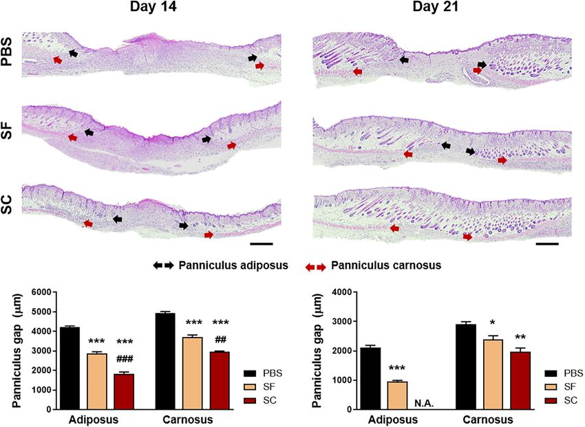
An et al.                                                                                                                     The ADSCs Secretome for Wound Healing

    SCHEME | The schematic illustration of the experimental procedure. The serum-containing (SC) and serum-free secretome (SF) were harvested by maturing the fully
    confluent adipose tissue-derived stem cells (ADSCs). The secretome was applied to the in vivo skin wound and tried to be delivered transdermally using the ex vivo
    porcine skin.

Data Analysis for Protein Profiling                                                     of Korea) powder was added to both secretome solutions at
The raw data were processed with MaxQuant software (version                             0.5% (w/v), followed by adding triethanolamine to prepare the
1.5.8.3) at default settings with unique peptide ≥ 2 and a                              gel formation by adjusting pH. For preparing the skin wound
minimum number of amino acid ≥ 6. Identified peaks were                                 model, the dorsal skin of mice was pierced using a 6-mm diameter
searched against a database of Homo sapiens from Uniprot1 .                             biopsy punch. The adhesive silicone chamber (CoverWellTM )
Output files generated from Maxquant were subjected to Perseus                          was adhered to peripheral regions of the wound to avoid wound
(version 1.6.2.2) to perform bioinformatics analysis.                                   contraction and 30 µl of the SF- and SC-containing gel was
                                                                                        applied, followed by covering the wound with TegadermTM film
In vivo Wound Healing Test                                                              dressing. At certain time points, the wound size was monitored
All the in vivo experimental procedures were approved by the                            and quantified compared with the initial wound size.
IACUC of the Seoul National University (approval number:
SNU-190916-2). The in vivo wound healing test was carried out                           Histological Analysis
using 8-week male balb/c-nude mice (OrientBio Co., Republic                             At 7 and 14 days posttreatment of the wounds, the skin tissues
of Korea). Carbomer (Polygel CA, Happycall Co., Ltd., Republic                          were collected and fixed in 4% paraformaldehyde, subsequently
                                                                                        processed to carry out the histological analysis. Hematoxylin
1
    https://www.uniprot.org/                                                            and eosin (H&E) and Masson’s trichrome staining (MTC)
                                                                                        were used to qualitatively compare the wound healing ability
                                                                                        of both SF and SC.
TABLE 1 | The concentration of growth factors in ADSC-derived
secretome (ng/ml).
                                                                                        Panniculus Gap Measurement
Samples                                             TGF-β1                  VEGF
                                                                                        Both the panniculus gap of adiposus and carnosus were analyzed
Serum-free secretome (SF)                             1.5                    24.9       at days 14 and 21 postwound, which are the gaps in length
Serum-containing secretome (SC)                       1.5                    19.5       between the edge of the regenerated adipose and muscle

Frontiers in Bioengineering and Biotechnology | www.frontiersin.org                 3                                             June 2021 | Volume 9 | Article 681501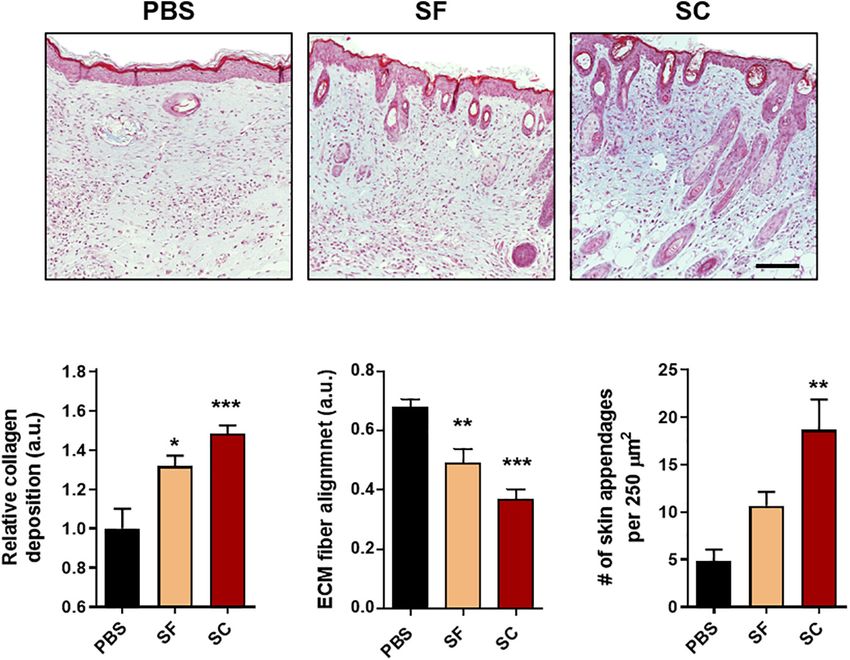
An et al.                                                                                                                 The ADSCs Secretome for Wound Healing

layer, respectively. Based on the H&E images, these gaps were                        Measurement of Collagen Deposition,
quantified using ImageJ (ImageJ Software).                                           Extracellular Matrix Fiber Alignment, and
                                                                                     Regeneration of Appendages
Epithelial Gap Measurement                                                           Collagen deposition, extracellular matrix (ECM) fiber alignment,
The epithelial gap of the wounds was measured based                                  and the number of skin appendages were measured based on
on the immunohistochemical (IHC) staining of cytokeratin-                            the MTC staining images at day 21 postwound. For the collagen
10 (ab76318, Abcam) at day 14 postwound. The gap in                                  deposition, blue coloration was separated, which indicates the
length between the edges of cytokeratin-10-stained regions was                       collagen, by thresholding of brightness, hue, and saturation
measured using ImageJ.                                                               in ImageJ. After which, the level of collagen deposition was

  FIGURE 1 | Proteomic analysis of SF. (A) LFQ intensity of representative extracellular matrix protein and growth factors, measured by LC/MS-MS. Gene ontology
  (GO) analysis representing the top 20 GO terms and its number of enrichment in (B) biological process and (C) molecular function.

Frontiers in Bioengineering and Biotechnology | www.frontiersin.org              4                                            June 2021 | Volume 9 | Article 681501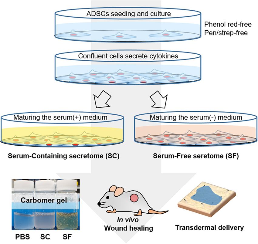
An et al. The ADSCs Secretome for Wound Healing
analyzed by measuring the intensity and compared with each Transdermal Delivery of the Secretome
other normalized with the PBS group. ECM fiber alignment The transdermal delivery of the secretome was tried using
was quantified using the OrientationJ plugin available on ImageJ a combination of physical penetration enhancements, i.e.,
software, as described in a previous research (Chantre et al., iontophoresis and a non-invasive metal roller. For visualizing
2018). Coherence values (dimensionless) were obtained from the delivered components, we conjugated the near-infrared dye,
the region of interest in each sample. Lastly, the number ZW800-1C-NHS ester, to SF by reacting 2 h at room temperature,
of skin appendages was manually counted per image (×20 followed by filtration against a 7-kDa MWCO centrifugal
magnification) using at least 15 images. membrane tube. The ZW800-1C-conjugated SF was applied to
ex vivo porcine skin and treated with physical enhancements.
In vivo Vessel Formation After 15 min and 1 h, the sample was wiped out, and skin
The in vivo angiogenic ability of each sample was evaluated surface was washed with saline, and the skin was imaged using the
by IHC staining of alpha-smooth muscle actin (α-SMA). The fluorescence-assisted resection and exploration (FLARE) system
number of newly formed blood vessels was counted in a high- with 3.6 mW/cm2 of 750 nm excitation light and white light (400–
power field (HPF, ×40 magnification) images at least 15 images. 650 nm) at 5,500 lux. The semiquantitative analysis was carried
out by measuring the integrated density of the fluorescent images
using Image J (Image J Software).
TABLE 2 | The representative extracellular matrix proteins and growth factors
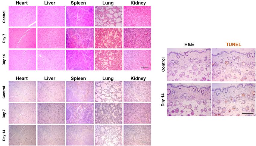
among the identified secreted molecules. Pathological Analysis of the Secretome
Protein name Abbreviation via Intraperitoneal Injection and Skin
Application
Extracellular matrix Fibronectin; Anastellin; Ugl-Y1; FN1
proteins Ugl-Y2; Ugl-Y3
Intraperitoneal injection (IP) was carried out by injecting 50 µl
Collagen alpha-1 (I) chain COL1A1 of SF into the 8-week female balb-c mice (OrientBio Co.) every
Collagen alpha-3 (VI) chain COL6A3 2 days to estimate the apoptotic response of the organs. After
Collagen alpha-2 (I) chain COL1A2 7 and 14 days postinjection, the mice were euthanized, and the
Vimentin VIM heart, liver, spleen, lung, and kidney were collected. The SF
Laminin subunit gamma-1 LAMC1 was also applied to the dorsal skin every 2 days for 14 days.
Laminin subunit beta-1 LAMB1 The collected tissues were processed to implement the H&E
Collagen alpha-1 (XII) chain COL12A1 and TUNEL staining.
Collagen alpha-1 (III) chain COL3A1
Collagen alpha-2 (VI) chain COL6A2 Statistical Analysis
Basement membrane-specific HSPG2
Statistical significance was determined by one-way analysis
heparan sulfate proteoglycan core
protein; Endorepellin; LG3 peptide
of variance (ANOVA) by Tukey’s multiple comparisons
Fibrillin-1 FBN1
method with GraphPad Prism 9 Software (Graphpad Software,
Extracellular matrix protein 1 ECM1
San Diego, CA, United States). All data are presented as
Alpha-actinin-1 ACTN1
mean ± standard deviation (SD).
Laminin subunit alpha-1 LAMA1
Growth factors Interleukin-6 IL6
Transforming growth TGFBI
factor-beta-induced protein ig-h3
RESULTS
Vascular endothelial growth factor VEGFC
C Preparation of the Serum-Free
Vascular endothelial growth factor A VEGFA Secretome and Serum-Containing
Transforming growth factor beta-1; TGFB1
Latency-associated peptide
Secretome
The SF and SC were prepared using the primary human ADSCs
Connective tissue growth factor CTGF
from independent donors. We harvested the secretome by
Platelet-derived growth factor C; PDGFC
Platelet-derived growth factor C, maturing ADSCs, which could maximize the secretion efficacy
latent form; Platelet-derived growth of the cytokines, growth factor, and protein molecules, according
factor C, receptor-binding form to the workflow in Scheme 1. Since both TGF-β1 and VEGF
Hepatocyte growth factor-like MST1 are essential in the wound healing process, we analyzed these
protein; Hepatocyte growth two factors to comparatively evaluate the in vivo wound healing
factor-like protein alpha chain;
Hepatocyte growth factor-like
effects. Initially, the SC exhibited the level of TGF-β1 and VEGF
protein beta chain of about 1.5 and 19.5 ng/ml, respectively. While the original
Multiple epidermal growth MEGF8 levels of the proteins in SF were far lower than those of the
factor-like domains protein 8 SC; however, we could concentrate the SF through a drying out
Hepatoma-derived growth factor HDGF method (Table 1).
Frontiers in Bioengineering and Biotechnology | www.frontiersin.org 5 June 2021 | Volume 9 | Article 681501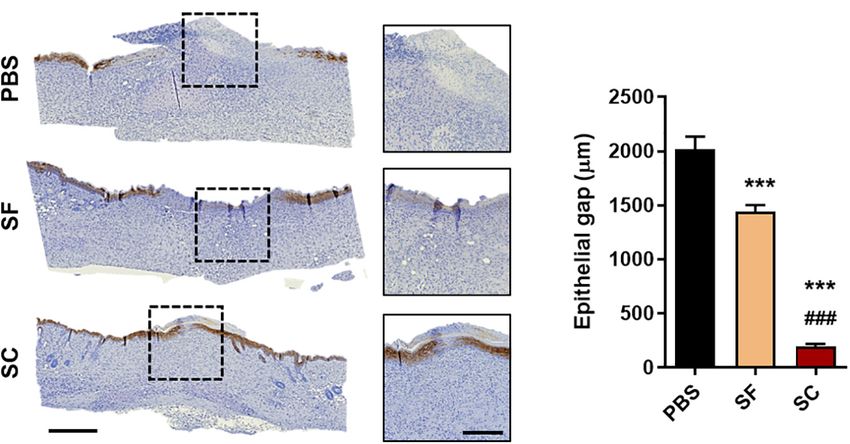
An et al.                                                                                                              The ADSCs Secretome for Wound Healing

Proteomic Analysis of the Serum-Free                                               extracellular matrix organization, angiogenesis, cell migration,
Secretome Reveals the Biological                                                   wound healing, etc.
Process Highly Involved in the Wound
Healing Process                                                                    The Serum-Containing Secretome Leads
The proteomic analysis of the SC revealed that a total of                          to Faster Wound Closure Than the
704 different proteins were consist of the secretomes in the                       Serum-Free Secretome
absence of the serum proteins. Among them, the representative                      We compared the wound closure rate between SF and SC with
20 proteins of the extracellular matrix and growth factors are                     tuning the level of TGF-β1 and VEGF through the drying out
shown in Figure 1A. The proteins that are involved in the                          process. When applying the SF and SC to the mice skin wound
matrix organization, e.g., fibronectin, collagen families, and                     every 2 days, the wound healing rate was significantly faster
vimentin, were mostly identified with strong signals, and a                        than that of the PBS-applied group (Figure 2). Although the
variety of growth factors, such as the VEGF family, TGF-β1,                        SF contained a similar level of TGF-β1 and even the higher
and connective tissue growth factor (CTGF), were observed.                         VEGF contents, the SC exhibited a much effective wound closure
The protein name, according to the abbreviation, is described                      ability than the SF.
in Table 2. In addition, by combining the proteomics with gene                        The result of the IHC staining of cytokeratin-10 supported
ontology (GO) analysis, we could comprehensively classify the                      the faster wound closure of the SC, which revealed the narrow
secreted proteins on the basis of both the biological process                      epithelial gap (Figure 3). In the case of SF, the epithelial gap was
and molecular function (Figures 1B,C). The top 20 of the                           significantly reduced compared with the PBS group; however,
biological process of SF revealed that it would be highly involved                 it did not show as much effect as SC. In addition, the H&E
in the wound healing process, exhibiting the GO terms of an                        staining showed that the SC provided outstanding wound healing

  FIGURE 2 | The ADSC secretome accelerated wound closure. (A) Photographs of the wound (rubber ring diameter = 9 mm). (B) Wound closure profiles by
  measuring the wound size (*compared with the PBS group; # compared with the SF group;*# p < 0.05; ## p < 0.01; ***p < 0.001).

  FIGURE 3 | Immunohistochemical staining of cytokeratin-10 on day 14. (A) Representative microscopy images (Scale bar = 500 µm in low magnified images and
  200 µm in high magnified images) and (B) the gap between the regenerated epithelial cells (***### p < 0.001).

Frontiers in Bioengineering and Biotechnology | www.frontiersin.org            6                                           June 2021 | Volume 9 | Article 681501
An et al. The ADSCs Secretome for Wound Healing
effects not only on the superficial wound closure but also on newly formed skin appendages was significantly higher in the SC
the regeneration of internal skin tissue (Figure 4). The adipose group than that of the SF group.
(adiposus) and muscle (carnosus) layers were regenerating in the Angiogenesis is one of the other criteria confirming tissue
wound bed, narrowing those gaps between the edges of native remodeling during the wound healing process (Tonnesen et al.,
tissue. The SC showed the narrowest gaps of both layers at 2000). The IHC staining of α-SMA indicated that SC induced
day 14 postwound; even the adiposus layer in SC was clearly the largest number of angiogenesis, and the SF also showed
regenerated. As a result, the SC has a superior wound healing the angiogenic ability but inferior to the SC (Figure 6). Also,
ability than SF with showing faster wound closure and internal the vasculature in the SC group was enlarged than that of
tissue regeneration. the SF group. As a result, it was proved that the SC showed
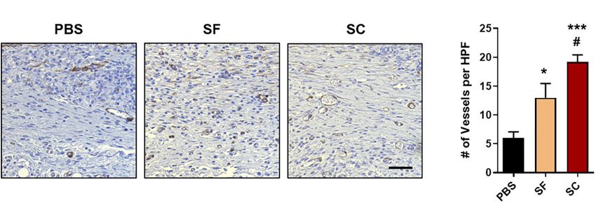
increasing collagen synthesis, the formation of skin appendages,
and promoted angiogenic response, that is, the SC enhances the
The Serum-Containing Secretome tissue remodeling process compared with the SF.
Enhances Tissue Remodeling Compared
With the Serum-Free Secretome Intraperitoneal Injection and
After the proliferative phase during the wound healing process, Pathological Analysis
the tissue granulation and remodeling process begin, and these To confirm the biosafety of the SF, we injected SF into the
are determined by the collagen synthesis and ECM remodeling peritoneal region in mice and carried out the pathological
(Gurtner et al., 2008). At 21 days postwound, the MTC showed analysis using H&E and TUNEL staining. After 7 and 14 days
that the collagen synthesis of the SC groups was highly increased postinjection, the tissues were harvested from the heart, the liver,
in the wound bed compared with both the SC and PBS groups the spleen, the lung, and the kidney. H&E staining demonstrated
(Figure 5). Moreover, the alignment of ECM fiber in the SC that the tissues of SF-injected mice showed similar microscopic
group revealed the lowest values, which indicates the wound bed morphology compared with those of healthy mice (control), and
became most similar to that of the native tissue (Ferguson and abnormal changes were not observed in the tissues (Figure 7A).
O’Kane, 2004; Chantre et al., 2018). Remarkably, the number of Moreover, to confirm the apoptosis level in the normal tissues,
FIGURE 4 | Hematoxylin and eosin (H&E) staining of the wound on days 14 and 21. (A) Light microscopy images of the wound bed representing the panniculus
adiposus (black arrow) and carnosus (red arrow) (Scale bar = 500 µm). (B) Quantitative measurement of the panniculus gap at days 14 and 21 (*compared with the
PBS group; # compared with the SF group; *p < 0.05; **## p < 0.01; ***### p < 0.001).
Frontiers in Bioengineering and Biotechnology | www.frontiersin.org 7 June 2021 | Volume 9 | Article 681501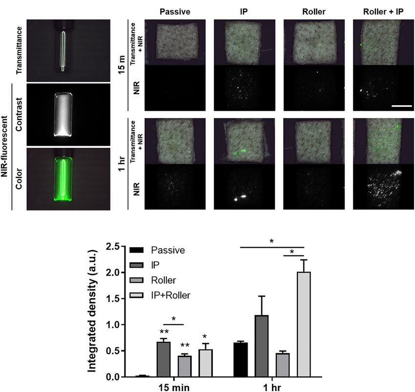
An et al.                                                                                                                 The ADSCs Secretome for Wound Healing

  FIGURE 5 | Masson’s trichrome (MTC) staining of the wounds at day 21. (A) Representative light microscopy images (Scale bar = 100 µm). The qualitative analysis
  of the skin regeneration (B) collagen deposition, (C) extracellular matrix (ECM) fiber alignment, and (D) the number of skin appendages (*compared with the PBS
  group; # compared with the SF group; ∗ p < 0.05; **p < 0.01; ***p < 0.001).

  FIGURE 6 | In vivo angiogenesis evaluation with the immunohistochemical staining (IHC) of alpha-smooth muscle actin (α-SMA). (A) Representative light microscopy
  images at regenerated wounds (Scale bar = 100 µm). (B) The number of newly formed vessels in the high-power field (HPF) magnification images (n = 10–15)
  (*compared with the PBS group; # compared with the SF group; ∗,# p < 0.05; ***p < 0.001).

the TUNEL assay was also performed against the heart, the liver,                     (PPEs), i.e., iontophoresis and non-invasive metal roller. For
the spleen, the lung, and the kidney (Figure 7B). Similar to the                     visualization, we labeled the secretome with NHS ester-
healthy group, there were few apoptotic cells in the harvest tissues                 conjugated ZW800-1C, near-infrared (NIR) fluorescent dye
of the SF-treated group, both on days 7 and 14; however, the                         (Figure 8A). This NIR-labeled secretome was applied to the
biosafety of the secretome should be further investigated in a                       ex vivo porcine skin, and PPEs were exerted on the skin, after
dose-dependent manner.                                                               15 min and 1 h, then the skin tissues using the FLARE system
                                                                                     (Figure 8B), followed by implementing the semiquantitative
                                                                                     analysis (Figure 8C). A little portion of the secretome was
Transdermal Delivery of Secretome                                                    absorbed into the skin in the passive and roller groups, where
Using Non-invasive Physical Penetration                                              the secretome was nearby the follicular regions. However, in
Enhancers                                                                            the case of the iontophoresis (IP) group, the fluorescent signals
We finally tried to deliver the molecules in the secretome                           appeared in several regions, even in other than follicular regions.
through transdermally using the physical penetration enhancers                       In addition, the most abundant secretome was absorbed into

Frontiers in Bioengineering and Biotechnology | www.frontiersin.org              8                                            June 2021 | Volume 9 | Article 681501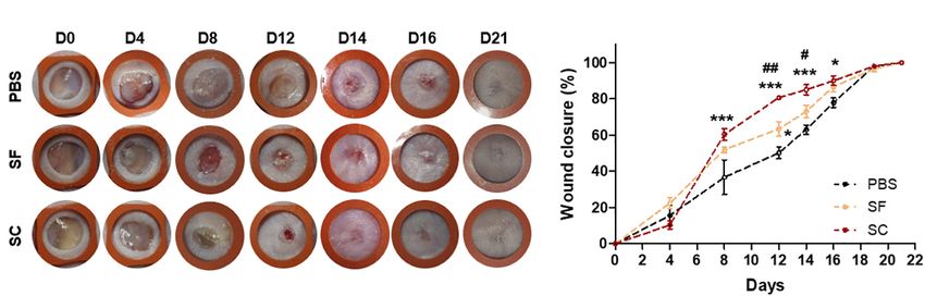
An et al.                                                                                                                      The ADSCs Secretome for Wound Healing

  FIGURE 7 | Pathological analysis of SF via intraperitoneal injection and applying to the skin surface. The 50 µl of SF was injected every other day until 2 weeks or
  applied to the dorsal skin surface of mice. (A) Hematoxyline and eosin (H&E) and (B) TUNEL staining of the tissues harvested from the heart, the liver, the spleen, the
  lung, and the kidney. (C) H&E- and TUNEL-stained images of the skin, which did not display any toxical responses (Scale bar = 200 µm).

  FIGURE 8 | Ex vivo transdermal delivery of NIR dye-labeled SF. (A) The NIR dye, ZW800-1C-NHS-ester, was conjugated with proteins in the secretome.
  (B) Macroscopic visualization of transport of secretome into the ex vivo porcine skin with the assistance of physical penetration enhancers (PPEs). (C) The
  quantification data based on the FLARE images indicated the combinatorial application of IP and roller enhanced the transport of secretome transdermally
  (*p < 0.05; **p < 0.01) (Scale bar = 1 cm).

Frontiers in Bioengineering and Biotechnology | www.frontiersin.org                 9                                               June 2021 | Volume 9 | Article 681501
An et al.                                                                                                    The ADSCs Secretome for Wound Healing

the skin when the combination of roller and IP was applied                    vimentin, etc. (Table 2). It suggests that the maturing culture
during 1 h. As a result, it was confirmed that the transdermal                of ADSCs promotes the secretion of ECM proteins as well as
delivery of the secretome was significantly increased in the usage            maximizes the productive efficacy of the secretome.
of additional stimulation, whereas hardly achieved by passive                    An apparent reason for obtaining a high level of secreted
diffusion alone.                                                              proteins is that the absolute amount of cells in this study (100%
                                                                              confluent) was higher than that of the conventional method (70–
                                                                              90% confluent)—the crucial point of this study to optimize the
DISCUSSION                                                                    maturing conditions where apoptosis did not occur. Besides, we
                                                                              considered that the maturing process-induced hypoxia might
The most crucial finding in this study is that we have developed              be the main reason for accelerating the secretion of a high
a maturing method that can maximize the productive efficiency                 amount of VEGF since the level of VEGF expression increased in
of the stem cell secretome. In this study, we produced the                    hypoxia conditions (Chang et al., 2013). Moreover, some works
ADSC secretome with a GMP-compliant process, which could                      demonstrated that secretomes from apoptotic cells had elevated
lower the possible contamination. It contained a remarkably                   levels of proangiogenic factors and also an impact on tissue
high amount of protein molecules for wound healing, i.e., TGF-                regeneration and anti-inflammatory functions (Beer et al., 2015;
β1 and VEGF. We could obtain the SF and concentrated it                       Simader et al., 2019; Alegre, 2020; Medina et al., 2020). We
in ambient conditions. This allowed the SF to have a similar                  hypothesize that ADSCs in the maturing process stress-related
level of proteins to that of SC, and their in vivo wound healing              signaling which may have enhanced the secretion of the biological
ability was evaluated.                                                        molecules that eventually facilitated the tissue repair process.
   The secreted substances from stem cells, such as exosome and                  Wound healing is a complex and orchestrated process that
secretome, possess the cellular information to exert biological               several processes are integrated and overlapped. For this reason,
activity. As a cell-free therapy, they have been extensively studied          secretome-based therapy is beneficial to treat the wound as it
not only for wound treatment but also administered through                    contains anti-inflammatory factors, promotes cell mitogenesis,
various routes for disease treatment (Beer et al., 2016; Konala               and induces neovascularization (Estrada et al., 2009; Mildner
et al., 2016; Guan et al., 2017; Laggner et al., 2020). In the                et al., 2013; Lombardi et al., 2019). We found out that both
case of secretome, both quality control and analysis are the                  SF and SC promoted overall wound healing, particularly wound
most significant barriers to their clinical usage. Although recent            closure (Figure 2), re-epithelization (Figure 3), proliferation
advances in analytical techniques make it feasible to determine               (Figure 4), tissue remodeling (Figure 5), and angiogenesis
the protein molecules, the serum-containing secretome is                      (Figure 6). The inflammatory response was not characterized.
susceptible to contamination, which interrupts mass production,               It was concerned that proinflammatory factors, interleukin (IL)-
storage, distribution, and commercialization (Shin et al., 2019).             6, and IL-8, were determined in proteomic analysis of SF, while
While the serum-free secretome is relatively less susceptible                 the anti-inflammatory factor, IL-10, was not (Supplementary
to contamination and has a diminished immune response, the                    Material). Thus, it should be further investigated whether
amount of secreted molecules is significantly reduced. Some                   the presence of these proinflammatory factors interrupts the
strategies, such as freeze-drying and supercritical fluid, have been          wound healing process (Balaji et al., 2015; Bai et al., 2020).
applied as a drying method to store the secretome and elevate                 Although the protein levels of TGF-β1 and VEGF were
the concentration of proteins (Bari et al., 2018); however, this              about the same between SF and SC, overall wound healing
procedure may also hinder the stability of the secretome proteins             capability seemed to be much enhanced by SC. We thought
(Bari et al., 2018).                                                          that both serum and secreted proteins of SC provide a
   The conventional method of harvesting a secretome (or                      more favorable environment for wound regeneration than SF;
conditioned medium) proceeds in cell confluency about 70–90%                  however, it is necessary to confirm how the composition of
up to 48 h (Kalinina et al., 2015; Park et al., 2018). In contrast, we        proteins other than TGF-β1 and VEGF is composed through
used the maturation culturing method by continuously culturing                proteomic analysis of SC.
the ADCSs from a fully confluent state and harvested the medium                  Recently, as stem cell secretome-based cosmetics have
after 7 days, which could be applicable to obtain the SF. To                  attracted attention, their transdermal delivery efficiency has
compare the yield of secretome, we chose two representative                   been disputed. Thus, we estimated the skin permeation of
growth factors, i.e., TGF-β1 and VEGF, since they exert crucial               the secretome (Figure 8). In general, it is known that
roles in the wound healing process. In previous studies, VEGF                 macromolecular drugs, such as peptides and proteins, are
contents in stem cell secretome (or conditioned medium) were                  impossible to pass through the stratum corneum and can be
at most under 0.5 ng/ml (Zisa et al., 2009; Ge et al., 2018).                 transported through the follicular route (Bos and Meinardi,
Interestingly, SC, in this study, represented the concentration               2000). However, due to its uneven distribution depending on the
of about 20 ng/ml without any additional processes (Table 1),                 regions, follicular delivery of macromolecules is still ambiguous
which is extraordinarily superior to other works. For TGF-β1, we              (Otberg et al., 2004). Likewise, we could observe the transdermal
could harvest about 1.5 ng/ml in pristine SC, but there were few              delivery of secretome using macroscopic NIR images and found
studies for comparison. Above all, in the proteomic analysis of SF            that the secretome was mainly distributed in the hair follicles
(Figure 1), we confirmed that there were lots of proteins relevant            and barely transported into the skin surface. There are many
to an ECM organization, e.g., fibronectin, collagen subunits,                 types of delivery enhancers to increase the delivery efficacy

Frontiers in Bioengineering and Biotechnology | www.frontiersin.org      10                                     June 2021 | Volume 9 | Article 681501
An et al. The ADSCs Secretome for Wound Healing
of macromolecules into the skin (An et al., 2020). It was ETHICS STATEMENT
evidently observed that the secretome was dispersed into the skin
besides the follicles when using the non-invasive dermal roller The animal study was reviewed and approved by Institutional
and iontophoresis. Considered that the NIR dye-conjugated Animal Care and Use Committe (IACUC) at Seoul
secretome proteins were filtrated through 7 kDa MWCO National University.
membrane, that is, the molecular weight of the transported
proteins was over 7 kDa, it was anticipated that the more active
ingredients of pristine secretome actually have an effect on the AUTHOR CONTRIBUTIONS
skin functions when delivered using penetration enhancers.
Y-HA: conceptualization, methodology, validation, writing–
original draft, and formal analysis. DHK, S-DK: project
administration and supervision. EL: methodology and validation
CONCLUSION of ADSC secretome. DL and DWK: methodology, validation, and
formal analysis of proteomics. MP and JHK: assisted in vivo tests
In this study, we demonstrated the harvesting and harnessing and data analysis. JWK, HH, YS, J-YC, and J-UP: writing–review
of the therapeutic potentials of ADSCs in the form of the and editing. NH: conceptualization, writing–review and editing,
secretome. Remarkably, Remarkably, we have firstly reported supervision, and project administration. All authors contributed
that the maturation process could achieve efficient extraction to the article and approved it for publication.
of secreted factors (i.e., extractomes) from the ADSCs that
contains a higher level of secreted molecules than conventional
methods. Furthermore, we confirmed that the ADSC secretome FUNDING
acted synergistically to restore skin defect by facilitating tissue
regeneration and preventing scar formation without any toxicity This work was financially supported by the Ministry
by comparing the SC and SF. Consequently, our study implies of Science and ICT (NRF-2016R1E1A1A01943393, NRF-
that ADSC secretome can be effectively produced via maturation 2017M3A9C6031786, NRF-2019M3A9G1023840, NRF-
process, and particularly, SF would be safely utilized to restore 2019R1I1A1A01059554, NRF-2019M3A9H1103786, and NRF-
damaged tissue architecture in clinical cases. 2020M3H1A1073304). The Institute of Engineering Research
at Seoul National University and Senior Science & Life, Inc.,
provided research facilities for this work.
DATA AVAILABILITY STATEMENT
SUPPLEMENTARY MATERIAL
The mass spectrometry proteomics data have been deposited
to the ProteomeXchange Consortium via the PRIDE (Perez- The Supplementary Material for this article can be found
Riverol et al., 2019) partner repository with the dataset identifier online at: https://www.frontiersin.org/articles/10.3389/fbioe.
PXD026436. 2021.681501/full#supplementary-material
REFERENCES Beer, L., Zimmermann, M., Mitterbauer, A., Ellinger, A., Gruber, F., Narzt,
M. S., et al. (2015). Analysis of the secretome of apoptotic peripheral blood
Alegre, M. L. (2020). The anti-inflammatory function of the apoptotic secretome. mononuclear cells: impact of released proteins and exosomes for tissue
Am. J. Transplant. 20, 1471–1471. doi: 10.1111/ajt.15990 regeneration. Sci. Rep. Uk. 16:5.
An, Y. H., Park, M. J., Lee, J., Ko, J., Kim, S. H., Kang, D. H., et al. Bos, J. D., and Meinardi, M. M. H. M. (2000). The 500 dalton rule for the skin
(2020). Recent advances in the transdermal delivery of protein therapeutics penetration of chemical compounds and drugs. Exp. Dermatol. 9, 165–169.
with a combinatorial system of chemical adjuvants and physical penetration doi: 10.1034/j.1600-0625.2000.009003165.x
enhancements. Adv. Ther. Germany 3:1900116. doi: 10.1002/adtp.201900116 Chan, B., Cadarette, S., Wodchis, W., Wong, J., Mittmann, N., and Krahn, M.
Bai, H. T., Kyu-Cheol, N., Wang, Z. H., Cui, Y. T., Liu, H., Liu, H., et al. (2020). (2017). Cost-of-illness studies in chronic ulcers: a systematic review. J. Wound
Regulation of inflammatory microenvironment using a self-healing hydrogel Care. 26, S4–S14.
loaded with BM-MSCs for advanced wound healing in rat diabetic foot ulcers. Chang, C. P., Chio, C. C., Cheong, C. U., Chao, C. M., Cheng, B. C.,
J. Tissue Eng. 11:2041731420947242. and Lin, M. T. (2013). Hypoxic preconditioning enhances the therapeutic
Balaji, S., King, A., Marsh, E., LeSaint, M., Bhattacharya, S. S., Han, N., et al. (2015). potential of the secretome from cultured human mesenchymal stem cells in
The role of interleukin-10 and hyaluronan in murine fetal fibroblast function experimental traumatic brain injury. Clin .Sci. 124, 165–176. doi: 10.1042/cs201
in vitro: implications for recapitulating fetal regenerative wound healing. PLoS 20226
One 7:10. Chantre, C. O., Campbell, P. H., Golecki, H. M., Buganza, A. T., Capulli, A. K.,
Bari, E., Perteghella, S., Di Silvestre, D., Sorlini, M., Catenacci, L., Sorrenti, M., et al. Deravi, L. F., et al. (2018). Production-scale fibronectin nanofibers promote
(2018). Pilot production of mesenchymal stem/stromal freeze-dried secretome wound closure and tissue repair in a dermal mouse model. Biomaterials 166,
for cell-free regenerative nanomedicine: a validated GMP-compliant process. 96–108. doi: 10.1016/j.biomaterials.2018.03.006
Cells. 7:190. doi: 10.3390/cells7110190 Damous, L. L., de Carvalho, A., Nakamuta, J. S., Shiroma, M. E., Louzada, A. C. S.,
Beer, L., Mildner, M., Gyongyosi, M., and Ankersmit, H. J. (2016). Peripheral Soares-Jr, J. M., et al. (2018). Cell-free therapy with the secretome of adipose
blood mononuclear cell secretome for tissue repair. Apoptosis 21, 1336–1353. tissue-derived stem cells in rats’ frozen-thawed ovarian grafts. Stem Cell Res.
doi: 10.1007/s10495-016-1292-8 Ther. 9:323.
Frontiers in Bioengineering and Biotechnology | www.frontiersin.org 11 June 2021 | Volume 9 | Article 681501An et al. The ADSCs Secretome for Wound Healing Del Papa, N., Caviggioli, F., Sambataro, D., Zaccara, E., Vinci, V., Di Luca, G., et al. as tissue messengers. Nature 580, 130–135. doi: 10.1038/s41586-020- (2015). Autologous fat grafting in the treatment of fibrotic perioral changes 2121-3 in patients with systemic sclerosis. Cell Transplant. 24, 63–72. doi: 10.3727/ Mildner, M., Hacker, S., Haider, T., Gschwandtner, M., Werba, G., Barresi, C., 096368914x674062 et al. (2013). Secretome of peripheral blood mononuclear cells enhances wound Estrada, R., Li, N., Sarojini, H., An, J., Lee, M. J., and Wang, E. (2009). Secretome healing. PLoS One 22:8. from mesenchymal stem cells induces angiogenesis via Cyr61. J. Cell Physiol. Otberg, N., Richter, H., Schaefer, H., Blume-Peytavi, U., Sterry, W., and Lademann, 219, 563–571. doi: 10.1002/jcp.21701 J. (2004). Variations of hair follicle size and distribution in different body sites. Ferguson, M. W. J., and O’Kane, S. (2004). Scar-free healing: from embryonic J. Invest. Dermatol. 122, 14–19. doi: 10.1046/j.0022-202x.2003.22110.x mechanisms to adult therapeutic intervention. Philos. T. Roy. Soc. B 359, Park, S. R., Kim, J. W., Jun, H. S., Roh, J. Y., Lee, H. Y., and Hong, I. S. (2018). Stem 839–850. doi: 10.1098/rstb.2004.1475 cell secretome and its effect on cellular mechanisms relevant to wound healing. Ge, Q. H., Zhang, H. W., Hou, J. X., Wan, L. F., Cheng, W. Z., Wang, X. Y., et al. Mol. Ther. 26, 606–617. doi: 10.1016/j.ymthe.2017.09.023 (2018). VEGF secreted by mesenchymal stem cells mediates the differentiation Perez-Riverol, Y., Csordas, A., Bai, J., Bernal-Llinares, M., Hewapathirana, S., of endothelial progenitor cells into endothelial cells via paracrine mechanisms. Kundu, D. J., et al. (2019). The PRIDE database and related tools and resources Mol. Med. Rep. 17, 1667–1675. in 2019: improving support for quantification data. Nucleic Acids Res. 47, Guan, L., Suggs, A., Galan, E., Lam, M., and Baron, E. D. (2017). Topical application D442–D450. doi: 10.1093/nar/gky1106 of ST266 reduces UV-induced skin damage. Clin. Cosmet. Inv. Derm. 10, Robert, A. W., Gomes, F. A., Rode, M. P., da Silva, M. M., Veleirinho, M. B. D., 459–471. doi: 10.2147/ccid.s147112 Maraschin, M., et al. (2019). The skin regeneration potential of a pro-angiogenic Gurtner, G. C., Werner, S., Barrandon, Y., and Longaker, M. T. (2008). Wound secretome from human skin-derived multipotent stromal cells. J. Tissue Eng. repair and regeneration. Nature 453, 314–321. 12:10. Hodgkinson, T., Wignall, F., Hoyland, J. A., and Richardson, S. M. (2020). Salgado, A. J., Reis, R. L., Sousa, N. J., and Gimble, J. M. (2010). Adipose High BMPR2 expression leads to enhanced SMAD1/5/8 signalling and tissue derived stem cells secretome: soluble factors and their roles in GDF6 responsiveness in human adipose-derived stem cells: implications regenerative medicine. Curr. Stem. Cell Res. Ther. 5, 103–110. doi: 10.2174/ for stem cell therapies for intervertebral disc degeneration. J. Tissue Eng. 157488810791268564 11:2041731420919334. Shin, J., Rhim, J., Kwon, Y., Choi, S., Shin, S., Ha, C. W., et al. (2019). Comparative Jarbrink, K., Ni, G., Sonnergren, H., Schmidtchen, A., Pang, C., Bajpai, R., et al. analysis of differentially secreted proteins in serum-free and serum-containing (2016). Prevalence and incidence of chronic wounds and related complications: media by using BONCAT and pulsed SILAC. Sci. Rep. 9:3096. a protocol for a systematic review. Syst. Rev. 5:152. Sielaff, M., Kuharev, J., Bohn, T., Hahlbrock, J., Bopp, T., Tenzer, S., et al. (2017). Kalinina, N., Kharlampieva, D., Loguinova, M., Butenko, I., Pobeguts, O., Evaluation of FASP, SP3, and iST protocols for proteomic sample preparation Efimenko, A., et al. (2015). Characterization of secretomes provides evidence in the low microgram range. J. Proteom. Res. 16, 4060–4072. doi: 10.1021/acs. for adipose-derived mesenchymal stromal cells subtypes. Stem. Cell Res. jproteome.7b00433 Therapy 11:6. Simader, E., Beer, L., Laggner, M., Vorstandlechner, V., Gugerell, A., Erb, M., et al. Konala, V. B. R., Mamidi, M. K., Bhonde, R., Das, A. K., Pochampally, R., and Pal, (2019). Tissue-regenerative potential of the secretome of gamma-irradiated R. (2016). The current landscape of the mesenchymal stromal cell secretome: a peripheral blood mononuclear cells is mediated via TNFRSF1B-induced new paradigm for cell-free regeneration. Cytotherapy 18, 13–24. doi: 10.1016/j. necroptosis. Cell Death Dis. 30:10. jcyt.2015.10.008 Tonnesen, M. G., Feng, X. D., and Clark, R. A. F. (2000). Angiogenesis in wound Laggner, M., Gugerell, A., Bachmann, C., Hofbauer, H., Vorstandlechner, V., healing. J. Invest. Derm. Symp. Proc. 5, 40–46. Seibold, M., et al. (2020). Reproducibility of GMP-compliant production of Tu, S. M. (2010). Origin of cancers. Clinical perspectives and implications of a therapeutic stressed peripheral blood mononuclear cell-derived secretomes, a stem-cell theory of cancer. Cancer Treat Res. 154, v–239. novel class of biological medicinal products. Stem. Cell Res. Therapy 3:11. Weng, Y., Sui, Z., Shan, Y., Jiang, H., Zhou, Y., Zhu, X., et al. (2016). In-depth Lee, M. J., Kim, J., Kim, M. Y., Bae, Y. S., Ryu, S. H., Lee, T. G., et al. (2010). proteomic quantification of cell secretome in serum-containing conditioned Proteomic analysis of tumor necrosis factor-alpha-induced secretome of human medium. Anal. Chem. 88, 4971–4978. doi: 10.1021/acs.analchem.6b00910 adipose tissue-derived mesenchymal stem cells. J. Proteome Res. 9, 1754–1762. Zisa, D., Shabbir, A., Suzuki, G., and Lee, T. (2009). Vascular endothelial growth doi: 10.1021/pr900898n factor (VEGF) as a key therapeutic trophic factor in bone marrow mesenchymal Lee, S. M., Lee, S. C., and Kim, S. J. (2014). Contribution of human stem cell-mediated cardiac repair. Biochem. Biophys. Res. Co. 390, 834–838. adipose tissue-derived stem cells and the secretome to the skin allograft doi: 10.1016/j.bbrc.2009.10.058 survival in mice. J. Surg. Res. 188, 280–289. doi: 10.1016/j.jss.2013. 10.063 Conflict of Interest: DHK, EL, JWK, and S-DK were employed by the company, Lindeman, G. J., and Visvader, J. E. (2010). Insights into the cell of origin in Senior Science & Life, Inc. breast cancer and breast cancer stem cells. Asia Pac. J. Clin. Oncol. 6, 89–97. doi: 10.1111/j.1743-7563.2010.01279.x The remaining authors declare that the research was conducted in the absence of Lombardi, F., Palumbo, P., Augello, F. R., Cifone, M. G., Cinque, B., and Giuliani, any commercial or financial relationships that could be construed as a potential M. (2019). Secretome of adipose tissue-derived stem cells (ASCs) as a novel conflict of interest. trend in chronic non-healing wounds: an overview of experimental in vitro and in vivo studies and methodological variables. Int. J. Mol. Sci. 30:20. Copyright © 2021 An, Kim, Lee, Lee, Park, Ko, Kim, Koh, Hong, Son, Cho, Park, Kim Marfia, G., Navone, S. E., Di Vito, C., Ughi, N., Tabano, S., Miozzo, M., et al. (2015). and Hwang. This is an open-access article distributed under the terms of the Creative Mesenchymal stem cells: potential for therapy and treatment of chronic non- Commons Attribution License (CC BY). The use, distribution or reproduction in healing skin wounds. Organogenesis 11, 183–206. doi: 10.1080/15476278.2015. other forums is permitted, provided the original author(s) and the copyright owner(s) 1126018 are credited and that the original publication in this journal is cited, in accordance Medina, C. B., Mehrotra, P., Arandjelovic, S., Perrys, J. S. A., Guo, Y. Z., with accepted academic practice. No use, distribution or reproduction is permitted Morioka, S., et al. (2020). Metabolites released from apoptotic cells act which does not comply with these terms. Frontiers in Bioengineering and Biotechnology | www.frontiersin.org 12 June 2021 | Volume 9 | Article 681501
You can also read