High Volume Liposuction in Tumescence Anesthesia in Lipedema Patients: A Retrospective Analysis - Matthias Sandhofer
←
→
Page content transcription
If your browser does not render page correctly, please read the page content below
doi:10.36849/JDD.2021.5828

                     March 2021                                                                            Volume 20 • Issue 3
                Copyright © 2021                         ORIGINAL ARTICLE                          Journal of Drugs in Dermatology
                                                          SPECIAL TOPIC

                High Volume Liposuction in Tumescence Anesthesia
                  in Lipedema Patients: A Retrospective Analysis
                         Matthias Sandhofer MD,a Victoria Hofer, a,b Martina Sandhofer,a Mindt Sonani,c
                                            Werner Moosbauer,a,d Martin Barscha
                     aDermatologie Zentrum für Haut, Ästhetik, Laser,Venen, Praxis Dr. Matthias Sandhofer/Dr. Martin Barsch;
                                                  Austrian Center for Lipedema, Wien/Linz, Austria
                             b
                              Danube Private University, Faculty of Medicine/Dental Medicine, Krems-Stein, Austria
                              c
                               Institute for Clinical Chemistry, University Hospital Mannheim, Mannheim, Germany
                        d
                         Kepler Universitätsklinikum GmbH, Anästhesiologie und Operative Intensivmedizin, Linz, Austria

 ABSTRACT
   Background: Lipedema is a chronic, progressive disease that occurs almost exclusively in women and leads to pathological, painful
   fat growths at the extremities. Only symptomatic therapy can be offered since the etiology of the disease has not yet been clarified.
   Liposuction in tumescent anesthesia has established itself as a surgical treatment method of choice. The complication rate associated
   with the procedure and the pharmacological course and safety of treatment in patients with lipedema has not yet been sufficiently
   studied. The aim of the study was to broaden the evidence on the safety of ambulatory high-volume liposuction in tumescent anesthesia
   in lipedema patients. Influencing factors of patients (weight, fat content, comorbidities) or the process technique (drug administration,
   volume of aspirates) should be investigated on the safety and risks of tumescent anesthesia. This was a retrospective data analysis in
   which data from 27 patients (40 liposuction procedures) treated at the Sandhofer and Barsch lipedema center between 2016 and 2018
   were evaluated. The liposuctions were carried out in tumescent anesthesia and using a Power-Assisted Liposuction system. Clinical
   examinations and regular blood samples were carried out before the procedure, intra- and postoperatively. The procedures lasted an
   average of 118 minutes and an average of 6111 ml of aspirate was removed. For tumescent anesthesia, patients were given an average
   lidocaine dose of 34.23 mg/kg body weight and an epinephrine dose of 0.11 mg/kg body weight. No relevant complications associated
   with drug side effects, hypovolemia or hypervolemia or blood loss were detected. Liposuction under high volume tumescent anesthesia
   for the treatment of lipedema patients is, even for major intervention, a safe procedure.

   J Drugs Dermatol. 2021;20(3):        doi:10.36849/JDD.2021.5828

 INTRODUCTION

L
        iposuction is one of the most common surgical procedures          of lipedema is based on conservative physical decongestive
        in aesthetic surgery worldwide and was first described            therapy and surgical liposuction of the pathological adipose
        out in the mid-1990s as a therapy for lipedema.1,2 During         tissue augmentation. The appropriate treatment method should
this time, liposuction could be established as a safe and effective       be determined individually for each patient.12 The excessive
therapeutic alternative in the treatment of lipedema, especially          increase in adipose tissue and the resulting restrictions in
by German, Austrian and Dutch operative dermatologists.3-9                mobility as well as the disproportionate appearance cannot
Further development of the surgical procedure using lymph-                be treated with conservative therapy,13 especially if there is no
friendly liposuction techniques with fine cannulas, liposuction           edema present. Manual lymphatic drainage (MLD), intermittent
has established itself as an important, minimally invasive                pneumatic compression, compression stockings, exercise,
therapeutic approach for lipedema.8 Several studies have                  and skin care are often used to control pain and symptoms in
shown that liposuction significantly reduced sensitivity to pain          lymphedema and therefore also in lipedema. Recent studies
and pressure as well as the tendency to hematoma.10 Improving             have shown that there is little or no lymphedema in lipedema.14,15
mobility after the procedure leads to an increase in energy               All over, the term lipedema is a misnomer, since more than
turnover, which can contribute to further weight loss.8 Several           90% of typical lipedema patients do not have any edema.
studies describe a significant improvement in the quality of              Especially the patients that are seen in the practice, in contrary
life due to the suction of the pathological fat tissue, which can         to the patients seen in specialized lymph clinics, where the
still be demonstrated eight years after the intervention.10,11 To         percentage of lipedema patients with an edema is a bit higher.
date a causal therapy for lipedema is not known. The treatment            Treatment with MLD showed no significant therapeutic effect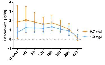
doi:10.36849/JDD.2021.5828

                              Journal of Drugs in Dermatology           M. Sandhofer,V. Hofer, M. Sandhofer, et al
                             March 2021 • Volume 20 • Issue 3

on lipedema apart from a short relief due to patients’ care. In the     previous fat analysis with the impedance measurement, to
contrary, seeing a few hundred lipedema patients a year in the          classify a metabolic disorder, accurately determine the total
Austrian lipedema center it is significant that a lot of lipedema       amount of fat in the patient. Since lidocaine is a lipophilic
patients report that the compression stocking aggravate pain            substance, from the amount of total fat we can predict the
in lipedema patients (own observation). Surgical treatment              appropriate lidocaine dose in tumescent solution. This method
involves sensitive, atraumatic lymph-sparing liposuction using          is also extremely helpful to control the postoperative course
tumescent local anesthesia. The treatment has been proven to            (weight and lifestyle). Jeffrey Klein published the first study on
be safe and effective for cosmetic indications and lipedema.5,16-19     the use of tumescent anesthesia in 1987. The anesthetic solution
Tumescent anesthesia is a special form of local anesthesia, in          described consisted of physiological saline (0.9% NaCl) with
which large amounts of an anesthetic solution, consisting of a          0.091% lidocaine (910 mg/l) and 0.91 mg/l epinephrine.20 Klein
physiological solution with local anesthetics and epinephrine,          modified the recommendations for the composition of the
are introduced into the subcutaneous adipose tissue. This local         anesthetic solution several times in subsequent studies. In
anesthesia makes large areas of the skin insensitive to pain and        1999 he published that the concentrations of the substances
surgical interventions can be carried out without an additional         dissolved in the anesthetic fluid should be made dependent
anesthetic procedure.20,21 In the early days of surgical liposuction,   on the region of the body to be treated.27 To date, there are no
bleeding and the associated high need for transfusion was               guidelines specifying the exact composition of the infiltration
the limiting factor in liposuction. By adding epinephrine to            solution. The lidocaine concentration used in the solution
the infiltration fluid, liposuction could be achieved without           varies, depending on the surgeon, between 500–1500 mg/l and
the need for a transfusion.22 Since nowadays high amount                the epinephrine concentration between 0.5–1.5 mg / l.4 No data
of fluid is used, the additional compression of the fluid on            are available for large-volume liposuction, as it is the case with
the vessels further decreases blood loss during intervention.           lipedema.
Examination of adipose tissue using lymphoscintigraphy and
immunohistochemistry after liposuction has demonstrated no              We are the first who examined lidocaine and epinephrine
significant damage to lymphatic vessels using tumescent local           in serum after high volume liposuctions in lipedema. In
anesthesia compared with traditional liposuction techniques             the present study we analyzed the safety and efficacy of
using general anesthesia. Liposuction techniques using radio            liposuction regarding different parameters. During the regular
frequency, ultrasound, or laser are not useful for lipedema             quality management, lidocaine and epinephrine levels were
patients because of possible damage to lymphatic vessels.13             determined intra-and post-operatively and the clinical course
The effectiveness of liposuction using tumescent anesthesia is          was examined. In this retrospective data analysis, randomized
based on the fact, that beside subtotal removement of fat, the          data were selectively collected from 27 patients who underwent
capillaries, including the well-known leaky vessels in lipedema,        40 liposuctions between 2016 and 2018, whereas 13 patients
can be removed. This results in no, or far less bruises for the         from this cohort underwent a second liposuction within 3 days.
patients after the liposuction and leads to the suppression of
angiogenesis and subsequently adipogenesis at the cellular               MATERIALS AND METHODS
level.23,24 That explains the sustainable impact as described by        Liposuction Procedure
Baumgartner and Schmeller.11 Klein et al looked at the serum            Depending on the severity of the lipedema, two or three
levels of the lidocaine on small liposuction volumes, with              procedures for the legs and one procedure for the arms, if
patients he injected tumescence solution and did not aspirate           affected, were planned for each patient. All interventions
and patients he injected tumescence solution and performed              were performed under tumescent anesthesia and superficial
a normal liposuction afterwards, resulting in lower lidocaine           sedoanalgesia, which was performed and monitored by an
serum levels when he aspirated.25 This is not comparable to             anesthetist. After premedication with midazolam 7.5 mg orally,
lipedema since the liposuction volume of lipedema patients is           the analgesia was carried out with 3–5 mg intravenous midazolam
extremely high. In general, a critical consideration regarding          and continuous administration of low dose remifentanil via a
local tumescence anesthesia should be given in terms of                 perfusor. In the case of marked anxiety, a further midazolam of
large amounts of tumescence infiltrates, which could lead to            1–2 mg or 30–60 µg clonidine was administered if necessary.
hypervolemia or pulmonary edema. Removing large amounts                 The patients were always responsive during liposuction and
of fat also creates the potential for hypovolemia (shock).              were able to independently change the position during the
The amount of local anesthetic and epinephrine can lead to              procedure and contract specific muscle groups if the surgeon
toxic reactions.25 There is also the fundamental question of            asks for it. This is necessary for a better esthetic outcome and
whether liposuction should be performed under general or                one of the major advantages of a sedoanalgesia compared to
local anesthesia with slight analgesic sedation.26 Necessarily,         general anesthesia. The tumescent fluid was freshly prepared
parameters such as blood loss, infection, and pain should be            before the procedure and warmed to 37°C and introduced into
taken under consideration An important point is the exact,              the subcutaneous adipose tissue by 2 persons simultaneously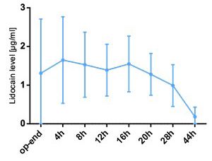
doi:10.36849/JDD.2021.5828

                              Journal of Drugs in Dermatology        M. Sandhofer,V. Hofer, M. Sandhofer, et al
                             March 2021 • Volume 20 • Issue 3

under pressure using a KMI Surgical Infusion/Irrigation Pump.        or additional medication administered were documented.
The infiltration cannulas were wiped like a wiper, starting in       The amount of oral fluid intake was recorded by the patients
the depth near the fascia and were then extended to the upper        themselves. The survey was carried out analogously to the
layers and continued until the infiltrated tissue developed a firm   blood tests performed before the operation, after the operation,
whitish skin turgor (state of tumescence). "Vivomed infiltration     and after 4, 8, 12, 16, 20, 28, and 44 hours. In the course of the
needles 1.2x100 mm" were used for infiltration. After a waiting      postoperative visits, the patients were asked to get up and walk
period of 30 minutes, liposuction was started. Liposuction was       a few rounds through the room.
carried out using the PAL Liposuction System from MicroAire.
In order to make liposuction gentler, the suction cannula was        Ultrasound Examination
set in motion. The cannulas with a diameter of 3 to 4 mm were        Preoperatively we looked for venous disorders, the quality
inserted into the subcutaneous fat tissue via small incisions of     and structure of fat, exclusion of edemas (lymphedema,
approximately 4 mm. The suction was carried out considering          phlebedema, lipolymphedema, and lipophlebolymphedema),
the position and course of the lymphatic vessels. If pain is         and the thickness of fat over the proximal and the distal tibia. In
indicated intraoperatively, a minimal secondary infiltration is      contrast to the hypoechoic structure of normal adipose tissue,
carried out with a blunt, 40 cm long infiltration cannula with       lipedema appears widened with hyperechoic connective tissue
a diameter of 2 mm. No intravenous fluid substitution was            septa and the typical image of a "snow storm pattern". In the
performed. All patients were only discharged when they met           case of an insufficient veins which needed to be treated, this
the discharge criteria.                                              was done at least 2 months before the liposuction. The thickness
                                                                     of the fat pretibial proximal and distal, the position of the small
Data Collection                                                      saphenous vein in the middle calf and the determination of
A detailed medical history and a physical examination were           the Marshall point as described elsewhere.28 as well as the
carried out preoperatively to confirm the diagnosis of all           exclusion of lymphedema confirmed the diagnosis of lipedema
patients. All patients were weighed using a body analyzer scale      (lipophobia of the tissue). A sonographic assessment of the
(impedance measurement) and the results regarding weight,            pleural space, the inferior caval vein and the heart was carried
body fat percentage and visceral fat, and BMI were recorded          out before the operation, at the end of the operation and after
in the medical history. In addition, an electrocardiogram (EKG)      20 hours post-surgery. It was recorded whether evidence of
examination was carried out to rule out cardiac problems.            hypervolemia with pulmonary edema or hypovolemia could
During the operation, the data on the amount of tumescent            be found. During echocardiography, the ejection fraction was
solution used and its composition was noted. A distinction was       determined and whether wall movement disorders occurred.
made between the infiltration phase and the actual liposuction.
The amount of the aspirate and the fat content in the aspirate       Electrocardiography (ECG)
were documented. During the operation and postoperative care,        A 12-lead ECG examination was carried out several times on
the vital parameters (blood pressure, pulse, oxygen saturation)      all patients. This was done before the operation, at the end of
were collected and documented several times. This was done           the operation and 20 hours after the procedure. To evaluate this
before and during the operation, as well as after 4, 8, 12, 16,      retrospective data analysis, the existing ECGs were presented
20, 28, and 44 hours after the procedure. All patients underwent     anonymously to an uninvolved cardiologist for evaluation. It
blood sampling after infiltration and before liposuction, after      was analyzed whether new ECG changes appeared.
liposuction and after 4, 8, 12, 16, 20, 28, and 44 hours. For
further analysis, the samples were sent to the Institute for          RESULTS
Clinical Chemistry at the University of Mannheim, where the          General Data on Patient, Operation, and Medication
lidocaine and epinephrine levels were determined. A blood            The study is a retrospective data analysis in which data from
count was determined from the first and the last sample in order     27 patients were evaluated who underwent liposuction at the
to examine the drop-in hemoglobin and hematocrit level. Heart        Sandhofer and Barsch Lipedema Center in 2016–2018. During the
enzymes and liver function parameters were determined at the         regular quality management, lidocaine and epinephrine levels
end of the operation and after 20 hours.                             were determined intra-and post-operatively by 27 patients and
                                                                     the clinical course was examined. According to the inclusion
Side Effects                                                         criteria, only these patients are used for the present data analysis.
During the postoperative visits, the patients were asked about       Only patients were included who had a complete record of the
their well-being, exercise capacity and postoperative pain on the    treatment including lidocaine and epinephrine levels, as well as
VAS scale (1–10). The information on the subjective assessment       post-operative clinical follow-up. Analyzing the treated patients
of side effects was divided into five categories. In addition,       master data revealed an average age of 41.7 years, body weight
complications observed postoperatively (nausea, tinnitus,            of 90.3 kg, and a height of 167.4 cm, which resulted in an average
circulatory problems, rapid heartbeat, shortness of breath)          BMI of 32.3 (Table 1). Regarding the general operation data, the
doi:10.36849/JDD.2021.5828
Journal of Drugs in Dermatology M. Sandhofer,V. Hofer, M. Sandhofer, et al
March 2021 • Volume 20 • Issue 3
TABLE 1.
FIGURE 1. The average lidocaine levels of all patients after the
Patient Master Data. The treated patients were on average 41.7 years
procedure and 4, 8, 12, 16, 20, 28, and 44 hours later. Mean ± SD. N=27.
old, weighted 90.3 kg and were 167.4 cm tall, which resulted in an
average BMI of 32.3.
Mean σ
Age 41.7 12.7
Body weight [kg] 90.3 16.4
Body fat [kg] 35.4 10.6
Visceral fat [%] 8.3 3.1
Body height [cm] 167.4 5.3
BMI 32.3 5.9
TABLE 2.
General Operation Data. The procedure lasted an average of 118 min-
utes. An average of 6111 ml was aspirated, of which an average of 5585
ml was fat. During tumescent anesthesia, an average of 11,404 ml of
anesthetic fluid was administered.
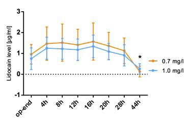
FIGURE 2. The average lidocaine levels separated according to the
Mean σ epinephrine concentration (0.7 and 1.0 mg/l) in the tumescent fluid after
the procedure and 4, 8, 12, 16, 20, 28, and 44 hours later. Mean ± SD.
Operation time [min] 118 17.7 N=27. Pdoi:10.36849/JDD.2021.5828
Journal of Drugs in Dermatology M. Sandhofer,V. Hofer, M. Sandhofer, et al
March 2021 • Volume 20 • Issue 3
FIGURE 3. Influence of the epinephrine dose (0.7 and 1.0 mg/l) on the Influence of the Operating Region on the Course of the Lidocaine
course of the lidocaine level (without arms) after the procedure and 4, Level (With Arms)
8, 12, 16, 20, 28, and 44 hours later. Mean ± SD. N=27. Pdoi:10.36849/JDD.2021.5828
Journal of Drugs in Dermatology M. Sandhofer,V. Hofer, M. Sandhofer, et al
March 2021 • Volume 20 • Issue 3
FIGURE 6. The maximum lidocaine level per patient. Horizontal line Influence of the Tumescent Solution on the Lidocaine Peak Level
shows the cut-off at which the lidocaine levels were regarded as To analyze which patients had an elevated level during treatment,
increased (3.0 µg/ml). the highest measured lidocaine concentration in the blood was
analyzed for each patient. Lidocaine levels of at least 3.0 µg/
ml were defined as increased. A total of five patents showed
elevated levels. The average lidocaine peak level was 1.98 µg /
ml (σ = 1.24) and occurred after 11.63 hours (σ = 9.58h). If patients
whose arms were treated were excluded from the analysis, the
average peak level was 1.59 µg / ml (σ = 0.67), which can be
found after 12.7 hours (σ = 9.66h).
Outliers: The two peak values above 5 µg/ml were found in
patients where a whole-arm type was aspirated, and venous
blood was drawn from the back of the hand: This serum level
shows a local artefact and does not represent the total serum
level Figure 6.
Postoperative Pain and Blood Loss
TABLE 4A. The side effects were analyzed. All patients showed good or
Results of Pain (a) and Blood Loss (b) in the Postoperative Course. very good exercise capacity during the 44 hours analyzed. At
Elevated subjective pain assessment in the postoperative course and
the end of the operation, the patients reported an average pain
how many patients received analgesics at each timepoint (post-opera-
tion, 4, 8, 12, 16, 20, 28, and 44 hours) (a). Comparison of hemoglobin sensation of 4.4 on the VAS scale. The postoperative sensation
and hematocrit before and after liposuction procedure and calculated of pain decreases in the further course. For a large proportion of
drop in hemoglobin (b). N=27. the patients, additional analgesic therapy is given in the course
VAS analgetic of postoperative care. To analyze the blood loss during the
Time [h]
Mean σ Yes No operation, the blood count before the operation and 44 hours
after the operation was compared. On average, a decrease
Post-op 4.44 2.46 -- --
in hemoglobin by 2.3 g / dl and in hematocrit by 6.7% was
4 3.70 1.94 4 19 observed.
8 3.12 1.60 13 10
Blood Pressure, Pulse, Respiratory Rate, SpO2, Heart Enzymes
12 2.20 1.31 8 13
The blood pressure values remained stable over the entire
16 2.05 1.17 6 16
investigation period. No increase in postoperative blood
20 1.77 1.15 0 20 pressure was found. The average heart rate increased during the
28 2.02 1.85 2 19 procedure and was significantly higher compared to the initial
44 2.46 1.82 2 9 value at the end of the operation. (Pdoi:10.36849/JDD.2021.5828
Journal of Drugs in Dermatology M. Sandhofer,V. Hofer, M. Sandhofer, et al
March 2021 • Volume 20 • Issue 3
above the starting point. 1500 mg/l and small liposuction with aspirate amounts of less
than three liters. With 11.4 l, the amount of tumescent solution
Eyelid Edema applied in this study is higher than in comparative studies. This
Minor eyelid edema was found in two patients after 20 hours. can be explained by the full tumescent technique, in which
The next time the patient was examined, the eyelid edema was tumescent liquid is introduced until the tissue has a firm turgor.
no longer visible after 28 hours. Due to the laxity of the skin, patients with lipedema, especially
after weight loss, need significantly more fluid until the tissue is
Drinking Amount full, this happens mostly in lipedema patients after bariatric
The patients drank 2.9 liters of water within the first 20 hours surgery. However, infiltration of high levels of tumescent fluid
postoperatively. In the following 24 hours it was only 2.38 liters. with dissolved lidocaine could increase the risk of lidocaine-
This results in a total liquid supply of 5.35 liters. associated toxicity. To reduce this risk, we reduced the lidocaine
concentration of the tumescent solution to 233 mg/l in this study
Diameter Vena Cava Inferior and since 2016 routinely. In the event of pain during suction, a
The diameter of the inferior vena cava was measured in the solution with 400 mg/l was added for the subsequent infiltration.
course of the ultrasound examinations. No difference was found The use of a TLA with 0.0233 lidocaine enables a larger volume
in the filling state and diameter of the vena cava inferior. liposuction, especially since a toxic lidocaine level is reached
much later than with the originally 0.04 concentration. The
Side Effects recommendations regarding a safe amount of lidocaine to be
Overall, mild side effects of the treatment were recorded in the administered vary widely. By Ostad et al a total of up to 76 mg
postoperative visits in 12 out of 27 patients. Nine patients said lidocaine per kg body weight was administered without any
they had a short episode with poor circulation. Five patients signs of toxicity.35 Many authors consider a lidocaine dose of up
showed up with a reddened and overheated skin color in the to 55 mg/kg body weight to be safe, while other investigators
course of a mild SIRS during the visit. consider this amount to be risky and recommend lower limit
values13,36 In a recent pharmacological study by Klein et al,
DISCUSSION lidocaine plasma levels were examined at different lidocaine
Although the amount of lidocaine administered with tumescent doses. As a result, he finds that a lidocaine amount of 55 mg/kg
anesthesia is many times higher than the maximum dose body weight is probably safe in most cases, but a maximum
recommended under local anesthesia, tumescent anesthesia dosage of 45 mg/kg body weight is recommended due to the
has proven to be a very safe anesthetic procedure.29,30 A major low safety reserve.25 With an infiltration of 34.23 mg / kg body
factor in the slow absorption of lidocaine is reduced blood flow weight lidocaine, the present study showed a maximum
in the operating area.31 Another factor that is important for the lidocaine concentration in plasma with an average of 1.58 µg /
absorption of lidocaine is the lipophilicity of the substance. ml, which was measured after an average of 11.8 hours. Klein
Adipose tissue has a very high binding capacity for lidocaine was able to observe average lidocaine levels after 12 to 14 hours
and only releases the bound active ingredient slowly. One gram using tumescent anesthesia with a lidocaine concentration of
of fat can bind up to one milligram of lidocaine.32 Several authors 1000 mg/l and a dosage of 34 mg/kg body weight.20 In a similar
suspect that the high lidocaine binding capacity of the adipose study involving twelve patients, peak lidocaine levels between
tissue is largely responsible for the slow absorption of the 0.6 and 3.6 µg/ml were measured six to twelve hours after the
lidocaine in the vascular system. If there is no firmness with TLA end of the infiltration.37 In both studies, however, only small
solution (watermelon consistence or state of tumescence) we amounts of tumescent solution were applied. The lidocaine
have to assume a situation of intramuscular and subcutaneous concentration in the tumescent solution and the chosen
injection with a high end of 7 mg/kg lidocaine.33 In contrast to anesthesia procedure could also be influencing factors on the
conventional local anesthesia, the drugs are used very diluted in absorption of lidocaine. There is evidence that a low-
tumescent anesthesia. While high peak levels can be observed concentration lidocaine solution causes delayed absorption of
quickly after intravenous or intramuscular administration of the lidocaine. Burk et al, who used a similarly low lidocaine
lidocaine, the absorption of lidocaine after subcutaneous concentration in the tumescent solution (250 mg/l) in the course
administration is slow at a constant rate and regardless of the of several interventions, found the maximum peak plasma
amount of lidocaine remaining in the adipose tissue.34 Klein levels of lidocaine after 12 hours with 21 mg/kg body weight in
compares the pharmacokinetics of lidocaine in tumescent an administered lidocaine dose between 0.6 and 1.6 µg/ml.38
anesthesia with the slow absorption of a substance from a depot Kenkel et al also administered a lidocaine dose of 21 mg/kg
injection or a drug with a sustained release effect or a continuous body weight with a lidocaine concentration of 300 mg/l. They
12–16 hour intravenous infusion.25 Previously published data on found 88 average peak values of 1.8 µg/ml, which were measured
the pharmacology of tumescent anesthesia come mainly from after 12.8 hours.39 Despite a significantly higher lidocaine dose
studies with small amounts of tumescent solution administered, of 34 mg/kg, the mean maximum lidocaine concentration of 1.58
high lidocaine concentration of the tumescent solution of 500– µg/ml is lower in this study than in Kenkel et al In the previouslydoi:10.36849/JDD.2021.5828
Journal of Drugs in Dermatology M. Sandhofer,V. Hofer, M. Sandhofer, et al
March 2021 • Volume 20 • Issue 3
mentioned studies, the intervention in general anesthesia and of the total fat. This particularly affects slim lipedema patients
"superwet" technique is carried out with the infiltration of small with a total fat of less than 12kg. The expected hypovolemia
amounts of liquid. Kenkel with 4.7 liters uses significantly less must be compensated by oral fluid administration, whereby
tumescent fluid than in this study (11.4 liters). Klein describes approx. 50-100% of the removed fat must be substituted
that the absorption of lidocaine is additionally reduced in the by fluid. The administration of a 0.0233% lidocaine dose
tumescence technique because the high tissue pressure further is sufficient for anesthesia, but the requirement is that the
reduces the blood circulation and the diffusion of the lidocaine subcutaneous fat tissue has a watermelon-like consistency -
into the vascular system is made more difficult due to the high full tumescence.13 The use of sedoanalgesia with short-acting
fluid content.27 Therefore, although the average lidocaine drugs (midazolam, propofol, remifentanil) administered by a
concentrations measured in both comparative studies are well specialist in anesthesia and intensive care medicine during the
below the pre-toxic limit of 3 µg/ml, the use of the tumescence procedure, also makes things easier for the patient. Follow-up
technique may increase safety. Although the administration of care and patient clearance should be carried out by the same
low-concentration tumescent solutions should delay the specialist. Between the individual procedures, there should be
absorption of lidocaine into the vascular system, the maximum a break of at least 4 weeks. The arms should be performed in
lidocaine levels are found very early in 35% of our patients. In 8 a separate procedure, especially since the increased resorption
out of 27 patients the lidocaine peak levels cannot be measured in the axillary area cannot be excluded. For patients over the
after 8 to 16 hours, as is typical for tumescent anesthesia, but age of 60, the pharmacological limits should be reduced by
within the first four hours. After the rapid rise within the first 30%. By the end of our study (January 2018) we had treated 580
four hours, this study shows almost a plateau until the levels patients with lipedema in an average of 3 sessions. Based on
begin to fall again after 16 hours. Oba et al found a maximum our results and the measures that followed, we have seen no
plasma level after only three hours when examining five patients more complications or postoperative inpatient stays in around
using a tumescent solution with 250 mg / l lidocaine and a 400 patients over the past 3 years.
lidocaine dose of 28 mg / kg body weight.39 The results of this
study confirm the safety of tumescent anesthesia in patients In this study it was shown for the first time that if the general
with lipedema. By using a tumescent solution of 233 mg guidelines for liposuction are adhered to, large interventions
lidocaine per liter, despite the administration of 11.4 liters of can also be carried out safely in ambulatory lipedema patients.
tumescent liquid, the safe total dose of 35 mg / kg lidocaine is This study thus contributes to strengthening the evidence on
not exceeded. Although the total amount of epinephrine the safety of liposuction in tumescent anesthesia in lipedema
supplied was significantly reduced by using a tumescent patients.
solution with 0.7 mg epinephrine per liter, the measured
epinephrine levels in the blood showed no significant differences DISCLOSURES
from a comparison group with a tumescent solution with 1 mg We exclude any conflict of interest and guarantee that all authors
epinephrine per liter. The infiltration of large amounts of are in complete agreement with the contents and submission of
tumescent fluid is well tolerated hemodynamically without this manuscript. Furthermore, we declare that our work is original
signs of hypervolemia. Postoperatively, the patients have good research, unpublished, and not submitted to another journal.
physical exercise capacity at all times. We not only analyzed the AQ: References were edited to conform to JDD style. Check
serum levels per kg body weight, but also per kg total fat. This REFERENCES references throughout text and confirm if accurate.
course is relatively parallel in the rather heavier patients. 1. Rudkin GH, Miller TA. Lipedema: a clinical entity distinct from lymphedema.
J Plast Reconstr Aesthet Surg. 1994;94:841-849.
However, slim patients with lipedema with a total fat of approx. 2. Sattler G, HE Rapprich S, Mössler K, Hagedorn M. Neue operative
10 kg, one has to be careful not to remove too much fat, Behandlungsmöglichkeiten bei benignen Fettgewebserkrankungen.
Z Hautkr. 1997;72:4.
especially since the storage capacity of the lidocaine is lost to 3. Cornely ME. Lymphologische Liposkulptur. Der Hautarzt. 2007;58:6.
the fat and the patients can develop circulatory problems and 4. Habbema, L. Safety of liposuction using exclusively tumescent local
toxicity. In patients with a BMI over 30, 15–20% of the total fat anesthesia in 3,240 consecutive cases. 2009;35:1728-1735.
5. Hanke CWB, G. Block S. Safety of liposuction in 15,336 patients. National
can be removed in one session. With lipedema where survey results. Dermatol Surg. 1995;21:4.
approximately 1.5–2 liters of tumescent solution per liter fat 6. Rapprich S, Dingler A, Podda M. Liposuction is an effective treatment
for lipedema-results of a study with 25 patients. J Dtsch Dermatol Ges.
extracted can be infiltrated.13 2011;9:33-40.
7. Reich-Schupke S, Schmeller W, Brauer WJ, et al. S1 guidelines: Lipedema.
CONCLUSION J Dtsch Dermatol Ges. 2017;15:758-767.
8. Sandhofer M, SP, Anderhuber F. Lipödem. J Ästhetische Chirurgie.
Based on our study and the results obtained, we primarily 2017;10:10.
used the measurement of the total fat content to evaluate the 9. Schmeller W, Meier-Vollrath, I. (2006). Tumescent liposuction: a new and
successful therapy for lipedema. J Cutan Med Surg. 2006;10:7-10.
lidocaine dose, but also the amount of fat to be removed. We 10. Dadras M, Mallinger PJ, Corterier CC, Theodosiadi S, Ghods M. liposuction
concluded that the amount of lidocaine per kg of fat and not in the treatment of lipedema: a longitudinal study. Arch Plast Surg.
2017;44:324-331.
just per kg of body weight has to be calculated. In addition, 11. Baumgartner A, Hueppe M, Schmeller W. (2016). Long-term benefit of
liposuction should not be performed with more than 15-20% liposuction in patients with lipoedema: a follow-up study after an average ofdoi:10.36849/JDD.2021.5828
Journal of Drugs in Dermatology M. Sandhofer,V. Hofer, M. Sandhofer, et al
March 2021 • Volume 20 • Issue 3
4 and 8 years. Br J Dermatol. 2016;174:1061-1067.
12. Fetzer A. Specialist approaches to managing lipoedema. Br J Community
Nurs. 2016;S30-35.
13. Sandhofer M, Hanke CW, Habbema L, et al. Prevention of progression of
lipedema with liposuction using tumescent local anesthesia: results of an
International Consensus Conference. Dermatol Surg. 2020;46:220-228.
14. Child AH, Gordon KD, Sharpe P, et al. (2010). Lipedema: an inherited
condition. Am J Med Genet A. 2010;152A:970-976.
15. Romeijn JRM, de Rooij MJM, Janssen, Martens, H. Exploration of patient
characteristics and quality of life in patients with lipoedema using a survey.
Dermatol Ther. 2018;8:303-311.
16. Bilancini S, Lucchi M, Tucci S, Eleuteri P. Functional lymphatic alterations in
patients suffering from lipedema. Angiology. 1995;46:333-339.
17. Dudek JE, Bialaszek W, Ostaszewski P. (2016). Quality of life in women with
lipoedema: a contextual behavioral approach. Qual Life Res. 2016;25:401-408.
18. Forner-Cordero I, Szolnoky G, Forner-Cordero A, and Kemeny L. (2012).
Lipedema: an overview of its clinical manifestations, diagnosis and treatment
of the disproportional fatty deposition syndrome - systematic review. Clin
Obes. 2012;2:86-95.
19. Lohrmann C, Foeldi E, Langer M. (2009). MR imaging of the lymphatic
system in patients with lipedema and lipo-lymphedema. Microvasc Res.
2019;77:335-339.
20. Klein JA. (1990a). Tumescent technique for regional anesthesia permits
lidocaine doses of 35 mg/kg for liposuction. J Dermatol Surg. 1990;16:248-263.
21. Klein JA. The tumescent technique. Anesthesia and modified liposuction
technique. Dermatol Clin. 1990;8:425-437.
22. Samdal F, Amland PF, Bugge JF. Blood loss during liposuction using the
tumescent technique. Aesthetic Plast Surg. 1994;18:157-160.
23. Priglinger E, Strohmeier K, Weigl M, et al. SVF-derived extracellular vesicles
carry characteristic miRNAs in lipedema. Sci Rep. 2020;10:7211.
24. Priglinger E, Wurzer C, Steffenhagen C, et al. (2017). The adipose tissue-
derived stromal vascular fraction cells from lipedema patients: Are they
different? Cytotherapy. 2017;19:849-860.
25. Klein JA, Jeske DR. Estimated Maximal Safe Dosages of Tumescent
Lidocaine. Anesth Analg. 2016:122:1350-1359.
26. Ghods M, Kruppa P. Surgical treatment of lipoedema. J Ästhetische
Chirurgie. 2018;50:400-411.
27. Klein JA. (1999). Anesthetic formulation of tumescent solutions. Dermatol
Clin. 1999;17:751-759,v-vi.
28. Marshall MS-S C. (2011). Prävalenz des Lipödems bei berufstätigen Frauen in
Deutschland: (Lipödem-3-Studie). Phlebologie. 2011;40:8.
29. Rubin JP, Bierman C, Rosow CE, et al (1999). The tumescent technique:
the effect of high tissue pressure and dilute epinephrine on absorption of
lidocaine. Plast Reconstr Surg. 1999;103:990-996; discussion 997-1002.
30. Thomasy M, Pypendop BH, Ilkiw JE, Stanley SD. Pharmacokinetics
of lidocaine and its active metabolite, monoethylglycinexylidide, after
intravenous administration of lidocaine to awake and isoflurane-anesthetized
cats. Am J Anim Vet Sci. 2005;66, 1162-1166.
31. Luby M, Rohrich RJ, Brown SA. (2004). Pharmacokinetics and safety of
lidocaine and monoethylglycinexylidide in liposuction: a microdialysis study.
Plast Reconstr Surg. 2004; 114, 516-524; discussion 525-516.
32. de Jong RH. Mega-dose lidocaine dangers seen in "tumescent" liposuction.
J Clin Monit Comput. 2000;16:77-79.
33. Miller A, Mandeville J. Predicting and measuring fluid responsiveness with
echocardiography. Echo Res Pract. 2016;3:G1-G12.
34. Conroy PH, O'Rourke J. Tumescent anaesthesia. Surg-J R Coll Surg E
2013;11: 210-221.
35. Ostad A, Kageyama N, Moy RL. Tumescent anesthesia with a lidocaine dose
of 55 mg/kg is safe for liposuction. Dermatol Surg. 1996;22: 921-927.
36. Haeck PC, Swanson JA, Gutowski KA, Basu CB, Wandel AG, Damitz LA,
Reisman NR, Baker SB, Committee APS. (2009). Evidence-based patient
safety advisory: liposuction. Plas Reconstr Surg. 2009;124:28S-44S.
37. Samdal F, Amland P, Bugge JF. Plasma lidocaine levels during suction-
assisted lipectomy using large doses of dilute lidocaine with epinephrine.
Plas Reconstr Surg. 1994;93:1217-1223.
38. Burk RW 3rd, Guzman-Stein G, Vasconez LO. Lidocaine and epinephrine
levels in tumescent technique liposuction. Plast Reconstr Surg.
1996;97:1379-1384.
39. Kenkel JM, Lipschitz AH, Shepherd G, Armstrong VW, Streit F, Oellerich, Oba
H. (2003). Large-volume tumescent anesthesia for extensive liposuction in
oriental patients: lidocaine toxicity and its safe dose level. Plast Reconstr
Surg. 2003;111:945-946.
AUTHOR CORRESPONDENCE
Matthias Sandhofer MD
E-mail:................……......................... dr.matthias@sandhofer.atYou can also read