Imaging and Spectroscopy at 9.4 Tesla: First Results on Patients and Volunteers
←
→
Page content transcription
If your browser does not render page correctly, please read the page content below
Technology

Imaging and Spectroscopy at 9.4 Tesla:
First Results on Patients and Volunteers
R. Pohmann1; G. Shajan1; J. Hoffmann1; J. Budde1; G. Hagberg1,2; O. Bieri3; S. Bisdas4; U. Ernemann4; M. Weigel5; Ph. Ehses1,2;
J. Hennig5; G. Chadzynski1,2, K. Scheffler1,2
1
  MRC Department, MPI for Biological Cybernetics, Tübingen, Germany
2
  Dept. Biomedical Magnetic Resonance, University of Tübingen, Tübingen, Germany
3
  Division of Radiological Physics, Department of Radiology, University of Basel Hospital, Basel, Switzerland
4
  Dept. of Neuroradiology, University of Tübingen, Tübingen, Germany
5
  Department of Radiology Medical Physics, University Medical Center Freiburg, Freiburg, Germany

Introduction
9.4 Tesla§ (T) is currently the highest             imaging (MRI) / MR spectroscopy (MRS)              specific absorption rate (SAR) and trans-
field strength to have been successfully            measurements at 9.4T began in Tübin-               mit homogeneity, as well as increased
used for imaging and spectroscopy in                gen, 261 healthy subjects have volun-              susceptibility effects. The next step from
humans, and the Max Planck Institute                teered to participate.                             7T to 9.4T is somewhat smaller and,
for Biological Cybernetics in Tübingen is           The big step from 3T to 7T has clearly             accordingly, further increased RF inho-
probably the first site to have enrolled            shown the advantages and problems at               mogeneity and susceptibility variations
patients for an ethically-approved study            very high fields. Most prominent are               have been observed, although these are
of brain tumors in November 2012.                   challenges related to the transmitted              qualitatively similar to those effects
Since 2008 when the first human MR                  radio frequency (RF) field in terms of             reported at 7T. So far, in our experience

1A                                                  1B                                   1C

    1 (1A) Photograph of the transmit array hardware. Integrated TR switches with preamplifiers enable the coil to be used in the transceive mode
    as well. (1B) Photograph of the 31-channel receive-only array. (1C) Final RF configuration with the receive array assembled inside the transmit
    array.

58 MAGNETOM Flash · 2/2013 · www.siemens.com/magnetom-world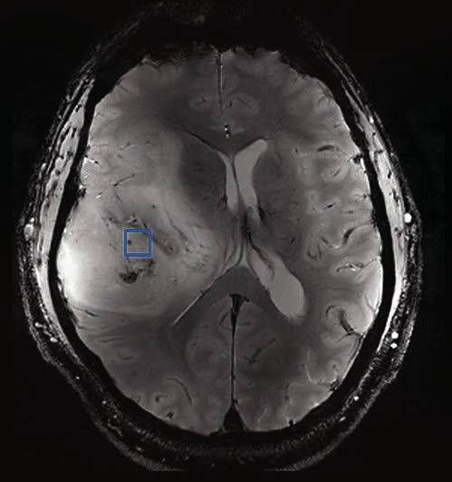
Technology

2

    2 A non-intensity corrected sagittal gradient echo image demonstrating whole brain coverage at 9.4T. Transmit phases of the 16 transmit elements
    were optimized for this slice. An in plane resolution of 280 μm with a slice thickness of 1 mm was obtained within 5 min, 33 s (TR 400 ms, TE 14 ms,
    nominal flip angle 30°).

the most striking differences between                local susceptibility changes is also highly        sequences, such as gradient echoes,
7T and 9.4T are the even shorter RF                  increased, as demonstrated in T2*-                 and to apply advanced techniques like
wave length and the significantly                    weighted BOLD experiments, and effects             TrueFISP and hyperechoes with reduced
increased susceptibility effects. The                related to oriental anisotropy become              SAR. Quantitative comparisons of signal
short wave length and thus locally very              clearly visible.                                   strength, relaxation times and contrast
confined excitation patterns actually                In this report we show examples of dif-            can be found in the referenced papers.
increase the flexibility and effectiveness           ferent imaging techniques and spectros-
of parallel transmit techniques and lead             copy that has been measured during                 Dedicated RF coils for 9.4T
us to the design of transmit arrays with             the last two years at 9.4T. The goal was           In addition to the static magnetic field
16 independent loops. The sensitivity to             to test the feasibility of basic imaging           strength (B0), RF coil performance is a

                                                                                  MAGNETOM Flash · 2/2013 · www.siemens.com/magnetom-world            61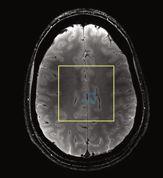
Technology

3A                                                                  3C

3B

  3 T2*-weighted magnitude (3A) and phase (3B) image of the same slice through the brain of a volunteer, acquired at 9.4T. (3C) Susceptibility-
  weighted image from a different volunteer. This image was acquired with a 16-channel microstrip transceiver coil and a spatial resolution of
  0.175 × 0.175 × 1.3 mm3.

dominant factor in determining the sen-          brain with often acceptable homogene-              improved by using a separate array of
sitivity of an MR experiment. The princi-        ity [2], static and dynamic RF shimming            receive coil detectors that closely follows
pal RF engineering challenge at high             approaches are the most promising tech-            the contours of the anatomy [5]. Parallel
B0 field is to achieve homogeneous exci-         niques to mitigate the B1+ field inhomo-           imaging benefits particularly from the
tation across the volume of interest.            geneities. They require an array of trans-         shorter wavelength at ultra-high field
As the wavelength in tissue approaches           mit elements with the possibility to               strength because of reduced inductive
sample dimensions, constructive and              individually control the amplitude and             coupling to the farther coil elements,
destructive interference of the electro-         phase of the current to each of these              resulting in distinct coil sensitivities and
magnetic field causes field concentra-           coil elements. An additional degree of             hence effective sensitivity encoding [6].
tion, but also signal voids, resulting in        freedom can be obtained by arranging               Our approach for human brain imaging
an inhomogeneous MR image [1]. At                the transmit elements in multiple rows             at 9.4T combines separate transmit and
9.4T, even in human brain MRI, severe            to extend the longitudinal coverage and            receive arrays to maximize the receive
field dropouts appear in the lower tem-          more importantly, to correct the inho-             sensitivity together with the ability to
poral lobe and cerebellum if the coil is         mogeneities in all three directions [3].           modulate the transmit field in three
driven in the conventional circularly            While transceiver coils, which combine             dimensions.
polarized (CP) mode, making it a chal-           multi-channel transmit and receive in              The transmit coil consists of 16 elements
lenging task to achieve whole-brain              the same array elements, are a popular             arranged in two rows of eight elements
excitation. While the traveling wave             design for ultra-high field applications [4],      each [3]. The lower row elements are
approach is able to extend the imaged            both signal-to-noise ratio (SNR) and               rotated by 22.5º with respect to the
volume into the lower regions of the             parallel imaging performance can be                upper row and all adjacent coil elements

60 MAGNETOM Flash · 2/2013 · www.siemens.com/magnetom-world
Technology
are inductively decoupled. For reception, r epetition time 28 ms, 21 slices) within variations as in venous blood. In a com-
31 elements are arranged symmetrically around 15 min, using the above parison to images acquired at 3T, an
in 4 rows on a close-fitting helmet for described combination of a 16-channel SNR gain of almost a factor of nine was
maximum sensitivity. A combination of transmit and a 31-channel receive array. found [10]. In addition, the high sensi-
inductive decoupling and geometric Even higher contrast-to-noise ratio can tivity towards susceptibility variations
overlap is used to minimize the induc- be reached by using the image phase as made it possible to distinguish venous
tive coupling between the coil elements completely B1-independent contrast structures at a considerably smaller size.
[7]. The transmit and receive arrays and parameter. Phase images (Fig. 3B) show These data show that anatomical imag-
the final setup are shown in Figure 1. good contrast between gray and white ing with high resolutions, great contrast
Unlike for the closely-coupled trans- matter, but are also able to depict intra- and high image quality is possible at
ceiver arrays, subject-specific adjust- cortical structures or oriented fibers. In 9.4T without the limitations due to SAR
ments of the transmit array are not addition, phase imaging at high fields is or B1-inhomogeneity. Modulations of the
required. Furthermore, the RF circuitry used to detect changes in iron or myelin magnetic susceptibility, especially, can
and low noise preamplifiers required for content [8, 9]. At 9.4T, the high sensitiv- be detected with high accuracy with
the receive elements are closely packed ity of phase imaging makes voxel sizes simple gradient echo sequences at 9.4T.
in the coil housing, thus providing a sim- below 20 nl feasible within reasonable Additionally, the strong susceptibility
ple and fast setup, comparable to clini- scan times of around 20 minutes. effect can help to improve the spatial
cal routine examinations. Due to the Finally, magnitude and phase informa- specificity of spin-echo EPI-based func-
dual row design of the transmit array, tion are combined in susceptibility- tional MRI studies [11].
the entire brain, including the cerebel- weighted imaging (SWI, Fig. 3C) to Successful balanced SSFP imaging is
lum, can be imaged. This is demon- specifically emphasize susceptibility commonly hampered by its prominent
strated in the sagittal image in Figure 2,
for which slice selective static phase
shimming was applied to optimize the
transmit phase.
4
High-resolution GRE, TrueFISP
and susceptibility-weighted
imaging
Due to the high sensitivity towards varia-
tions in the magnetic susceptibility at
9.4T, improved and even novel contrast
mechanisms can be achieved. The pulse
sequences required to exploit these
advantages are based on gradient-echo
techniques and are relatively insensitive
towards SAR limitations and inhomoge-
neities of the transmit field. T2*-weighted
images (Fig. 3A) already show excellent
image contrast and can replace T1 or
T2-weighted imaging techniques that
suffer from serious SAR-limitations and
B1-dependent contrast variations in
many applications. In addition, these
techniques allow for fast acquisition
with parameters that are optimized for
maximum SNR. Thus, using flip angles
close to the Ernst-angle and carefully
adjusting bandwidth and echo time
makes it possible to reach high spatial
resolutions in acceptable scan times.
4 Axial sample image from an isotropic 3D whole brain balanced SSFP scan acquired
The image in Fig. 3A was acquired with at 9.4T (other scan parameters were: 1.5 mm isotropic, 144 × 144 × 96 matrix,
a voxel volume of 40 nl (voxel size TR 1.6 ms, TE 0.6ms, BW 1825 Hz/Pixel, flip angle ~10°, TA 9.5 sec.).
0.2 × 0.2 × 1 mm3, echo time 20 ms,
MAGNETOM Flash · 2/2013 · www.siemens.com/magnetom-world 61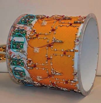
Technology

5A                                                  5B                                                      5C

      5 Maximum Intensity Projections across 40 mm for axial (left) and sagittal multi-slab 3D ToF data sets acquired with FA/TE: (5A) 14°/4.55 ms;
     (5B) 24°/4.55 ms; (5C) 24°/10ms, all from a healthy volunteer.

     sensitivity to local frequency offsets              in vivo applications to about 10°. For            of the flowing blood, allowing it to cross
     (off-resonances) that may lead to pro-              the typical low T2/T1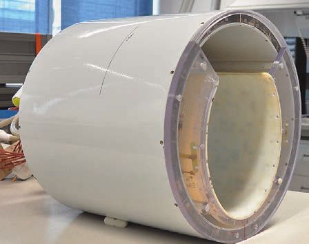
6A
syngo MR B15 sequence was to allow a venous blood in the sagittal sinus
variable duration of the excitation pulse. without substantially compromising
Slab-wise imaging (2.4 cm axial FOV) the visibility of the arteries (Fig. 5C).
with a 3D gradient echo sequence, a TR
of 20 ms, a GRAPPA factor of 4 and 32 Hyperecho imaging
reference lines, and an in-plane voxel Turbo spin echo (TSE, RARE, FSE)
size of 0.5 × 0.5 mm with a slice thick- sequences [17] find widespread applica-
ness of 0.4 mm was used. The echo tion in clinical routine MRI, the main
time (3.8; 4.55; 10 ms), flip-angle (FA reason being that they combine the
15 –36°) and the duration of the excita- diagnostically relevant T2 contrast with
tion pulse (1024–4096 μs) were varied. the robust signal behavior of spin echo
The acquisition time for each slab was refocusing. T2 contrast is highly sensi- 6B
between 1 ½–3 ½ min. tive to a broad variety of pathologies
Post-processing consisted of removing such as inflammation and is an indis-
the scalp signal originating from the pensable tool for examination of various
subcutaneous fat using BET (Brain pathologies of the CNS. Spin echo refo-
Extraction Tool, FSL$), and intensity cor- cusing facilitates a relative insensitivity
rection, obtained after thresholding out to susceptibility and field inhomogene-
the angiographic information and fitting ity, i.e. B0 effects. Additionally, TSE
the background signal with a 2nd order sequences produce a high amount of
polynomial. Maximum Intensity Projec- stimulated echo contributions that are
tions were performed across 40 mm. We T1-weighted. Since T1 >> T2 for most
found that a reasonable flip angle at biological tissues, particularly at ultra-
9.4T is 20–30° to optimize the signal for high fields, these stimulated echo con-
blood that remains within the slab for tributions partially counterbalance the
about 5 excitations. signal loss from refocusing flip angles
The improved contrast-to-background deviating from 180° due to B1 variations.
ratio that we expected was not achieved All in all, these reasons seem to make
with the standard TONE pulse due to TSE sequences the ideal technique for
SAR restrictions. Even in the absence of clinical imaging at (ultra-)high fields.
pulses for the suppression of venous However, a major drawback is the high 6C
blood or magnetization transfer, we RF power deposition caused by the
came close to the SAR limits (95–99%) multiple refocusing RF pulses, which can
with low flip angles of 14–16°. Under lead to severe tissue heating. Thus, the
these conditions, the venous signal was RF power deposition, usually quantified
prominent and the image contrast was in terms of SAR has to be strictly limited.
poor (Fig. 5A). By increasing the dura- Since SAR increases quadratically with
tion of the RF pulses, the flip angle could field strength, the same image acquired
be increased to 24°–32°, yielding an at 9.4T requires almost ten times the SAR
improved image contrast (Fig. 5B). Never- as at 3T. Therefore, the fast multiple refo-
theless there was no room for additional cusing of magnetization with high turbo
venous suppression pulses, not even by factors in modern TSE sequences severely
the reduced flip angle approach that has hampers their use at ultra-high fields.
been successfully introduced at 7T [15]. Common ways of mitigating SAR prob-
At 9.4T, not only the T1 but also the T2 lems include reducing the number of
relaxation times are changed. The T2 in slices, shortening the echo train via a
arterial blood is similar to tissue (about smaller turbo factor, increasing the repe-
40 ms) while in venous blood it is sub- tition time TR, changing the shape of
stantially shorter (5–9 ms dependent on the RF pulses (e.g. using a Gaussian
the fractional oxyhemoglobin content shaped pulse) and increasing the dura- 6 Three out of five slices from the
[16]). In line with these observations we tion of the RF pulses. No matter which yperTSE experiment performed at 9.4T
h
in Tübingen.
found that increasing the TE from 4.5 (combination of) option(s) is chosen,
to 10 ms was sufficient to suppress the some compromise has to be made in
MAGNETOM Flash · 2/2013 · www.siemens.com/magnetom-world 63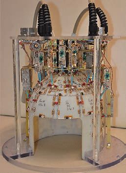
Technology
7A 7B 7C
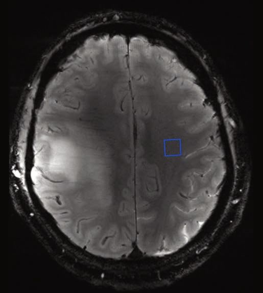
7 Localizer (7A), single spectrum (7B) and all the spectra within the VOI (7C) acquired from the brain of a healthy volunteer. Localizations of VOI
and single spectrum were marked with yellow and blue squares respectively.
terms of SNR, acquisition time, volume Whereas the Hyperecho-scheme makes suboptimal image and SAR properties.
coverage and image quality. use of symmetry relations between the Basically, there are two recipes to
Another effective means to reduce SAR flip angles to reinstate the full signal at calculate suitable flip angles for TRAPS:
is to lower the refocusing flip angle in a some given point of time (echo) within 1. Setting the flip angles directly via
TSE sequence. However, for conven- the TSE train [18], TRAPS allows an some support points defined by func-
tional TSE sequences with constant refo- almost free variation of refocusing flip tions, etc. This AUTO-TRAPS approach
cusing flip angles, this method results in angles along the TSE train after a pre- has the advantage that the flip angle
lower SNR and changes the contrast of paration of magnetization close to the courses can be adapted in a flexible
the resulting images, particularly in case so-called static pseudo steady state way to consider protocol changes
of the vast flip angle reductions that are (SPSS) [19, 22, 23]. In the SPSS, the made by the user, and the AUTO-
necessary for TSE imaging at 9.4T. echo intensity always stays close to the TRAPS recipe allows a quite direct
Methods that employ variable instead of optimal value that can be reached for a control of SAR [21].
constant refocusing flip angles along the given flip angle. Any loss of coherence 2.More sophisticated refocusing flip
TSE train have been introduced in order would lead to signal fluctuations and angle schemes can be obtained by
to allow SAR reductions while maintain- inevitable signal loss, resulting in image calculating the flip angles from a pre-
ing a T2-like contrast and high SNR. artifacts. Since the variation of signal in defined signal course or echo inten-
These novel TSE sequences are usually k-space directly influences the point sity course along the echo train [24].
referred to as hyperTSE-, Hyperecho- spread function (PSF), flip angle varia- This method has the advantage that
TSE-, and smooth transitions between tions can also be used to improve the dedicated image properties can be set
pseudo steady states (TRAPS)-sequences PSF. Thus, TRAPS, which offers the via the predefined signal course [24].
[18, 19, 20, 21]. Generally, high signal unique possibility to vary flip angles in a However, there is no unique solution
for the echoes encoded for the center of flexible way in hyperTSE sequences, for the flip angles and it is more
k-space is maintained, thus, warranting allows both considerable SAR savings as complex to adapt the settings to any
a high SNR for the reconstructed image. well as the optimization of image prop- protocol changes made by the user.
The signal in the peripheral parts of erties for TSE [19, 20, 21, 24]. For our proof of concept study at 9.4T,
k-space may be reduced slightly due to A great emphasis in TRAPS lies on the a flexible AUTO-TRAPS approach was
lower flip angles; however, strong stimu- flip angle train, which has to be properly used [21]. The TRAPS based flip angle
lated echo contributions still maintain adapted to any changes of the protocol method was implemented in a self-made
sufficient signal levels at ultra-high settings that influence the timing or the TSE sequence within the IDEA platform
fields due to T1 >> T2, as mentioned view ordering / reordering of the TSE syngo MR B15. A T2-weighted TSE proto-
above. sequence. ‘Unwise variations’ can lead to col with the following parameters was
64 MAGNETOM Flash · 2/2013 · www.siemens.com/magnetom-worldTechnology
set up: FOV 220 × 175 mm2, 5 slices with sition was preceded by an Actual Flip Fig. 8A. MR scanning showed the pres-
1 mm slice distance, voxel size angle Mapping (AFI) scan [25, 26]. ence of an extended tumorous mass
0.58 × 0.58 × 1.0 mm3, turbo factor 17, Figure 7 shows spectra acquired from a in the right hemisphere, pressing on
TE 48 ms, TR 8.94 s. Due to additional healthy volunteer (31 years old). In this the cerebral midline that was deviated
T1-weighted stimulated echo contribu- case, an 8-channel transmit coil com- towards the left hemisphere. In the
tions, hyperTSE sequences (as all low bined with a 24-channel receive array antero-posterior direction the tumor
flip angle TSE sequences) show a was used for data acquisition. Here, the extended from the frontal to the parietal
reduced T2 contrast compared to a TSE spectra were acquired from a superior lobe, and in the cranio-caudal direction
180° sequence at a given echo time. region of the human brain. The VOI (yel- from the hand area in the primary motor
In order to compensate for this effect, low square, Fig. 7A) was placed in the cortex, across the lateral fissure into the
TE was increased from the originally axial plane, parallel to the line between temporal cortex. The tumor texture was
desired value [20]. Here, please note the anterior and posterior commissures heterogenous and showed calcifications,
that T2 relaxation times are considerably (ac-pc line). A typical spectrum from confirmed by CT scans, indicating an
shorter at 9.4T than at 3T or even 1.5T. healthy brain tissue (localization within oligodendroglioma.
Thus, a comparatively low TE already the VOI marked with blue square) is pre- Here all the CSI data were collected with
gives a substantial T2 contrast. sented in Fig. 7B. The following brain a 16-channel transmit coil combined
The used hyperTSE protocol demon- metabolites have been identified: myo- with a 31-channel receiving helmet.
strated a relative SAR of 33.7%, i.e. a Inositol (Ins, marked with 1), methylene Spectra were collected at two different
SAR saving of 66.3% compared to a con- (CH2) and methyl groups (CH3) of cre- locations: within the lesion (Fig. 8A),
ventional TSE sequence. This allowed the atine and phosphocreatine (Cr and PCr, and contralaterally, in healthy tissue
acquisition of 5 slices instead of only 1. #2), overlapped peaks of glutamine and (Fig. 8B). Analysis of the acquired spec-
Fig. 6 shows the first hyperTSE images glutamate (Gln and Glu, #3), taurine tra revealed a strong decrease in the sig-
from a patient at 9.4T. The used AUTO- (Tau, #4), choline containing com- nal of NAA and an increase in signals of
TRAPS based hyperTSE demonstrates pounds (tCho, #5), aspartate (Asp, #6), tCho, Glu, Gln, and Ins within the lesion
very good imaging behavior and sug- N-acetylaspartate (NAA, #7), Gln (#8), (Fig. 8A) compared to the contralateral
gests that hyperTSE sequences are Glu (#9), γ-Aminobutryic acid (GABA, side (Fig. 8B). Observations made here
highly suitable for acquiring ultrahigh #10), N-acetylaspartylglutamate (NAAG, are in agreement with the previous clini-
resolution images at 9.4T that offer #11) and macromolecules (#12). cal studies performed at lower magnetic
promising diagnostic value. Improved spectral resolution and SNR fields (1.5 and 3T) [28, 29]. Further-
make it possible to differentiate between more, our results suggest an increase in
Proton spectroscopy in metabolites which are typically over- signals of Tau and scyllo-inositol (Scyllo)
patients and volunteers lapped at lower field strengths, particu- within the tumor tissue (Figs. 8A and 8B,
Chemical shift imaging (CSI) data were larly the resonances between ~4 and marked with yellow arrow). According to
collected with a Stimulated Acquisition ~3.2 ppm, and ~2.9 and 2.1 ppm (Ins, the latest research those play an impor-
Mode sequence (STEAM) with the fol- Glu, Gln, GABA, NAA and NAAG). Those tant role in distinction between different
lowing parameters: TE 20 ms; TR 2000 findings are in agreement with results tumor types or grades [32, 33] and may
ms; TM 11 ms; FOV 160 × 160 mm2; vol- described in previous studies [27, 28]. serve as prognostic markers [34]. How-
ume of interest (VOI) 60 × 60 × 10 mm3 Analysis of the spectra acquired from ever, the accurate detection of Scyllo
and 40 × 40 × 10 mm3 (volunteer and the entire VOI (Fig. 7C) shows that the and Tau with 1H MRS at lower magnetic
patient respectively); voxel size 10 mm3 spectra from the central 4 × 4 voxel field strengths could be difficult as they
isotropic; spectral bandwidth 4000 Hz. region have excellent quality. Special- are strongly overlapped with the neigh-
In order to keep the reference voltage ized sequences [29, 30] and parallel boring resonances of tCho and Ins.
below the hardware limits, the 90° transmit techniques [31] will further
h-sinc RF pulses (sinc pulses with 4 side improve the spectra by reducing the Summary
lobes) within the standard STEAM chemical shift displacement, which is Although inhomogeneity in B1 and B0 is
sequence had to be replaced by 90° her- still significant for some of the peaks further increased at 9.4T compared to
mite pulses. Additionally, their RF band- (tCho, Cr and NAA), as well the depen- 7T resulting variations in signal strength
width has been increased to 3100 Hz. dence of the signal amplitude on the across the image is acceptable and com-
This allowed minimizing the influence of spatially inhomogeneous transmit field. parable to 7T. We currently investigate
chemical shift displacement. In order to Spectra acquired from the brain of a the possible gain in SNR going from
determine the reference voltage neces- 43-year-old patient with clinically con- 3T to 7T and to 9.4T, and preliminary
sary for the STEAM sequence, CSI acqui- firmed oligodendroglioma are shown in results indicate the expected linear
MAGNETOM Flash · 2/2013 · www.siemens.com/magnetom-world 65Technology
8A
8B
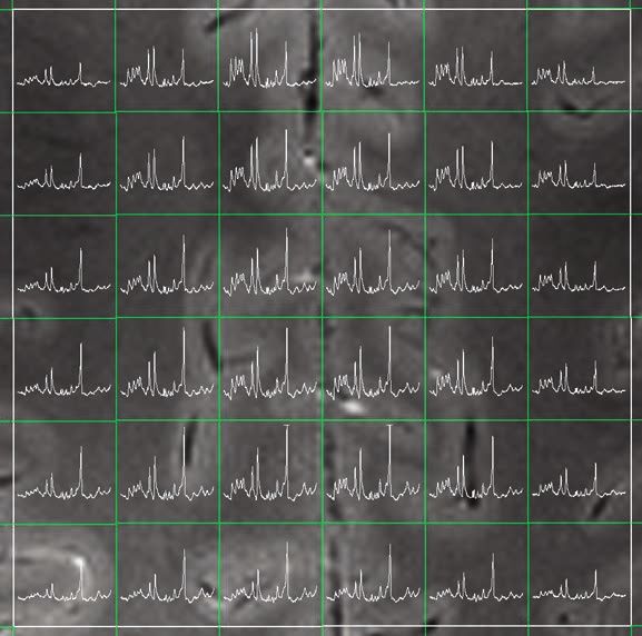
8 Localizer and single spectrum acquired from the lesion (8A) and from the contralateral side (8B). Blue squares denote the localization of the
spectra whereas the yellow arrows indicate the position of Tau and Scyllo.
increase. However, a solid comparison is of the most important steps in the near Acknowledgement
quite difficult due to very different coil future in our lab will be the implementa- Dr. Mathias Nittka, Siemens Healthcare,
geometries and technologies used at tion and application of parallel transmit Erlangen.
these different field strengths. concepts and corresponding RF coil
§
The 7T and 9.4T systems are investigational devices.
Macroscopic homogeneous B1 and B0 technology, as well as systems to
The products mentioned herein are still under
fields are the prerequisite for the study improve and control B0 homogeneity development and not commercially available yet.
of microscopic susceptibility changes such as dynamic shimming with high- Their future availability cannot be ensured.
$
Brain Extraction Tool, FSL is not a Siemens Healthcare
within tissue, which are significantly order shim inserts.
Product. Siemens bears no responsibility for this prod-
enhanced at ultra-high fields. Thus, one uct including, but not limited to, its regulatory status.
66 MAGNETOM Flash · 2/2013 · www.siemens.com/magnetom-worldTechnology
References:
14 Cerebral TOF Angiography at 7T: Pushing the 29 Henning A, Fuchs A, Murdoch JB, Boesinger P.
1 Van de Moortele PF, Akgun C, Adriany G,
Limits to Reap the Benefi ts of Ultra-High Field Slice-selective FID acquisition, localized by outer
Moeller S, Ritter J, Collins CM, Smith MB,
Imaging, Issue 49 of MAGNETOM Flash · 1/2012 volume suppression (FIDLOWS) for 1H MRSI of
Vaughan JT and Uğurbil K. B1 Destructive
· www.siemens.com/magnetom-world by: the human brain at 7 T with minimal signal loss.
Interferences and Spatial Phase Patterns at 7T
Schmitter S, Wu X, Pfeuffer J, Hamm M, Uğurbil NMR Biomed 2009; 22:683-696.
with a Head Transceiver Array Coil. Magn Reson
K, Van de Moortele PF. 30 Boer VO, van Lier ALHMW, Hoogduin JM, Wijnen
Med 2005; 54:1503-1518.
15 Johst S, Wrede KH, Ladd ME, Maderwald S. JP, Luijten PR, Klomp DWJ. 7-T 1H MRS with
2 Hoffmann J, Shajan G, Budde J, Scheffler K,
Time-of-Flight magnetic resonance angiography adiabatic refocusing at short TE using radiofre-
Pohmann R: Human Brain Imaging at 9.4 Tesla
at 7 T using venous saturation pulses with quency focusing with a dual-channel volume
Combining a Tunable Patch Antenna for
reduced flip angles. Invest Radiol 2012;47: transmit coil. NMR Biomed 2011; 24)1038-1046.
Transmission. Magn. Reson. Med., ePub ahead.
445-450. 31 Hetherington HP, Avdievich NI, Kuznietsov AM,
DOI: 10.1002/mrm.24367 (2012).
16 Lee S-P, Silva AC, Uğurbil K, Kim SG. Diffusion- Pan JW. RF shimming for spectroscopic localiza-
3 Shajan G, Hoffmann J, Scheffler K, and
weighted spin-echo fMRI at 9.4T: Microvascular/ tion in the human brain at 7T. Magn Reson Med
Pohmann R. A 16-Element Dual-row Transmit
Tissue contribution to BOLD signal changes. 2010; 63:9-19.
Coil Array for 3D RF Shimming at 9.4T. In:
Magn Reson Med 1999;42:919-928. 32 Rijpkema M, Schuuring J, van der Meulen Y,
Proceedings of the 20th Annual Meeting of
17 Hennig J, Nauerth A, et Friedburg H: RARE van der Graaf M, Bernsen H, Boerman R,
ISMRM, Melbourne, 2012, p 308.
imaging: a fast imaging method for clinical MR, van der Kogel A, Heerschap A. Characterization
4 Shajan G, Hoffmann J, Budde J, Adriany G,
Magn Reson Med 1986; 3: 823-833. of oligodendrogliomas using short echo
Uğurbil K, Pohmann R: Design and Evaluation
18 Hennig J and Scheffler K, Hyperechoes, Magn time 1H MR spectroscopic imaging. NMR
of an RF Front-End for 9.4 T Human MRI. Magn.
Reson Med 2001; 46:6-12. Biomed, 2003; 16:12-18.
Reson. Med. 66, 594-602 (2011).
19 Hennig J, Weigel M, Scheffler K. Multiecho 33 Chawla S, Oleaga L, Wang S, Krejza J, Wolf RL,
5. Wiggins GC, Triantafyllou C, Potthast A,
sequences with variable refocusing flip angles: Woo JH, O’Rourke DM, Judy KD, Grady MS,
Reykowski A, Nittka M and Wald LL. 32-Channel
Optimization of signal behavior using smooth Melhem ER, Poptani H. Role of proton magnetic
3 Tesla Receive-Only Phased Array Head Coil
transitions between pseudo steady states resonance spectroscopy in differentiating
with Soccer-Ball Element Geometry. Magn
(TRAPS). Magn Reson Med 49: 527-535 (2003). oligodendrogliomas from astrocytomas.
Reson Med 2006; 56:216-223.
20 Weigel M and Hennig J. Contrast Behavior and J Neuroimaging, 2010; 20:3-8.
6 Wiesinger F, Van de Moortele PF, Adriany G,
Relaxation Effects of Conventional and Hyper- 34 Wilson M, Cummins CL, MacPherson L,
Zanche ND, Ugurbil K and Pruessmann KP.
echo-Turbo Spin Echo Sequences at 1.5T and 3T. Sun Y, Natarajan K, Grundy RG, Arvanitis TN,
Potential and feasibility of parallel MRI at high
Magn Reson Med 55: 826-835 (2006). Kauppinen RA, Peet AC. Magnetic resonance
field. NMR Biomed 2006; 19:368-378.
21. Weigel M and Hennig J. Development and Opti- spectroscopy metabolite profiles predict survival
7 Shajan G, Hoffmann J, Scheffler K and Pohmann
mization of T2 weighted Methods with Reduced in pediatric brain tumors. Eur J Cancer, 2013;
R. A 31-Element Receive Array for Human Brain
RF Power Deposition (Hyperecho-TSE) for 49:457-464.
Imaging at 9.4 T. No. 351, Proceedings of the
Magnetic Resonance Imaging. Z Med Phys 18:
2012 ESMRMB Annual Meeting.
151-161 (2008).
8 G. A. Lodygensky, J. P. Marques, R. Maddage, E.
22 Alsop DC. The sensitivity of low flip angle RARE Contact
Perroud, S. V. Sizonenko, P. S. Hüppi, R. Gru-
imaging. Magn Reson Med. 1997; 37:176-84. Professor Klaus Scheffler, Ph.D.
etter: In vivo assessment of myelination by
23 Hennig J, Scheffler K, Easy Improvement of MRC Department
phase imaging at high magnetic field. Neuroim-
Signal-to-Noise in RARE sequences with Low Max Planck Institute for Biological
age 59, 1979-1987 (2012).
Refocusing Flip Angles Magn Reson Med. 2000; Cybernetics
9 M. Fukunaga, T.Q. Li , P. van Gelderen, J.A. de
983-985. Department of Biomedical Magnetic
Zwart, K. Shmueli, B. Yao, J. Lee, D. Maric, M.A.
24 Hennig J, Weigel M, Scheffler K. Calculation of Resonance
Aronova, G. Zhang, R.D. Leapman, J.F. Schenck,
Flip Angles for Echo Trains With Predefined Center for Integrative Neuroscience,
H. Merkle, J.H. Duyn: Layer-specific variation
Amplitudes With the Extended Phase Graph CIN University of Tübingen
of iron content in cerebral cortex as a source of
(EPG)-Algorithm: Principles and Applications to Spemannstr. 41
MRI contrast. Proc. Natl. Acad. Sci. USA 107,
Hyperecho and TRAPS Sequences. Magn Reson 72076 Tübingen
3834–3839 (2010).
Med 51: 68-80 (2004). Germany
10 J. Budde, G. Shajan, J. Hoffmann, K. Uğurbil, R.
25 Yarnykh VL. Optimal radiofrequency and gradi- Phone: +49 (0)7071 601-700/701
Pohmann: Human Imaging at 9.4 T Using T2*-,
ent spoiling for improved accuracy of T1 and B1 Fax: +49 (0)7071 601-702
Phase- and Susceptibility-Weighted Contrast.
Magnetic Resonance in Medicine 65, 544-550 measurements using fast steady-state tech- klaus.scheffler@tuebingen.mpg.de
(2011). niques. Magn Reson Med, 2010; 57(1):192-200.
11 Budde J, Shajan G, Zaitsev M, Scheffler K, 26 Pohmann R, Scheffler K. A theoretical and
Pohmann R: Functional MRI in human subjects experimental comparison of different tech-
with gradient-echo and spin-echo EPI at 9.4 T. niques for B1 mapping at very high fields.
Magn. Reson. Med., in press (2013). NMR Biomed, 2012; e-pub ahead of print.
12 Conolly D, Nishimura D, Macovski A, Glover G. 27 Avdievich NI, Pan JW, Baehring JM, Spencer DD,
Variable-rate selective excitation. J Magn Reson Hetherington HP. Short echo spectroscopic
1988;78:440-477. imaging of the human brain at 7T using trans-
13 Schmitter S, Bock M, Johst S, Auerbach EJ, ceiver arrays. Magn Reson Med 2009; 62:17-25.
Uğurbil K, Van de Moortele PF. Contrast en- 28 Deelchand DK, Van de Moortele PF, Adriany G,
hancement in TOF cerebral angiography at 7 T Iltis I, Andersen P, Strupp JP, Vaughan T, Uğurbil
using saturation and MT pulses under SAR K, Henry PG. in vivo 1H NMR spectroscopy of
constraints: impact of VERSE and sparse pulses. the human brain at 9.4 T: initial results. J Magn
Magn Reson Med 2012;68(1):188-197. Reson 2010; 206:74-80.
MAGNETOM Flash · 2/2013 · www.siemens.com/magnetom-world 67You can also read