Integrated Bacterial and Fungal Diversity Analysis Reveals the Gut Microbial Alterations in Diarrheic Giraffes
←
→
Page content transcription
If your browser does not render page correctly, please read the page content below
ORIGINAL RESEARCH
                                                                                                                                            published: 12 August 2021
                                                                                                                                      doi: 10.3389/fmicb.2021.712092

                                             Integrated Bacterial and Fungal
                                             Diversity Analysis Reveals the Gut
                                             Microbial Alterations in Diarrheic
                                             Giraffes
                                             Aoyun Li 1,2† , Bingxian Liu 3† , Feiran Li 2 , Yuanyuan He 2 , Lei Wang 4 ,
                                             Muhammad Fakhar-e-Alam Kulyar 2 , Huade Li 5 , Yuhang Fu 2 , Huaisen Zhu 2 ,
                                             Yaping Wang 2 and Xiong Jiang 1*
                                             1
                                              Hubei Three Gorges Polytechnic, Yichang, China, 2 College of Veterinary Medicine, Huazhong Agricultural University,
                                             Wuhan, China, 3 College of Veterinary Medicine, South China Agricultural University, Guangzhou, China, 4 Animal Husbandry
                                             Station of Bijie City, Bijie, China, 5 Sichuan Academy of Grassland Science, Chengdu, China

                                             Gut microbiota has been demonstrated to be associated with multiple gastrointestinal
                                             diseases, but information regarding the gut microbial alternations in diarrheic giraffe
                          Edited by:         remains scarce. Here, 16S rDNA and ITS gene amplicon sequencing were conducted
                Ernesto Perez-Rueda,
      Universidad Nacional Autónoma
                                             to investigate the gut microbial composition and variability in diarrheic giraffes. Results
                   de México, Mexico         demonstrated that Firmicutes and Proteobacteria were the most dominant phyla in the
                       Reviewed by:          gut bacterial community, whereas Ascomycota and Basidiomycota were observed to
                 Sonia Dávila-Ramos,
                                             be predominant in the gut fungal community regardless of health status. However,
    Universidad Autónoma del Estado
                  de Morelos, Mexico         the species and relative abundance of preponderant bacterial and fungal genera in
       Mario Alberto Martínez Núñez,         healthy and diarrheic giraffes were different. In contrast to the relatively stabilized gut
     Universidad Nacional Autónoma
                   de México, Mexico
                                             fungal community, gut bacterial community displayed a significant decrease in the
                     *Correspondence:
                                             alpha diversity, accompanied by distinct changes in taxonomic compositions. Bacterial
                            Xiong Jiang      taxonomic analysis revealed that the relative abundances of eight phyla and 12 genera
                  jiangx1025@sina.com
                                             obviously increased, whereas the relative abundances of two phyla and eight genera
     † These   authors have contributed
                                             dramatically decreased during diarrhea. Moreover, the relative richness of five fungal
                    equally to this work
                                             genera significantly increased, whereas the relative richness of seven fungal genera
                   Specialty section:        significantly declined in diarrheic giraffes. Taken together, this study demonstrated that
         This article was submitted to
         Microorganisms in Vertebrate
                                             diarrhea could cause significant alternations in the gut microbial composition of giraffes,
                    Digestive Systems,       and the changes in the gut bacterial community were more significant than those in
               a section of the journal      the gut fungal community. Additionally, investigating the gut microbial characteristics of
             Frontiers in Microbiology
                                             giraffes in different health states is beneficial to provide a theoretical basis for establishing
           Received: 19 May 2021
           Accepted: 16 July 2021            a prevention and treatment system for diarrhea from the gut microbial perspective.
         Published: 12 August 2021
                                             Keywords: gut microbiota, diarrhea, 16S rDNA, ITS, giraffe
                               Citation:
        Li A, Liu B, Li F, He Y, Wang L,
  Fakhar-e-Alam Kulyar M, Li H, Fu Y,
   Zhu H, Wang Y and Jiang X (2021)
                                            INTRODUCTION
       Integrated Bacterial and Fungal
    Diversity Analysis Reveals the Gut
                                            Ruminant intestines are colonized by trillions of microbes which play crucial roles in immune
      Microbial Alterations in Diarrheic    system maturation, intestinal epithelial mucosal barrier maintenance, metabolism, and nutrient
Giraffes. Front. Microbiol. 12:712092.      absorption (Garrett et al., 2010; Arumugam et al., 2011; Tremaroli and Backhed, 2012;
    doi: 10.3389/fmicb.2021.712092          Backhed et al., 2015). Statistical analysis indicates that approximately 98% of intestinal microbes

Frontiers in Microbiology | www.frontiersin.org                                    1                                         August 2021 | Volume 12 | Article 712092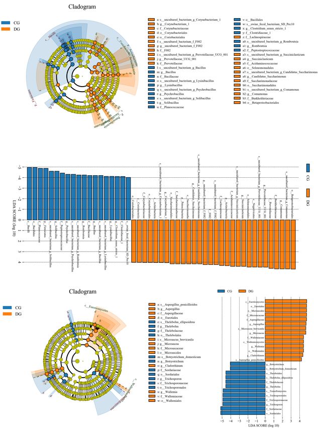
Li et al.                                                                                                       Gut Microbiota in Diarrheic Giraffes

are bacteria, whereas the remaining contains fungi (about 0.1%),          This species has been introduced into zoos around the world but
protozoa, and viruses (Garrett et al., 2010; Tremaroli and                showed a high diarrheic rate due to the alterations in habitat
Backhed, 2012; Backhed et al., 2015). These microbes display              and survival environment (Brown et al., 2007; Mulherin et al.,
a symbiotic relationship with the host through complicated                2008; AlZahal et al., 2016). However, to date, the relationship
networks of interactions and crosstalk with each other (Aziz              between gut microbial composition and diversity and diarrhea
et al., 2013; Guo et al., 2015). Remarkably, some potentially             in giraffes is still unclear. Taking advantage of this gap, we
pathogenic microorganisms may also inhabit as parts of normal             investigated the composition and variability of gut bacterial and
gut microbiota but may take opportunity to cause disease, e.g.,           fungal communities in the healthy and diarrheal giraffes by 16S
gut microbial alteration and immune dysregulation of host (Aziz           rDNA and ITS2 amplicon sequencing.
et al., 2013; Guo et al., 2015). Gut microbial community evolves
with host and has become a vital organ affecting its health (Cryan
and Dinan, 2012; Zhang et al., 2020). Several studies provided            MATERIALS AND METHODS
supporting evidence that shifts in the gut microbial composition
that could extend its adverse effects beyond the gastrointestinal         Sample Acquisition
system and affect the functions of extra-intestinal organs, such as       In this study, a total of twelve 4-year-old giraffes inhabiting in
the brain and liver (Zhang et al., 2020). Gut microbial community         Yichang zoo (Yichang, China) were used for sample collection,
has been demonstrated to be involved in the development of                including six healthy giraffes and six diarrheic populations.
multiple diseases such as colorectal cancer, diabetes, obesity,           Additionally, the proportion of male and female in both
and dyspepsia (Delzenne et al., 2011; Musso et al., 2011;                 groups was 1:1. These giraffes were maintained under the
Ambalam et al., 2016). Although gut bacterial importance has              same conditions and possessed the same immune procedure.
been well demonstrated by numerous studies, analyses regarding            Prior to the sample acquisition, all the giraffes were observed
the relationship between gut fungal communities and host health           and diagnosed by professional veterinarians to determine their
have been insufficient to date.                                           health status and diarrheic populations without any medicinal
    Diarrhea is one of the leading causes of decreased productivity       treatment. Sufficient feed and water were provided ad libitum for
and death in ruminants that has been considered a vital factor            all giraffes throughout the experimental period. One day prior to
impeding animal husbandry development in many countries                   sample acquisition, healthy and diarrheic giraffes were placed in
(Musso et al., 2011; Ambalam et al., 2016). Early investigations          separate pens to prevent infection and sample contamination. Six
revealed that diarrhea was present in almost all ruminants and            individual fresh fecal samples were collected from each giraffe via
especially epidemic in neonatal goat, sheep, cattle, and yak with         using sampler the following morning. Subsequently, the obtained
immature gastrointestinal tract, which caused approximately half          rectal feces were resampled from the intermediate portion to
of all ruminant deaths (Li et al., 2018; Bu et al., 2020; Xue             minimize pollution through bedding and flooring. Finally, all
et al., 2020). Previous research has indicated that some intestinal       the samples were placed immediately in sterile plastic containers,
microbes including bacteria and fungi of ruminants alternate              snap-frozen using liquid nitrogen and stored at −80◦ C for
between preponderant and weak populations accompanied by                  subsequent DNA extraction.
diarrheic symptoms (Yang et al., 2017; Wang et al., 2018; Wang
et al., 2019a). Therefore, some inevitable associations may be            16S rDNA and ITS Gene Amplification
present between alternations in gut microbial community and               and Sequencing
diarrhea, but its specific connections and laws remain to be              The bacterial and fungal genomic DNA was extracted from feces
determined. Previously, numerous studies were performed on                of giraffes using QIAamp DNA Mini Kit (QIAGEN, Hilden,
pathogenic bacteria, parasite, and virus to reveal the cause of           Germany) following suggested instructions of manufacturer.
ruminant diarrhea (Gallardo et al., 2020; He et al., 2020). Recent        The total microbial DNA was quantified via utilizing UV-Vis
studies on gut microbiota of goat, yak, and mice have provided            spectrophotometer (NanoDrop 2000, United States), and DNA
evidence that gut microbial dysbiosis may be one of the reasons           integrity was evaluated by 0.8% agarose gel electrophoresis.
of diarrhea (Han et al., 2017; Shao et al., 2020).                        Bacterial 16S rDNA (338F: ACTCCTACGGGAGGCAGCA
    Metagenomic analysis based on high-throughput sequencing              and 806R: GGACTACHVGGGTWTCTAAT) and fungal ITS
technology is an efficient tool for characterizing gut microbial          gene primers (ITS5F: GGAAG TAAAAGTCGTAACAAGG and
composition and diversity differences after suffering certain             ITS2R: GCTGCGTTCTTCATCGA TGC) with special barcodes
diseases and has made possible in systematically investigating            were synthesized on the basis of the conservative regions to
the relationship between host diseases and gut microbiota (Han            amplify the V3/V4 and ITS2 regions, respectively. To ensure
et al., 2017; Shao et al., 2020). Moreover, in-depth comparison           the accuracy of the results, PCR amplification was conducted
and analysis of obtained gut microbial information contributed            in triplicates under the same conditions. The agarose gel
to further understand the mechanisms causing ill health and               electrophoresis (2%) and AxyPrep DNA Gel Extraction Kit
develop the strategies to minimize the collateral damage (Han             (Axygen, Union City, CA, United States) was employed to
et al., 2017; Shao et al., 2020). Giraffe (Giraffa camelopardalis)        evaluate PCR amplification product and recycle target fragment,
mainly inhabiting the African continent is the tallest terrestrial        respectively. PCR-recycled products were performed fluorescent
ruminant in the world and displays a complicated gastrointestinal         quantitation on Microplate reader (BioTek, FLx800) based on
microbial ecosystem (Brown et al., 2007; AlZahal et al., 2016).           the original electrophoretic results. According to the fluorescence

Frontiers in Microbiology | www.frontiersin.org                       2                                   August 2021 | Volume 12 | Article 712092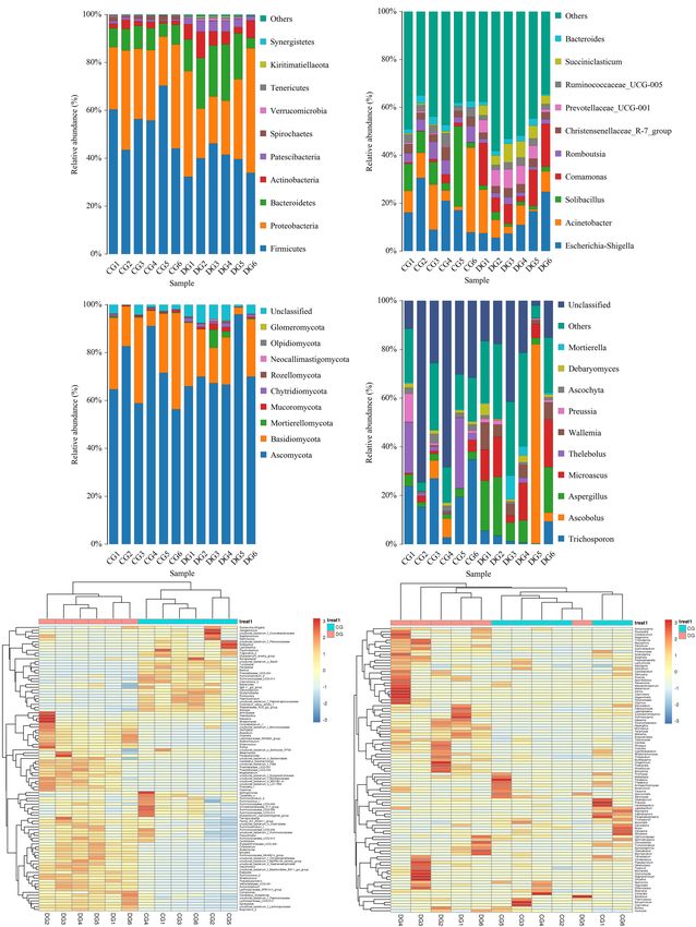
Li et al. Gut Microbiota in Diarrheic Giraffes
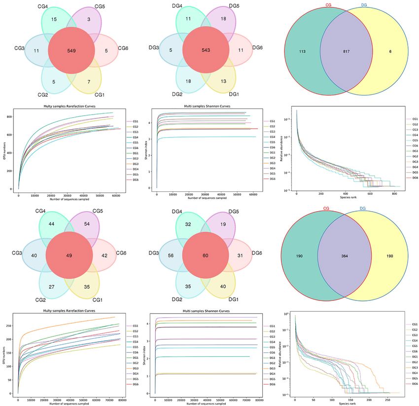
quantitative results and the requirements of sequencing quantity, flat and showed a tendency to saturate characteristics, indicating
the samples were mixed in corresponding proportions. The that nearly all the bacterial and fungal species were identified
obtained PCR products were used to construct the sequencing in fecal samples of giraffes (Figures 1D,E,J,K). Furthermore, the
library via using TruSeq Nano DNA LT Library Prep Kit rank abundance curves for all samples were wide and fell gently,
(Illumina, San Diego, CA, United States). Meanwhile, the 2% suggesting satisfactory evenness and abundance (Figures 1F,L).
agarose gel electrophoresis was used for the final fragment
selection and purifying the library. The quality inspection and Shifts in Gut Microbial Diversities With
fluorescence quantification of the sequencing libraries were
the Effect of Diarrhea
performed before sequencing. The qualified libraries were diluted
To further dissect the alternations of gut bacterial and fungal
and then mixed in corresponding proportions based on the
communities in diarrheic giraffe, the qualified sequences were
required sequencing quantity. The final libraries were subjected
aligned to estimate alpha and beta diversity indices. Gut bacterial
to high-throughput sequencing via MiSeq sequencing machine.
and fungal alpha diversity could be characterized by sequencing
depth (Good’s coverage), species abundance (ACE), and species
Bioinformatics and Statistical Analysis diversity (Shannon and Simpson). Good’s coverage estimates
The original data produced from high-throughput sequencing
varied from 99.79 to 99.96%, suggesting that the majority
were performed by quality screening using QIIME software
of bacterial and fungal phenotypes presented in each sample
(Qiime1.9.1) to achieve reliable results in the subsequent
were detected (Figures 2A,E). There was statistically significant
bioinformatics analysis. The interrogative sequences including
differences in the gut bacterial ACE (834.53 ± 56.67 versus
short sequences (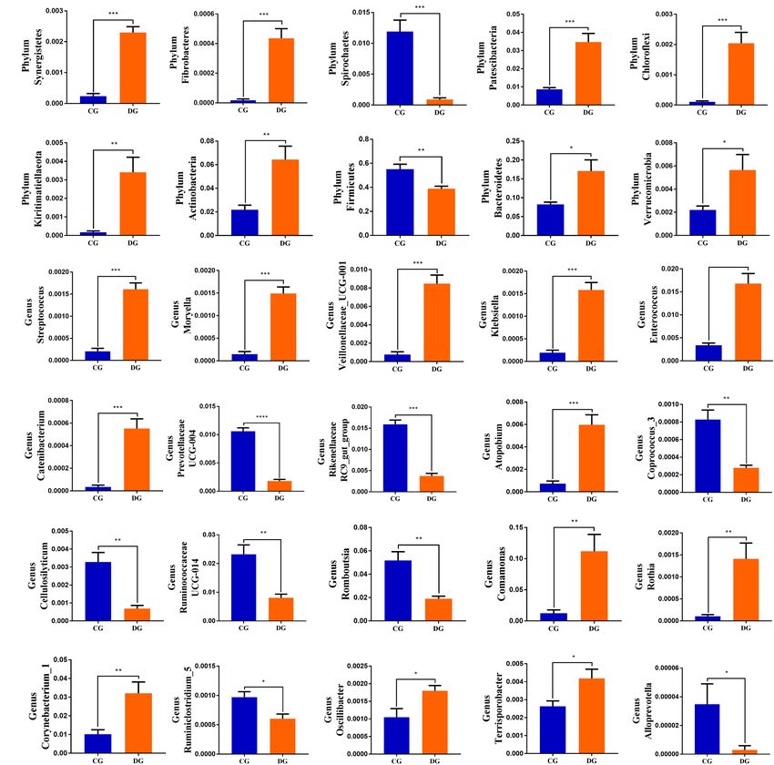
Li et al. Gut Microbiota in Diarrheic Giraffes
TABLE 1 | The bacterial sequence information of each sample.
Sample Raw reads Clean reads Effective reads AvgLen (bp) GC (%) Effective (%)
CG1 79,898 79,389 70,069 422 53.25 87.7
CG2 79,747 79,277 68,993 422 53.65 86.51
CG3 79,912 79,401 68,577 420 53.05 85.82
CG4 80,066 79,565 70,631 418 53.71 88.22
CG5 79,912 79,286 70,064 424 53.28 87.68
CG6 80,125 79,660 69,945 420 52.79 87.29
DG1 79,811 79,378 69,500 421 54.07 87.08
DG2 79,677 79,196 71,013 419 54.38 89.13
DG3 79,983 79,528 72,110 418 54.42 90.16
DG4 79,699 79,288 72,501 419 54.2 90.97
DG5 80,049 79,603 72,133 419 54.21 90.11
DG6 79,983 79,497 71,908 421 54.6 89.90
TABLE 2 | The fungal sequence information of each sample.
Sample Raw reads Clean reads Effective reads AvgLen (bp) GC (%) Effective (%)
CG1 80,228 79,749 79,352 228 44.76 98.91
CG2 80,062 79,593 79,557 251 50.72 99.37
CG3 79,851 79,325 79,310 230 44.32 99.32
CG4 79,832 78,434 74,738 255 50.45 93.62
CG5 80,134 79,617 79,494 237 46.7 99.20
CG6 80,282 79,841 79,836 231 46.01 99.44
DG1 80,110 79,630 78,957 237 47.03 98.56
DG2 79,829 79,269 79,242 251 49.41 99.26
DG3 79,679 79,155 79,060 243 47.78 99.22
DG4 80,093 79,569 79,500 246 47.59 99.26
DG5 79,911 79,085 79,057 256 42.15 98.93
DG6 80,339 79,787 79,772 240 48.26 99.29
effect of diarrhea on taxonomic compositions, 194 genera were eight bacterial genera dramatically decreased under the influence
detected from the gut microbiota of giraffes. Among these of diarrhea (Figure 4B). Considering that this discriminant
genera identified, Escherichia-Shigella (16.98%) was the most analysis cannot distinguish the dominant taxon, linear
predominant bacterial genus in the CG group, followed by discriminant analysis effect size (LEfSe) analysis coupled with
Acinetobacter (13.11%) and Solibacillus (11.63%) (Figure 3B). linear discriminant analysis (LDA) was conducted to identify the
However, Escherichia-Shigella (12.41%), Acinetobacter (7.71%), specific bacteria related to diarrhea (Figure 5). Results revealed
and Comamonas (11.36%) were abundantly present in the DG that at the phylum level, the abundance of Actinobacteria and
group, accounting for over 31% of overall bacterial composition. Patescibacteria in the DG group were significantly preponderant
The genus-level cluster analysis employing heatmap revealed than the CG group, while the Firmicutes was lower. At the
the distribution of the bacterial genus in different samples and genus level, the DG group was significantly enriched for
indicated the influence of diarrhea on the bacterial genus-level Comamonas, Prevotellaceae_UCG_001, Succiniclasticum, and
compositions (Figure 3E). Corynebacterium_1, whereas the CG group showed a significantly
To further dissect the shifts in taxonomic compositions higher abundance of Clostridium_sensu_stricto_1, Lysinibacillus,
of giraffes in different health states, Metastats analysis was Bacillus, Romboutsia, Psychrobacillus, and Solibacillus.
performed for different classification levels. A comparison
of the DG and CG groups revealed a significant increase
(p < 0.05 or p < 0.01) in the abundance of Synergistetes,
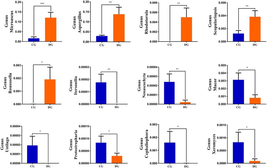
Significant Alterations in Gut Fungal
Fibrobacteres, Patescibacteria, Chloroflexi, Kiritimatiellaeota, Taxonomic Compositions in Diarrheic
Actinobacteria, Bacteroidetes, and Verrucomicrobia, as well as a Giraffe
distinct decrease (p < 0.01) in the abundance of Spirochaetes and There were nine phyla and 262 genera identified in the gut
Firmicutes (Figure 4A). At the genus level, 20 genera were totally fungal communities of all giraffes using RDP classifier. The
identified to be dramatically different between both groups. Of top 10 phyla and 10 genera of gut fungal community in both
these discriminatory taxa, the relative abundances of 12 bacterial groups are presented in Figures 3C,D. The phyla Ascomycota
genera significantly increased, whereas the relative abundances of (CG = 70.63%, DG = 72.63%) and Basidiomycota (CG = 25.84%,
Frontiers in Microbiology | www.frontiersin.org 4 August 2021 | Volume 12 | Article 712092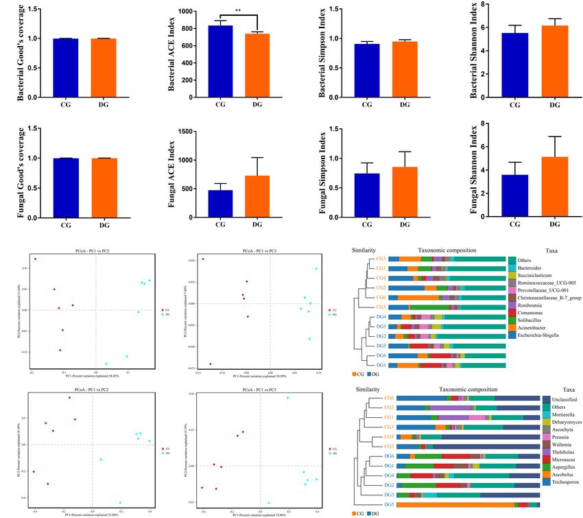
Li et al.                                                                                                                              Gut Microbiota in Diarrheic Giraffes

  FIGURE 1 | Gut bacterial and fungal OTU distribution and feasibility analysis. (A–C) Venn diagrams for gut bacterial OTU distribution. (D–F) Bacterial rarefaction and
  rank abundance curves were used for assessing the quality of sequencing including depth, abundance, and evenness. (G–I) Venn diagrams for gut fungal OTU
  distribution. (J–L) Fungal rarefaction and rank abundance curves were used to evaluate the quality of sequencing including depth, abundance, and evenness. Each
  colored curve displayed in the figures represents a sample.

DG = 18.01%) were the most dominant fungi in giraffes                                   (13.81%), and Microascus (12.14%) were enriched in the DG
regardless of health status, which together consisted of over                           group (Figure 3D). The heatmap showed a higher similarity
90% of the fungal composition (Figure 3C). Other fungal phyla                           of the individuals within group than that among groups and
such as Chytridiomycota (0.28%, 1.07%), Rozellomycota (0.16%,                           revealed the alternations in fungal genus-level compositions
0.25%), Neocallimastigomycota (0.11%, 0.18%), Olpidiomycota                             under the effect of diarrhea (Figure 3F).
(0.01%, 0.06%), and Glomeromycota (0.02%, 0.05%) in the CG                                 Using Metastats analysis to investigate the fungal genus-level
and DG groups were represented with a lower abundance.                                  taxonomic compositions in both groups, we observed that the
At the genus level, the dominant fungal genera observed in                              relative abundances of five genera (Microascus, Aspergillus,
the CG group were Trichosporon (20.76%), Aspergillus (2.95%),                           Rhodotorula, Scopulariopsis, and Roussoella) significantly
and Thelebolus (9.44%), whereas Ascobolus (14.33%), Aspergillus                         increased, whereas the relative abundances of seven genera

Frontiers in Microbiology | www.frontiersin.org                                     5                                           August 2021 | Volume 12 | Article 712092
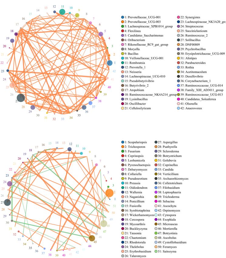
Li et al.                                                                                                                             Gut Microbiota in Diarrheic Giraffes

  FIGURE 2 | The alternations of gut bacterial and fungal diversities during diarrhea. Gut bacterial and fungal alpha diversities can be evaluated by Good’s coverage
  (A,E), ACE (B,F), Simpson (C,G), and Shannon (D,H). (I,J) Principal coordinate (PCoA) analysis based on the weighted and unweighted UniFrac distance of gut
  bacterial community. (L,M) Principal coordinate (PCoA) analysis of gut fungal community. (K,N) Gut bacterial and fungal clustering analysis based on unweighted
  pair-group method with arithmetic means (UPGMA). All of the data represent means ± SD. **p < 0.01.

(Itersonilia, Neoascochyta, Mucor, Ustilago, Protostropharia,                           Correlation Network Analysis
Cephaliophora, and Xeromyces) obviously decreased during                                Network analysis was conducted utilizing Python to illuminate
diarrhea (Figure 6). Among them, two fungal genera (Itersonilia                         linkages among different bacterial and fungal genera in gut
and Ustilago) even cannot be found in the gut fungal communities                        microbiota (Figure 7). The bacterial network consisted of
of diarrheic giraffes. LEfSe was applied to generate a cladogram                        80 nodes and 1,608 edges, whereas the fungal network was
to further investigate the variation in the fungal taxa composition                     composed of 80 nodes and 266 edges. Results revealed
(Figures 5C,D). Besides the above-mentioned significantly                               that Ruminococcaceae_UCG-014 was positively associated with
different funguses, we also observed that several funguses such as                      Rikenellaceae_RC9_gut_group (0.9231) and Alistipes (0.9371).
Wallemia and Cladorrhinum were the most dominant microbiota                             Rikenellaceae_RC9_gut_group was positively associated with
in the feces of patients in the DG group, whereas Botryotrichum,                        Alistipes (0.9790). Microascus was positively associated with
Thelebolus, and Trichosporon were significantly overrepresented                         Rhodotorula (0.8774). Aspergillus was positively associated with
in the CG group.                                                                        Rhodotorula (0.8194).

Frontiers in Microbiology | www.frontiersin.org                                     6                                          August 2021 | Volume 12 | Article 712092
Li et al.                                                                                                                        Gut Microbiota in Diarrheic Giraffes

  FIGURE 3 | The composition and relative abundance of the gut microbial community at the phylum and genus levels. (A,B) The gut bacterial relative abundance and
  composition of healthy and diarrheic giraffes at the phylum and genus levels. (C,D) The gut fungal relative abundance and composition of healthy and diarrheic
  giraffes at the phylum and genus levels. (E,F) Heatmap of the 50 most abundant gut bacterial and fungal genera in healthy and diarrheic giraffes.

DISCUSSION                                                                          diseases (Dumas et al., 2006; Wang et al., 2019b; Xiang et al.,
                                                                                    2020). Thus, investigating the gut microbial composition and
Accumulating evidence demonstrated that the stable gut                              diversity may contribute to expand our understanding of
microbiota was a vital barrier for host against the invasion                        disease etiology and provide convenient method to evaluate
and colonization of foreign pathogens, whereas gut microbial                        the health status of host. Gut microbiota is intimately involved
alternations may be the driving or central factor of multiple                       in several other important activities including maintaining the

Frontiers in Microbiology | www.frontiersin.org                                 7                                         August 2021 | Volume 12 | Article 712092
Li et al. Gut Microbiota in Diarrheic Giraffes FIGURE 4 | Significant alternations in the gut bacterial abundance at the level of phylum (A) and genus (B) during diarrhea. All of the data represent means ± SD. *p < 0.05; **p < 0.01; ***p < 0.001 and ****p < 0.0001. immunity, metabolism, and host health, which in turn depends variabilities of microbial community structure. However, studies on normal intestinal morphology (Li et al., 2021a). Diarrhea regarding diarrheic influence on gut microbiota in giraffe is one of the most common diseases of animals regardless have been insufficient to date. Taking advantage of this gap, of age and species, posing a great threat to public health, we first characterized the variability of gut microbiota in animal welfare, and animal husbandry. Moreover, the occurrence diarrheic giraffes. of diarrhea is inevitably related to gastrointestinal damage, Generally, gut microbiota is dynamically diverse within limits indicating that the gut microbiota may be also altered. To date, and influenced by multiple factors such as species, age, sex, research into the gut microbial composition and distribution diet, and health status (Manichanh et al., 2012; Wang et al., in different health status has covered many species including 2020; Xiong et al., 2020; Zhu et al., 2020). Several studies have goat, piglet, yak, and mice and demonstrated the significant indicated that diarrhea was able to cause a significant decrease Frontiers in Microbiology | www.frontiersin.org 8 August 2021 | Volume 12 | Article 712092
Li et al.                                                                                                                                Gut Microbiota in Diarrheic Giraffes

  FIGURE 5 | LEfSe analysis integrated with LDA scores revealed differential biomarkers associated with diarrhea in giraffes. (A,C) Cladogram revealing the
  phylogenetic distribution of intestinal bacteria and fungus correlated with the control or diarrheic groups. (B,D) The differences in the relative abundance of bacteria
  and fungi between the control and the diarrheic groups. LDA scores > 4 was considered statistically significant.

in the diversity of gut bacterial community as well as shifts in                          et al. (2020) demonstrated a decreased alpha diversity of the
intestinal functions (Barash et al., 2017; He et al., 2020). Ma et al.                    gut bacterial community in diarrheic piglets. Consequently,
(2020) revealed that the diversity of gut bacterial community                             diarrheic giraffes may be accompanied by significant changes
in diarrheic rats was significantly decreased. Additionally, He                           in the gut microbial composition and structure. Considering

Frontiers in Microbiology | www.frontiersin.org                                       9                                           August 2021 | Volume 12 | Article 712092
Li et al.                                                                                                                         Gut Microbiota in Diarrheic Giraffes

  FIGURE 6 | Significant changes in the gut fungal abundance at the level of phylum and genus during diarrhea. *p < 0.05; **p < 0.01 and ***p < 0.001 represent
  distinct difference between control and diarrheic groups.

the particularity of the species and the availability of samples,                     the diversity of the gut fungal community between diarrheic
we selected fecal samples as the research object to assess                            and healthy yaks. Moreover, Sangster et al. (2016) observed
the gut microbial composition and diversity. Consistent with                          that the gut fungal community of diarrheic patients induced
previous studies, this study demonstrated a dramatically declined                     by Clostridium difficile infection did not change significantly
ACE index in the gut bacterial community of giraffe during                            as compared with healthy population. In this study, we also
diarrhea, implying the disorder of gut bacterial community.                           observed that the differences of gut fungal diversity between
Several previous studies have demonstrate that the abundance                          diarrheic and healthy giraffes were not significant. Therefore, we
and diversity of gut microbiota was positively related to intestinal                  speculated that gut bacterial community played a major role in
functions, thereby the intestine with higher microbial diversity                      the occurrence of giraffe diarrhea, whereas gut fungi community
and abundance favors the energy utilization and performing                            was secondary. Moreover, PCoA also was performed to evaluate
complicated physiological functions (Barash et al., 2017; He                          the differences in the main components of the gut bacterial and
et al., 2020). Furthermore, imbalanced gut microbiota may result                      fungal communities between both groups. Results indicated that
in increased intestinal permeability and decreased immunity,                          the individuals of control group were clustered together and
which may further promote the invasion by members of                                  separated from the diarrhea group, implying a distinct difference
pathogenic bacteria and conditioned pathogen (Barash et al.,                          in the primary composition of the gut bacterial and fungal
2017). Therefore, diarrheic giraffe may also be at risk of                            communities between both groups. This study indicated that
intestinal dysfunction and other complications under situation of                     despite of shared environment and diets, the giraffes showed
decreased gut bacterial diversity. Gut fungi, as a key component                      significant alterations in the gut bacterial and fungal communities
of the microbial community, was also considered to be an                              during diarrhea. Therefore, we speculated that diarrhea was the
important contributor to intestinal health and function (Barash                       primary driving force for changes in gut bacterial and fungal
et al., 2017). Previous research has indicated that the gut                           communities of giraffes.
fungal diversity of patients with diarrhea-predominant irritable                         This study demonstrated that Firmicutes, Proteobacteria, and
bowel syndrome was significantly different from that of healthy                       Bacteroidetes were the most dominant bacterial phyla, whereas
population (Shukla et al., 2015; Hong et al., 2020). Li et al.                        Ascomycota and Basidiomycota were the most preponderant
(2018) indicated that there was no significant difference in                          fungal phyla in gut microbial community of giraffes, regardless

Frontiers in Microbiology | www.frontiersin.org                                  10                                         August 2021 | Volume 12 | Article 712092
Li et al.                                                                                                                               Gut Microbiota in Diarrheic Giraffes

  FIGURE 7 | Correlation network reveals the correlation among the different bacterial (A) and fungal (B) genera. The circles with different colors indicate the name of
  bacterial and fungal genus, and their sizes represent relative abundance. The strength of the correlation between both genera is positively related to the thickness of
  the line. The green line between both genera represents a positive correlation, whereas the orange line indicates a negative correlation.

of health status. These bacterial and fungal phyla were also                             (Li et al., 2018, 2021b). We further investigated the gut
found to be abundantly presented in the gut microbiota of                                microbial variabilities of this common diarrhea of giraffe.
cattle, goat, and yak, which was shown to be the major                                   The differences of specific bacteria and fungi can intuitively
characteristic of the gut microbial community in ruminants                               indicate the potential relationship between gut microbiota and

Frontiers in Microbiology | www.frontiersin.org                                     11                                           August 2021 | Volume 12 | Article 712092
Li et al.                                                                                                           Gut Microbiota in Diarrheic Giraffes

diarrhea. Our results revealed distinct increases in the relative            growth performance and decrease of gastrointestinal diseases
abundances of eight bacterial phyla (Synergistetes, Fibrobacteres,           of animals (Tan et al., 2014). These produced metabolites play
Patescibacteria, Chloroflexi, Kiritimatiellaeota, Actinobacteria,            an important role in improving intestinal environment and
Bacteroidetes, and Verrucomicrobia) and significant declines                 maintaining intestinal health. Given this phenomenon, we
in the relative abundances of two phyla (Spirochaetes and                    speculated that those genera seem to participate as key factors
Firmicutes) during diarrhea. Firmicutes, consisting of a large               in maintaining the balance of gut microbiota and modulating
amount of gram-positive bacteria, are responsible for converting             intestinal physiological activities to further prevent diarrhea.
complicated carbohydrates into reabsorbable substrates (Li                   The higher abundances of Streptococcus, Moryella, Klebsiella,
et al., 2018, 2021b). Moreover, most members of Firmicutes                   Comamonas, Oscillibacter, and Rothia in the gut microbiota were
are considered beneficial bacteria, which helps in regulating                closely related to many diseases including septicemia, bacteremia,
systemic immune responses, maintaining intestinal environment                endocarditis, pneumonia, and cellulitis (Boudewijns et al., 2003;
and inhibiting opportunistic pathogens (Sun et al., 2016).                   Fendukly et al., 2003; Broutin et al., 2020; Kjaer et al., 2020;
Actinobacteria synergy with one partner or host can easily be                Liu X. J. et al., 2020; Palacio et al., 2020). Veillonellaceae may
transformed into pathogenic interactions with another (Miao                  promote the development of inflammation and its abundance
and Davies, 2010). Consistent with our findings, Wang et al.                 increase significantly in patients with inflammatory bowel
(2018) also observed that the abundances of Actinobacteria and               disease and irritable bowel syndrome (Boudewijns et al., 2003;
Verrucomicrobia in the gut microbiota of diarrheic goats were                Fendukly et al., 2003; Broutin et al., 2020; Kjaer et al., 2020; Liu
significantly increased. Synergistetes has been shown to cause               Z. et al., 2020; Palacio et al., 2020). Klebsiella, a gram-negative
periodontal disease (Vartoukian et al., 2009; McCracken and                  pathogenic bacterium, mainly distributed in the respiratory
Nathalia, 2020).                                                             tract and intestine, which may cause pneumonia, hysteritis,
    Importantly, this study also found a high variation in                   mastitis, and other suppurative inflammation (Nordmann et al.,
some bacterial and fungal genera between healthy and                         2009; Qin et al., 2020). Enterococcus has been shown to result in
diarrheic giraffes and this variation may play crucial roles                 life-threatening meningitis, endocarditis, and sepsis (Mohanty
in intestinal ecosystem and function. This study demonstrated                et al., 2005). Moreover, many antibiotics frequently used in
significant increases in the relative abundances of 12 genera                the clinic failed treating Enterococcus infection because of the
(Streptococcus, Moryella, Veillonellaceae_UCG-001, Klebsiella,               inherent and acquired drug resistance (Arias and Murray, 2012).
Enterococcus, Catenibacterium, Atopobium, Comamonas, Rothia,                 Catenibacterium was closely related to morbid obesity and
Corynebacterium_1, Oscillibacter, and Terrisporobacter) as                   metabolic syndrome (Perler et al., 2021). The relative abundance
well as significant declines in the relative abundances of eight             of Atopobium was significantly increased in the patients with
genera (Prevotellaceae_UCG-004, Rikenellaceae_RC9_gut_group,                 esophageal cancer (Deng et al., 2021). Corynebacterium can
Coprococcus_3, Cellulosilyticum, Ruminococcaceae_UCG-014,                    result in lung abscess and caseous lymphadenitis, which was
Romboutsia, Ruminiclostridium_5, and Alloprevotella) with the                widespread in small ruminant populations (Samies et al., 1986;
effect of diarrhea. Prevotellaceae and Cellulosilyticum have been            Dorella et al., 2006). Terrisporobacter, an emerging anaerobic
reported to be involved in the degradation and digestion of                  pathogen, can cause surgical site infection (Cheng et al., 2016).
carbohydrate, pectin, and cellulose (Cai et al., 2010; De Filippo                Gut fungal community also plays important roles in intestinal
et al., 2010; O’Keefe et al., 2015). Rikenellaceae can degrade               function and host health, which are in line with gut bacterial
plant-derived polysaccharides and limit the development of                   community (Paterson et al., 2017). Interestingly, although the
colitis via stimulating T-regulatory cell differentiation (He et al.,        differences in gut fungal diversity between both groups were
2015; Peng et al., 2015; Dubin et al., 2016). Ruminococcaceae                not significant, the proportion of some intestinal fungus was
has long been regarded as a potential beneficial bacterium due               altered. We observed that the diarrheic giraffes displayed
to the positive regulation of the immune system and intestinal               increased Microascus, Aspergillus, Rhodotorula, Scopulariopsis,
environment (Shang et al., 2016). Furthermore, Ruminococcaceae               and Roussoella and decreased Itersonilia, Neoascochyta, Mucor,
has been reported to be negatively associated with liver cirrhosis,          Ustilago, Protostropharia, Cephaliophora, and Xeromyces as
non-alcoholic fatty liver, and increased intestinal permeability             compared with healthy populations. Microascus has been
(Huang et al., 2015; Shang et al., 2016). Romboutsia, an obligate            demonstrated to cause life-threatening brain abscess and
anaerobe, contains multiple metabolic capabilities associated                pneumonia (Baddley et al., 2000; Mohammedi et al., 2004;
with carbohydrate utilization and fermentation of single amino               Ustun et al., 2006; Paterson et al., 2017). Aspergillus was
acids (Ricaboni et al., 2016; Xin et al., 2019). Alloprevotella              closely related to multiple respiratory disease, while Rhodotorula
can produce moderate amounts of acetate and succinate and                    can cause fungemia (Zaas et al., 2003; Viegas et al., 2021).
decrease lifetime cardiovascular disease risk (Downes et al., 2013;          Scopulariopsis is characterized by its inherent resistance to the
Ricaboni et al., 2016; Xin et al., 2019). Some genera such as                available antifungal drugs and could result in pulmonary and
Coprococcus and Ruminiclostridium have been demonstrated to                  disseminated infections (Zaas et al., 2003; Kammoun et al., 2018;
produce short-chain fatty acids, which is beneficial to regulate             Viegas et al., 2021). Roussoella is a novel opportunistic pathogen,
energy intake and maintain the morphology and function of                    which can cause subcutaneous mycoses (Ahmed et al., 2014;
intestine and intestinal epithelial cells (Tan et al., 2014; Ye              Vasant et al., 2017). Diarrheic giraffes with altered gut microbiota
et al., 2021). Moreover, Ruminiclostridium, as an intestinal                 may also be accompanied by weakened immunity and disease
beneficial bacterium, is involved in the positive regulation of the          resistance. Consequently, some opportunistic pathogens may

Frontiers in Microbiology | www.frontiersin.org                         12                                    August 2021 | Volume 12 | Article 712092
Li et al. Gut Microbiota in Diarrheic Giraffes
also display pathogenicity, which may worsen the condition or giraffes. Results demonstrated that the gut bacterial community
cause other diseases. in diarrheal giraffe undergoes significant changes, characterized
Microbes inhabiting in the gastrointestinal tract such as by a decreased gut bacterial diversity and altered gut bacterial
bacteria and fungus can form a stabilized ecosystem that plays composition. Moreover, although diarrhea did not change the
vital roles in disease prevention, pathogenic growth inhibition, gut fungal diversity of giraffe, the types and proportions of some
and gastrointestinal homeostasis (Ahmed et al., 2014; Hu et al., fungus have changed significantly. These results also enriched
2017; Vasant et al., 2017). Generally, the intestinal bacteria and the knowledge of the gut bacterial and fungal in giraffe and
fungi can interact in a commensal, symbiotic, or antagonistic convey an important message that the altered gut bacterial and
relationship, causing a stabilized ecosystem (Aziz et al., 2013; fungal community may be one of the causes for the occurrence
Dabke et al., 2019). The damaged stable state of gut microbial or aggravation of diarrhea. This study also provides a theoretical
community was regarded as the pathological mediators of basis for alleviating diarrhea from the gut microbial perspective.
multiple diseases (Sircana et al., 2018). Therefore, the altered
intestinal bacteria and fungi may affect other bacterial and
fungal functions, which further affect the overall intestinal DATA AVAILABILITY STATEMENT
functions and aggravate the gut microbial alternations (Lozupone
et al., 2012). We observed that the Ruminococcaceae_UCG-014, The datasets presented in this study can be found in online
Rikenellaceae_RC9_gut_group, and Alistipes were dramatically repositories. The names of the repository/repositories and
affected by diarrhea, and there is a significant positive correlation accession number(s) can be found below: https://www.ncbi.nlm.
among them, which implied that their functions could be further nih.gov/, PRJNA727450.
affected. In the fungal community, some pathogenic bacteria
such as Microascus, Aspergillus, and Rhodotorula were also
significantly affected by diarrhea, and those fungi also displayed a ETHICS STATEMENT
strong positive correlation. Therefore, these pathogenic bacteria
may interact with each other, which further increase their The animal study was reviewed and approved by the
pathogenicity. This study conveyed a crucial message that Animal Welfare and Ethics Committee of Hubei Three
diarrhea not only directly changed the gut microbial diversity and Gorges Polytechnic.
abundance but also indirectly affected other functional bacteria
and fungus, which may affect intestinal functions.
AUTHOR CONTRIBUTIONS
XJ and AL provided the research idea. YW, BL, YF, YH,
CONCLUSION HL, FL, HZ, and LW contributed to reagents, materials, and
analysis tools. AL wrote the manuscript. MF-e-AK and AL
In summary, this study characterized the dynamic alternations revised the manuscript. All authors participated in writing and
of gut bacterial and fungal communities during diarrhea in reviewing the manuscript.
REFERENCES Baddley, J. W., Moser, S. A., Sutton, D. A., and Pappas, P. G. (2000). Microascus
cinereus (Anamorph scopulariopsis) brain abscess in a bone marrow transplant
Ahmed, S. A., Stevens, D. A., van de Sande, W. W., Meis, J. F., and de Hoog, recipient. J. Clin. Microbiol. 38, 395–397. doi: 10.1128/jcm.38.1.395-397.2000
G. S. (2014). Roussoella percutanea, a novel opportunistic pathogen causing Barash, N. R., Maloney, J. G., Singer, S. M., and Dawson, S. C. (2017). Giardia alters
subcutaneous mycoses. Med. Mycol. 52, 689–698. doi: 10.1093/mmy/myu035 commensal microbial diversity throughout the murine gut. Infect. Immun.
AlZahal, O., Valdes, E. V., and McBride, B. W. (2016). Analysis of the distal 85:e00948–16. doi: 10.1128/IAI.00948-16
gut bacterial community by 454-pyrosequencing in captive giraffes (Giraffa Boudewijns, M., Magerman, K., Verhaegen, J., Debrock, G., Peetermans, W. E.,
camelopardalis). Zoo Biol. 35, 42–50. doi: 10.1002/zoo.21252 Donkersloot, P., et al. (2003). Rothia dentocariosa, endocarditis and mycotic
Ambalam, P., Raman, M., Purama, R. K., and Doble, M. (2016). Probiotics, aneurysms: case report and review of the literature. Clin. Microbiol. Infect. 9,
prebiotics and colorectal cancer prevention. Best Pract. Res. Clin. Gastroenterol. 222–229. doi: 10.1046/j.1469-0691.2003.00503.x
30, 119–131. doi: 10.1016/j.bpg.2016.02.009 Broutin, L., Deroche, L., Michaud, A., Le Moal, G., Burucoa, C., Gayet, L. E., et al.
Arias, C. A., and Murray, B. E. (2012). The rise of the enterococcus: beyond (2020). First description of bacteremia caused by Oscillibacter valericigenes in
vancomycin resistance. Nat. Rev. Microbiol. 10, 266–278. doi: 10.1038/ a patient hospitalized for leg amputation. Anaerobe 64:102244. doi: 10.1016/j.
nrmicro2761 anaerobe.2020.102244
Arumugam, M., Raes, J., Pelletier, E., Le Paslier, D., Yamada, T., Mende, D. R., Brown, D. M., Brenneman, R. A., Koepfli, K. P., Pollinger, J. P., Mila, B., Georgiadis,
et al. (2011). Enterotypes of the human gut microbiome. Nature 473, 174–180. N. J., et al. (2007). Extensive population genetic structure in the giraffe. BMC
doi: 10.1038/nature09944 Biol. 5:57. doi: 10.1186/1741-7007-5-57
Aziz, Q., Dore, J., Emmanuel, A., Guarner, F., and Quigley, E. M. (2013). Gut Bu, D., Zhang, X., Ma, L., Park, T., Wang, L., Wang, M., et al. (2020).
microbiota and gastrointestinal health: current concepts and future directions. Repeated inoculation of young calves with rumen microbiota does not
Neurogastroenterol. Motil. 25, 4–15. doi: 10.1111/nmo.12046 significantly modulate the rumen prokaryotic microbiota consistently but
Backhed, F., Roswall, J., Peng, Y., Feng, Q., Jia, H., Kovatcheva-Datchary, P., et al. decreases diarrhea. Front. Microbiol. 11:1403. doi: 10.3389/fmicb.2020.01403
(2015). Dynamics and stabilization of the human gut microbiome during the Cai, S., Li, J., Hu, F. Z., Zhang, K., Luo, Y., Janto, B., et al. (2010). Cellulosilyticum
first year of life. Cell Host Microbe 17, 690–703. doi: 10.1016/j.chom.2015. ruminicola, a newly described rumen bacterium that possesses redundant
04.004 fibrolytic-protein-encoding genes and degrades lignocellulose with multiple
Frontiers in Microbiology | www.frontiersin.org 13 August 2021 | Volume 12 | Article 712092Li et al. Gut Microbiota in Diarrheic Giraffes carbohydrate- borne fibrolytic enzymes. Appl. Environ. Microbiol. 76, 3818– Hu, L., Geng, S., Li, Y., Cheng, S., Fu, X., Yue, X., et al. (2017). Exogenous fecal 3824. doi: 10.1128/AEM.03124-09 microbiota transplantation from local adult pigs to crossbred newborn piglets. Cheng, M. P., Domingo, M. C., Levesque, S., and Yansouni, C. P. (2016). Front. Microbiol. 8:2663. doi: 10.3389/fmicb.2017.02663 A case report of a deep surgical site infection with Terrisporobacter Huang, C., Song, P., Fan, P., Hou, C., Thacker, P., and Ma, X. (2015). Dietary glycolicus/T. Mayombei and review of the literature. BMC Infect. Dis 16:529. sodium butyrate decreases postweaning diarrhea by modulating intestinal doi: 10.1186/s12879-016-1865-8 permeability and changing the bacterial communities in weaned piglets. J. Nutr. Cryan, J. F., and Dinan, T. G. (2012). Mind-altering microorganisms: the impact 145, 2774–2780. doi: 10.3945/jn.115.217406 of the gut microbiota on brain and behaviour. Nat. Rev. Neurosci. 13, 701–712. Kammoun, S., Rekik, M., Trabelsi, H., Neji, S., Feki, J., and Ayadi, A. (2018). Orbital doi: 10.1038/nrn3346 cellulitis secondary to a fungal sinusitis caused by Scopulariopsis: the first case Dabke, K., Hendrick, G., and Devkota, S. (2019). The gut microbiome and in Tunisia. J. Mycol. Med. 28, 384–386. doi: 10.1016/j.mycmed.2018.04.006 metabolic syndrome. J. Clin. Invest. 129, 4050–4057. doi: 10.1172/JCI129 Kjaer, H. S., Lofberg, S. V., Nielsen, D. K., Kobbero, H., and Justesen, U. S. 194 (2020). Bacteraemia with moryella indoligenes and Fastidiosipila sanguinis: a De Filippo, C., Cavalieri, D., Di Paola, M., Ramazzotti, M., Poullet, J. B., Massart, S., case report. Access Microbiol. 2:acmi000108. doi: 10.1099/acmi.0.000108 et al. (2010). Impact of diet in shaping gut microbiota revealed by a comparative Li, A., Wang, Y., He, Y., Liu, B., Iqbal, M., Mehmood, K., et al. (2021a). study in children from Europe and rural Africa. Proc. Natl. Acad. Sci. U.S.A. 107, Environmental fluoride exposure disrupts the intestinal structure and gut 14691–14696. doi: 10.1073/pnas.1005963107 microbial composition in ducks. Chemosphere 277:130222. doi: 10.1016/j. Delzenne, N. M., Neyrinck, A. M., and Cani, P. D. (2011). Modulation of the gut chemosphere.2021.130222 microbiota by nutrients with prebiotic properties: consequences for host health Li, A., Yang, Y., Zhang, Y., Lv, S., Jin, T., Li, K., et al. (2021b). Microbiome analysis in the context of obesity and metabolic syndrome. Microb. Cell Fact. 10 Suppl reveals the alterations in gut microbiota in different intestinal segments of 1:S10. doi: 10.1186/1475-2859-10-S1-S10 Yimeng black goats. Microb. Pathog. 155:104900. doi: 10.1016/j.micpath.2021. Deng, Y., Tang, D., Hou, P., Shen, W., Li, H., Wang, T., et al. (2021). Dysbiosis of 104900 gut microbiota in patients with esophageal cancer. Microb. Pathog. 150:104709. Li, K., Mehmood, K., Zhang, H., Jiang, X., Shahzad, M., Dong, X., et al. (2018). doi: 10.1016/j.micpath.2020.104709 Characterization of fungus microbial diversity in healthy and diarrheal yaks Dorella, F. A., Pacheco, L. G., Oliveira, S. C., Miyoshi, A., and Azevedo, V. (2006). in Gannan region of Tibet Autonomous Prefecture. Acta Trop. 182, 14–26. Corynebacterium pseudotuberculosis: Microbiology, biochemical properties, doi: 10.1016/j.actatropica.2018.02.017 pathogenesis and molecular studies of virulence. Vet. Res. 37, 201–218. doi: Liu, X. J., Qiao, X. W., Huang, T. M., Li, L., and Jiang, S. P. (2020). Comamonas 10.1051/vetres:2005056 kerstersii bacteremia. Med. Mal. Infect. 50, 288–290. doi: 10.1016/j.medmal. Downes, J., Dewhirst, F. E., Tanner, A., and Wade, W. G. (2013). Description of 2019.12.005 Alloprevotella rava gen. Nov., Sp. Nov., Isolated from the human oral cavity, Liu, Z., Li, A., Wang, Y., Iqbal, M., Zheng, A., Zhao, M., et al. (2020). Comparative and reclassification of Prevotella tannerae Moore et al. 1994 as Alloprevotella analysis of microbial community structure between healthy and Aeromonas tannerae gen. Nov., Comb. Nov. Int. J. Syst. Evol. Microbiol. 63, 1214–1218. veronii-infected Yangtze finless porpoise. Microb. Cell Fact. 19:123. doi: 10. doi: 10.1099/ijs.0.041376-0 1186/s12934-020-01383-4 Dubin, K., Callahan, M. K., Ren, B., Khanin, R., Viale, A., Ling, L., et al. Lozupone, C. A., Stombaugh, J. I., Gordon, J. I., Jansson, J. K., and Knight, R. (2016). Intestinal microbiome analyses identify melanoma patients at risk (2012). Diversity, stability and resilience of the human gut microbiota. Nature for checkpoint-blockade-induced colitis. Nat. Commun. 7:10391. doi: 10.1038/ 489, 220–230. doi: 10.1038/nature11550 ncomms10391 Ma, Z., Wang, H.-J., Ma, X.-J., Li, Y., and Yang, H.-J., Li, H., et al. (2020). Dumas, M. E., Barton, R. H., Toye, A., Cloarec, O., Blancher, C., Rothwell, A., Modulation of gut microbiota and intestinal barrier function during alleviation et al. (2006). Metabolic profiling reveals a contribution of gut microbiota to of antibiotic-associated diarrhea with Rhizoma Zingiber officinale (Ginger) fatty liver phenotype in insulin-resistant mice. Proc. Natl. Acad. Sci. U.S.A. 103, extract. Food Funct. 11, 10839–10851. doi: 10.1039/d0fo01536a 12511–12516. doi: 10.1073/pnas.0601056103 Manichanh, C., Borruel, N., Casellas, F., and Guarner, F. (2012). The gut microbiota Fendukly, F., Karlsson, I., Hanson, H. S., Kronvall, G., and Dornbusch, K. (2003). in IBD. Nat. Rev. Gastroenterol. Hepatol. 9, 599–608. doi: 10.1038/nrgastro. Patterns of mutations in target genes in septicemia isolates of Escherichia 2012.152 coli and Klebsiella pneumoniae with resistance or reduced susceptibility to McCracken, B. A., and Nathalia, G. M. (2020). Phylum Synergistetes in the oral ciprofloxacin. Apmis 111, 857–866. doi: 10.1034/j.1600-0463.2003.1110904.x cavity: a possible contributor to periodontal disease. Anaerobe 68:102250. doi: Gallardo, P., Izquierdo, M., Vidal, R. M., Soto, F., Ossa, J. C., and Farfan, M. J. 10.1016/j.anaerobe.2020.102250 (2020). Gut Microbiota-Metabolome changes in children with diarrhea by Miao, V., and Davies, J. (2010). Actinobacteria: the good, the bad, and the ugly. diarrheagenic E. coli. Front. Cell Infect. Microbiol. 10: 485. doi: 10.3389/fcimb. Antonie Van Leeuwenhoek 98, 143–150. doi: 10.1007/s10482-010-9440-6 2020.00485 Mohammedi, I., Piens, M. A., Audigier-Valette, C., Gantier, J. C., Argaud, Garrett, W. S., Gordon, J. I., and Glimcher, L. H. (2010). Homeostasis and L., Martin, O., et al. (2004). Fatal Microascus trigonosporus (anamorph inflammation in the intestine. Cell 140, 859–870. doi: 10.1016/j.cell.2010.01.023 Scopulariopsis) pneumonia in a bone marrow transplant recipient. Eur. J. Clin. Guo, W., Li, Y., Wang, L., Wang, J., Xu, Q., Yan, T., et al. (2015). Evaluation of Microbiol. Infect. Dis. 23, 215–217. doi: 10.1007/s10096-003-1096-y composition and individual variability of rumen microbiota in yaks by 16S Mohanty, S., Dhawan, B., Kapil, A., Das, B. K., Pandey, P., and Gupta, A. (2005). rRNA high-throughput sequencing technology. Anaerobe 34, 74–79. doi: 10. Brain abscess due to Enterococcus avium. Am. J. Med. Sci. 329, 161–162. doi: 1016/j.anaerobe.2015.04.010 10.1097/00000441-200503000-00011 Han, Z., Li, K., Shahzad, M., Zhang, H., Luo, H., Qiu, G., et al. (2017). Analysis Mulherin, E., Bryan, J., Beltman, M., O’Grady, L., Pidgeon, E., Garon, L., et al. of the intestinal microbial community in healthy and diarrheal perinatal yaks (2008). Molecular characterisation of a bovine-like rotavirus detected from a by high-throughput sequencing. Microb. Pathog. 111, 60–70. doi: 10.1016/j. giraffe. BMC Vet. Res. 4:46. doi: 10.1186/1746-6148-4-46 micpath.2017.08.025 Musso, G., Gambino, R., and Cassader, M. (2011). Interactions between gut He, B., Nohara, K., Ajami, N. J., Michalek, R. D., Tian, X., Wong, M., et al. (2015). microbiota and host metabolism predisposing to obesity and diabetes. Annu. Transmissible microbial and metabolomic remodeling by soluble dietary fiber Rev. Med. 62, 361–380. doi: 10.1146/annurev-med-012510-175505 improves metabolic homeostasis. Sci. Rep. 5:10604. doi: 10.1038/srep10604 Nordmann, P., Cuzon, G., and Naas, T. (2009). The real threat of Klebsiella He, K., Yan, W., Sun, C., Liu, J., Bai, R., Wang, T., et al. (2020). Alterations in the pneumoniae carbapenemase-producing bacteria. Lancet Infect. Dis. 9, 228–236. diversity and composition of gut microbiota in weaned piglets infected with doi: 10.1016/S1473-3099(09)70054-4 Balantioides coli. Vet. Parasitol. 288:109298. doi: 10.1016/j.vetpar.2020.109298 O’Keefe, S. J., Li, J. V., Lahti, L., Ou, J., Carbonero, F., Mohammed, K., et al. (2015). Hong, G., Li, Y., Yang, M., Li, G., Qian, W., Xiong, H., et al. (2020). Fat, fibre and cancer risk in african americans and rural africans. Nat. Commun. Gut fungal dysbiosis and altered bacterial-fungal interaction in patients 6:6342. doi: 10.1038/ncomms7342 with diarrhea-predominant irritable bowel syndrome: an explorative study. Palacio, R., Cabezas, L., Cornejo, C., and Seija, V. (2020). [Comamonas Neurogastroenterol. Motil. 32:e13891. doi: 10.1111/nmo.13891 kerstersii bacteremia in a young man with acute appendicitis]. Rev. Frontiers in Microbiology | www.frontiersin.org 14 August 2021 | Volume 12 | Article 712092
Li et al.                                                                                                                                            Gut Microbiota in Diarrheic Giraffes

    Chilena. Infectol. 37, 182–185. doi: 10.4067/s0716-101820200002                              Wang, J., Zhu, G., Sun, C., Xiong, K., Yao, T., Su, Y., et al. (2020). TAK-242
    00182                                                                                           ameliorates DSS-induced colitis by regulating the gut microbiota and the
Paterson, M. J., Oh, S., and Underhill, D. M. (2017). Host-microbe interactions:                    JAK2/STAT3 signaling pathway. Microb. Cell Fact. 19:158. doi: 10.1186/s12934-
    commensal fungi in the gut. Curr. Opin. Microbiol. 40, 131–137. doi: 10.1016/j.                 020-01417-x
    mib.2017.11.012                                                                              Wang, Y., Li, A., Liu, J., Mehmood, K., Wangdui, B., Shi, H., et al. (2019a). L.
Peng, B., Huang, S., Liu, T., and Geng, A. (2015). Bacterial xylose isomerases from                 Pseudomesenteroides and L. Johnsonii isolated from yaks in Tibet modulate
    the mammal gut Bacteroidetes cluster function in Saccharomyces cerevisiae for                   gut microbiota in mice to ameliorate enteroinvasive Escherichia coli-induced
    effective xylose fermentation. Microb. Cell Fact. 14:70. doi: 10.1186/s12934-015-               diarrhea. Microb. Pathog. 132, 1–9. doi: 10.1016/j.micpath.2019.04.020
    0253-1                                                                                       Wang, Y., Li, A., Zhang, L., Waqas, M., Mehmood, K., Iqbal, M., et al.
Perler, B. K., Reinhart, E. M., Montgomery, M., Maynard, M., Shapiro, J. M.,                        (2019b). Probiotic potential of Lactobacillus on the intestinal microflora against
    Belenky, P., et al. (2021). Evaluation of the microbiome in men taking pre-                     Escherichia coli induced mice model through high-throughput sequencing.
    exposure prophylaxis for HIV prevention. AIDS Behav. 25, 2005–2013. doi:                        Microb. Pathog. 137:103760. doi: 10.1016/j.micpath.2019.103760
    10.1007/s10461-020-03130-7                                                                   Wang, Y., Zhang, H., Zhu, L., Xu, Y., Liu, N., Sun, X., et al. (2018). Dynamic
Qin, X., Wu, S., Hao, M., Zhu, J., Ding, B., Yang, Y., et al. (2020). The                           distribution of gut microbiota in goats at different ages and health states. Front.
    colonization of carbapenem-resistant klebsiella pneumoniae: epidemiology,                       Microbiol. 9:2509. doi: 10.3389/fmicb.2018.02509
    resistance mechanisms, and risk factors in patients admitted to intensive care               Xiang, L., Wu, Q., Osada, H., Yoshida, M., Pan, W., and Qi, J. (2020). Peanut
    units in china. J. Infect. Dis. 221, S206–S214. doi: 10.1093/infdis/jiz622                      skin extract ameliorates the symptoms of type 2 diabetes mellitus in mice by
Ricaboni, D., Mailhe, M., Khelaifia, S., Raoult, D., and Million, M. (2016).                        alleviating inflammation and maintaining gut microbiota homeostasis. Aging
    Romboutsia timonensis, a new species isolated from human gut. New Microbes                      (Albany NY) 12, 13991–14018. doi: 10.18632/aging.103521
    New Infect. 12, 6–7. doi: 10.1016/j.nmni.2016.04.001                                         Xin, J., Chai, Z., Zhang, C., Zhang, Q., Zhu, Y., Cao, H., et al. (2019). Comparing
Samies, J. H., Hathaway, B. N., Echols, R. M., Veazey, J. J., and Pilon, V. A.                      the microbial community in four stomach of dairy cattle, yellow cattle and
    (1986). Lung abscess due to Corynebacterium equi. Report of the first case in                   three yak herds in Qinghai-Tibetan plateau. Front. Microbiol. 10:1547. doi:
    a patient with acquired immune deficiency syndrome. Am. J. Med. 80, 685–688.                    10.3389/fmicb.2019.01547
    doi: 10.1016/0002-9343(86)90825-9                                                            Xiong, L., You, J., Zhang, W., Zhu, Q., Blachier, F., Yin, Y., et al. (2020).
Sangster, W., Hegarty, J. P., Schieffer, K. M., Wright, J. R., Hackman, J., Toole, D. R.,           Intrauterine growth restriction alters growth performance, plasma hormones,
    et al. (2016). Bacterial and fungal microbiota changes distinguish C. Difficile                 and small intestinal microbial communities in growing-finishing pigs. J. Anim.
    infection from other forms of diarrhea: results of a prospective inpatient study.               Sci. Biotechnol. 11:86. doi: 10.1186/s40104-020-00490-x
    Front. Microbiol. 7:789. doi: 10.3389/fmicb.2016.00789                                       Xue, N. Y., Liu, F., Tao, W. F., Zhao, Q., Qiu, H. Y., Hu, Y., et al. (2020). Molecular
Shang, Q., Shan, X., Cai, C., Hao, J., Li, G., and Yu, G. (2016). Dietary                           detection of Cryptosporidium spp. And Enterocytozoon bieneusi in longjiang
    fucoidan modulates the gut microbiota in mice by increasing the abundance                       wagyu cattle in Northeastern China. Microb. Pathog. 149:104526. doi: 10.1016/
    of Lactobacillus and Ruminococcaceae. Food Funct. 7, 3224–3232. doi: 10.1039/                   j.micpath.2020.104526
    c6fo00309e                                                                                   Yang, Q., Huang, X., Zhao, S., Sun, W., Yan, Z., Wang, P., et al. (2017). Structure and
Shao, H., Zhang, C., Xiao, N., and Tan, Z. (2020). Gut microbiota characteristics in                function of the fecal microbiota in diarrheic neonatal piglets. Front. Microbiol.
    mice with antibiotic-associated diarrhea. BMC Microbiol. 20:313. doi: 10.1186/                  8:502. doi: 10.3389/fmicb.2017.00502
    s12866-020-01999-x                                                                           Ye, X., Zhou, L., Zhang, Y., Xue, S., Gan, Q. F., and Fang, S. (2021). Effect of host
Shukla, R., Ghoshal, U., Dhole, T. N., and Ghoshal, U. C. (2015). Fecal microbiota                  breeds on gut microbiome and serum metabolome in meat rabbits. BMC Vet.
    in patients with irritable bowel syndrome compared with healthy controls using                  Res. 17:24. doi: 10.1186/s12917-020-02732-6
    Real-Time polymerase chain reaction: an evidence of dysbiosis. Dig. Dis. Sci. 60,            Zaas, A. K., Boyce, M., Schell, W., Lodge, B. A., Miller, J. L., and Perfect, J. R. (2003).
    2953–2962. doi: 10.1007/s10620-015-3607-y                                                       Risk of fungemia due to Rhodotorula and antifungal susceptibility testing of
Sircana, A., Framarin, L., Leone, N., Berrutti, M., Castellino, F., Parente, R., et al.             Rhodotorula isolates. J. Clin. Microbiol. 41, 5233–5235. doi: 10.1128/jcm.41.11.
    (2018). Altered gut microbiota in type 2 diabetes: just a coincidence? Curr. Diab.              5233-5235.2003
    Rep. 18:98. doi: 10.1007/s11892-018-1057-6                                                   Zhang, L., Jiang, X., Li, A., Waqas, M., Gao, X., Li, K., et al. (2020).
Sun, B., Wang, X., Bernstein, S., Huffman, M. A., Xia, D. P., Gu, Z., et al.                        Characterization of the microbial community structure in intestinal segments
    (2016). Marked variation between winter and spring gut microbiota in free-                      of yak (Bos grunniens). Anaerobe 61:102115. doi: 10.1016/j.anaerobe.2019.
    ranging Tibetan Macaques (Macaca thibetana). Sci. Rep. 6:26035. doi: 10.1038/                   102115
    srep26035                                                                                    Zhu, L., Xu, F., Wan, W., Yu, B., Tang, L., Yang, Y., et al. (2020). Gut microbial
Tan, J., McKenzie, C., Potamitis, M., Thorburn, A. N., Mackay, C. R., and Macia, L.                 characteristics of adult patients with allergy rhinitis. Microb. Cell Fact. 19:171.
    (2014). The role of short-chain fatty acids in health and disease. Adv. Immunol.                doi: 10.1186/s12934-020-01430-0
    121, 91–119. doi: 10.1016/B978-0-12-800100-4.00003-9
Tremaroli, V., and Backhed, F. (2012). Functional interactions between the                       Conflict of Interest: The authors declare that the research was conducted in the
    gut microbiota and host metabolism. Nature 489, 242–249. doi: 10.1038/                       absence of any commercial or financial relationships that could be construed as a
    nature11552                                                                                  potential conflict of interest.
Ustun, C., Huls, G., Stewart, M., and Marr, K. A. (2006). Resistant Microascus
    cirrosus pneumonia can be treated with a combination of surgery, multiple anti-              Publisher’s Note: All claims expressed in this article are solely those of the authors
    fungal agents and a growth factor. Mycopathologia 162, 299–302. doi: 10.1007/                and do not necessarily represent those of their affiliated organizations, or those of
    s11046-006-0067-0                                                                            the publisher, the editors and the reviewers. Any product that may be evaluated in
Vartoukian, S. R., Palmer, R. M., and Wade, W. G. (2009). Diversity and                          this article, or claim that may be made by its manufacturer, is not guaranteed or
    morphology of members of the phylum “synergistetes” in periodontal health                    endorsed by the publisher.
    and disease. Appl. Environ. Microbiol. 75, 3777–3786. doi: 10.1128/AEM.02
    763-08                                                                                       Copyright © 2021 Li, Liu, Li, He, Wang, Fakhar-e-Alam Kulyar, Li, Fu, Zhu, Wang
Vasant, J. A., Maggiani, F., and Borman, A. M. (2017). Subcutaneous mycotic                      and Jiang. This is an open-access article distributed under the terms of the Creative
    cyst caused by roussoella percutanea in a UK renal transplant patient.                       Commons Attribution License (CC BY). The use, distribution or reproduction in
    Mycopathologia 182, 721–725. doi: 10.1007/s11046-017-0121-0                                  other forums is permitted, provided the original author(s) and the copyright owner(s)
Viegas, C., Caetano, L. A., and Viegas, S. (2021). Occupational exposure to                      are credited and that the original publication in this journal is cited, in accordance
    Aspergillus section Fumigati: tackling the knowledge gap in Portugal. Environ                with accepted academic practice. No use, distribution or reproduction is permitted
    Res 194, 110674. doi: 10.1016/j.envres.2020.110674                                           which does not comply with these terms.

Frontiers in Microbiology | www.frontiersin.org                                             15                                               August 2021 | Volume 12 | Article 712092
You can also read


