Is That Near My Hand? Multisensory Representation of Peripersonal Space in Human Intraparietal Sulcus
←
→
Page content transcription
If your browser does not render page correctly, please read the page content below
The Journal of Neuroscience, January 24, 2007 • 27(4):731–740 • 731

Behavioral/Systems/Cognitive

Is That Near My Hand? Multisensory Representation of
Peripersonal Space in Human Intraparietal Sulcus
Tamar R. Makin,1 Nicholas P. Holmes,3 and Ehud Zohary1,2
1   Neurobiology Department, Life Sciences Institute, and 2Interdisciplinary Center for Neural Computation, Hebrew University, Jerusalem 91904, Israel, and
3   Institut National de la Santé et de la Recherche Médicale Unité 534, Espace et Action, 69676 Bron, France

Our ability to interact with the immediate surroundings depends not only on an adequate representation of external space but also on our
ability to represent the location of objects with respect to our own body and especially to our hands. Indeed, electrophysiological studies
in monkeys revealed multimodal neurons with spatially corresponding tactile and visual receptive fields in a number of brain areas,
suggesting a representation of visual peripersonal space with respect to the body. In this functional magnetic resonance imaging study,
we localized areas in human intraparietal sulcus (IPS) and lateral occipital complex (LOC) that represent nearby visual space with respect
to the hands (perihand space), by contrasting the response to a ball moving near-to versus far-from the hands. Furthermore, by inde-
pendently manipulating sensory information about the hand, in the visual (using a dummy hand) and proprioceptive domains (by
changing the unseen hand position), we determined the sensory contributions to the representation of hand-centered space. In the
posterior IPS, the visual contribution was dominant, overriding proprioceptive information. Surprisingly, regions within LOC also
displayed visually dominant, hand-related activation. In contrast, the anterior IPS was characterized by a proprioceptive representation
of the hand, as well as showing tactile hand-specific activation, suggesting a homology with monkey parietal hand-centered areas. We
therefore suggest that, whereas cortical regions within the posterior IPS and LOC represent hand-centered space in a predominantly
visual manner, the anterior IPS uses multisensory information in representing perihand space.
Key words: body schema; fMRI; LOC; proprioception; rubber hand; vision

Introduction                                                                                                         spatially overlapping RFs have been reported in the ventral in-
Peripersonal space is the multisensory space immediately sur-                                                        traparietal area (VIP), most commonly centered on the head
rounding our body, or more specifically the sector of space that                                                     (Colby et al., 1993; Duhamel et al., 1997, 1998; Avillac et al., 2005;
closely surrounds a certain body part (Rizzolatti et al., 1981,                                                      Schlack et al., 2005); larger tactile RFs with closely matching vi-
1997). A crucial factor that distinguishes the perception of our                                                     sual RFs, centered on the arm, torso, and face have been reported
immediate surroundings from that of more distant space is our                                                        in area 7b (Leinonen and Nyman, 1979; Robinson and Burton,
potential ability to interact with (i.e., to reach and grasp, or to                                                  1980a,b; Hyvärinen, 1981; Graziano and Gross, 1994), and some
avoid) objects within peripersonal space. Electrophysiological                                                       neurons in areas 2 and 5 have visual RFs that have been reported
studies in macaque monkeys suggest that multisensory periper-                                                        to follow the hand when it was moved to a new position in space
sonal space is represented in body part-centered coordinate                                                          (Obayashi et al., 2000).
frames, within both subcortical structures (the putamen), and in                                                         In humans, most of the evidence for the existence of a multi-
cortical regions of the frontal and parietal lobes (Hyvärinen,                                                      sensory system representing peripersonal space comes from neu-
1981; Rizzolatti et al., 1981; Colby et al., 1993; Graziano and                                                      ropsychological studies with patients suffering from cross-modal
Gross, 1993). Multisensory neurons within these areas have vi-                                                       extinction after a right hemisphere stroke. In studies of cross-
sual and, less commonly, also auditory receptive fields (RFs)                                                        modal extinction, a visual stimulus presented near to the patient’s
mapping the space surrounding the monkey’s body in close prox-                                                       ipsilesional (right) hand often extinguished the perception of a
imity to the somatosensory RF of the neuron.                                                                         simultaneous tactile stimulus on the patient’s contralesional
   In the monkey parietal cortex, several areas are related to                                                       (left) hand. When the right visual stimulus was presented far
peripersonal space processing: neurons with multisensory and                                                         from the patient’s hand, however, the degree of extinction was
                                                                                                                     reduced (di Pellegrino et al., 1997; Làdavas et al., 1998). Further-
Received Aug. 23, 2006; revised Nov. 8, 2006; accepted Nov. 28, 2006.
                                                                                                                     more, when the hands were held in a crossed position (such that
   This work was supported in part by Center of Excellence Grant 80009 from the Israel Academy of Sciences. N.P.H.
was supported by a Science Research Fellowship from the Royal Commission for the Exhibition of 1851, Imperial        the left hand was positioned in the right hemispace and vice
College, London, United Kingdom. We thank Prof. Marshall Devor, Prof. Shaul Hochstein, Ayelet McKyton, and Lior      versa), visual stimulation near the right hand still induced signif-
Shmuelof for their useful suggestions.                                                                               icant extinction of left hand tactile stimuli. These findings are
   Correspondence should be addressed to Tamar R. Makin, Neurobiology Department, Life Sciences Institute,
Hebrew University, Jerusalem 91904, Israel. E-mail: tamarmakin@pob.huji.ac.il.
                                                                                                                     consistent with the electrophysiological findings from monkeys
   DOI:10.1523/JNEUROSCI.3653-06.2007                                                                                suggesting that the representation of peripersonal space is body
Copyright © 2007 Society for Neuroscience 0270-6474/07/270731-10$15.00/0                                             part (i.e., hand) centered (Farnè et al., 2005). Other than these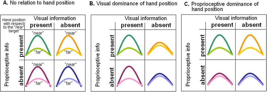
732 • J. Neurosci., January 24, 2007 • 27(4):731–740                                                                              Makin et al. • Multisensory Hand-Centered Space

neuropsychological findings, and some behavioral evidence
showing cross-modal interactions in near space (Spence et al.,
2004), little is known about the nature of the representation of
peripersonal space in humans, and of perihand space in particu-
lar (Culham et al., 2006).
    We set out to find cortical areas in humans that represent
visual information in hand-centered coordinates, by indepen-
dently manipulating visual stimulus distance and both visual and
proprioceptive feedback of hand position. We report here the
finding of a transition, within the parietal cortex, from a strictly
visual representation of the hand in the posterior intraparietal
sulcus (IPS), to a representation based on both visual and propri-
oceptive information in the anterior IPS.

Materials and Methods                                                             Figure 1. Experimental setup: a 2 ⫻ 2 ⫻ 2 factorial design. A schematic illustration of the
Subjects. Eleven healthy volunteers without histories of neurological, psy-       experimental conditions is shown. In each experiment, a moving ball was presented in either of
chiatric, or visual deficits were recruited (25–33 years of age; three fe-        two positions, near or far (with respect to the hand), while the subject was maintaining fixation
males). All of the subjects were naive regarding the purpose of the study.        between the two targets. Proprioceptive and visual information about hand position with re-
All procedures were approved by the Tel-Aviv Sourasky Medical Center              spect to the near condition were manipulated between experiments. A, Real-hand experiment:
Ethics Committee, and subjects gave written informed consent before the           having congruent visual and proprioceptive information. B, Occluded-hand experiment: pro-
experiment.                                                                       prioceptive information only. C, Dummy-hand experiment: illusory visual information. D,
   Magnetic resonance imaging. All experiments were conducted on a                Retracted-hand experiment: with the hand away from the near target.
whole-body 1.5 T, SIGMA Horizon, LX8.25 General Electric (Milwau-
kee, WI) scanner, equipped with a quadrature surface coil (Nova Medi-             termine whether the trajectory of the ball would hit the center of the
cal, Wakefield, MA). Blood oxygenation level-dependent (BOLD) con-                target or not, by covertly responding “yes” or “no.” Subjects practiced
trast was obtained with multislice gradient echo-planar (EPI) sequence            this task before each scan, and were able to maintain visual fixation while
(repetition time, 2000 ms; echo time, 55 ms; flip angle, 70°; field of view,      performing the task, and reported responding approximately equally
21 ⫻ 21 cm 2; matrix size, 64 ⫻ 64), using a real-time system. The scanned        “yes” and “no.” Eye movements were not monitored during scanning.
volume included 19 near-axial slices of 4 mm thickness with 1 mm inter-              This procedure was repeated four times for each subject, under differ-
slice gap, covering the whole brain (except for one subject, for whom a           ent experimental conditions (for experimental setup, see Fig. 1). In the
slice thickness of 4.5 mm was required). T1-weighted high-resolution              “real-hand experiment,” the subject’s left hand was placed (palm up) on
(1.2 ⫻ 1.2 mm 2) anatomical images with the same orientation and slice            the table and within view. The near target was positioned on the subject’s
thickness as the EPI slices were also acquired after each scan to improve         hand, such that the moving ball was 2–5 cm away at its closest point. The
the quality of the spatial matching of the functional data with the ana-          subject’s right hand rested comfortably by their right side (Fig. 1 A). In
tomical data. The functional magnetic resonance imaging (fMRI) images             the “retracted-hand experiment,” the subject’s left hand was retracted
were superimposed on a high-resolution (1 ⫻ 1 ⫻ 1 mm 3) whole-brain               toward their left shoulder and covered from sight. The near target was
spoiled gradient (3D-SPGR) sequence (using a standard head coil), al-             placed on the table at the same position as in the real-hand experiment
lowing accurate cortical segmentation, reconstruction, and volume-                (Fig. 1 D). In the “occluded-hand experiment,” the subject’s left hand was
based statistical analysis.                                                       placed on the table in the same position as in the real-hand experiment,
   Apparatus and procedure. Because of mounting evidence from neuro-              but this time it was occluded from the subject’s sight by a cardboard
psychological research showing disorders in perception of peripersonal            shield, on which the near target was laid (Fig. 1 B). In the “dummy-hand
space after right hemisphere injuries (i.e., causing deficits for the left side   experiment,” the subject’s left hand was again retracted to their left
of the body, specifically for the left hand), we chose to study the perihand      shoulder and covered. This time, a left-hand-and-wrist cosmetic pros-
space of the left hand. We hoped that this procedure would enable us to           thetic dummy hand was placed on the table, on which the near target was
better identify brain areas in the contralateral (right) hemisphere and           laid (Fig. 1C). After the dummy-hand experiment, subjects were inter-
therefore be useful in comparison with the data from patients with right          viewed to determine whether they experienced the “rubber hand illu-
hemisphere lesions.                                                               sion” during the scan (Botvinick and Cohen, 1998; Ehrsson et al., 2004).
   The subjects lay in the magnetic resonance imaging scanner and                 None reported experiencing any such illusion. For six of the subjects, we
viewed the apparatus through a mirror placed above their faces. Two               repeated the procedure with the left hand retracted and the near target
cardboard targets were hung in the subject’s field of view: one on a table        placed on the table. In this “far-dummy-hand experiment,” the dummy
above the subject’s left thigh (“near” target) and another suspended cen-         hand was suspended in midair behind the far target. A sample of the
trally in their upper visual field, 70 cm further from the near position          experiments was videotaped and analyzed off-line to assess whether the
toward their feet (“far” target). A fixation point was positioned halfway         visual stimuli were similar in frequency across experiments (see supple-
between the two targets, on which the subject was instructed to fixate            mental data, available at www.jneurosci.org as supplemental material).
through all of the experiments (except for the “tactile test,” in which the          For nine of the subjects, we performed an additional tactile test at the
subject’s eyes were closed and covered) (see below).                              end of these scans, in which they laid both of their hands palm-up with
   The visual stimulus was a ball attached to a 70-cm-long stick, which           their eyes covered. In a block design, the 2.5 cm plastic ball-on-stick was
was moved toward (and stopping 2–5 cm from the target) and away from              moved at 1 Hz touching either the left hand or the right hand, in a similar
one of the targets at a frequency of ⬃1 Hz. The visual stimuli were               manner as described above for the near target stimulus. The subjects were
delivered by a trained experimenter, who listened to a metronome with a           asked to keep their eyes closed and to assess on each trial if the touch they
frequency of 1 Hz. The experimenter stood to the right of the subject,            felt was in the same spatial position as the trial before (i.e., a one-back
occluded from their sight by a curtain. The subject could not see the             task).
experimenter’s hand and could see only the moving ball attached to the               Experimental design. The experiments followed a repeated-measures
stick. The diameters of the balls (2.5 and 4 cm) were matched so that             block design. In each scan, two conditions (near target and far target for
the visual image on the retina was of approximately the same size for the         the majority of experiments, and “left hand” and “right hand” for the
near and far targets, respectively (their retinal positions, however, were        tactile test) were interleaved and repeated five times per block, with a
different). During the stimulation periods, subjects were required to de-         randomized order of presentation between subjects. In the real-hand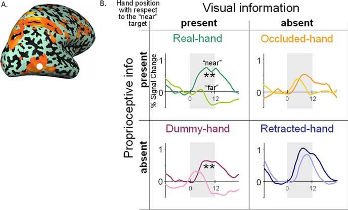
Makin et al. • Multisensory Hand-Centered Space J. Neurosci., January 24, 2007 • 27(4):731–740 • 733
experiment, a third condition was added in which the visual stimulus cedure included segmentation of the white matter using a grow-region
actually touched the subject’s left hand at the end of each trajectory. In all function, the smooth covering of a sphere around the segmented region,
experiments, each block lasted 12 s and was followed by a rest (i.e., and the expansion of the reconstructed white matter into the gray matter.
fixation) period of 10 s. The first and last rest periods were longer (24 and The surface was then unfolded, cut along the calcarine sulcus, and
14 s, respectively). The order of the experiments (real hand, retracted flattened.
hand, dummy hand, occluded hand, and far dummy hand) was random-
ized between subjects. The tactile test was always performed after com-
pletion of the other scans.
Results
Data analysis. Analysis was performed using BrainVoyager 4.96 and We first localized cortical areas that selectively responded to a
BrainVoyager QX (BV QX) software packages (Brain Innovation, Maas- visual stimulus (a moving ping-pong ball attached to a stick)
tricht, The Netherlands; 2000). The first seven images of each functional (Graziano et al., 1997) when approaching the immediate space
scan were discarded to allow for signal stabilization. Preprocessing in- surrounding the hand (perihand space). For this purpose, we
cluded head motion correction, slice-time correction, and high-pass contrasted the BOLD response to the ball approaching and reced-
temporal smoothing in the frequency domain (three cycles per total scan ing from the left hand, with the response to a ball approaching
time) to remove drifts and to improve the signal-to-noise ratio. The and receding from a distant target far from the hand (near vs far
functional images were superimposed on two-dimensional anatomical conditions in the real-hand experiment) (for experimental setup,
images and incorporated into the three-dimensional data sets through
see Fig. 1 A). Because the retinal projection of the ball in the two
trilinear interpolation. The complete data set was transformed into stan-
dard space (Talairach and Tournoux, 1988) and Z-normalized for the
conditions was different, the preference for the near over the far
whole time course. A correction for temporal autocorrelation was ap- ball could have resulted from low-level visual differences unre-
plied. To compute statistical parametric maps for the individual and lated to distance from the hand (such as the distance from the
group analyses, we applied a general linear model (GLM) using predic- eyes, for example). To control for this, we repeated the procedure
tors convolved with the canonical hemodynamic response function in the retracted-hand experiment (Fig. 1 D): areas that show pref-
(Boynton et al., 1996). erence for the near ball when the subject’s hand is retracted away
Statistical significance was assessed using a cluster approach, in which from both targets are likely to be sensitive to such changes in the
the initial voxelwise threshold criterion was p ⬍ 0.01, and a subsequent retinal input. We therefore focus our attention on regions that
spatial extent threshold of 20 – 40 contiguous functional voxels (986 – show preference for the near stimulus (compared with the far),
1972 mm 3), resulting in a whole-brain corrected value of p ⬍ 0.05 for
only when it is near the hand, suggesting that this preference is
each experiment (Forman et al., 1995) (implemented by Monte Carlo
simulations in BV QX).
indeed hand related.
The activation time courses from individual subjects’ data were ob- Second, we wanted to determine whether the preference for
tained from statistically significant clusters only [voxelwise t test, p ⬍ the near ball depended only on proprioceptive information con-
0.05, corrected for a minimum spatial extent of 10 functional voxels (488 cerning the hand, or also on visual information. To this end, we
mm 3) in each region of interest (ROI)], after applying the GLM analysis. occluded the subject’s hand from sight and repeated the experi-
The average percentage signal change (between 8 and 16 s after block ment (Fig. 1 B). In this occluded-hand experiment, although the
onset) was calculated per condition for all subjects showing significant subject’s hand was placed close to the near target, no visual feed-
activation in the real-hand experiment ROI. Paired two-tailed t tests were back of hand position was available, so that any specificity for the
applied to assess significant differences between the mean percentage near ball was likely to be based on proprioceptive information
signal change across conditions and experiments (using a derived pref- (about the occluded hand’s location). Additionally, we investi-
erence index) (see below). Voxels were selected on an individual basis if
gated the role of vision of a “hand” in the representation of peri-
they showed a preference for the near condition (over the far condition),
in the real-hand experiment, and were within a given cortical area ac- hand space in another experiment (dummy-hand experiment)
cording to anatomical markers. Because of extended activation for some by placing a realistic dummy hand by the near target, while the
of the subjects, the size of each ROI was restricted to a maximum of 15 subject’s own hand was retracted away. This created the illusory
mm from the anatomical marker in each axis. For the lateral occipital visual input of a hand positioned close to the near target, thus
complex (LOC) ROI, instead of the anatomical marker, we used a func- conflicting with veridical information from proprioception re-
tional marker from a visual object localizer (taken from Culham et al., garding the subject’s actual hand position (Fig. 1C).
2003) and combined it with the preference for the near stimulus in the
real-hand experiment. The Talairach and Tournoux coordinates were Model predictions
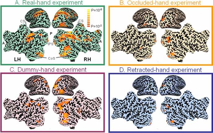
determined for the mean center of gravity of each ROI across subjects: Figure 2 depicts three models that allowed us to determine
aIPS, the anterior and inferior end of the IPS (10 subjects; center x ⫽ 34,
whether an area is hand related, and if so, if it was visually or
y ⫽ ⫺37, z ⫽ 43); pIPS, the posterior activation found in the middle of
the superior–inferior axis (11 subjects; 22, ⫺66, 47); LOC (9 subjects; 43,
proprioceptively driven. If neither visual nor proprioceptive
⫺66, ⫺5). To quantify the preference for the near over the far stimulus hand position information affected stimulus processing, the pref-
between experiments, we calculated a standardized preference index erence for the near over the far ball in a given area would be
adapted from Field and Wann (2005). The index was measured as the constant and significant in all four experiments. Such an out-
difference between the average percentage signal change (within a sub- come is depicted in Figure 2 A.
ject) for the near minus far conditions, divided by the averaged percent- Another possibility is that the preference for the near ball in
age signal change (between subjects) for the near condition (near subject ⫺ perihand space (real-hand experiment) would not be replicated
far subject/near group). when the hand was retracted away, suggesting that the preference
Across-subjects statistical parametric maps were calculated using a for the near stimulus is modulated by proprioceptive and/or vi-
hierarchical random-effects model analysis (Friston et al., 1999), after sual hand position information. If the preference for the near ball
spatial smoothing with a three-dimensional Gaussian kernel of 6 mm
is recovered when a dummy hand is visible close to the near
(full-width at half-maximum). For the far-dummy-hand experiment,
which was performed in six subjects, we used a fixed effect model analy- target, and abolished again when no hand is visible, we can con-
sis, corrected for false positives (whole-brain cluster analysis with the clude that the area has a visually determined representation of
same initial t value cutoff, and p values as reported before). The statistical hand position (Fig. 2 B). Finally, if the preference for the near ball
parametric maps were overlaid on a representative inflated cortical sur- is significant when the real hand is placed by the near target, but
face map, reconstructed from the T1-weighted 3D-SPGR scan. This pro- not when it is retracted away (regardless of any visual informa-
734 • J. Neurosci., January 24, 2007 • 27(4):731–740                                                                                                 Makin et al. • Multisensory Hand-Centered Space

Figure 2. ROI analysis: model predictions. A schematic illustration of the expected hemodynamic relationship between the near (light lines) and far (dark lines) conditions across four different
experiments. A, When the preference for the near condition is consistent regardless of visual and proprioceptive hand position information. B, When the preference for the near condition is observed
only when visual information of the hand (real or dummy) indicates its position close to the near target. C, If the preference for the near condition is present only when there is proprioceptive
information that the hand is positioned close to the near target.

tion), we can conclude that the area has a
proprioceptively determined representa-
tion of hand position (Fig. 2C). These dif-
ferent models guided our interpretation of
the experimental results as described
below.

Statistical parametric maps
(group analysis)
Localizing areas that represent visual
information with respect to the hand
Our first step was to delineate hand-
related areas, using a group analysis statis-
tical parametric map approach. Areas with
significantly higher BOLD signal in re-
sponse to a visual stimulus moving near
the hand, compared with stimuli moving
far from the hand (real-hand experiment)
are shown in Figure 3A and Table 1, exper-
iment A. Overall, the activation was more
prominent in the right hemisphere (RH) Figure 3. Group results: determining the relative contributions of visual and proprioceptive information to the hand schema.
than in the left hemisphere (LH). This is in fMRI differential activation (whole brain corrected, p ⬍ 0.05) for near versus far stimuli on representative inflated and unfolded
accordance with the fact that the near ball maps of the right hemisphere (RH) and left hemisphere (LH). Shown are the areas with preference for the ball approaching the
                                                 near target. A, When next to the subject’s hand. B, When the subject’s hand was occluded from sight. C, When a dummy hand was
was presented in the left visual field (near placed at the same position as the occluded hand, while the subject’s own hand was retracted. D, When the subject’s hand was
the subject’s left hand). In the right hemi- retracted away from the near target. The comparison between the activation preference in the different experiments enables
sphere, activation was found in the occip- identification of putative hand-related areas in the cortex, as well as the factors (visual or proprioceptive) governing the hand
ital cortex, spreading from the calcarine position-related representation. Note that the mere presence of the dummy hand modulated parietal areas in a similar way to the
sulcus (CalS) to the posterior collateral real hand. A, Anterior; P, posterior; CS, central sulcus; ColS, collateral sulcus.
sulcus (pColS), and to the most caudal
part of the IPS (intersecting with the trans-                                      clude (based on the model prediction in Fig. 2A) that this preference
verse occipital sulcus [intraparietal transverse occipital (IPTO)]).               is not related to the position of the ball with respect to the hand (i.e.,
Activation was also prominent in the parietal cortex along the                     perihand space). However, this is not the case in other areas, such as
IPS, in the frontal cortex around the middle frontal sulcus, and in                the IPS, LOC, and the frontal areas, for which the preference for the
the ventral premotor cortex (PMv), and in the right LOC.                           near ball in the real-hand experiment was lost when the hand was
    Clusters of voxels showing preference for the near ball posi-                  retracted. These areas, showing preference for the near ball only
tion over the “far,” even when the hand was retracted away from                    when presented close to the hand, can be regarded as representing
the near target, are shown in Figure 3D and Table 1, experiment                    visual information with respect to hand position.
B. In the right hemisphere, such near preference was seen only in                  Determining the relative contributions of visual and
the occipital cortex, in the CalS, pColS, and in the IPTO. Because                 proprioceptive information to the hand schema
these areas showed preference for the near stimulus over the far in                The hand schema is defined here as the cortical representation of
both the real-hand and retracted-hand experiments, we can con-                     the hand across sensory modalities. To study the relative contri-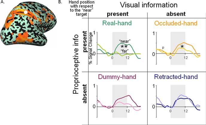
Makin et al. • Multisensory Hand-Centered Space J. Neurosci., January 24, 2007 • 27(4):731–740 • 735
Table 1. Group results: fMRI peak activation coordinates within significant clusters of voxels for the contrast between near and far conditions across four experiments (A–D)
Experiment A: real hand Experiment B: retracted hand Experiment C: occluded hand Experiment D: dummy hand
Area BA x y z t x y z t x y z t x y z t
pColS 19 20 ⫺65 ⫺9 7.8 21 ⫺64 ⫺6 5.4 20 ⫺66 ⫺6 6.1 17 ⫺67 ⫺5 8.2
19 ⫺23 ⫺66 ⫺12 6.6 ⫺19 ⫺71 ⫺13 3.7
IPTO 19 24 ⫺79 35 8.3 24 ⫺79 31 4.3 21 ⫺79 33 6.8 24 ⫺82 28 8.9
19 ⫺30 ⫺79 24 5.0
pIPS 7 15 ⫺63 53 6.0 18 ⫺62 50 6.9
7 ⫺21 ⫺61 45 5.0
aIPS 40 39 ⫺39 43 6.8
40 ⫺40 ⫺37 42 6.9
Superior parietal gyrus 5 29 ⫺41 57 8.0 25 ⫺43 55 6.4
5 ⫺35 ⫺43 57
Occipitotemporal/lateral occipital sulci 37 42 ⫺65 ⫺5 6.2 39 ⫺69 ⫺1 3.6 37 ⫺67 ⫺1 6.2
Postcentral gyrus 2 57 ⫺22 36 7.1
⫺58 ⫺29 34 6.0 ⫺51 ⫺26 38 5.0
Inferior frontal gyrus (PMv) 44 52 7 20 7.1 53 12 11 2.6 53 11 12 2.6
Inferior frontal sulcus 9 37 27 28 6.1
Middle frontal sulcus 9 31 32 33 5.7
Cingulate sulcus 32 4 21 40 6.2
Putamen 28 7 8 4.0 29 0 13 5.1
Middle temporal gyrus 21 60 ⫺40 ⫺2 5.7
63 ⫺38 ⫺7 4.0
Insula 13 40 ⫺33 19 5.6
All clusters were maintained from a p ⬍ 0.01 threshold (corrected for whole brain) except for t values emphasized by bold letters (p ⬎ 0.01) and underlined (uncorrected cluster). BA, Brodmann’s area; x, y, z, Talairach and Tournoux (1988)
peak voxel coordinates in millimeters; t, peak voxel t value.
bution of proprioceptive information to the hand schema, we
isolated the proprioceptive contribution to the observed BOLD
response, by occluding the subject’s hand from sight. The con-
trast between the near and far conditions for the occluded-hand
experiment is shown in Figure 3B and Table 1, experiment C.
When no visual information concerning hand position was avail-
able, activation was restricted to the occipital cortex, again along
the CalS, the pColS, and the IPTO. Because these areas showed a
similar preference in the retracted-hand experiment, we con-
clude that their near preference resulted from low-level visual
differences between the conditions. At a lower threshold (voxel-
wise p ⬍ 0.04), we also noticed a small cluster in area PMv located
Figure 4. Tactile properties of the hand-related visual areas. Activation on an inflated
in the inferior frontal gyrus (Table 1, experiment C), suggesting whole-brain map (shown in orange) represents areas that significantly responded to a tactile
that in this area proprioceptive information contributes to the stimulus on the subject’s left hand. We present only voxels that also show a preference for the
hand schema. near ball over the far in the real-hand experiment (in the absence of any tactile input; green
To determine the contribution of visual information to the lines). Areas showing preference for the near ball with illusory visual information of the hand
hand schema, we placed a dummy hand in a natural position by position (dummy-hand experiment) are shown in pink. Note that there is little overlap between
the near target. The results for the contrast between the near and the tactile areas and the areas modulated by the seen (dummy or real) hand position. LH, Left
far conditions in the dummy-hand experiment are shown in Fig- hemisphere; RH, right hemisphere; CS, central sulcus. Cluster corrected (p ⬍ 0.05).
ure 3C and Table 1, experiment D. In addition to the occipital
areas (CalS, pColS, and IPTO), activation selective for the near
ies in monkeys, human ventral premotor cortex might be sensi-
condition was also restored in the right hemisphere LOC and
tive both to visual and proprioceptive information regarding
posterior parietal cortex, primarily in the posterior part of the
hand position. Given the marginal statistical significance of this
IPS. Because these areas showed a preference for the near ball in
result, however, this conclusion should be taken with caution.
perihand space, and did not show this preference when the hand
was retracted away or occluded from sight, we can conclude Tactile representation of the hand
(based on the model prediction in Fig. 2 B) that the visual infor- To study further the multisensory properties of areas identified as
mation from the dummy hand was sufficient to reactivate them. potentially representing perihand space, we conducted a tactile
This suggests that the response of these areas to visual stimuli experiment in nine of the subjects, in which the experimenter
(placed in perihand space) is based primarily on visual informa- repeatedly touched the subjects’ hands with the same ball previ-
tion about hand position, regardless of veridical but conflicting ously used as the visual stimulus, while subjects kept their eyes
information from proprioception. None of the frontal activation closed. When masked inclusively with the areas showing prefer-
specific to the near ball in the real-hand experiment was retained ence for the near stimulus in the real-hand experiment, the post-
in the dummy-hand experiment. However, at a reduced thresh- central sulcus and the anterior section of the IPS also responded
old (voxelwise p ⬍ 0.03, uncorrected), we did find a small cluster significantly to the tactile stimulation (Fig. 4). Generally, these
in PMv, which did not survive the whole brain cluster-size cor- areas were not modulated by illusory visual information (i.e.,
rection (see Materials and Methods and Table 1, experiment D). they did not show a preference for the near ball in the dummy-
This suggests that, in accordance with electrophysiological stud- hand experiment) (a small overlap with the areas activated in the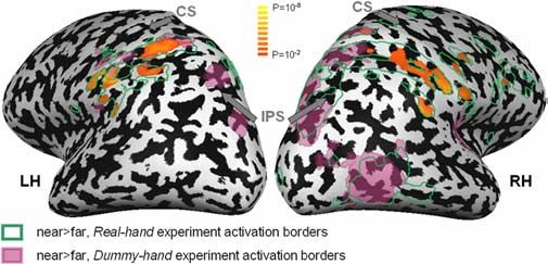
736 • J. Neurosci., January 24, 2007 • 27(4):731–740                                                                                  Makin et al. • Multisensory Hand-Centered Space

dummy-hand experiment was found in
the right superior parietal gyrus and left
inferior postcentral sulcus). Other sites
with overlapping tactile and near-hand vi-
sual processing were found in the right
central sulcus, posterior superior temporal
sulcus, inferior frontal gyrus, and in the
left parietal operculum (see supplemental
Table 1, available at www.jneurosci.org as
supplemental material).

ROI analysis
The results described above were derived
from statistical maps and are therefore
threshold dependent. To further confirm
the representation of visual information
with respect to the hand, we conducted an
ROI analysis, focusing on some of the
higher-order visual areas activated in the
real-hand experiment. The regions were Figure 5. ROI analysis: posterior IPS. A, Areas showing significant preference for the near over the far condition in the real-hand
chosen for each individual subject accord- experiment superimposed on a representative inflated right hemisphere. The white-filled circle represents the average location
ing to anatomical markers, as well as for (center of gravity) of the ROI between subjects. B, Averaged hemodynamic response curves of the percentage signal change for
their preference for the near over the far the near condition (dark colors) and the far condition (light colors). For each subject, clusters of voxels showing significantly
stimulus condition in the real-hand exper- greater activation for the near (compared with the far) condition in the real-hand experiment (within the posterior IPS) were
iment (see Materials and Methods). selected as the ROI. These clusters were then closely examined in the three other experiments. The gray background denotes the
Within three selected areas, the percentage time of presentation of the visual stimuli (in seconds). Asterisks denote statistical significance between the averaged time courses:
BOLD signal change, relative to stimulus *p ⬍ 0.05; **p ⬍ 0.01. Note that the visual presence of the (dummy or real) hand modulates the response of the region to the
                                                 visual stimuli.
onset, was compared between conditions
and experiments to determine how visual
                                                                                the dummy-hand and retracted-hand experiments (see Materials
and proprioceptive hand position information contributed to the
                                                                                and Methods). Because the only difference between these two
hand schema (i.e., to the difference between near and far
                                                                                experiments was the presence of the dummy hand positioned by
conditions).
                                                                                the near ball, the significant difference in the preference index
                                                                                between these experiments (paired t test, p ⫽ 0.0002) indicates
Intraparietal sulcus
                                                                                that the visual information concerning the position of the hand
Unlike the caudal part of the IPS (IPTO), which showed a signif-
                                                                                (real or illusory) was the main factor governing the response in
icant preference for the near ball in all contrast maps (for an ROI
                                                                                the pIPS. (Note that there was little effect of proprioceptive in-
analysis, see supplemental Fig. S1, available at www.jneurosci.org
                                                                                formation in these areas: the retracted-hand and the occluded-
as supplemental material), the main section of the IPS showed
                                                                                hand experiments yielded similar preference indices, paired t test,
hand-related properties (i.e., a preference for the near ball only
                                                                                p ⫽ 0.23)
when the hand was present). We focused on two ROIs in the right
                                                                                    In the anterior part of the IPS, the contrast maps showed a
(contralateral) hemisphere: The posterior aspect of the IPS
                                                                                preference for the near stimulus only in the real-hand experi-
(pIPS) (not to be confused with the most caudal part of IPS), and
                                                                                ment, suggesting that converging and matching proprioceptive
the anterior aspect of IPS (aIPS). ROI analyses of the anterior and
                                                                                and visual inputs are needed to generate activity in this region.
posterior IPS in the left hemisphere can be found in the supple-
                                                                                However, in the ROI analysis (Fig. 6), we found a small but sta-
mental information (supplemental Fig. S3, available at www.
                                                                                tistically significant preference for the near ball when the hand
jneurosci.org as supplemental material).
                                                                                was present but not seen (occluded-hand experiment; p ⫽ 0.029).
   The posterior aspect of the IPS had shown a preference for the
                                                                                A comparison between the preference indices for the occluded-
near ball only with concurrent visual information about hand
                                                                                hand and retracted-hand experiments (which differ only in the
position (i.e., in both the real-hand and dummy-hand experi-
                                                                                proprioceptive information regarding hand position) yielded a
ments) (Fig. 3 A, C). The ROI analysis for this area (Fig. 5) con-
                                                                                significant difference between the two (paired t test, p ⫽ 0.01). In
firmed this finding: the preference for the near ball was abolished
                                                                                the same area, a comparison between the preference for the near
when the hand was retracted ( p ⫽ 0.10, t test comparing the area
                                                                                ball in the dummy-hand and retracted-hand experiments (which
under the average hemodynamic response curve between the
                                                                                differ only in visual aspects of hand position) also showed a sig-
near and far conditions) (see Materials and Methods) and was
                                                                                nificant difference ( p ⫽ 0.02). We conclude that, in the aIPS, in
recovered when a dummy hand was placed instead of the real
                                                                                addition to visual information, there is an important propriocep-
hand ( p ⫽ 0.0001). Without visual feedback of the hand, the
                                                                                tive contribution to the determination of hand position for peri-
preference for the near ball in the occluded-hand experiment
                                                                                hand space representation.
remained nonsignificant (although p ⫽ 0.07). This pattern of
results is consistent with the model suggested in Figure 2 B, de-               Lateral occipital complex
scribing an area that represents visual stimuli in hand-centered                Another ROI was aimed to study further the LOC. Surprisingly,
coordinates, and in which hand position information is princi-                  our group results showed enhanced activation in LOC for visual
pally determined by vision. This result was further confirmed by                stimuli near the hand, compared with far stimuli. This result was
a comparison between the preference indices for the near ball in                unexpected because this area is regarded as a higher-level visual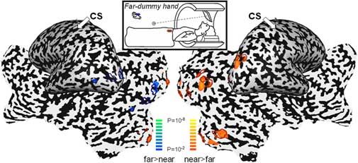
Makin et al. • Multisensory Hand-Centered Space J. Neurosci., January 24, 2007 • 27(4):731–740 • 737
Controlling for possible confounding effects
of attention
One possible explanation for the observed
selectivity of the BOLD response for visual
stimuli approaching the subject’s real
hand (or dummy hand) is that the hand
(or dummy hand) naturally captured the
subject’s visual attention. As a result,
the representation of any stimulus near the
hand might have been enhanced. This ex-
planation emphasizes the hand as an ob-
ject of interest, per se, rather than as a body
part. Such an interpretation would there-
fore predict that any object of interest (re-
gardless of whether it resembled the hand)
would generate the same preference for
stimuli near the attended object.
To account for these possible con-
founding effects of attention, for six of the
Figure 6. ROI analysis: anterior IPS. Notations are as in Figure 5. Note that the proprioceptive information of hand position
subjects we conducted another experi-
(next to the near target) modulates the response of the region to the visual stimuli. ment, in which we placed the far target on
a dummy hand positioned far from the
subject (far-dummy-hand experiment).
Under such circumstances, the dummy-
hand position clearly does not correspond
anatomically with the normal body
scheme. If the preference for the near ball
in the hand-related areas resulted from re-
ferring attention to the hand (or dummy)
as an object of interest, rather than as a
hand per se, one would predict that the
preference for the near ball should switch
to the far ball (which now approaches the
far dummy hand). However, if the prefer-
ence for the near ball in the main experi-
ment was attributable to these regions’
representation of the hand (or dummy) as
a body part, no such preference should be
seen when the dummy is placed in the far
position.
The statistical parametric maps for the
far-dummy-hand experiment are pre-
Figure 7. ROI analysis: LOC. Notations are as in Figure 5.
sented in Figure 8. Preference for the near
area, which is not usually considered as being related to spatial ball (over the far) was observed bilaterally
processing (Grill-Spector and Malach, 2004). As far as we know, in early visual areas, as well as in the right posterior collateral
LOC has never before been examined in this respect (i.e., modu- sulcus and the IPTO. Conversely, the preference for the far ball
lation of activity dependent on the location of visual stimuli with was significant only in occipital visual areas in the left hemisphere
respect to body parts). In our group results, a preference for the (in accordance with the position of the ball in the right visual
near stimulus was shown only with available visual hand position field). We did not find significant differences between the BOLD
information (i.e., in the real- and dummy-hand experiments). responses to near and far stimuli in any of the previously identi-
The ROI analysis in LOC (Fig. 7) replicated these findings fied hand-related areas. This result suggests that the preference
(dummy-hand experiment, paired t test for “near” vs “far,” p ⫽ for the near ball in the hand-related areas was specific only for
0.008). Without visual information regarding hand position, the hands in near space, and argues strongly against an attentional
preference for the near ball was no longer significant, regardless interpretation of our principal results.
of the actual (i.e., proprioceptive) hand position (retracted-hand
experiment, p ⫽ 0.13; occluded-hand experiment, p ⫽ 0.25). Discussion
When compared with the retracted-hand experiment, only the Several cortical areas showed preference for the near ball when it
preference for the near ball in the dummy-hand experiment was approached the left hand (compared with the far ball), but not
significant (preference indices: retracted hand vs dummy hand, when the same stimulus was presented with the hand retracted
p ⫽ 0.04; retracted hand vs occluded hand, p ⫽ 0.53), suggesting away (Fig. 3). Because the only variable changing between these
that in LOC, visual processing of perihand space is modulated by two experiments was the position of the hand, this result can be
the seen position of the hand (whether real or illusory), although explained in terms of hand position-dependent modulations of
this effect was less prominent than in the aIPS. the sensitivity to visual stimuli moving within perihand space. By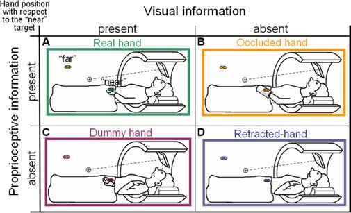
738 • J. Neurosci., January 24, 2007 • 27(4):731–740                                                                                     Makin et al. • Multisensory Hand-Centered Space

                                                                                                       experiment) (Fig. 8). We therefore conclude that the preference
                                                                                                       for the near ball in the hand-related areas was not simply attrib-
                                                                                                       utable to the representation of the hand (or dummy) as an atten-
                                                                                                       tionally capturing object per se, but rather was specific for hands
                                                                                                       in near space. Whether this preference is unique to the represen-
                                                                                                       tation of the hand, or may also be modulated by the presence of
                                                                                                       other functionally equivalent objects in their spatial surrounding
                                                                                                       (such as suggested by the literature on tool use) (Maravita and
                                                                                                       Iriki, 2004), is an important area for future research. For addi-
                                                                                                       tional discussion of the rubber-hand illusion and the dummy-
                                                                                                       hand effect in other research, see the supplemental information
Figure 8. Attentional control: placing the dummy hand outside perihand space. Areas show-              (available at www.jneurosci.org as supplemental material).
ing preference for the near over the far ball (reds) and for the far ball (blues) over the near ball
in the far-dummy-hand experiment (solid patches) and in the retracted-hand experiment (out-            Visually based hand schema: posterior IPS
line patches) are shown. Note that the two experiments differ only in the presence of the              Activation in the posterior aspect of the medial IPS in humans has
dummy hand by the far target. The overlap in responses in the two experiments suggests that,           been shown to play a role in tasks requiring visuomotor coordi-
when the dummy hand is placed out of peripersonal space, it does not affect hand-related areas,        nation of hand movements with respect to targets (Chaminade
as would be expected if the modulation was attributable to attention referred to the dummy             and Decety, 2002; Simon et al., 2002; Grefkes et al., 2004). The
hand. CS, Central sulcus.                                                                              same area has been reported to show a topographic mapping of
                                                                                                       space both for saccades and for pointing to targets, which is up-
                                                                                                       dated with eye movements (Medendorp et al., 2003, 2005). Ac-
independently manipulating visual and proprioceptive informa-
                                                                                                       cording to Ehrsson et al. (2004), activity in the medial wall of the
tion concerning hand position, we were able to identify three
                                                                                                       IPS reflects the seen position of the hand. These, as well as our
main patterns of visual processing: areas that represent visual
                                                                                                       present findings, support the potential role of pIPS as an area that
information regardless of hand position (in the occipital cortex)
                                                                                                       integrates visual and spatial information in hand-centered coor-
(supplemental Fig. S1, available at www.jneurosci.org as supple-
                                                                                                       dinates. Because this area was not modulated to the same extent
mental material); areas modulated by purely visual aspects of
                                                                                                       by proprioceptive cues in the absence of visual feedback, we
hand position [in the posterior IPS (Fig. 5) and in LOC (Fig. 7)];
                                                                                                       suggest that, in this hand-related area, the coordinate system is
and regions modulated by both proprioceptive and visual infor-
                                                                                                       dominated by visual, rather than by somatosensory information
mation regarding hand position [in the anterior IPS (Fig. 6) and
                                                                                                       concerning the hand. It is possible, however, that some proprio-
the ventral premotor cortex (Table 1)]. In the next section, we
                                                                                                       ceptive information concerning hand position can also be de-
discuss the effect of the dummy hand in determining visually
                                                                                                       rived from the pIPS.
based hand representation.
                                                                                                       LOC: a new candidate region for a visually based hand schema?
Visually based hand schema                                                                             The other region that showed modulation of visual responses
Perihand neurons in macaque ventral premotor cortex, as well as                                        with respect to the seen position of the hand (or dummy hand)
postural neurons in area 5, responded to a realistic dummy mon-                                        regardless of proprioception was LOC. Peripersonal space has
key hand, even if the position of the dummy conflicted with                                            typically been associated with vision-for-action in the dorsal
proprioceptive information regarding the true position of the                                          stream (Graziano and Cooke, 2006). Why, therefore, would the
monkey’s hand (Graziano, 1999; Graziano et al., 2000). Similarly,                                      ventral stream be involved in spatial representation of objects
in studies of cross-modal extinction in neuropsychological pa-                                         with relation to the hand? Several studies have demonstrated that
tients, placing a dummy hand near to an ipsilesional visual stim-                                      LOC shows sensitivity to disparity-defined shape (Gilaie-Dotan
ulus can increase the proportion of extinguished contralesional                                        et al., 2002), achieved by combining different depth cues (Brou-
tactile stimuli during simultaneous presentation, although the                                         wer et al., 2005; Welchman et al., 2005). This suggests that LOC
patient’s ipsilesional hand was retracted away from both the vi-                                       may be sensitive not only to depth within objects but also in
sual stimulus and the dummy hand (Farnè et al., 2000). In healthy                                     space. Moreover, a recent fMRI study showed the involvement of
human subjects, viewing a dummy hand being stroked by a paint-                                         ventral stream visual areas in peripersonal space: Lloyd et al.
brush in synchrony with feeling similar strokes on their own                                           (2006) investigated the neural responses to noxious stimuli com-
occluded hand can create both an illusion of ownership of the                                          pared with innocuous stimuli, both of which touched a dummy
dummy hand, and a change in felt hand position (the “rubber-                                           hand. In addition to parietal and frontal areas, the fusiform gyrus
hand illusion”) (Botvinick and Cohen, 1998).                                                           also showed differential activation to the noxious stimuli only
    In our study, the mere presence of the dummy hand in front of                                      when the dummy hand was aligned with the subject’s shoulders
the subjects’ retracted hands modulated the preference for a near                                      (compared with the same procedure repeated with the dummy
stimulus in the posterior IPS and the LOC (Fig. 3C). This result                                       hand in an unnatural position). This, in addition to the present
was obtained although all subjects reported that they did not                                          results, suggests that the ventral stream might also be involved in
sense an illusion of ownership over the dummy hand. Therefore,                                         the representation of stimuli in peripersonal space.
this activation cannot be explained by the conscious perception                                            Our results revealed a second type of visual processing with
of the rubber-hand illusion.                                                                           respect to the hand: hand-related visual representation, which is
    Furthermore, we excluded the possibility that the mere pres-                                       modulated mainly by proprioception. In the next section, we will
ence of the hand (or dummy hand) covertly captured the sub-                                            discuss these results.
jects’ visual attention, thereby enhancing the representation of
the near ball. In the far-dummy-hand experiment, although the                                          Proprioceptively based hand schema
far ball was approaching the dummy hand, there was no selective                                        Neurons in macaque area 5 are predominantly modulated by
preference for it (unlike the near preference in the dummy-hand                                        proprioceptively determined hand position (i.e., are sensitive to
Makin et al. • Multisensory Hand-Centered Space J. Neurosci., January 24, 2007 • 27(4):731–740 • 739
hand position while it is hidden from view) but may also encode rather to a general dominance of the right hemisphere in spatial
the position of the monkey’s arm visually (Graziano et al., 2000). processing (Jager and Postma, 2003). This is particularly interest-
Some neurons in the ventral premotor cortex that respond to an ing, given the presumed functional dissociation between the dor-
approaching visual stimulus in perihand space also respond when sal and ventral visual streams (Shmuelof and Zohary, 2005,
the hand is occluded from sight (Graziano, 1999). In humans, 2006).
Làdavas et al. (2000) have shown proprioceptive information of
the hand alone does not suffice to modulate multisensory pro- Conclusions
cessing in peripersonal space. We wanted to see whether the Using both group statistical parametric maps and ROI analyses,
hand-related areas from our experiments would continue to re- we found a decreasing gradient in the dominance of visual infor-
spond selectively to a visual stimulus when it was presented close mation along the IPS: in the most caudal part of the IPS (adjoin-
to the subjects’ occluded hand. ing the transverse occipital sulcus), processing of visual informa-
tion was independent of hand position; in the posterior IPS, as
Proprioceptive prevalence in hand representation: anterior IPS well as in clusters within LOC, we found that visual processing
In the ROI analysis, the most anterior part of the IPS showed a was modulated primarily by the visible position of the hand and,
significant preference for the near ball (over the far) when the to a much lesser extent (if at all), by its veridical (proprioceptive)
hand was occluded (Fig. 6). The same area was sensitive to the position. We therefore conclude that these areas may represent
visual position of the hand (the preference indices in the dummy- perihand space in visually driven hand coordinates (regardless of
hand experiment were significantly greater than in the retracted- whether the hand is real or illusory). Finally, the anterior part of
hand experiment) and was activated by tactile stimulation ap- the IPS showed multisensory properties: along with a weakened
plied to the left hand (Fig. 4). We therefore suggest that clusters visual modulation of hand-related processing, both significant
within human aIPS are multisensory, showing perihand proper- proprioceptive modulation of visual processing and a significant
ties similar to those reported in single-unit studies of perihand response to purely tactile stimulation of the hand were revealed.
space in macaque frontal and parietal cortices. We therefore conclude that aIPS represents perihand space in
What do we know about aIPS from imaging studies? Consis- both somatosensory and visual hand coordinates.
tent with the macaque anterior intraparietal (AIP) area, the hu-
man analog hAIP (Culham et al., 2006) is highly activated by References
visuomotor tasks such as visually guided grasping (Binkofski et Avillac M, Deneve S, Olivier E, Pouget A, Duhamel JR (2005) Reference
al., 1998; Shikata et al., 2003; Frey et al., 2005) and also responds frames for representing visual and tactile locations in parietal cortex. Nat
Neurosci 8:941–949.
to hand manipulation without visual feedback (Binkofski et al.,
Binkofski F, Dohle C, Posse S, Stephan KM, Hefter H, Seitz RJ, Freund H-J
1999; Jäncke et al., 2001; Stoeckel et al., 2003). These reports fit (1998) Human anterior intraparietal area subserves prehension: a com-
nicely with our results showing visual responses modulated by bined lesion and functional MRI activation study. Neurology
unseen (i.e., proprioceptive) changes in hand position. Further- 50:1253–1259.
more, Grefkes et al. (2002) and Macaluso et al. (2003) both found Binkofski F, Buccino G, Stephan KM, Rizzolatti G, Seitz RJ, Freund H-J
visual–tactile integration in this area, in addition to activations (1999) A parieto-premotor network for object manipulation: evidence
contingent on motor responses. Finally, our Talairach and Tour- from neuroimaging. Exp Brain Res 128:210 –213.
Botvinick MM, Cohen JD (1998) Rubber hands “feel” touch that eyes see.
noux coordinates for the ROI analysis (averaged center of mass Nature 391:756.
between subjects, x ⫽ 34, y ⫽ ⫺37, z ⫽ 43) approximately match Boynton GM, Engel SA, Glover GH, Heeger DJ (1996) Linear systems anal-
those of the possible human VIP homolog (x ⫽ 38, y ⫽ ⫺44, z ⫽ ysis of functional magnetic resonance imaging in human V1. J Neurosci
46), which responded to multisensory stimuli approaching sub- 16:4207– 4221.
ject’s faces (Bremmer et al., 2001). Bremmer F, Schlack A, Shah NJ, Zafiris O, Kubischik M, Hoffmann K-P,
Zilles K, Fink GR (2001) Polymodal motion processing in posterior pa-
rietal and premotor cortex: a human fMRI study strongly implies equiv-
A possible homolog to monkey PMv
alencies between humans and monkeys. Neuron 29:287–296.
The activation patterns in PMv [consistent with the functional
Brouwer GJ, van Ee R, Schwarzbach J (2005) Activation in visual cortex
localization of Mayka et al. (2006)] in our study naturally raise correlates with the awareness of stereoscopic depth. J Neurosci
special interest, because this cortical area has been recognized as a 25:10403–10413.
region containing perihand neurons in monkeys. In humans, it Chaminade T, Decety J (2002) Leader or follower? Involvement of the infe-
has also been associated with the rubber-hand illusion (Ehrsson rior parietal lobule in agency. NeuroReport 13:1975–1978.
et al., 2004), suggesting that it might be highly influenced by the Colby CL, Duhamel J-R, Goldberg ME (1993) Ventral intraparietal area of
the macaque: anatomic location and visual response properties. J Neuro-
visually inferred position of the hand. In our study, the effects in
physiol 69:902–914.
this region were not as prominent as in the other areas we focused Culham JC, Danckert SL, DeSouza JF, Gati JS, Menon RS, Goodale MA
on. We found some preference for the near ball in the absence of (2003) Visually guided grasping produces fMRI activation in dorsal but
visual feedback of hand position, and to a lesser extent with only not ventral stream brain areas. Exp Brain Res 153:180 –189.
visual information available (Table 1). Similarly to the aIPS, PMv Culham JC, Cavina-Pratesi CC, Singhal A (2006) The role of parietal cortex
also responded to tactile stimuli applied to the hand (Fig. 4). It is in visuomotor control: what have we learned from neuroimaging? Neu-
therefore possible that strong PMv activation requires concur- ropsychologia 44:2668 –2684.
di Pellegrino G, Làdavas E, Farnè A (1997) Seeing where your hands are.
rent visual and tactile stimulation within perihand space. Nature 388:730.
Duhamel JR, Bremmer F, BenHamed S, Graf W (1997) Spatial invariance of
Laterality effects for peripersonal space visual receptive fields in parietal cortex neurons. Nature 389:845– 848.
Our results show a clear right hemisphere prevalence (Fig. 3) in Duhamel JR, Colby CL, Goldberg ME (1998) Ventral intraparietal area of
the macaque: congruent visual and somatic response properties. J Neu-
terms of the magnitude and extent of the resultant activation. It rophysiol 79:126 –136.
remains to be seen whether this laterality is attributable to the Ehrsson HH, Spence C, Passingham RE (2004) That’s my hand! Activity in
predominantly contralateral representation of the stimulus next premotor cortex reflects feeling of ownership of a limb. Science
to the left hand, to the position of the hand in the visual field, or 305:875– 877.You can also read