Isolation and Characterization of Nocardiae Associated with Foaming Coastal Marine Waters
←
→
Page content transcription
If your browser does not render page correctly, please read the page content below
pathogens
Article
Isolation and Characterization of Nocardiae Associated with
Foaming Coastal Marine Waters
Luke Wright, Mohammad Katouli                       and D. İpek Kurtböke *

                                          Genecology Research Centre, School of Science, Technology and Engineering, University of the Sunshine Coast,
                                          Maroochydore, QLD 4558, Australia; Luke.Wright@research.usc.edu.au (L.W.); mkatouli@usc.edu.au (M.K.)
                                          * Correspondence: ikurtbok@usc.edu.au; Tel.: +61-(07)-5430-2819

                                          Abstract: Nocardiosis is an infectious disease caused by Nocardia species that occurs worldwide,
                                          albeit more prevalently in tropical/subtropical regions. It can appear as either acute, subacute or as
                                          a chronic infection mostly with those with a compromised/weakened immune system. Inhalation
                                          of spores and or mycelium fragments is the main transmission route for developing pulmonary
                                          nocardiosis. In contrast, cutaneous nocardiosis usually occurs via direct contact. In the subtropical
                                          region of the Sunshine Coast in Australia foaming events with thick and persistent and orange-brown
                                          color foam have been observed during summer seasons in the near shore marine environments.
                                          This study reports the existence of nocardiae in these near shore marine environments by the use of a
                                          novel isolation method which used the gas requirements of nocardiae as a selective battery. A total of
                                          32 nocardiae were isolated with the use of this novel method and subsequently conducted molecular
                                          identification methods confirmed that the isolates belonged to the genus Nocardia. Twenty-one isolates
                                out of the 32 were closely related to N. nova strains MGA115 and one was related to CBU 09/875,

                                          in addition when compared with human pathogenic nocardiae twenty of the isolates were found to be
Citation: Wright, L.; Katouli, M.;        related to N. nova strain JCM 6044. Isolates displayed varied resistance against some of the antibiotics
Kurtböke, D.İ. Isolation and
                                          tested when interpretation threshold recommended the Comite de L’Antibiogramme de la Societe
Characterization of Nocardiae
                                          Francaise de Microbiologie were used. The highest level of resistance against cefotaxime (n = 27) and
Associated with Foaming Coastal
                                          ceftriaxone (n = 24). Some of the isolates (n = 6) that displayed resistance to selected antibiotics also
Marine Waters. Pathogens 2021, 10,
                                          possessed potential human pathogenic characteristics such as adherence and translocation through
579. https://doi.org/10.3390/
pathogens10050579
                                          human long epithelial cells as well as displaying phage resistance (n = 26). They might thus present
                                          a potential public health risk if frequently encountered through exposure to aerosols generated by
Academic Editor:                          the foam as well as direct contact through a wound. Preventative measures to control the growth of
David Rodríguez-Lázaro                    nocardiae in such environments such as the control of pollutants, might prevent potential infections
                                          that might be caused by these bacteria in humans as well as in marine animals.
Received: 15 February 2021
Accepted: 6 May 2021                      Keywords: Nocardia; Actinobacteria; pathogenicity; foaming coastal waters; antibiotic resistance;
Published: 10 May 2021                    Nocardia-phage

Publisher’s Note: MDPI stays neutral
with regard to jurisdictional claims in
published maps and institutional affil-
                                          1. Introduction
iations.
                                               Members belonging to the genus Nocardia of the phylum Actinobacteria and the or-
                                          der Corynebacteriales [1] are classified as Gram-positive (although many species display
                                          a level of acid fastness) and filamentous bacteria [2]. They are widely distributed [3–5],
                                          mostly saprophytic and ubiquitous in the environment and commonly found in soil,
Copyright: © 2021 by the authors.
                                          water bodies, and decaying vegetation [6]. One source where nocardiae are most pre-
Licensee MDPI, Basel, Switzerland.
                                          dominant is wastewater recycling plants, specifically, in aerated activated sludge treat-
This article is an open access article
                                          ments [7–10]. The wastewater recycling industry normally utilizes nocardiae and other
distributed under the terms and
                                          filamentous microorganisms for bioremediation in the anoxic phase of their treatment
conditions of the Creative Commons
Attribution (CC BY) license (https://
                                          plants to metabolize dissolved nitrates (NO3 - ) [7,11,12]. However, if the denitrifying pro-
creativecommons.org/licenses/by/
                                          cess in the anoxic phase is inadequate, brownish colored foam/sludge may form on the
4.0/).
                                          surface of the final clarifier [13].

Pathogens 2021, 10, 579. https://doi.org/10.3390/pathogens10050579                                       https://www.mdpi.com/journal/pathogens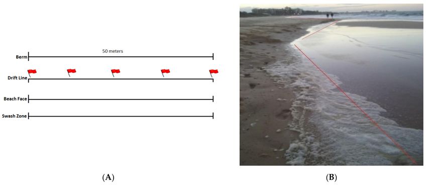
Pathogens 2021, 10, 579 2 of 17
Some of the foaming events in aquatic environments have been linked to the pres-
ence of pollutants [14]. Similarly, the foaming events in the Sunshine Coast region of
Australia along coastal shorelines, lakes and streams were frequently coincided with ad-
verse/turbulent weather conditions when excess pollution in effluent water runoff at
near-shore marine environments was observed https://www.qld.gov.au/environment/
pollution/management/disasters/flood-impacts They carried similar characteristics of
the foam color and density reported by Fryer and Gray [15]. Kurtböke [16–18] reported
the existence of nocardiae in the foaming coastal marine waters as identified via the use of
molecular methods. Some of the isolates were closely related to human pathogenic ones
reported in other parts of the world [17].
Although, species belonging to the Nocardia cluster causing human diseases are gener-
ally considered opportunistic pathogens, nocardiosis is reported to manifest in immuno-
competent people and according to the Genetic and Rare Diseases Information Center [19]
approximately one third of infected individuals are immune-competent. Although the
number of known Nocardia species responsible for disease in humans is also increasing [20],
a review of the literature on nocardiosis in humans by Kandi [21] emphasized otherwise.
One of the reasons for this fact might be due to the lack of epidemiological data on the
incidence of nocardiosis which may not be truly reflective of the pathogenic potential of No-
cardia species. This might be partly due to the fastidious growth patterns of certain species
of Nocardia or lack of rapid diagnostic tests required for their detection and subsequent
isolation in a clinical laboratory [22].
Numerous studies to date have identified three main virulence factors that pathogenic
Nocardia species may use to overcome host’s immune system and cause disease [23–25].
These factors include the presence of trehalose 6-6’-dimycolate (TDM), superoxidase dismu-
tase and the well-categorized antioxidant enzyme catalase. TDM is a glycolipid molecule
found in the cell walls of numerous Nocardia species. TDM is shown to reduce and inhibit
the phagocytic process of the host’s immune cells e.g., macrophages. It has been suggested
that the trehalose levels in microorganisms may vary considerably depending on the envi-
ronmental conditions, and higher levels of trehalose may increase the virulence potential
of a microorganism [26]. Superoxidase dismutase and catalase are potent antioxidant
enzymes that offer a level of resistance against oxidative enzymes produced by foreign or
host cells.
Adhesion, invasion and translocation assays in vitro using human cell lines are typ-
ically used to assess and quantify the protective capability of cells against a pathogenic
organism as well as the ability of the invading organism to adhere and navigate the host’s
immune defense. Whilst some studies have focused on using murine and human cell lines
to assess the adhesion and translocation properties of pathogenic Nocardia species [27–29],
cultured strains isolated from diseased human tissue were used in the tests. Hence, little is
known about these pathogenic characteristics of Nocardia species isolated from environ-
mental samples; particularly from coastal foaming marine waters and sandy beaches that
are commonly used for recreational activities.
In the view of the above, this study was undertaken to determine the presence of
nocardiae in foaming near shore marine waters with use of a novel selective isolation
method. Isolated nocardiae was subsequently investigated for their pathogenic properties
using Calu-3 cells (ATCC HTB-55) and their antibiotic and phage susceptibility patterns
were also determined.
2. Results
2.1. Selective Isolation and Molecular Level Identification of Nocardiae
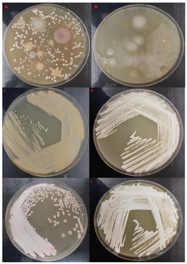
The novel selective isolation method employed resulted in the removal of background
microbiota and successful isolation of Nocardia species. Facultative anaerobic character-
istics of nocardiae resulted in their survival in the anaerobic chamber and clearance of
background bacterial flora (Figure 1 A–F) which would under aerobic conditions would
overgrow nocardiae colonies and render them unculturable.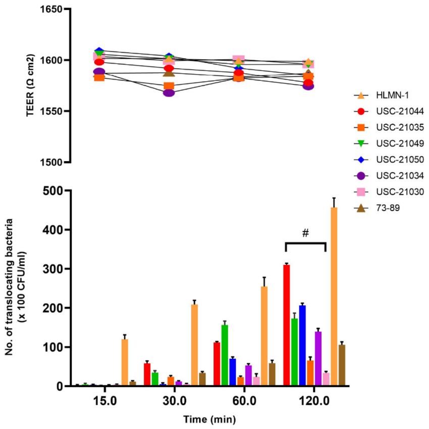
Pathogens 2021, 10, 579                                                                                                 3 of 17

                               When isolation plates were subjected to anaerobic conditions first 62.50% of nocardiae
                          were cultured compared to the 37.5% when direct aerobic conditions were used. A total
                          number of 32 confirmed Nocardia species were isolated following molecular level character-
                          ization (Figure 2, Table S1) and some of them were closely related to the pathogenic species
                          of nocardiae (Figure 3).

                          Figure 1. Isolation of nocardiae via exposure to anaerobic (A) and anaerobic (B) conditions first and
                          then incubating them under aerobic conditions. Typical white/pinkish/salmon colored colonies of
                          Nocardia isolates from the coastal foaming marine waters on GYM-Streptomyces medium: (C) USC-
                          21021, (D) USC-21044, (E) USC-21038 and (F) USC-21046.

                               Twenty-one isolates out of the 32 were closely related to N. nova strains MGA115
                          and one was related to CBU 09/875 (Figure 2), in addition when compared with human
                          pathogenic nocardiae twenty of the isolates were found to be related to N. nova strain JCM
                          6044 (Figure 3).
Twenty-one isolates out of the 32 were closely related to N. nova strains MGA115 and
one was related to CBU 09/875 (Figure 2), in addition when compared with human path-
Pathogens 2021, 10, 579 ogenic nocardiae twenty of the isolates were found to be related to N. nova strain4 ofJCM
17
6044 (Figure 3).
Figure
Figure 2. Phylogenetictree
2. Phylogenetic treeofofthe
the32
32isolates
isolates using
using their
their16s
16srRNA
rRNAgenegenesequences
sequencesin relation to their
in relation to
closest relatives. Bootstrap values ( ≥ 50%) are indicated at nodes. The scale bar represents
their closest relatives. Bootstrap values (≥50%) are indicated at nodes. The scale bar represents per- percentage
(%) divergence.
centage The accession
(%) divergence. number
The accession of the of
number closest relatives
the closest is included.
relatives Mycobacterium
is included. tuberculosis
Mycobacterium
was chosen
tuberculosis as chosen
was an out-group
as an sequence
out-group to sequence
root the tree. *: isolated
to root using
the tree. aerobic conditions,
*: isolated **: isolated
using aerobic condi-
by exposing
tions, anaerobic
**: isolated conditions
by exposing first followed
anaerobic conditions by incubation
first followedat aerobic conditions.
by incubation at aerobic condi-
tions.Pathogens
Pathogens 10, x10,
2021,
2021, FOR579PEER REVIEW 5 of517of 17
Figure 3. Phylogenetic
Figure treetree
3. Phylogenetic of the Nocardia
of the isolates
Nocardia using
isolates their
using 16s 16s
their rRNA gene
rRNA sequences
gene in relation
sequences in relation
to their closest
to their relatives
closest identified
relatives as being
identified clinically
as being significant
clinically and grouped
significant according
and grouped to seven
according to seven
antimicrobial susceptibility patterns [30]. Bootstrap values (≥50%) are indicated at nodes. The scale
antimicrobial susceptibility patterns [30]. Bootstrap values (≥50%) are indicated at nodes. The scale
bar represents percentage (%) divergence. The accession number of the closest relatives is in-
bar represents percentage (%) divergence. The accession number of the closest relatives is included.
cluded. Mycobacterium tuberculosis was chosen as an out-group sequence to root the tree.
Mycobacterium tuberculosis was chosen as an out-group sequence to root the tree.
Nocardiae presence was greater in the summer months confirming the temperature
Nocardiae presence was greater in the summer months confirming the temperature
dependent existence of the taxon members in the near-shore marine environments. In
dependent existence of the taxon members in the near-shore marine environments. In win-
winter months no nocardiae were isolated when both aerobic and anaerobic isolation
ter months no nocardiae were isolated when both aerobic and anaerobic isolation methods
methods were(Figure
were used used (Figure 4,S2).
4, Table Table S2).Pathogens 2021, 10, 579 6 of 17
Figure 4. The total number of Nocardia species isolated during a 1-year period from the foaming
marine waters of the Sunshine Coast region.
2.2. Antibiotic Susceptibility Profiles of the Isolates
Nocardia isolates displayed resistance against some of the antibiotics tested with
cefotaxime, and ceftriaxone showing the highest level of resistance (Table 1, Table S2).
Table 1. Susceptibility of nocardiae isolates (n = 32) to various antibiotics.
Antibiotic % Resistance
Ampicillin (10 µg) 15.7 (n = 5)
Ceftriaxone (30 µg) 75 (n = 24)
Cefotaxime (30 µg) 84.4 (n = 27)
Imipenem (10 µg) 6.3 (n = 2)
Amikacin (30 µg) −
Minocycline (30 µg) 6.3 (n = 2)
Sulfamethoxazole/ trimethoprim (1.25/23.75 µg) 50 (n = 16) *
Erythromycin (15 µg) 6.3 (n = 2)
Tobramycin (10 µg) 63 (n = 20)
−: No resistance was detected, *: E-strip test for confirmation of the susceptibility against this antibiotic is required
if the inhibition zone is 99%) per Calu-3
cell. The degree of adhesion for the positive control E. coli strain HLMN-1 was 21.8 ± 0.6
(90%) whereas for the USC-21034 isolate it was 15.5 ± 0.8 CFU/cell.
2.4. Translocation of Nocardia Isolates
The confluency and the integrity of the Calu-3 cells were initially tested by measuring
the Transepithelial electrical resistance (TEER) value of the culture over 16 days. The results
indicated that cells reach its maximum confluency after 10 days (Figure S1) and therefore
all translocation experiments (maximum 2 h for each assay) were done on day 10 when the
TEER value reached 1588 Ω cm2 .
The six selected Nocardia isolates with the highest degree of adhesion per cell as
well as the highest degree of colonization were tested for their rate of translocation via
Calu-3 cells. The overall translocation ability of the isolate USC-21044 was significantly
greater (p = 0.0164) compared to the other five Nocardia isolates difference in degrees ofPathogens 2021, 10, 579 7 of 17
translocation of the Nocardia isolates over the 120 min assay compared to the controls was
also significant (p = 0.054) (Figure 5). During the translocation assays TEER values remained
constant indicating that the translocation occurred via Calu-3 cells and not between the
cells (Figure 5).
Figure 5. The rate of translocation of six selected nocardiae isolates over 120 min using Calu-3 cell line.
Escherichia coli strains (HMLN-1 and 73–89) were used as positive and negative control respectively.
Data shown are from triplicate experiments and plotted as the mean ± SEM. Transepithelial resistance
remained stable throughout the test.
2.5. Detection of Nocardia-Specific Phage Susceptibility of the Isolates
Majority of the nocardiae isolates were found to be resistant to the nocardiae-specific
phages (Table 2).
Table 2. Nocardia specific phage susceptibility of the isolates.
Strain Code Phage Susceptibility
USC-21042, USC-21037 +++
USC-21044, USC-21021, USC-21046 ++
USC-21018 ±
USC-21034, USC-21050, USC-21049, USC-21035, USC-21030,
USC-21025, USC-21006, USC-21038, USC-21039, USC-21036,
USC-21048, USC-21017, USC-21028, USC-21027, USC-21010, −
USC-21029, USC-21047, USC-21026, USC-21022, USC-21040,
USC-21043, USC-21043, USC-21016, USC-21012, USC-21011, USC-21024
+++: Highly susceptible (complete lysis), ++: Susceptible (complete partial lysis), +: Moderately susceptible
(lysis and single plaques), ±: Low susceptibility (lysis but regrowth of the host), −: Not susceptible.Pathogens 2021, 10, 579 8 of 17
3. Discussion
Investigative studies were conducted to determine whether nocardiae would be
present in the foaming coastal marine waters increasingly encountered at the Sunshine
Coast region of Australia [18,32,33] similar to the nocardiae regularly detected in activated
sludge systems [8]. To determine the presence of the species of the genus Nocardia in the
foam samples a novel selective isolation method was used. The selective battery used
stemmed from the exploitation of the gas requirements of nocardiae during their laboratory
growth. The novel method developed allowed successful isolation of nocardiae without
background contamination thus further facilitating their transfer onto purification media.
When anaerobic conditions are used 62.50% of nocardiae were cultured compared to the
37.5% when aerobic conditions were used, confirming the effectivity of the novel method
developed in the study. Selective isolation of microorganisms is of significant value as
it generates information on the morphologies and growth patterns of microorganisms
under laboratory conditions [18,32,33]. The successful isolation of nocardiae in this study
thus furthered information on the in vitro characteristics of the local isolates which are of
importance in terms of definition of chemotherapy regimens in the event of local infections.
Moreover, laboratory studies on the cultured representatives of nocardiae can provide
further information such as the detection of pathogenicity and hydrolytic capabilities of
the organisms. Such information can further be used for pollution control in coastal marine
waters where nocardiae with such characteristics can survive and persist [34]. In addition,
sewage outflows during cyclonic events can introduce human pathogenic nocardiae into
recreational environments such as local beaches [35]. N. farcinica as an example has been
reported to be an opportunistic pathogen in activated sludge plants [36]. Vautrin et al. [35]
have recently indicated that urban infiltration basins can accumulate urban pollutants and
favor the growth of potentially pathogenic biological agents as Nocardia.
The majority of the nocardiae isolates were also found to be resistant to the phages in
the study. Since these phages were originally isolated using type strains of non-Australian
origin, the resistance observed by the isolates might indicate endemicity for some of the
local isolates. If bacteriophages are used to control nocardia infections data like the one
obtained in this study can provide background information when phage cocktails are
designed for therapy purposes which are of importance for the acceptance and implemen-
tation of phage therapy [37,38].
Over 70% of resistance was detected for cefotaxime and ceftriaxone among the local
isolates when interpretation threshold recommended the Comite de L’Antibiogramme
de la Societe Francaise de Microbiologie were used [39,40]. Currently, there are different
tests used to determine nocardiae antibiotic susceptibility patterns. These tests are (i) disk
diffusion, (ii) broth dilution, (iii) agar dilution, (iv) epsilometer test (E-test) and (v) the
BACTEC radiometric method. Recent study by Lebaux et al. [39] concludes that the Antibi-
otic Susceptibility Test (AST) performed by disk diffusion method appears to be a reliable
method for routine testing of Nocardia. They, however, recommend that for trimethoprim-
sulfamethoxazole, if the inhibition zone isPathogens 2021, 10, 579 9 of 17
events and an increase in human pulmonary and or cutaneous nocardiosis cases despite the
fact that pathogenic Nocardia species are known to become aerosolised and infect a human
host [42]. The primary site of infection for nocardiosis to manifest in humans is in the lungs
via inhalation of Nocardia cells and spores, and with direct skin contact; a primary site that
has the potential to spread to other parts of the body (disseminated nocardiosis). In this
study, Nocardia species isolated from beach foam were found to adhere and translocate
with a varying degree of efficiency in Calu-3 cells in vitro. Findings are of significance for
contact tracing if disease occurs as well as informing swimmers about the health risks of
nocardiae who may be exposed to such species during shoreline foaming events.
From the six isolates chosen for the translocation assay, two isolates (USC-21044 and
USC-21050) displayed a reasonably high level of translocation. Isolate USC-21044’s closest
relative is Nocardia niigatensis and there is little to no evidence in the scientific literature
of Nocardia niigatensis causing pulmonary nocardiosis in humans. However, pulmonary
nocardiosis is known to mimic pulmonary tuberculosis in clinical manifestations, such as
in its signs and symptoms, radiological and laboratory results [43]. Hence, pulmonary
nocardiosis has the potential to be misdiagnosed as pulmonary tuberculosis or vice versa.
A recent study by Muricy et al. [43] reported isolating nocardiae from sputum samples
from patients that were initially thought to be suffering from an infection caused by
Mycobacterium tuberculosis. Isolate USC-21050’s closest relative is Nocardia flavorosea and is
not again a typically isolated Nocardia species from patients suffering nocardiosis. Although,
a study by Tan et al. [44] after having reviewed medical records from the National Taiwan
University Hospital (between 1998–2008), noted that Nocardia flavorosea had been isolated
from four patients suffering from nocardiosis.
Interestingly, numerous isolates in the study whose closest relative was identified as
being a Nocardia nova species/strain displayed significant less adhesion and ultimately
less translocation potential in contrast to the other two isolates USC-21044 and USC-
21050. There is, however, little to no data available in the scientific literature on the
adhesion, and translocation capabilities of pathogenic Nocardia species associated with
coastal foaming marine waters using a human cell lung culture model in vitro.
According to the Public Health Agency of Canada [45] the infectious dose (ID) and the
incubation period of pathogenic Nocardia species in human hosts is unknown. Accordingly,
data like the one presented in this study might be of importance indicting infectious
capacities of locally isolated nocardiae in human hosts. Terrigenous origin actinomycetes
can adapt to marine environments with diversified functions and adaptability [46] and
increasing presence of fish pathogenic nocardiae in marine and aquaculture environments
reported in other parts of the world might be an indication of such adaptation [47].
A review of the literature on nocardiosis in humans by Kandi [21] emphasized the
lack of epidemiological data on the incidence of nocardiosis, and how this lack of data
may not be truly reflective of the pathogenic potential of Nocardia species. This might be
due to lack of sensitive identification tests and or the fastidious growth conditions certain
species of Nocardia require for cultivation, isolation [48] and identification [49] in a clinical
laboratory. However, as recently indicated by Mehta and Shamoo [22] advancements in
rapid molecular diagnostic technology will soon place nocardiae in the “extended pantheon
of medically important pathogens”. Accordingly, studies like the one presented here will
generate further information on the occurrence and diversity of nocardiae in different
environments and might have significance in linking the clinical data to the presence of
disease causative species in the environment by the public health authorities.
4. Materials and Methods
4.1. Sampling Sites and Sample Types
Five locations along the Sunshine Coast beaches (1) Mooloolaba (26◦ 400 58.6” S 153◦ 070 30.1”
E), (2) Maroochydore (26◦ 390 17.7” S 153◦ 060 13.6” E), (3) Cotton Tree (26◦ 390 10.1” S 153◦ 050 56.9”
E), (4) Currimundi (26◦ 460 03.8” S 153◦ 080 12.9” E) and (5) Warana (26◦ 430 09.9” S 153◦ 080 07.7”
E) were selected for foam sampling. These beach locations were selected due to their popu-Pathogens 2021, 10, 579 10 of 17
larity with swimmers, varying beach conditions and history of foam formation. All beach
locations were sampled over a one-year period during the spring, summer, autumn and
winter to allow for a greater assessment of the nocardiae occurrence and diversity in near
shore-foaming marine waters of this region in Australia.
A 50 m stretch along the drift line of each beach location at 10 m intervals/transects
was selected for foam sampling (Figure 6A,B). The samples were collected on the surface
of the drift line and collected in 60 mL sterile specimen containers and stored on ice before
being transported to the University of the Sunshine Coast (USC) for processing within 4 h
of collection.
Figure 6. (A) Foam sampling design and (B) an example site of sampling along the drift line.
4.2. Isolation of Nocardiae Using a Novel Isolation Technique
Each foam sample (1 mL) was serially diluted and the aliquots of 0.2 mL from each
dilution were transferred and spread onto six duplicates of the DSMZ medium #65 (GYM-
Streptomyces medium [50] containing 50 ppm of cycloheximide and nystatin respectively to
remove fungal contaminants, and allowed to dry for 20 min. One set of duplicate plates
were pre-treated in an anoxic (CO2 ) environment (OXOID, Melbourne, Australia) at 28 ◦ C
for 48 h prior to being incubated and subsequently cultured at 28 ◦ C in an O2 environment
(standard atmospheric conditions). The remaining duplicates were directly incubated at
28 ◦ C in an O2 environment without being pre-treated with CO2 . Following ~2–3 weeks of
incubation, colonies were selected based on typical Nocardia morphology as described in
the Wink compendium [51].
Each selected isolate was then purified using DSMZ medium #65, and the plates were
incubated at 28 ◦ C in an O2 environment for ~2–3 weeks. Subsequently, following observ-
able Nocardia morphology and nil signs of contamination, the isolates were cryopreserved
in tryptic soy broth (TSB, OXOID) supplemented with 20% glycerol and stored at −20 ◦ C
prior to undergoing molecular identification.
4.3. Molecular Characterization of the Isolates
Direct colony polymerase chain reaction (PCR) adapted from Gathogo et al. [52] was
used to extract the chromosomal DNA and amplify the 16s rRNA gene from the Nocardia
isolates. The complete 16S rRNA gene was amplified using the HotStarTaq® Multiplex PCR
Kit (QIAGEN, Melbourne, Australia) for the extension of all primers. The amplification of
the 16S rRNA gene was obtained by using the bacterial specific and universal primers 27F
(50 -AGAGTTTGATCCTGGCTCAG-30 ) and 1492-R (50 GGTT ACCTTGTTACGACTT-30 ) as
per Madueno et al. [53]. The PCR setting consisted of initial chromosomal DNA extractionPathogens 2021, 10, 579 11 of 17
and denaturation step at 95 ◦ C for 15 min, followed by 34 cycles of 94 ◦ C for 30 s, 60 ◦ C for
1.5 min, 72 ◦ C for 1.5 min, and the final extension was programmed at 72 ◦ C for 10 min
and hold on completion at 12 ◦ C. The amplified PCR products were gel electrophoresed at
90 v for 90 min on a 1.0% agarose (Molecular Grade, Bioline, Sydney, Australia) in 0.6×
TrisBase EDTA (TBE) stained with ethidium bromide and viewed under a UV source for
the presence of a 1500 bp DNA product; the PCR products were sequenced at Macrogen
in Korea [31].
Forward and reverse sequences in FASTA format (including chromatogram) of the
32 Nocardia isolates were imported into DNA Baser Assembler (Version 5.15.0.087) [54] for
contiguous (contig) ambiguity correction and sequence assembly (de novo).
The 16s rRNA gene sequences (contigs) of the 32 isolates and reference strains were
imported into MEGA6 [55] in FASTA format and aligned according to MUSCLE [56];
Reference strain contigs were imported from the National Center for Biotechnology Infor-
mation [57].
The phylogenetic analysis/evolutionary history was inferred by using the maximum
likelihood method based on the Tamura-Nei model [58]. Sequence alignment and evolu-
tionary analyses were conducted in MEGA6 [55] and the phylogenetic tree was constructed
accordingly.
4.4. Detection of Antibiotic Susceptibility Patterns of the Isolates
Antibiotic susceptibility of the isolates and type strains were tested according to
the disc diffusion method as outlined by the Clinical and Laboratory Standards Insti-
tute (CLSI) [59]; Staphylococcus aureus ATCC® 25923, and Escherichia coli ATCC® 25922
(only for imipenem 10 µg) were used as controls [39,40]. The 32 isolates, and type strains
used as controls were tested in duplicates against ten different antibiotics (OXOID) (ampi-
cillin (AMP: 10 µg), ceftriaxone (CRO: 30 µg), cefotaxime (CTX: 30 µg), imipenem (IMP:
10 µg), amikacin (AK: 30 µg), minocycline (MH: 30 µg), sulphamethoxazole/trimethoprim
(SXT: 1.25/23.75 µg), erythromycin (E: 15 µg) and tobramycin (TOB: 10 µg). Results
were recorded according to the zone diameter interpretive standards recommended the
Comite de L’Antibiogramme de la Societe Francaise de Microbiologie, Recommendations
2013 [39,40].
4.5. Calu-3 Cell Line
The Calu-3 cells (ATCC HTB-55) obtained from the American Type Culture Collection
(ATCC, Manassas, VA, USA) were maintained at 37 ◦ C and 5% CO2 in a 50 mL culture
flask in Eagle’s Essential Medium (EMEM, ATC302003). The medium was supplemented
with 15% (v/v) fetal bovine serum (FBS, Moregate Biotech, Brisbane, Australia), and 1%
(v/v) penicillin-streptomycin. Media was sterile filtered through a Millipore Express PES
membrane (0.22 µm). The cell culture media was monitored and changed every 48 h until
cell growth reached 95% confluence (~2.5 × 10⁶ cells) using a disposable hemocytometer
(INCYTO-C-Chip, ProSciTech, Townsville, Australia).
The Nocardia isolates were grown in 10 mL of TSB (OXOID) in an incubator shaker
for 8 days at 28 ◦ C and at 165 rpm. Prior to the adhesion assay, the Nocardia suspensions
were centrifuged at 3500 rpm for 10 min and the supernatant discarded. The pellets were
re-suspended in PBS (pH 7.4) at adjusted to the desired cell density (1.0 × 10⁹ CFU mL−1 ,
OD = 1 at 600 nm); cell count was also verified by a cell plate count). E. coli strain HMLN-1
a professional translocating strain isolated from a mesenteric lymph-node of a fetal case
of pancreatitis [60] and 73–89, originally isolated from ceacal contents of a hemorrhaged
rat [61] were used as positive and negative controls respectively. They were grown in
10 mL of TSB in an incubator shaker for 2 days at 37 ◦ C and at 165 rpm. Prior to the
assay, the suspensions of control strains were centrifuged at 3500 rpm for 10 min and the
supernatant discarded. The pellets were re-suspended in PBS (pH 7.4) at the desired cell
density (1.0 × 10⁹ CFU mL−1 , OD = 1 at 600 nm; cell counts were also verified by a cell
plate count.Pathogens 2021, 10, 579 12 of 17
4.6. Adhesion and Translocation Assays
The adhesion and translocation assays performed were adapted from Vollmerhausen
et al. [62]. Calu-3 cells were seeded into 8 well chamber slides (Lab-Tek II chamber, Thermo
Fisher Scientific, Melbourne, Australia) filled with 0.3 mL of growth medium supple-
mented with 15% (v/v) FBS and 1% (v/v) penicillin-streptomycin and inoculated with a
1.7 × 10⁵ mL cells in each of the 8 chambers. The cells were grown to 100% confluence and
prior to the adhesion assay, the medium was removed, and each chamber was washed
three times with phosphate-buffer saline (PBS) with pH 7.4, Antibiotic free medium was
then added to each chamber. Furthermore, to obtain an accurate multiplicity of infection
ratio (MOI) the average of three chambers cell count at 100% confluence was used. It was
established that the mean number of cells at 100% confluence per chamber was 200,000 cells;
this cell count was used to determine the number of bacterial cells required to achieve a
1:50 MOI [62].
Subsequently, 10 µL of bacterial suspension (1.0 × 10⁹ CFU mL, OD = 1 at 600 nm)
was inoculated into each chamber and incubated for 90 min at 37 ◦ C. Cells were then fixed
with 95% ethanol (v/v) for 5 min and stained using Giemsa stain for a period of 30 min,
and observed using a light microscopy. All tests were performed in triplicate.
Bacterial adhesion per cell was determined by counting the number of adhering
bacteria per 25 randomly selected Calu-3 cells. The percentage of bacteria colonizing
Calu-3 cells was determined by randomly counting 100 Calu-3 cells and the mean number
of cells showing bacterial adhesion was calculated after three replicate counting. Isolates
showing greater than 50% adhesion to Calu-3 cells were selected for the translocation assay.
These isolates also showed to have a higher number of bacteria per cells.
4.7. Trans-Epithelial Electrical Resistance Measurements and Translocation Assay
TEER was initially measured during the growth of Calu-3 cells using a Millicell-ERS
voltammeter (MERS00001, Millipore, Melbourne, Australia); over 16 post seeding days
to establish the TEER value of a confluence growth of Calu-3 cells. The 24-well plate was
pre-filled with EMEM containing 15% FBS and 1% (v/v) penicillin-streptomycin at 37 ◦ C.
TEER between the inner and outer wells was measured and calculated using the following
equation: measured monolayer resistance—measured blank resistance (filter without cells)
× area of the filter (0.6 cm2 ) = trans-monolayer resistance (Ω cm−2 ) [63,64].
For the translocation assay, the Calu-3 Cells were seeded at 5 × 10⁴ cells into a 0.8 µm
insert (12 mm diameter, Millicell inserted within a 24 well culture plate (NUNC, Melbourne,
Australia). The inner well was maintained with 400 µL and the outer well contained 600 µL
of culture media (EMEM + 15% FBS + 1% (v/v) penicillin-streptomycin). Cells were grown
to confluence and reached a stable TEER value (≥1500 Ω cm2 ) over 10 days which was
established from the initial assay. Next, both wells (inner and outer) had their media
removed, washed three times with PBS (pH 7.4) and replaced with antibiotic free media.
The suspensions of 10 µL (1.0 × 10⁹ CFU mL, OD = 1 at 600 nm) of bacterial isolates
were inoculated into corresponding wells and incubated at 28 ◦ C. Samples (100 µL) were
collected from the outer well following 15, 30, 60 and 120 min of incubation, serially
diluted using PBS and spread onto GYM agar. The culture media taken from the outer
well was replaced at each sampled time interval. Plates were incubated for 8 days at 28 ◦ C,
and colony growth was checked every 24 h. The translocating bacteria were calculated and
presented as the mean ± SEM [65,66].
4.8. Detection of Nocardiae Specific Phage Susceptibility of the Isolates
The six phages previously isolated from marine environments located on the Sunshine
Coast where foam was evident [17,18] and had been stored in the University of the Sunshine
Coast-Microbial Library (USC-ML) were selected for this assay (Table 3). All of the phages
were in the Siphoviridae morphology group [67] with broad activity spectra within the
genus Nocardia (Table 4).Pathogens 2021, 10, 579 13 of 17
Table 3. Details of the Nocardia-specific phages.
Phage Codes Propagation Hosts Used to Isolate the Phages
Ø1 Nocardia soli (DSMZ-44490)
Ø2 Nocardia soli (DSMZ-44490)
Ø3 Nocardia soli (DSMZ-44490)
Ø4 Nocardia asteroides (ACM-2963) *
Ø5 Nocardia soli (DSMZ-44490)
Ø6 Nocardia asteroids (ACM-2963) *
* ACM: Australian Collection of Microorganisms.
Table 4. Susceptibility of the Nocardia type strains to the composite phage suspension containing all
six phages.
Type Strain IDs Phage Susceptibility
N. shimofusensis (DSMZ 44733) −
N. takedensis (DSMZ 44802) ±
N. cumidelens/soli (DSMZ 44488) ++
N. soli (DSMZ 44490) +++
N. uniformis (DSMZ 43136) ++
N. salmonicida (DSMZ 40472) ++
N. pseudovaccinii (DSMZ 43406) ++
N. veterana (DSMZ 44445) ++
N. fluminea (DSMZ 44489) −
N. flavorosea (DSMZ 44480) +++
N. asteroides (ACM 131) ++
N. asteroides (ACM 2963) ++
+++: Highly susceptible (complete lysis), ++: Susceptible (complete partial lysis), +: Moderately susceptible
(lysis and single plaques), ±: Low susceptibility, −: Not susceptible.
Phages were propagated in peptone-yeast extract calcium (PYCa) broth [68], and rou-
tinely seeded with their designated propagation hosts (PH) until~1.0 × 10⁸ plaque forming
units/ml for each phage isolate was achieved. A composite sample of six different phages
was prepared using equal volumes of each phages and the phage susceptibility of the
isolates was determined using spot test assay as described by Thomas et al. [8]. Lysis zones
were evaluated using the index described by Jonns et al. [69].
4.9. Statistical Analysis
The total number of isolates were presented in the format of a bar and pie chart accord-
ing to beach location, season, and isolation technique used. All charts were constructed
by GraphPad Prism statistical software package (version 8.4.2, Graphpad, San Diego,
CA, USA).
For the adhesion and translocation assays data were presented as mean ± standard
deviation (SD), percentage (%), mean and standard error of the mean (SE). Data were anal-
ysed using one-way Brown-Forsythe and Welch’s ANOVA and differences were deemed
statistically significant if p < 0.05; The Brown-Forsythe and Welch’s ANOVA was employed
due to the heterogeneity of group variance. The Post-Hoc analysis was performed by
Dunnett’s T3 multiple comparisons test to identify where the difference amongst the means
occurred (significant if p < 0.05).
The translocation assay results were analysed using traditional ANOVA in conjunc-
tion with Tukey’s multiple comparisons test due to smaller numbers and meeting the
requirements of equal variance amongst the groups. Comparison between the Nocardia
and the control group was conducted by means of an unpaired t-test with Welch correction,
and deemed significantly different if p < 0.05. The GraphPad Prism statistical software
package (version 8.4.2) was used for all statistical analysis.Pathogens 2021, 10, 579 14 of 17
5. Conclusions
In conclusion, the results obtained from this study further increases our understanding
of the occurrence, diversity and human pathogenic potential of Nocardia isolates obtained
from foaming coastal marine waters in the Sunshine Coast region of Australia. From the
human health point of view the presence of such isolates in the coastal foams may present
a risk, especially swimmers or individuals with underlying immune conditions, thus iden-
tification of such nocardiae might lead to the development of preventative measures by
the local authorities. In addition, as indicated by Porri et al. [70] sea foam is important
for “retaining larvae of polychaetes, mussels and barnacles near to the shore” thus foam’s
unpolluted condition might be of significance for the health of near shore marine life
as well.
Supplementary Materials: The following are available online at https://www.mdpi.com/article/10
.3390/pathogens10050579/s1. Table S1: Details of the Nocardia isolates; a Closest relative based on 16s
rRNA gene sequence similarity. b +++: Highly susceptible (complete lysis), ++: Susceptible (complete
partial lysis), +: Moderately susceptible (lysis and single plaques), ±: Low susceptibility (Lysis but
regrowth of the host), −: Not susceptible. c Marine/beach locations situated on the Sunshine Coast,
QLD, Australia; Table S2: S, susceptible. I, Intermediate. R, resistant. Footnote: S, susceptible.
I, Intermediate. R, resistant, ¥: inhibition zone isPathogens 2021, 10, 579 15 of 17
5. Hashemi-Shahraki, A.; Heidarieh, P.; Bostanabad, S.Z.; Hashemzadeh, M.; Feizabadi, M.M.; Schraufnagel, D.; Mirsaeidi, M.
Genetic diversity and antimicrobial susceptibility of Nocardia species among patients with nocardiosis. Sci. Rep. 2015, 5, 17862.
[CrossRef]
6. Desai, H.; Wickstrom, K.; Low, S.; Mahmoud, N. Nocardia: An unusual bacteria in an unexpected patient. Am. J. Respir. Crit.
Care Med. 2016, 193, 1–18.
7. Pitt, P.; Jenkins, D. Causes and control of Nocardia in activated sludge. J. Water Pollut. Control Fed. 1990, 62, 143–150.
8. Thomas, J.A.; Soddell, J.A.; Kurtböke, D.I. Fighting foam with phages? Water Sci. Technol. 2002, 46, 511–518.
9. Ovez, S.; Orhon, D. Microbial Ecology of Bulking and Foaming Activated Sludge Treating Tannery Wastewater. J. Environ. Sci.
Health Part A 2005, 40, 409–422. [CrossRef]
10. Pajdak-Stós, A.; Kocerba-Soroka, W.; Fyda, J.; Sobczyk, M.; Fiałkowska, E. Foam-forming bacteria in activated sludge effectively
reduced by rotifers in laboratory- and real-scale wastewater treatment plant experiments. Environ. Sci. Pollut. Res. 2017, 24,
13004–13011. [CrossRef]
11. Wandl, G.; Müller-Rechberger, H.; Matsché, N.; Svardal, K.; Winkler, S. Two stage activated sludge plants—Influence of different
operational modes on sludge bulking and nitrification. Water Sci. Technol. 2002, 46, 479–486. [CrossRef]
12. DeWitt, D.; Wagoner, D.L.; Jarrell, J. Use of On-Line Analyzers—Identification of New Approaches for WWTP Process Control
and Optimization. Proc. Water Environ. Fed. 2013, 2013, 6766–6776. [CrossRef]
13. Jenkins, D.; Richard, M.G.; Daigger, G.T. Manual on the Causes and Control of Activated Sludge Bulking, Foaming, and Other Solids
Separation Problems; Lewis Publishers: Boca Raton, FL, USA, 2004.
14. Schilling, K.; Zessner, M. Foam in the aquatic environment. Water Res. 2011, 45, 4355–4366. [CrossRef]
15. Fryer, M.; Gray, N. Foaming Scum Index (FSI)—A new tool for the assessment and characterisation of biological mediated
activated sludge foams. J. Environ. Manag. 2012, 110, 8–19. [CrossRef]
16. Kurtböke, D.I. (Lab Report) ’Chocolate mousse’ on Sunshine Coast beaches. Microbiol. Aust. 2008, 29, 104–105.
17. Kurtböke, D.I. Actinomycetes in Biodiscovery: Genomic Advances and New Horizons. In The Handbook of Microbial Resources;
Gupta, V.K., Sharma, G.D., Tuohy, M.G., Gaur, R., Eds.; CAB International Publications: Oxfordshire, UK, 2016; pp. 567–590.
18. Kurtböke, D.I. Ecology and Habitat Distribution of Actinobacteria. In Biology and Biotechnology of Actinobacteria; Wink, J., Hamedi,
J., Eds.; Springer: Berlin, Germany, 2017; pp. 123–149.
19. Genetic and Rare Diseases Information Center (GARD). Available online: https://rarediseases.info.nih.gov/diseases/7210
/nocardiosis (accessed on 15 March 2020).
20. Schlaberg, R.; Fisher, M.A.; Hanson, K.E. Susceptibility Profiles of Nocardia Isolates Based on Current Taxonomy. Antimicrob.
Agents Chemother. 2013, 58, 795–800. [CrossRef]
21. Kandi, V. Human Nocardia Infections: A Review of Pulmonary Nocardiosis. Cureus 2015, 7, 1–6. [CrossRef]
22. Mehta, H.H.; Shamoo, Y. Pathogenic Nocardia: A diverse genus of emerging pathogens or just poorly recognized? PLoS Pathog.
2020, 16, e1008280. [CrossRef]
23. Beaman, B.L.; Beaman, L. Nocardia Asteroides as an Invasive, Intracellular Pathogen of the Brain and Lungs. In Bacterial Invasion
into Eukaryotic Cells; Oelschlaeger, T.A., Hacker, J., Eds.; Springer: New York, NY, USA, 2000; pp. 167–197.
24. Trevino-Villarreal, J.H.; Vera-Cabrera, L.; Valero-Guillén, P.L.; Salinas-Carmona, M.C. Nocardia brasiliensis Cell Wall Lipids
Modulate Macrophage and Dendritic Responses That Favor Development of Experimental Actinomycetoma in BALB/c Mice.
Infect. Immun. 2012, 80, 3587–3601. [CrossRef]
25. Christova, N.; Lang, S.; Wray, V.; Kaloyanov, K.; Konstantinov, S.; Stoineva, I. Production, Structural Elucidation, and In Vitro
Antitumor Activity of Trehalose Lipid Biosurfactant from Nocardia farcinica Strain. J. Microbiol. Biotechnol. 2015, 25, 439–447.
[CrossRef]
26. Tournu, H.; Fiori, A.; Van Dijck, P. Relevance of Trehalose in Pathogenicity: Some General Rules, Yet Many Exceptions. PLoS Pathog.
2013, 9, e1003447. [CrossRef]
27. Beaman, B.L. Differential binding of Nocardia asteroides in the murine lung and brain suggests multiple ligands on the nocardial
surface. Infect. Immun. 1996, 64, 4859–4862. [CrossRef]
28. Tam, S.; Barry, D.P.; Beaman, L.; Beaman, B.L. Neuroinvasive Nocardia asteroides GUH-2 Induces Apoptosis in the Substantia
Nigra in Vivo and Dopaminergic Cells in Vitro. Exp. Neurol. 2002, 177, 453–460. [CrossRef]
29. Barry, D.P.; Beaman, B.L. Nocardia asteroides strain GUH-2 induces proteasome inhibition and apoptotic death of cultured cells.
Res. Microbiol. 2007, 158, 86–96. [CrossRef]
30. Brown-Elliott, B.A.; Conville, P.; Wallace, R.J. Current Status of Nocardia Taxonomy and Recommended Identification Methods.
Clin. Microbiol. Newsl. 2015, 37, 25–32. [CrossRef]
31. Macrogen. 2020. Available online: http://www.macrogen.com (accessed on 24 March 2020).
32. Kurtböke, D.I. Selective Isolation of Rare Actinomycetes; Queensland Complete Printing Services: Nambour, Australia, 2003.
33. Kurtböke, D.I. Biodiscovery from rare actinomycetes: An eco-taxonomical perspective. Appl. Microbiol. Biotechnol. 2012, 93,
1843–1852. [CrossRef]
34. Azadi, D.; Shojaei, H. Biodegradation of polycyclic aromatic hydrocarbons, phenol and sodium sulfate by Nocardia species
isolated and characterized from Iranian ecosystems. Sci. Rep. 2020, 10, 1–12. [CrossRef]Pathogens 2021, 10, 579 16 of 17
35. Vautrin, F.; Pujic, P.; Paquet, C.; Bergeron, E.; Mouniée, D.; Marchal, T.; Salord, H.; Bonnet, J.-M.; Cournoyer, B.; Winiarski, T.; et al.
Microbial risk assessment of Nocardia cyriacigeorgica in polluted environments, case of urban rainfall water. Comput. Struct.
Biotechnol. J. 2021, 19, 384–400. [CrossRef]
36. Stratton, H.; Seviour, R.; Soddell, J.; Blackall, L.; Muir, D. The opportunistic pathogen Nocardia farcinica is a foam-producing
bacterium in activated sludge plants. Lett. Appl. Microbiol. 1996, 22, 342–346. [CrossRef]
37. Alavidze, Z.; Aminov, R.; Betts, A.; Bardiau, M.; Bretaudeau, L.; Caplin, J.; Nino, C.; Coffey, A.; Cooper, I.; De Vos, D.; et al.
Silk route to the acceptance and re-implementation of bacteriophage therapy-expert round table on acceptance and re-
implementation of bacteriophage therapy. Biotechnol. J. 2016, 11, 595–600.
38. Sybesma, W.; Rohde, C.; Bardy, P.; Pirnay, J.-P.; Cooper, I.; Caplin, J.; Chanishvili, N.; Coffey, A.; De Vos, D.; Scholz, A.H.; et al.
Silk Route to the Acceptance and Re-Implementation of Bacteriophage Therapy—Part II. Antibiotics 2018, 7, 35. [CrossRef]
39. Lebeaux, D.; Bergeron, E.; Berthet, J.; Djadi-Prat, J.; Mouniée, D.; Boiron, P.; Lortholary, O.; Rodriguez-Nava, V. Antibiotic
susceptibility testing and species identification of Nocardia isolates: A retrospective analysis of data from a French expert
laboratory, 2010–2015. Clin. Microbiol. Infect. 2019, 25, 489–495. [CrossRef]
40. Bonnet, R.; Caron, F.; Cavallo, J.; Chardon, H.; Chidiac, C.; Courvalin, P.; Drugeon, H.; Dubreuil, L.; Jarlier, V.; Jehl, F. Comité de
l’Antibiogramme de la Société Française de Microbiologie. Recommandations. 2013. Available online: https://resapath.anses.fr/
resapath_uploadfiles/files/Documents/2013_CASFM.pdf (accessed on 13 April 2021).
41. Tan, Y.E.; Chen, S.C.-A.; Halliday, C.L. Antimicrobial susceptibility profiles and species distribution of medically relevant Nocardia
species: Results from a large tertiary laboratory in Australia. J. Glob. Antimicrob. Resist. 2020, 20, 110–117. [CrossRef]
42. Navarrete-Navarrete, N.; Sevilla, J.E.; García, M.T.; Urbano, F.; Sabio, J.M.; Jiménez-Alonso, J. A Man with Unilateral Endoph-
thalmitis: A Case of Disseminated Nocardiosis. Case Rep. Infect. Dis. 2015, 2015, 1–2. [CrossRef]
43. Muricy, E.C.M.; Lemes, R.A.; Bombarda, S.; Ferrazoli, L.; Chimara, E. Differentiation Between Nocardia spp. and Mycobacterium
spp.: Critical Aspects for Bacteriological Diagnosis. Revista Instituto Medicina Tropical São Paulo 2014, 56, 397–401. [CrossRef]
44. Tan, C.-K.; Lai, C.-C.; Lin, S.-H.; Liao, C.-H.; Chou, C.-H.; Hsu, H.-L.; Huang, Y.-T.; Hsueh, P.-R. Clinical and microbiological
characteristics of Nocardiosis including those caused by emerging Nocardia species in Taiwan, 1998–2008. Clin. Microbiol. Infect.
2010, 16, 966–972. [CrossRef]
45. Public Health Agency of Canada. Nocardia. Available online: http://www.phac-aspc.gc.ca/lab-bio/res/psds-ftss/nocardia-eng.
php (accessed on 16 April 2020).
46. Kurtböke, D.; Okazaki, T.; Vobis, G. Actinobacteria in Marine Environments: From Terrigenous Origin to Adapted Functional
Diversity. In Encyclopedia of Marine Biotechnology; Kim, S.K., Ed.; John Wiley & Sons: Hoboken, NJ, USA, 2020; pp. 1951–1978.
47. Han, H.; Kwak, M.; Ha, S.; Yang, S.; Kim, J.D.; Cho, K.; Kim, T.; Cho, M.Y.; Kim, B.; Jung, S.; et al. Genomic characterization of
Nocardia seriolae strains isolated from diseased fish. Microbiology 2018, 8, e00656. [CrossRef]
48. Das, A.K.; Nandy, S.; Dudeja, M.; Tiwari, R.; Alam, S. The Incidence of Nocardiosis at Pulmonary and Extra-Pulmonary Sites.
J. Clin. Diagn. Res. 2013, 7, 1427–1429. [CrossRef]
49. Currie, B.J.; Carapetis, J.R. Skin infections and infestations in Aboriginal communities in northern Australia. Australas. J. Dermatol.
2000, 41, 139–143. [CrossRef]
50. Leibniz Institute DSMZ—German Collection of Microorganisms and Cell Cultures. 2007. Available online: https://www.dsmz.
de/microorganisms/medium/pdf/DSMZ_Medium65.pdf (accessed on 1 January 2020).
51. Global Biodiversity Information Facility (GBIF). 2014. Available online: http://www.gbif-prokarya.de/microorganisms/wink.
html (accessed on 14 January 2020).
52. Gathogo, E.W.N.; Waugh, A.C.W.; Perić, N.; Redpath, M.B.; Long, P.F. Colony PCR amplification of actinomycete DNA. J. Antibiot.
2003, 56, 423–424. [CrossRef]
53. Madueño, L.; Coppotelli, B.; Alvarez, H.; Morelli, I. Isolation and characterization of indigenous soil bacteria for bioaugmentation
of PAH contaminated soil of semiarid Patagonia, Argentina. Int. Biodeterior. Biodegrad. 2011, 65, 345–351. [CrossRef]
54. DNA Baser. Sequence Assembly Software, 2020. Available online: http://www.dnabaser.com (accessed on 8 March 2020).
55. Tamura, K.; Stecher, G.; Peterson, D.; Filipski, A.; Kumar, S. MEGA6: Molecular evolutionary genetics analysis version 6.0.
Mol. Biol. Evol. 2013, 30, 2725–2729.
56. Edgar, R.C. MUSCLE: Multiple sequence alignment with high accuracy and high throughput. Nucleic Acids Res. 2004, 32,
1792–1797. [CrossRef]
57. National Center for Biotechnology Information (NCBI). BLAST: Basic Local Alignment Tool. 2020. Available online: http:
//blast.ncbi.nlm.nih.gov/Blast.cgi (accessed on 4 November 2020).
58. Tamura, K.; Nei, M. Estimation of the number of nucleotide substitutions in the control region of mitochondrial DNA in humans
and chimpanzees. Mol. Biol. Evol. 1993, 10, 512–526. [CrossRef]
59. CLSI. Performance Standards for Antimicrobial Susceptibility Testing M100S; Clinical and Laboratory Standards Institute: Wayne, PA,
USA, 2016.
60. Nettelbladt, C.G.; Katouli, M.; Bark, T.; Svenberg, T.; Möllby, R.; Ljungqvist, O. Evidence of Bacterial Translocation in Fatal
Hemorrhagic Pancreatitis. J. Trauma Inj. Infect. Crit. Care 2000, 48, 314–315. [CrossRef]
61. Katouli, M.; Nettebladt, C.G.; Muratov, V.; Ljungqvist, O.; Bark, T.; Svenberg, T.; Möllby, R. Selective translocation of coliform
bacteria adhering to caecal epithelium of rats during catabolic stress. J. Med. Microbiol. 1997, 46, 571–578. [CrossRef]Pathogens 2021, 10, 579 17 of 17
62. Vollmerhausen, T.L.; Woods, J.L.; Faoagali, J.; Katouli, M. Interactions of uroseptic Escherichia coli with renal (A-498) and
gastrointestinal (HT-29) cell lines. J. Med. Microbiol. 2014, 63, 1575–1583. [CrossRef]
63. Dekali, S.; Gamez, C.; Kortulewski, T.; Blazy, K.; Rat, P.; Lacroix, G. Assessment of an in vitro model of pulmonary barrier to
study the translocation of nanoparticles. Toxicol. Rep. 2014, 1, 157–171. [CrossRef]
64. Maher, S.; McClean, S. Investigation of the cytotoxicity of eukaryotic and prokaryotic antimicrobial peptides in intestinal epithelial
cells in vitro. Biochem. Pharmacol. 2006, 71, 1289–1298. [CrossRef]
65. Ramos, N.L.; Lamprokostopoulou, A.; Chapman, T.A.; Chin, J.C.; Römling, U.; Brauner, A.; Katouli, M. Virulence characteristics
of translocating Escherichia coli and the interleukin-8 response to infection. Microb. Pathog. 2011, 50, 81–86. [CrossRef]
66. Katouli, M.; Ramos, N.L.; Nettelbladt, C.G.; Ljungdahl, M.; Robinson, W.; Ison, H.M.; Brauner, A.; Möllby, R. Host species-specific
translocation of Escherichia coli. Eur. J. Clin. Microbiol. Infect. Dis. 2009, 28, 1095–1103. [CrossRef]
67. Kurtböke, D.I. Bioactive Actinomycetes: Reaching Rarity Through Sound Understanding of Selective Culture and Molecular
Diversity. In Microbial Resources; Kurtböke, D.I., Ed.; Elsevier: London, UK, 2017; pp. 45–76.
68. Bradley, S.; Anderson, D.; Jones, L. Phylogeny of actinomycetes as revealed by susceptibility to actinophage. J. Ind. Microbiol.
1961, 2, 223–237.
69. Jonns, J.A.; Brooks, P.R.; Exley, P.; Poole, S.; Kurtböke, D.I. Streptophage-mediated control of off-flavour taint producing
streptomycetes isolated from barramundi ponds. Synth. Syst. Biotechnol. 2017, 2, 105–112. [CrossRef]
70. Porri, F.; Puccinelli, E.; Weidberg, N.; Pattrick, P. Lack of match between nutrient-enriched marine seafoam and intertidal
abundance of long-lived invertebrate larvae. J. Sea Res. 2021, 170, 102009. [CrossRef]You can also read