Lingering dynamics in microvascular blood flow
←
→
Page content transcription
If your browser does not render page correctly, please read the page content below
Lingering dynamics in microvascular blood flow
                                                           A. Kihm,1 S. Quint,1, 2 M. W. Laschke,3 M. D. Menger,3 T. John,1 L. Kaestner,1, 4 and C. Wagner1, 5, a)
                                                           1)
                                                              Department of Experimental Physics, Saarland University, 66123 Saarbruecken,
                                                           Germany
                                                           2)
                                                              Cysmic GmbH, 81379 München
                                                           3)
                                                              Institute for Clinical and Experimental Surgery, Saarland University, 66421 Homburg,
                                                           Germany
                                                           4)
                                                              Theoretical Medicine and Biosciences, Saarland University, 66421 Homburg,
                                                           Germany
                                                           5)
                                                              Physics and Materials Science Research Unit, University of Luxembourg, Luxembourg
                                                           (Dated: February 26, 2021)
                                                           The microvascular networks in the body of vertebrates consist of the smallest vessels such as arterioles,
arXiv:2102.12929v1 [physics.bio-ph] 25 Feb 2021

                                                           capillaries, and venules. The flow of RBCs through these networks ensures the gas exchange in as well as the
                                                           transport of nutrients to the tissues. Any alterations in this blood flow may have severe implications on the
                                                           health state. Since the vessels in these networks obey dimensions similar to the diameter of RBCs, dynamic
                                                           effects on the cellular scale play a key role. The steady progression in the numerical modeling of RBCs, even
                                                           in complex networks, has led to novel findings in the field of hemodynamics, especially concerning the impact
                                                           and the dynamics of lingering events, when a cell meets a branch of the network. However, these results
                                                           are yet to be matched by a detailed analysis of the lingering experiments in vivo. To quantify this lingering
                                                           effect in in vivo experiments, this study analyzes branching vessels in the microvasculature of Syrian golden
                                                           hamsters via intravital microscopy and the use of an implanted dorsal skinfold chamber. It also presents a
                                                           detailed analysis of these lingering effects of cells at the apex of bifurcating vessels, affecting the temporal
                                                           distribution of cell-free areas of blood flow in the branches, even causing a partial blockage in severe cases.
                                                           Keywords: Microvasculature, Lingering, in vivo, blood flow, red blood cells

                                                  INTRODUCTION                                                   to mesh-like networks, where the subsequent diameters of
                                                                                                                 vessels at bifurcations satisfy, in general, Murray’s law,
                                                     The steady transport of nutrients to the tissues of the     although deviations are well-known10,11 . Compared to
                                                  body, as well as the delivery of oxygen, is crucial for the    macrovascular blood flow, effects arising from the par-
                                                  health state in animals and humans. However, in uni-           ticulate nature of blood are more pronounced in the mi-
                                                  cellular living beings, this task can be achieved by pure      crocirculation. Considering the volume fraction of RBCs
                                                  diffusion. Active transport is necessary to ensure this        in whole blood suspensions, the so-called hematocrit, a
                                                  exchange in vertebrates. This process is realized by the       temporal heterogeneity, is given in the capillary vessels
                                                  synergistic action of the heart, blood, and vasculature1 .     contrasting a temporal homogeneity ubiquitous in large
                                                  A key setting is the pressure difference between the aorta     vessels such as arteries (cf. Movie S2). Similarly, the
                                                  and the vena cava2 . In between, the microvasculature is       vessel diameter in which the blood flows has been found
                                                  present, consisting of the smallest vessels such as the ar-    to have an impact on the local hematocrit12,13 . This
                                                  terioles, capillaries, and venules. The diameters of these     so-called Fåhræus effect can be explained by the lateral
                                                  vessels in the microcirculation are in the range of the        migration of RBCs to the vessel centerline, leaving a cell-
                                                  size of individual RBCs, which highlights the importance       depleted layer close to the vessel walls. As a result, RBCs
                                                  of the deformation ability of RBCs. Due to the over-           obey a higher speed on average in the Poiseuille profile
                                                  all length of the microvasculature and the present cross-      than the bulk speed of the plasma. In the seminal work
                                                  sections of vessels, it is responsible for the largest re-     by Fåhræus and Lindqvist14 , the former effect could be
                                                  sistance and hence the largest dissipation of energy in        identified as one of the major causes of the dependence
                                                  the blood flow3–5 . Since the microcirculation is embed-       on the vessel diameter of apparent viscosity in blood so-
                                                  ded in the tissues and ensures the perfusion of these and      lutions. Apart from these observations, phase separation
                                                  the organs, any alteration in blood flow may have severe       in the microvasculature becomes apparent, leading to a
                                                  consequences on the actual health state. Indeed, several       nonhomogeneous distribution of RBCs within the vessels.
                                                  pathological states are linked to the disturbance in the       This phase separation is partially caused by the Fåhræus
                                                  microcirculation, such as Alzheimer’s disease6,7 . Angio-      effect, especially in conjunction with bifurcations, albeit
                                                  genesis and angioadaptation are hereby prone to fulfil the     it is not the only origin. The entirety of the described ef-
                                                  metabolic needs of the respective organs and tissues8,9 .      fects highlighting the significant impact of the bi-phasic
                                                  The observed architectures range from tree-like networks       composition of blood also show the necessity to model
                                                                                                                 blood in this way.
                                                                                                                 Recent advances in the modeling of the microvascula-
                                                  a) c.wagner@uni-saarland.de
                                                                                                                 ture in mammals have led to findings of the dynamics of
2
RBCs, as well as other cellular components in silico. The Experimental setup
complexity of the networks in these studies ranges from
one symmetrical bifurcation15,16 to the mimicking of an For intravital microscopic analyses of the microcir-
anatomically accurate in vivo network consisting of an in- culation, the hamster is anesthetized by an intraperi-
terconnected mesh-like structure with various branches, toneal injection of 100 mg/kg ketamine and 10 mg/kg
confluences, and bifurcations17–20 . In ref.20 , such com- xylazine, and 0.1 ml of the blood plasma marker 5 %
plex networks have been investigated on a scale that fluorescein isothiocyanate (FITC)-labeled dextran (150
both allows for dense suspensions of RBCs as well as kDa, Sigma-Aldrich, Taufkirchen, Germany) is injected
yielding discrete datapoints of every single RBC, includ- into the retrobulbar venous plexus for contrast enhance-
ing their individual shape. As a result, deformation and ment. Subsequently, the animal is fixed on a stage, al-
dynamic characteristics of RBCs in the vicinity of bifur- lowing for horizontal positioning of the desired field of
cations have been studied, leading to the observation of view under the objective of an upright microscope (Zeiss
the so-called lingering events. This phenomenon of RBCs AG, Oberkochen, Germany), as previously described for
resting at the apex of bifurcations is well known in phys- mice22 . Due to the geometry of the observation window
iology; however, to our knowledge, no systematic studies (circular with a diameter of 10 mm) and the attached
have been carried out to address this phenomenon. Fur- snap ring of the chamber, the use of liquid immersion ob-
ther, the results found in ref.20 are still unmatched by jectives is provided. To maximize the observation area,
any means in vivo. Thus, this study aims to elucidate one ideally uses narrow objectives since the objective may
the fundamental dynamics of these lingering events in collide with the frame by examining an area close to the
vivo. By means of intravital fluorescence microscopy, it boundaries of the observation window.
can extract position data out of flowing RBCs in the mi- In this study, two different objectives are used: a water
crocirculatory system of hamsters. Additionally, we have immersion objective for investigations of capillary blood
developed algorithms that allow to separate the effect of flow with a magnification of 63× and an air objective with
lingering on the flow of subsequent RBCs. Specifically, a magnification of 50× (both Zeiss). Its high magnifica-
we use this approach to show the impact of lingering on tion allows for tracking and analyzing individual RBCs
the void duration, i.e., cell-free areas in the bloodstream. (e.g., the cell shape evolution while passing through con-
fluences or bifurcations). On the contrary, an air objec-
tive with increased working distance is used to record
blood flow in vessel geometries of bigger dimensions (en-
MATERIALS AND METHODS
larged field of view) or embedded in deeper tissue lay-
ers. The recorded image series consist of up to 6,000
Permissions images, leading to a time coverage of 4 − 20 s depending
on the actual frame rate of the camera (ORCA-Flash4.0
All the conducted experiments were approved by the V3, Hamamatsu Photonics K.K., Hamamatsu, Japan).
local government animal protection committee (permis-
sion number: 25/2018) and were performed in accordance
with the German legislation on the protection of animals Image processing
and the NIH Guidelines for the Care and Use of Labora-
tory Animals. Due to the inherent properties of a dynamical system,
the amount of fluorescent dye in the field of view is highly
time-dependent. This fact results in a flickering motion
in the original footage. Histogram matching has been
Animal preparation
applied for uniform white balance throughout the images
and to suppress these flickering events. A Gaussian blur
We briefly describe the necessary steps in the prepa- was then applied to despeckle the images before applying
ration protocol (for a more detailed description, see21 ). a binary mask to disregard the background and enhance
Syrian golden hamsters (Mesocricetus auratus) with a the contrast of the image series. An example of this mask
bodyweight of 60–80 g are equipped with a dorsal skinfold is depicted in Fig. (2), where the original in vivo geom-
chamber, consisting of two symmetrical titanium frames etry can be found along with the corresponding mask,
with a total weight of approx. 4 g. For this purpose, the created by averaging all images and tracing the resulting
animals are anesthetized and from their depilated and mean image. Particle tracking has been carried out via a
disinfected back, one layer of skin and subcutis with the custom-tailored Matlab® (MathWorks, Massachusetts,
panniculus carnosus muscle, as well as the two layers of USA) script (see also16,23 ). By this technique, we were
the retractor muscle, are completely removed within the able to extract the position data of the moving cells. We
area of the observation window of the chamber. A total of stress that due to abundant breathing movements, not all
five hamsters were prepared to neglect any interindivid- images of a series can be analyzed but rather split into
ual effects and to observe different phenomena in various subseries of images where no displacements of the vessels
geometries. are visible. However, alterations in tissue thickness may3
lead to deficiencies in image quality. The same holds for bifurcation has been recorded. We analyzed a variety
the fact that vessels are winding in a three-dimensional of different geometries and hamster models (see Supple-
topology, and thus, the focal plane will only capture a cer- mentary Information). Most of the analyzed geometries
tain part of the geometry. Therefore, a complete autom- exhibit one apex with two branching vessels; however,
atized analysis is complicated and possible in only some we have presented here the interesting data of the more
peculiar cases. For most analyses, manual adjustments complex geometry with four apices and a total of seven
and evaluations must be carried out. Since the plasma branching vessels (see Fig. 2). Based on the geometry
in the hamsters is stained with a fluorescent dye, the ap- depicted in Fig. 2, we calculate the integrated brightness
parent RBCs obey lower brightness values than cell-free signal along a line perpendicular to the respective cen-
(plasma rich) areas. From the corresponding temporal terline of the vessel.
brightness distributions, one can therefore define voids To take the lingering into account, we further applied
and the passing RBCs. For uniform characterization of a particle-tracking algorithm yielding trajectories of indi-
void durations, we binarized the signal with respect to vidual RBCs. Out of these tracking data, we can extract
the mean value, i.e., all the signals above the mean will detailed knowledge of the RBC velocities. Since a linger-
be considered as void. In each branch, the mean pas- ing event is defined by an RBC resting at the apex of a bi-
sage time of RBCs are determined and denoted by τRBC . furcation, we analyzed the velocity data in a small region
For a better comparison within a geometry, we seek to around the apex of the respective bifurcation. If, in this
achieve a normalized void duration. Therefore, we have region, the speed of passing RBCs obeys a severe drop,
divided the calculated void durations by τRBC in the re- we call this a lingering event. The lingering duration
spective branch to achieve a normalization by the flow is quantized as a time interval when vRBC < 30 µm/s.
rate. These normalized void durations are then sorted in This value is significantly lower than that of typical cell
ascending order to obtain the empirical cumulative distri- speeds in the microvasculature, which are in the range of
bution function. We postulate this empirical cumulative vRBC = 100 µm/s. We did not set this lingering speed
distribution function to be
represented
by a log-normal to zero because the detected center of mass may shift
τn
distribution function cdf τRBC , slightly in consecutive images according to fluctuations.
We want to stress that the cause of this adaptation is
τn
exclusively due to the experimental nature and does not
log − µ̂
contradict a lingering event, as defined in20 .
τn 1 τRBC
cdf = 1 + erf √ , (1)
τRBC 2 2σ̂ In general, the combined application of both the anal-
ysis of brightness signals and particle tracking is needed
with the error function erf(·), and parameters µ̂, σ̂ ∈ since it cannot be guaranteed that the trajectories cover
R, σ̂ > 0. These paramaters are estimated based on our the whole distance the single RBCs are travelling due
dataset using the maximum-likelihood approach, yielding to limited image resolution. On the other hand, the sole
evaluation of brightness signals along the vessel centerline
1 X
N
τn
will not be sufficient to detect lingering events due to the
µ̂ = log , complex dynamics of RBCs at the apex. Thus, we used a
N n=1 τRBC
combination of both techniques in the sense that we ana-
1 X
N
τn
2 lyzed void durations by evaluating brightness signals and
σ̂ 2 = log − µ̂ . (2) applying the particle tracking data as a filter to separate
N −1 n=1 τRBC the influence of lingering on the distribution of voids in
each branch of the given microvascular network. Due to
Probability density distributions of the normalized void experimental restrictions, we have to deal with not all
durations are given by differentiation of Eq. [1]. The pos- visible parts of a given vessel being situated in the focal
tulation of the log-normal distribution of normalized void plane since they are exploiting a three-dimensional topol-
durations has been verified a posteriori by a Kolmogorov- ogy. To overcome this drawback, we obtained the previ-
Smirnov test. For the geometry in Fig. 2, the correspond- ously described cumulative brightness signal at a vessel
ing cumulative densities of void durations are depicted in segment that is in focus and thus corrected for the spatio-
Fig. 3. temporal shift of lingering RBCs at the bifurcation apex
and the influence on the flow field thereof at a position
further downstream. This shift is computed by the aver-
RESULTS age flow speed in the vessel segment. As a result, the im-
pact of distinct lingering scenarios can be associated with
We analyze bifurcating vessels in the microvascular the formation of voids at a given position in the daugh-
system of hamsters (arterioles, capillaries, and venules) ter branches. Using the maximum-likelihood approach,
with varying diameter and bifurcating angles (details are we estimated the parameters of our empirical distribu-
given in the Materials and Methods section). A typical tion, cf. Materials and Methods section. In Fig. 3, the
scenario of a lingering event is shown in Fig. 1, where the good agreement between the dataset and the estimated
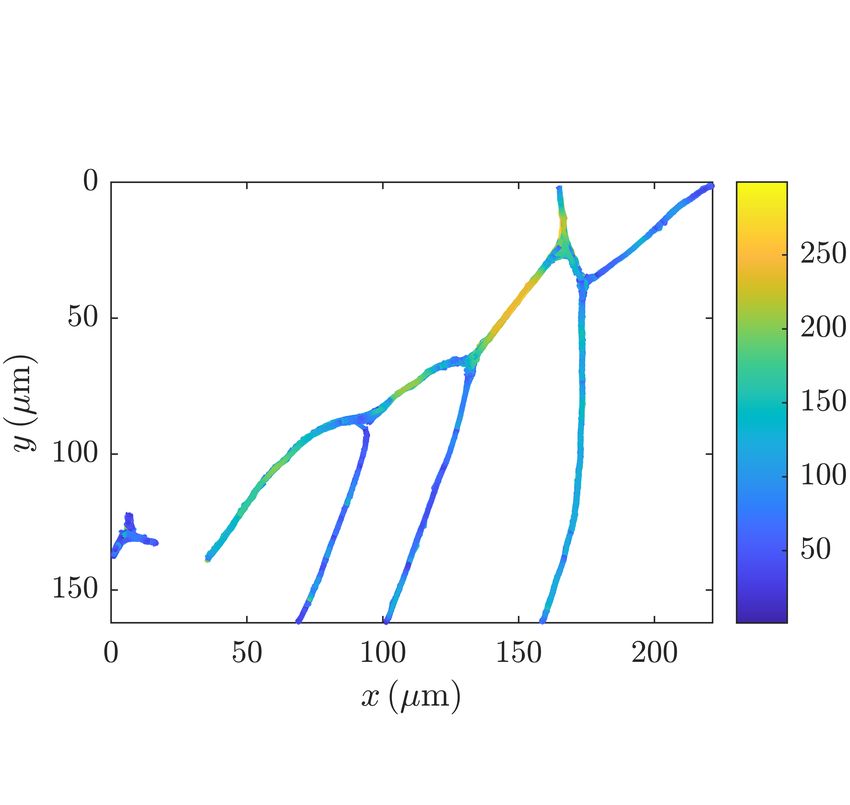
temporal evolution of a lingering RBC in an arteriolar cumulative distribution function is shown. The corre-4 Figure 1. Time series of a lingering RBC at an arterial bifurcation for a time interval of t = 380 ms. The plasma was fluorescently labeled, and therefore, RBCs appear as dark spots. At t = 0 ms, an RBC is touching the apex of the bifurcation, marked by the arrow. The cell starts to deform and linger around this apex, leading to a partial blockage with decreased flow rate, as can be seen in the upper daughter vessel for all subsequent images. Finally, the cell is detached from the apex at t = 380 ms. The scale bar is 10 µm in width. Additional data is provided in Fig. S1.2. Figure 2. (a) Results of particle tracking of RBCs in the given geometry. The flow is coming from top (mother vessel “M” in Figure (b)) and exits in all other branches. The colorbar corresponds to the tracked velocities and depicted is the superposition of 500 tracks. (b) Snapshot of the geometry with flowing RBCs (red). To enhance the contrast and visibility, false color images are shown. The daughter branches are labeled in ascending order from rightmost to leftmost and will be referred to in the main text. (c) Distribution of voids within a branch (1) of a bifurcation. The graph corresponds to the measured integrated intensity along a perpendicular line segment with respect to the centerline of this branch (red line segment in branch (1) in (b)). Values above the mean value (red dash-dotted line) can be regarded as voids, i.e., an absence of cells, whereas values below the mean value correspond to passing cells. On average, voids have a duration of approx. 100 ms; however, due to partial blockage caused by lingering RBCs, void formation can exceed multiple times the average duration, as can be seen at t ≈ 4.7 s, where a void with a duration of 350 ms is formed. sponding probability density functions of voids for the median values of the empirical probability distrubution geometry in Fig. 2 are given in Figs. 4 and 5, resp. In the for all graphs are narrowly distributed. Contrasting this first case, only void durations associated to non-lingering state, for voids associated with lingering events (Fig. 5), events were taken into account, whereas void durations we find a shift of medians toward higher void durations. exclusively associated with lingering events were taken A severe case of this observation can also be seen in the into account in the latter case. By comparing the graphs inset graph of Fig. 5, where the median void duration for each branch in both the figures, the influence of lin- was more than double in the lingering case with respect gering on the void durations is obvious. In Fig. 4, the to non-lingering events.
5

Figure 3. Cumulative distribution functions of void durations    Figure 5. Probability density functions of scaled void dura-
τ for all branches of the geometry in Fig. 2. The temporal       tions for all branches if only lingering events are taken into
length of the voids is hereby scaled for each branch by the      account. We define a lingering event to occur if the speed of an
average time of a RBC to pass, τRBC . The data points cor-       RBC is lower than vRBC ≤ 30µm/s in the vicinity of a bifur-
respond to measured void durations, whereas the solid line       cation apex. The legend is identical to the one in Fig. 3. The
corresponds to the respective log-normal distributions with      inset graph shows both the probability densities in the case
estimated parameters µ̂ and σ̂, as in Eq. (1).                   of lingering and non-lingering, respectively, for the geometry
                                                                 in Fig. 1 to represent extreme cases. Additional information
                                                                 about this geometry can also be found in Fig. S1.2. Filled
                                                                 circles in matching colors denote median values of normal-
                                                                 ized void durations, obtained from estimated parameters in
                                                                 Eq. [2].

                                                                 pressibility of the fluid. Even though the size of RBCs is
                                                                 comparable to the apparent vessel diameters, their speed
                                                                 may serve as a good approximation of the mean speed of
                                                                 the surrounding fluid (plug flow); hence, we find

                                                                          Q = ∆V /∆t = A vfluid ' A lRBC /τRBC ,             (3)

                                                                 with the time-averaged flowrate Q, the volume element
                                                                 ∆V , the cross-sectional area A, mean speed of the fluid
                                                                 vfluid , the length of the major axis of the circumscribing
                                                                 ellipse of RBCs lRBC and the average cell passage time
                                                                 τRBC , as introduced in previous paragraphs. Among all
Figure 4. Probability density functions of void durations for    the investigated pairs of bifuracting vessels, we find the
all branches as in Fig. 3 in the case of non-lingering events.   lingering frequency to be higher in the one with lower
The temporal length of the voids is hereby scaled for each       flowrates with respect to its counterpart with a higher
branch by the average time of a RBC to pass, τRBC . Me-          flowrate. Apart from lingering at bifurcations, RBCs
dian values obtained from estimated parameters in Eq. [2] are    may also deform in the vicinity of branches, i.e., bifurca-
indicated by filled circles in the respective color code.        tions or confluences. While lingering implies the strong
                                                                 interaction of the vessel walls, the sole presence of junc-
                                                                 tions may induce shape changes for approaching or dis-
   In addition to the probability densities of void dura-        tancing RBCs. The main difference is the reduction of
tions, we can also define a so-called lingering frequency as     the speed, which is significant in the case of lingering,
the fraction of voids not associated with lingering events       but adapted to the flow rates in the respective branch
and the total number of occurring voids in a branch of           in the latter case, although slight deviations may oc-
the network. Fig. 6 shows the calculated lingering fre-          cur. To analyze the spatio-temporal evolution of this
quencies of all the analyzed vessels in relation to the nor-     deformation, we calculated the circumscribing ellipse for
malized mean flowrate in the respective vessel. The nor-         each individual RBC for all consecutive images, yielding
malization factor is given by the mean flowrate of the           both the centroid position as well as the eccentricity of
mother or feeding vessel, which is equal to the sum of           the cell, given as the ratio of the distance between the
the flowrates of all draining vessels due to the incom-          two foci and the length of its major axis. Fig. 7 shows
6
Figure 6. Lingering frequencies of the detected voids in re- Figure 7. Eccentricity ε of RBCs as a function of the cen-
lation to the normalized mean flowrate in a distinct vessel. troid position within the geometry shown as the inset. The
The lingering frequency is hereby defined as the fraction of eccentricity is hereby calculated as the ratio of the distance
the void count associated with a lingering event and the total between the two foci and the length of its major axis of an el-
void count in the vessel. Further, we define the normalized lipse with identical second moments for each individual RBC
mean flowrate as a fraction of the flowrate in a daughter ves- for all consecutive images. The thick red solid line repre-
sel Qi and the mother vessel QM . Identical color codes belong sents the average of all individual graphs (thin lines). For the
to pairs of vessels branching from the same apex; the dashed analysis, only single RBCs are considered, whereas trains of
lines connect the data points of vessels. flowing RBCs are neglected. The offset of the centroid po-
sition is chosen in a way that the bifurcation apex xb is at
position zero.
the measured eccentricity values for the flowing RBCs in
the confluence-bifurcation geometry, both individually as
tial void lengths are highly correlated, and this implies
well as the average curve. The corresponding geometry
a breakup of clusters of RBCs approaching a bifurcation
exhibiting both a bifurcation and a confluence is shown
apex. The term cluster hereby implies the state of RBCs
as an inset of Fig. 7. From the average curve, one can
moving in a chain where the intercellular distance is in
clearly see the transition of cell shapes RBCs undergo
the order of the cellular size, where hydrodynamic inter-
while flowing. At the position of the confluence apex
actions are abundant24,25 . However, we emphasize that
xc , the mean eccentricity exhibits the global minimum,
the increase of the median void durations in the case of
implying the roundest obtained shape. Similarly, at the
lingering RBCs does not hold for all the analyzed geome-
position of the bifurcation apex xb , the mean eccentric-
tries (cf. Supplementary Materials).
ity exhibits a local minimum, indicating a transformation
Further, we also notice a suppression of very short
from an elongated to a more spherical shape when ap-
void durations, as can be seen from the comparison
proaching the apex and again elongating when entering
of Figs. 4 and 5. To quantify this statement, we cal-
one daughter branch.
culated the probabilities for void durations being less
than 0.5 τRBC . In the case of non-lingering, integra-
tion of the corresponding probability densities yields for
DISCUSSION the probabilities Pi (τvoid < 0.5 τRBC ) = {0.23, 0.47, 0.38,
0.31, 0.45, 0.27, 0.40}, i ∈ {1, . . . , 7}, where i denotes the
Since we analyzed the void formation downstream in branch identifier according to Fig. 2. Similarly, we obtain
a bifurcating vessel geometry, the apparent increase in P̃i (τvoid < 0.5 τRBC ) = {0.02, 0.30, 0.00, 0.03, 0.01, 0.46,
median void durations originates from two possible sce- 0.10}, i ∈ {1, . . . , 7} with the probabilities P̃i in the case
narios. One contribution is given by the redistribution of lingering. If one compares these values for each branch,
of consecutive RBCs into the adjacent daughter vessel. it is obvious that void durations less than or equal to
The second contribution is given by a change in void 0.5 τRBC are suppressed drastically in all but for i = 6.
speed due to an altered flow rate in the vessel that has In some of the graphs showing the probability densi-
an impact on the temporal void duration. In the latter ties of the void durations, rather long-tailed distributions
case, the spatial distance between the consecutive RBCs are present. We stress that these tails arise inherently
would sustain, outmatching the observed state. Indeed, due to the heterogeneous distribution of RBCs in the mi-
by considering the standard deviation of the speeds of crovascular networks, leading to cell-depleted sequences
passing RBCs in a vessel, variations of the flow rates in branches and thus long void durations in absence of
are negligible. Thus, temporal void durations and spa- lingering cells.7
One crucial question is the dependence of the linger- which is found to be higher in the branching vessel with
ing frequency on the flow properties. Fig. 6 shows a de- the higher flow rate compared to the adjacent vessel. All
creased frequency for the branch with the higher flowrate the presented results of our study show a good qualita-
with respect to the adjacent vessel. In the prevalent tive agreement with in silico results in ref. 20 although, in
low Reynolds number regime, RBCs follow merely the contrast to the well-defined boundary conditions in silico,
streamlines of the surrounding plasma, and thus, one the major experimental drawback is the limited insight
finds fewer cells in the vessel transporting less volume. into the whole model system. These limitations are given
However, the interaction of cellular compounds with the by a limited field of view and the sheer complexity of the
endothelial walls of the vessels is complex26 , and there- living hamster model and all its parameters. Neverthe-
fore, it is highly non-intuitive to observe this circum- less, we can assess the impact of lingering RBCs on the
stance. It is even more remarkable given the broad distri- flow behavior of subsequent cells in vivo. We can pro-
bution of opening angles in all the analyzed geometries. vide evidence to show that these lingering events cause
For normalized flowrates close to 0.5 QM , we obtained a breakup of trains of RBCs as well as redistribution in
very similar lingering frequencies for both the connected the branching vessels. Even though these effects seem to
vessels. Nevertheless, the overall magnitude of the lin- be rather fine-grained, the impact on the whole organism
gering frequency seems to be unaffected by this observa- may be severe, given the importance of blood flow to the
tion and rather depends on the cutting angle between the health state.
daughter vessels of the geometry in a way that small an-
gles exhibit higher lingering frequencies than large ones
in the majority of cases. Other flow parameters such as AUTHOR CONTRIBUTIONS
absolute flow rates or curvature of the bifurcation apex
may also influence the lingering frequency. M.W.L., M.D.M., L.K., and C.W. designed the re-
So far, we have focused on the impacts of lingering search; A.K., M.W.L., and S.Q. performed the research;
RBCs on the microvascular blood flow in vivo. However, A.K., and T.J. analyzed the data; A.K. and C.W. wrote
the physical prerequisites to obey lingering have not been the paper; and M.W.L, S.Q., M.D.M., L.K., and T.J.
discussed yet. RBCs obey an inner network of spectrin provided feedback and insights.
fibers, as they are responsible for their biconcave shape
at rest. Due to the flexibility of this spectrin network,
RBCs can pass through constrictions much smaller than ACKNOWLEDGMENTS
their size at rest27,28 . Yet, not only constrictions alter the
shape of RBCs, but also the complex structure of the vas- AK, TJ, LK, and CW gratefully acknowledge support
cular network itself, exhibiting merging and bifurcating from the research unit DFG FOR 2688 - Wa1336/12 of
vessels. The shape of RBCs undergoes a characteristic the German Research Foundation. MWL and MDM
deformation when approaching the apex of a bifurcation received support from the research unit DFG FOR
or a confluence, respectively (cf. Fig. 7). Recently, this 2688 - LA2682/9-1 of the German Research Founda-
behavior was reproduced in silico for a varying number tion. This work was supported by the European Union’s
of passing cells15 . Whereas this alteration of the shape Horizon 2020 research and innovation programme un-
is due to increasing or decreasing confinements depend- der the Marie Skłodowska-Curie grant agreement No.
ing on the geometry, it is responsible for the observed 860436 – EVIDENCE (SQ, LK, CW). CW, TJ, SQ,
lingering behavior. Particles such as hard spheres obey and AK kindly acknowledge the support and funding
a less severe coupling with the fluid, and we assume the of the “Deutsch-Französische-Hochschule” (DFH) DFDK
deformation and the strong fluid-cell interaction of RBCs CDFA-01-14 “Living fluids”.
is the major cause of lingering29 .
SUPPLEMENTARY MATERIAL
CONCLUSION
The final manuscript including all supplementary data
We used cutting edge intravital microscopy in conjunc- is accessible via doi:10.1016/j.bpj.2020.12.012.
tion with a combined sophisticated signal processing al-
gorithm and particle tracking to obtain detailed informa-
tion of flowing RBCs in living hamster models. Based on ACKNOWLEDGMENTS
this data we define and detect so called lingering events,
i.e. RBCs resting at a bifurcation apex of branching ves- AK, TJ, LK, and CW gratefully acknowledge support
sels. We show, that these lingering events particularly from the research unit DFG FOR 2688 - Wa1336/12 of
cause a redistribution of subsequent RBCs in the adja- the German Research Foundation. MWL and MDM
cent daughter vessels and lead to a break-up of trains received support from the research unit DFG FOR
of RBCs. We further analyze the ratio of lingering cells 2688 - LA2682/9-1 of the German Research Founda-
and all traversing RBCs, the so called lingering frequency, tion. This work was supported by the European Union’s8
Horizon 2020 research and innovation programme un- Gekle. Antimargination of microparticles and platelets in the
der the Marie Skłodowska-Curie grant agreement No. vicinity of branching vessels. Biophysical Journal, 115(2):411–
860436 – EVIDENCE (SQ, LK, CW). CW, TJ, SQ, 425, 2018.
17 C. Pozrikidis. Numerical simulation of blood flow through mi-
and AK kindly acknowledge the support and funding crovascular capillary networks. Bulletin of Mathematical Biology,
of the “Deutsch-Französische-Hochschule” (DFH) DFDK 71(6):1520–1541, 2009.
18 Prosenjit Bagchi. Mesoscale simulation of blood flow in small
CDFA-01-14 “Living fluids”.
vessels. Biophysical Journal, 92(6):1858–1877, 2007.
19 Peter Balogh and Prosenjit Bagchi. A computational approach to
modeling cellular-scale blood flow in complex geometry. Journal
REFERENCES of Computational Physics, 334:280–307, 2017.
20 Peter Balogh and Prosenjit Bagchi. Direct numerical simulation
1 Lars Kaestner. Calcium signalling Approaches and Findings in of cellular-scale blood flow in 3d microvascular networks. Bio-
the Heart and Blood. Springer Berlin Heidelberg, 2013. physical Journal, 113(12):2815–2826, 2017.
2 Aleksander S. Popel and Paul C. Johnson. Microcirculation and 21 Matthias W. Laschke, B. Vollmar, and Michael D. Menger. The
hemorheology. Annual Review of Fluid Mechanics, 37(1):43–69, dorsal skinfold chamber: window into the dynamic interaction of
2005. biomaterials with their surrounding host tissue. European Cells
3 J.L.M. Poiseuille. Recherches sur les causes du mouvement du
and Materials, 22:147–167, 2011.
sang dans les veines. 1832. 22 Laura Hertz, Sandra Ruppenthal, Greta Simionato, Stephan
4 A R Pries, T W Secomb, T Gessner, M B Sperandio, J F Gross,
Quint, Alexander Kihm, Asena Abay, Polina Petkova-Kirova, Ul-
and P Gaehtgens. Resistance to blood flow in microvessels in rich Boehm, Petra Weissgerber, Christian Wagner, Matthias W.
vivo. Circulation Research, 75(5):904–915, 1994. Laschke, and Lars Kaestner. The evolution of erythrocytes be-
5 Ian Gopal Gould, Philbert Tsai, David Kleinfeld, and Andreas
coming red in respect to fluorescence. Frontiers in Physiology,
Linninger. The capillary bed offers the largest hemodynamic 10, 2019.
resistance to the cortical blood supply. Journal of Cerebral Blood 23 Achim Guckenberger, Alexander Kihm, Thomas John, Christian
Flow & Metabolism, 37(1):52–68, 2017. Wagner, and Stephan Gekle. Numerical–experimental observa-
6 Eugenio Gutiérrez-Jiménez, Hugo Angleys, Peter M. Rasmussen,
tion of shape bistability of red blood cells flowing in a microchan-
Mark J. West, Laura Catalini, Nina K. Iversen, Morten S. Jensen, nel. Soft Matter, 14(11):2032–2043, 2018.
Sebastian Frische, and Leif Østergaard. Disturbances in the con- 24 Viviana Clavería, Othmane Aouane, Marine Thiébaud, Manouk
trol of capillary flow in an aged APPswe/PS1$\delta$e9 model Abkarian, Gwennou Coupier, Chaouqi Misbah, Thomas John,
of alzheimer’s disease. Neurobiology of Aging, 62:82–94, 2018. and Christian Wagner. Clusters of red blood cells in microcapil-
7 J. C de la Torre and G. B Stefano. Evidence that alzheimer’s
lary flow: hydrodynamic versus macromolecule induced interac-
disease is a microvascular disorder: the role of constitutive nitric tion. Soft Matter, 12(39):8235–8245, 2016.
oxide. Brain Research Reviews, 34(3):119–136, 2000. 25 M. Brust, O. Aouane, M. Thiébaud, D. Flormann, C. Verdier,
8 Axel R. Pries and Timothy W. Secomb. Making microvascular
L. Kaestner, M. W. Laschke, H. Selmi, A. Benyoussef, T. Pod-
networks work: Angiogenesis, remodeling, and pruning. Physi- gorski, G. Coupier, C. Misbah, and C. Wagner. The plasma
ology, 29(6):446–455, 2014. protein fibrinogen stabilizes clusters of red blood cells in micro-
9 Axel R. Pries, Timothy W. Secomb, and Peter Gaehtgens. Design
capillary flows. Scientific Reports, 4(1):4348, 2014. Number: 1
principles of vascular beds. Circulation Research, 77(5):1017– Publisher: Nature Publishing Group.
1023, 1995. 26 A. R. Pries and T. W. Secomb. Microvascular blood viscosity
10 Cecil D Murray. The physiological principle of minimum work.
in vivo and the endothelial surface layer. American Journal
i. the vascular system and the cost of blood volume. 12:8, 1926. of Physiology-Heart and Circulatory Physiology, 289(6):H2657–
11 T. F. Sherman. On connecting large vessels to small. the meaning
H2664, 2005.
of murray’s law. The Journal of General Physiology, 78(4):431– 27 N Mohandas and E Evans. Mechanical properties of the red cell
453, 1981. membrane in relation to molecular structure and genetic defects.
12 Robin Fåhraeus. The suspension stability of the blood. Physio-
Annual Review of Biophysics and Biomolecular Structure, 23(1):
logical Reviews, 9(2):241–274, 1929. 787–818, 1994. Publisher: Annual Reviews.
13 Pries A R, Secomb T W, Gaehtgens P, and Gross J F. Blood flow 28 Volkmar Heinrich, Ken Ritchie, Narla Mohandas, and Evan
in microvascular networks. experiments and simulation. Circula- Evans. Elastic thickness compressibilty of the red cell membrane.
tion Research, 67(4):826–834, 1990. Biophysical Journal, 81(3):1452–1463, 2001.
14 Robin Fåhræus and Torsten Lindqvist. The viscosity of the blood 29 J. Happel and H. Brenner. Low Reynolds number hydrodynam-
in narrow capillary tubes. American Journal of Physiology- ics: with special applications to particulate media. Prentice-Hall
Legacy Content, 96(3):562–568, 1931. international series in the physical and chemical engineering sci-
15 Ting Ye and Lina Peng. Motion, deformation, and aggregation
ences. Prentice-Hall, 1965.
of multiple red blood cells in three-dimensional microvessel bi-
furcations. Physics of Fluids, 31(2):021903, 2019.
16 Christian Bächer, Alexander Kihm, Lukas Schrack, Lars Kaest-
ner, Matthias W. Laschke, Christian Wagner, and StephanYou can also read


