Management of Gastrointestinal Foreign Bodies with Brief Review of the Guidelines
←
→
Page content transcription
If your browser does not render page correctly, please read the page content below
Pediatr Gastroenterol Hepatol Nutr. 2023 Jan;26(1):1-14
https://doi.org/10.5223/pghn.2023.26.1.1
pISSN 2234-8646·eISSN 2234-8840

Review Article                                   Management of Gastrointestinal
                                                 Foreign Bodies with Brief Review of
                                                 the Guidelines

                                                 Kaan Demiroren

                                                 Department of Pediatric Gastroenterology, University of Health Sciences, Yuksek Ihtisas Training and
                                                 Research Hospital, Bursa, Turkey

Received: Jan 25, 2022
1st Revised: Jul 9, 2022
                                                 ABSTRACT
2nd Revised: Jul 31, 2022
Accepted: Aug 30, 2022                           Foreign body (FB) ingestion is a common health problem that affects children more than
Published online: Jan 10, 2023                   adults. According to gastroenterologists’ guidelines, the management of FB ingestion
                                                 differs slightly between adult and children. This review aimed to compile adult and children
Correspondence to
Kaan Demiroren
                                                 guidelines and establish an understandable association to reveal the requirements and timing
Department of Pediatric Gastroenterology,        of the endoscopic procedure, which is the most effective and least complicated technique for
University of Health Sciences, Yuksek Ihtisas    gastrointestinal FBs. Coins, pins, and chicken and fish bones have been the most commonly
Training and Research Hospital, Emniyet          ingested FBs. However, with their increasing use in recent years, large batteries with lithium-
Street, Bursa 16310, Turkey.                     ion conversion, stronger magnets composed of rare earth metals, such as neodymium,
Email: kaandemiroren@yahoo.com
                                                 and superabsorbent objects have become the most morbid and mortal, necessitating new
       demirorenkaan@gmail.com
                                                 management strategies. Although the approach to gastrointestinal FBs is controversial,
Copyright © 2023 by The Korean Society of        with different treatment options available in different disciplines, many studies have
Pediatric Gastroenterology, Hepatology and       demonstrated the efficacy and safety of endoscopic procedures. Many factors influence the
Nutrition
                                                 timing of endoscopy, including the nature, size, and location of the ingested object and the
This is an open-access article distributed
under the terms of the Creative Commons
                                                 patient’s clinical condition.
Attribution Non-Commercial License (https://
creativecommons.org/licenses/by-nc/4.0/)
                                                 Keywords: Battery; Blunt; Button; Coin; Cylindrical; Magnet, Food impaction; Pointed;
which permits unrestricted non-commercial        Sharp; Superabsorbent
use, distribution, and reproduction in any
medium, provided the original work is properly

                                                 INTRODUCTION
cited.

ORCID iDs
Kaan Demiroren                                   The treatment of gastrointestinal (GI) foreign bodies (FBs) is controversial, with different
https://orcid.org/0000-0003-1137-1715
                                                 options available for surgical and nonsurgical specialties. Furthermore, gastroenterologists’
Conflict of Interest                             guidelines for adult and pediatric age groups differ in their approach to FB ingestion and
The author has no financial conflicts of         adult gastroenterologists must perform endoscopy on children in centers without pediatric
interest.                                        gastroenterologists. This review aimed to compile guidelines for adult and pediatric age
                                                 groups and establish a clear association. Furthermore, it was intended to examine less
                                                 discussed issues, such as superabsorbent objects, and to draw attention to some studies
                                                 that raised concerns about the guidelines. A PubMed/MEDLINE search was conducted using
                                                 the terms “foreign body AND gastrointestinal and children OR adult” and “foreign body
                                                 AND gastrointestinal AND guideline AND children OR adult”. The subject of “Endoscopic
                                                 removal for GI FBs” was reviewed, including the current literature. In particular, the North
                                                 American Society for Pediatric Gastroenterology, Hepatology and Nutrition (NASPGHAN)

https://pghn.org                                                                                                                                        1
Gastrointestinal Foreign Bodies

                                  [1], the European Society for Paediatric Gastroenterology Hepatology and Nutrition
                                  (ESPGHAN) [2], the European Society of Gastrointestinal Endoscopy (ESGE) [3], the
                                  American Society for Gastrointestinal Endoscopy (ASGE) [4], the Italian Society of Pediatric
                                  Gastroenterology Hepatology and Nutrition (SIGENP), and the Italian Association of
                                  Hospital Gastroenterologists and Endoscopists (AIGO) guidelines [5] in either children or
                                  adults are line with the following plan:
                                     1. Frequency of FB ingestion
                                     2. Diagnosis of FB ingestion
                                     3. Preparing for endoscopic removal
                                     4. Button and cylindrical batteries ingestion
                                     5. Magnet ingestion
                                     6. Pointed or sharp object ingestion
                                     7. Esophageal food impaction
                                     8. Coin and blunt object ingestion
                                     9. Superabsorbent object ingestion

                                  FREQUENCY OF FB INGESTION
                                  FB ingestion is a common health problem worldwide, and is more common in children than
                                  in adults. It is commonly accidental in children; however, in adults, it is caused by suicide,
                                  psychiatric illness, mental retardation, and secondary gain [6]. Incidents of child abuse are
                                  uncommon, and men and children under five years are most likely to ingest FB [7-10]. The
                                  American Association of Poison Control Centers recorded 94,051 FB ingestion in 2019 among
                                  all age groups (67,186 in children under 5 years and 12,223 in adults above 20 years). Three of
                                  the under 5 years patients who ingested batteries died [11].

                                  While 80–90% of GI FBs are spontaneously egested, 10-20% require endoscopic removal,
                                  and less than 1% require surgery to remove the FB or treat associated complications [3,4].
                                  Endoscopic FB removal has been reported to have a success rate of 88.5–100% [8,12-14].

                                  DIAGNOSIS OF FB INGESTION
                                  Patients who ingest an FB may develop dysphagia, odynophagia, retrosternal pain, stridor,
                                  FB sensation, hypersalivation, irritability, chest or abdominal pain, refusal to eat, wheezing,
                                  and dyspnea [1,3]. First, a detailed anamnesis covering the nature, number, time, and cause
                                  of FB ingestion and symptoms should be undertaken, followed by a physical examination.
                                  In a clinical setting, the ABC rules of resuscitation are valid. Aspiration occurs if a patient’s
                                  secretions become excessive and cannot be swallowed, The evaluation and the choice to
                                  perform endoscopy should be completed as soon as possible.

                                  Even if the patient is asymptomatic, all patients with suspected FB ingestion should be
                                  admitted to the emergency department and have a radiographical examination. If necessary,
                                  bidirectional neck, chest, abdomen, and pelvis radiographs should be taken. For radiolucent
                                  foreign entities, computed tomography (CT) may be performed. Magnetic resonance imaging
                                  (MRI) is ineffective for detecting FBs. There are no data on the use of ultrasonography and
                                  metal detectors. Radiographs should be used to evaluate mediastinal and peritoneal free air
                                  and the presence and number of FBs. However, plain radiography may fail to demonstrate
                                  subdiaphragmatic free air. In cases of doubt, it should be evaluated using CT. Before battery
                                  removal, MRI scans should never be performed [1,3,15]. Positive predictive values for
                                  radiographs are 100% for metallic objects, 43% for glass objects, 26% for fish bones, and 0%

https://pghn.org                  https://doi.org/10.5223/pghn.2023.26.1.1                                                            2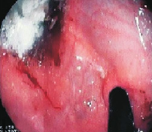
Gastrointestinal Foreign Bodies

                                  for splinters and woody objects [1]. The false-negative rate for food bolus impaction is 87%
                                  [3]. Point-of-care ultrasound (POCUS) is effective in identifying and monitoring ingested FBs
                                  in children [16]. POCUS use in children will increase in the future, as it is in adults [17,18].

                                  PREPARING FOR ENDOSCOPIC REMOVAL
                                  The timing of the endoscopic procedure is determined by many factors, including the nature
                                  and size of the FB, its location in the GI tract, the patient’s clinical condition, the time
                                  elapsed after possible ingestion, the patient’s nil per os (NPO) status, family’s anxiety, and
                                  the endoscopist experience [1,5].

                                  The timing of the endoscopic procedure can be defined as follows [1]:
                                    • Emergent: Within 2 hours of arrival, regardless of the NPO rules
                                    • Urgent: Within 24 hours of arrival, following the required NPO rules

                                  ESGE/ESPGHAN recommends endoscopic removal in children under general anesthesia or,
                                  if general anesthesia is not possible, with deep sedation [2]. SIGENP/AIGO recommends
                                  that an emergency endoscopy must be performed under general anesthesia with airways
                                  protection [5]. For children with complete airway protection, general anesthesia with
                                  endotracheal intubation is recommended, which is ideal for most emergency procedures
                                  [1,3,5]. Alternatively, endotracheal intubation has been recommended in selected cases such
                                  as cases with a high risk of aspiration (i.e., full stomach, proximal esophageal location of the
                                  FB, food bolus impaction), objects that are difficult to remove, and multiple objects [3,4].

                                  For FBs at or above the level of the cricopharyngeus muscle, otorhinolaryngology (ENT)
                                  consultation is advised [4]. Pediatric, ENT, or cardiothoracic surgeons use a rigid endoscope
                                  to remove FBs in some regions and countries in the upper esophagus [9,10,19-22]. In some
                                  institutes, these procedures are routine processes in all cases. The rigid endoscope is may be
                                  only safe for proximal esophageal objects [2]. Upper esophageal FBs can be easily removed
                                  with Magill forceps or balloon (e.g., Foley catheter) extraction using fluoroscopy rather than
                                  endoscopy. These methods were followed successfully by several institutions [19,22-24].

                                  Before performing an endoscopic treatment on an unfamiliar FB, a sample of objects of
                                  similar size and thickness should be rehearsed outside the patient using appropriate forceps.
                                  Radiographs should be performed again to rule out the possibility of FB displacement,
                                  before beginning the endoscopic removal procedure. Almost all FBs can be extracted with
                                  rat-toothed forceps and a net. Therefore, both types of forceps should be provided in the
                                  endoscopic unit. Except for the larger one, the endoscopy unit should have all conceivable FB
                                  gripping forceps and a gastroscope with a diameter of 6 mm [5].

                                  BUTTON AND CYLINDRICAL BATTERY INGESTION
                                  The emergency endoscopic procedure is indicated in children with button batteries stuck
                                  in their esophagus because large-diameter batteries lodged in the esophagus can lead to
                                  severe complications. Electrolytic activity hydrolyzes tissue fluids producing hydroxide at the
                                  negative end of the battery, resulting in alkali injury. Furthermore, the intensity of physical
                                  pressure increases the alkali injury (Fig. 1B, C). The increased size of batteries and their
                                  transformation into lithium cells pose an increased risk of mucosal injury. An alkaline caustic
                                  injury, especially with lithium-cell batteries larger than 20 mm, can occur within 30 minutes
                                  and last hours or days, resulting in perforation and aortoenteric fistulas [25].

https://pghn.org                  https://doi.org/10.5223/pghn.2023.26.1.1                                                       3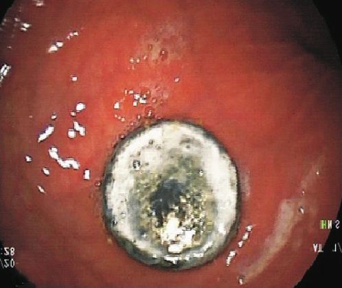
Gastrointestinal Foreign Bodies
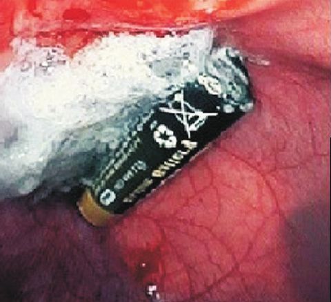
A B C
D E F
G H I
Fig. 1. (A) A button battery on radiograph (double contours/surrounding halo appearance is notable), (B)
endoscopic image of the battery and the mucosal mess, and (C) the areas of esophageal mucosal necrosis that
were healing during the follow-up endoscopy performed a week after the removal. (D) A button battery >2 cm
in the stomach, (E, F) necrotic foci and ulcer area at 3 hours after ingestion. (G, H) Appearance of endoscopy
performed at 24 hours after ingestion in a patient who ingested a cylindrical battery, area of erosive foci, and (I)
removal by a grasping forceps with a net.
In an extensive study, 0.8% of patients had tracheoesophageal fistula, esophageal
perforation, esophageal stricture, vocal cord paralysis, mediastinitis, cardiac arrest,
pneumothorax, aortoenteric fistula, and 0.15% died [26].
A complete history of ingested objects is not always recorded. Therefore, radiographs should
be carefully examined. It is necessary to determine whether a spherical radiopaque object in
the esophagus is a battery. The double-contour (halo) view around the object indicated the
presence of a battery (Fig. 1A).
Unless the batteries are buried in the mucosa, they can be removed using grasping forceps.
It is recommended that the batteries in the esophagus and batteries in the stomach should
be removed using rat-toothed forceps and grasping forceps with a net, respectively. The
surgeon should use a rigid endoscope if batteries are embedded in the mucosa. A battery in
the patient’s stomach did not suggest that the patient was safe and the battery could have
caused harm to the esophagus before it entered the stomach [27]. Therefore, although the
button battery is in the stomach, the danger of esophageal perforation should be considered.
Furthermore, if the button battery remains in the stomach without moving, it causes injury to
the gastric mucosa [27-29].
https://pghn.org https://doi.org/10.5223/pghn.2023.26.1.1 4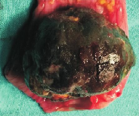
Gastrointestinal Foreign Bodies

                                  Table 1 [1-4,30] shows the management of ingested batteries. A battery stuck in the
                                  esophagus is the most emergency indication for endoscopic removal. Regardless of the NPO
                                  guidelines, the endoscopic procedure should be performed emergently by intubation under
                                  general anesthesia [2,3,5]. If the patient has a suspicion of perforation or bleeding, a surgeon
                                  and a cardiovascular surgeon should perform the endoscopic procedure in the operating
                                  room. If there is a suspicious endoscopic appearance after endoscopic removal, symptomatic
                                  patients should be evaluated using CT or MRI. If batteries >2 cm in diameter are found in
                                  the stomach, especially in young children, they should be removed endoscopically. However,
                                  smaller batteries can be clinically monitored.

                                  The ASGE guidelines recommend removing a ≥20 mm battery if it is still in the stomach 48
                                  hours after ingestion [4]. However, the NASPGHAN recommends removal within 24–48
                                  hours based on its algorithm [1]. Contrary to the ASGE and NASPGHAN guidelines, two
                                  studies [28,29] reported that gastric batteries, especially those smaller than 20 mm, should
                                  be removed within the first 24 hours to reduce complications.

                                  Endoscopy should be considered an emergency in all symptomatic cases, as indicated in
                                  the Table. An emergency endoscopy was performed on a symptomatic 2.5-year-old boy who
                                  had ingested a button battery >2 cm. Although endoscopy was performed within 3 hours of
                                  ingestion, necrotic foci and ulcerated regions were visualized in the stomach (Fig. 1D-F).

                                  Despite their large size, cylindrical batteries easily pass through the gastrointestinal tract
                                  because of their shape and cause less mucosal injury owing to their structure [31]. The ESGE
                                  and the ESPGHAN suggest that if a single cylindrical battery in the stomach is not passed
                                  in the stool, the patient can be followed up with radiographs for 7–14 days with a weak
                                  recommendation and low quality evidence [2]. However, in the case of a cylindrical battery,
                                  attention must be paid to the type of battery, the voltage type (such as A23 or A27), and how
                                  long it has been in use [32]. The cylindrical battery in the stomach should be removed within

                                  Table 1. Management of battery ingestion
                                  Clinical scenario                                                                    Procedure
                                  Button battery in the esophagus
                                    • The patient is stable                                       • Emergent endoscopy
                                    • The patient is unstable or there is bleeding                • Emergent endoscopy by a pediatric surgeon and a
                                                                                                    cardiovascular surgeon in the operating room
                                    • There is a suspicion of aortic injury (in the presence of  • CT angiography or MRI*
                                       bleeding, drop in hemoglobin, extensive and deep ulcer)
                                    • After battery removal                                      • Treatment and follow-up in terms of corrosive injury
                                                                                                   and perforation (if symptomatic, CT/MRI scans)
                                  Button battery in the stomach or below
                                    • Patient aged 5 years and battery ≥20 mm in size           • Endoscopy within 48 hours
                                    • Patient aged >5 years and battery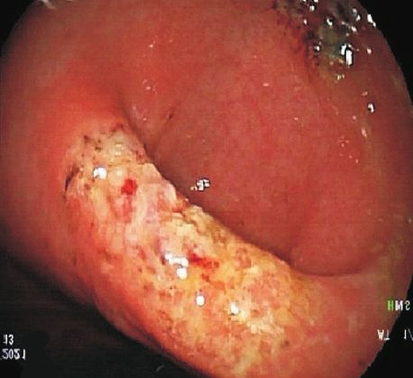
Gastrointestinal Foreign Bodies

                                  24 hours, especially if the patient is symptomatic. Fig. 1G and H show gastric hemorrhagic
                                  ulceration in a patient who ingested a cylindrical battery. Twenty-four hours after intake,
                                  an endoscopy was performed. A large-netted grasping forceps could be used to extract a
                                  cylindrical battery (Fig. 1I).

                                  MAGNET INGESTION
                                  The greatest risk of ingesting magnets is the binding of the two magnets by intercepting
                                  intestinal loops, which can result in enteroenteric fistula, perforation, peritonitis, ischemia,
                                  and necrosis.

                                  Rare earth metal magnets composed of neodymium, iron, and boron are more than ten times
                                  stronger than standard ferrite fridge magnets [6,33]. These magnets are now frequently
                                  used in desk toys and novelty items. Magnet ingestion and associated complications have
                                  significantly decreased with the withdrawal of such products from the market [34], only
                                  to increase again after the reintroduction of high-power magnet sets into the market [33].
                                  Nevertheless, magnet ingestion is increasing every year, and most cases are unreported.
                                  According to a recent survey [35], it is estimated that 50-70 cases of magnet ingestion occur
                                  in Japan annually.

                                  According to a large-scale survey, magnet removal was done using 52% endoscopy, 20%
                                  endoscopy, and surgery combined, 8% surgery, and 15% observation alone. Of the patients,
                                  41% had no perforation or fistula repair, whereas 22% had various degrees of intestinal
                                  resection [1]. Thirty-nine percent of 89 patients who ingested magnets were symptomatic,
                                  40% required stomach surgery, and all patients who required abdominal surgery swallowed
                                  more than one magnet or a metal object with the magnet [36].

                                  In the case of magnet ingestion, the following things should be considered: (A) if it is visible
                                  on plain radiography, lateral radiographs should be taken to confirm the number; (B) if
                                  multiple magnets are ingestion, they should be removed “urgently” even if the patient is
                                  asymptomatic; (C) esophagogastroduodenoscopy or colonoscopy should be performed
                                  depending on the position of the magnet; (D) although the forceps used for removal may
                                  vary depending on the shape and size of the magnet, grasping forceps with a net is ideal;
                                  (E) laparotomy or laparoscopy is required in institutions without endoscopy [1]. The
                                  management of magnet ingestion is presented in Table 2 [1,2,4].

                                  Surgery should be considered for symptomatic patients who ingest more than one magnet.
                                  Fig. 2 shows cases of symptomatic presentation direct magnet ingestion. In the first image,
                                  there were two magnets, one in the stomach and the other in the duodenum, and in the
                                  second image, 17 magnets that caused ileal perforation were seen.

                                  POINTED OR SHARP OBJECT INGESTION
                                  Perforation, extraluminal migration, abscess, peritonitis, fistula, appendicitis, necrotizing
                                  fasciitis, liver, bladder, heart, and lung penetration, incarcerated umbilical hernia, common
                                  carotid artery rupture, aortoesophageal fistula, and death can result from the ingestion
                                  of pins, sewing needles, and safety pins, nails, screws, toothpicks, and bone [37-49]. The
                                  average transit time of an object in children is 3.6 days, while the average perforation time is
                                  10.4 days [1,2].

https://pghn.org                  https://doi.org/10.5223/pghn.2023.26.1.1                                                           6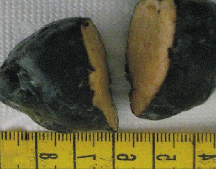
Gastrointestinal Foreign Bodies

                                  Table 2. Management of magnet ingestion
                                  Clinical scenario                                                           Procedure
                                  If it is the only magnet
                                      • In the esophagus or stomach          • It may be removed endoscopically or
                                                                             • If the family cooperates, education (removal of the magnet and
                                                                               metal objects from surroundings and clothes) and follow-up with
                                                                               serial radiographs
                                      • Below the stomach                    • Removed by endoscopy if possible, or
                                                                             • Follow-up with serial radiographs and parental education (removal
                                                                               of the magnet and metal objects from surroundings and clothes)
                                                                             • If passage is delayed, accelerate with PEG 3350/other laxatives
                                  Multiple magnets or a metal object ingested with one magnet
                                      • All in the esophagus or stomach      • Endoscopic removal within the first 12 hr
                                                                             • Referral if endoscopy is not possible
                                                                             • Consulting surgery if transfer >12 hr
                                                                             • Surgical intervention if endoscopic removal is not successful
                                      • Below the stomach
                                         Symptomatic                         • Surgery
                                         Asymptomatic                        • If there are no signs of obstruction and/or perforation on the
                                                                               radiograph, endoscopy (enteroscopy/colonoscopy) or
                                                                             • Follow-up with radiograph every 4–6 hr
                                                                             • If there is no progress, administration of PEG 3350/laxative
                                                                             • If not, endoscopy
                                                                             • If still not removed, surgery
                                  PEG: polyethylene glycol.

                                                                           A                                                              B

                                  Fig. 2. (A) Two magnets, one in the stomach and the other in the duodenum, (B) 17 magnets that caused ileal
                                  perforation.

                                  Appropriate protective equipment, such as an overtube, protective cap, or distal transparent
                                  caps used for band ligation, is recommended to protect the esophagus from trauma during
                                  endoscopic removal. A direct laryngoscope is an alternative removal tool for objects in the
                                  cricopharyngeal area [2].

                                  If an asymptomatic sharp object is found in the esophagus or stomach, it should be removed
                                  within the first 24 hours after proper NPO condition. During an endoscopic procedure, it
                                  may be difficult to locate and grab, such as an object in a stomach full of food. If the patient
                                  is symptomatic, the object should be removed emergently, as with other FBs. Table 3 [1-4]
                                  shows how to manage cases of ingestion of a sharp object.

                                  Safety pins are among the most commonly ingested FBs in several parts of the world,
                                  especially in infants. Complications of safety pin ingestion include carotid artery rupture,

https://pghn.org                  https://doi.org/10.5223/pghn.2023.26.1.1                                                                      7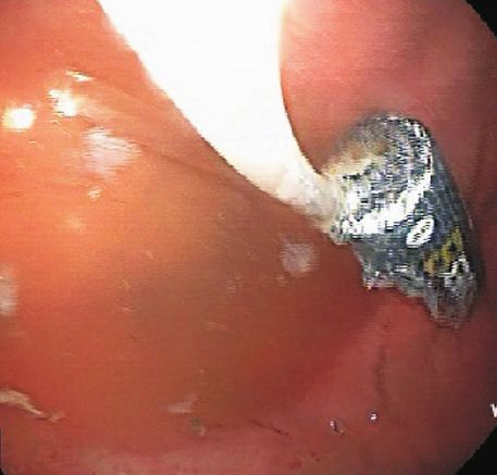
Gastrointestinal Foreign Bodies

                                  Table 3. Management of pointed object ingestion
                                  Clinical scenario                                                               Procedure
                                  Radiopaque
                                    • In the esophagus                             • Emergent/urgent endoscopy
                                    • In the stomach                               • Urgent/emergent endoscopy
                                    • In the small intestine (if under Treitz lig)
                                       Symptomatic                                 • Enteroscopy or surgery
                                       Asymptomatic                                • Clinical follow-up with serial radiographs
                                                                                   • Surgery if the patient becomes symptomatic or the object is not
                                                                                     removed/passed out for more than 3 d
                                  Radiolucent (foreign body thought to have been ingested by self-statement/witness statement)
                                    Symptomatic                                    • Emergent endoscopy
                                    Asymptomatic                                   • Endoscopy if evidence of esophagogram or CT; clinical follow-up
                                                                                     if there is no evidence of esophagogram or CT
                                  CT: computed tomography.

                                  hemopericardium, cardiac tamponade, duodenocolic fistula, and incarcerated umbilical
                                  hernia [50-53]. While approximately 30% of cases were operated by surgeons during
                                  follow-up [54], Almost all of the safety pins, which can be achieved by flexible endoscopic
                                  procedures performed by pediatric gastroenterologists, are easily removed [55,56]. Because
                                  safety pins are easily removed endoscopically, it may be suggested that the best option is to
                                  remove the safety pin using endoscopy while it is still in the esophagus and stomach.

                                  In some regions, fishbone was the most commonly ingested FBs by children, and fishbones
                                  were removed by scope in 59% of 416 children [57].

                                  ESOPHAGEAL FOOD IMPACTION
                                  Food impaction is the most common esophageal FB found in adults [4]. Furthermore, it
                                  is commonly observed in children with eosinophilic esophagitis, reflux esophagitis, post-
                                  anastomotic stenosis after tracheoesophageal fistula repair, achalasia, and other motility
                                  disorders. In endoscopy, an overtube is very useful in adults; however, its usage in children
                                  is a risk of perforation. During the endoscopic procedure, biopsies should be taken from the
                                  esophagus for eosinophilic esophagitis, and patients should also be evaluated for stenosis [1,2].

                                  Acceptable methods for managing esophageal food impactions have been proposed,
                                  including en bloc removal, piecemeal removal, and gentle push technique [4]. If the patient
                                  cannot tolerate secretions, endoscopic procedure should be performed emergently as
                                  removing or gently pushing. If the patient tolerates secretions, endoscopic removal should
                                  be performed urgently together with adequate preparation and/or possibility of spontaneous
                                  passage [1-3].

                                  COINS AND BLUNT OBJECT INGESTION
                                  Coins should be removed from the esophagus within 24 hours. First, coins should be
                                  distinguished from batteries on radiographs. In the case of battery ingestion, a double
                                  contour appearance on the edge was visible. A lateral radiograph can be taken to be sure. In
                                  younger children, objects >2.5 cm in diameter cannot pass through the pylorus, while objects
                                  >6 cm cannot pass through the duodenum. Therefore, such large and long objects should be
                                  removed, even if they are in the stomach [1-4].

                                  For coins and other blunt objects in the esophagus, rat-toothed forceps are appropriate,
                                  and grasping forceps with a net are suitable for those in the stomach. If the coin is stuck
                                  horizontally in the esophagus, symptoms such as hypersalivation and difficulty swallowing

https://pghn.org                  https://doi.org/10.5223/pghn.2023.26.1.1                                                                         8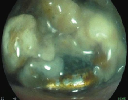
Gastrointestinal Foreign Bodies

                                  may occur. If endoscopic removal is impossible, the coin may be moved vertically using
                                  a nasogastric tube. The coin may fall into the stomach during this procedure, in which
                                  case the urgency of the treatment is no longer necessary. In some institutions, a coin in
                                  the upper esophagus is extracted with a Foley catheter under anesthesia and fluoroscopy.
                                  Before extracting the FB from the esophagus using a nasogastric tube or Foley catheter, care
                                  should be taken to ensure that the FB is not a button battery. A coin-size button battery may
                                  have been buried in the mucosa, causing esophageal necrosis, which may have resulted in
                                  perforation due to interference with tube or catheter.

                                  If the coin is symptomatic, it should be removed emergently; otherwise, it should be done as
                                  soon as possible. If a coin is in the stomach or below, it may remain for a few weeks. However,
                                  endoscopy may not be conducted in the stomach if its diameter is >2.5 cm. Laxatives can be
                                  very effective for coins and other blunt objects in the stomach, as well as sharp objects in the
                                  stomach, especially below the stomach. Table 4 [1,2,4] shows the management of ingested
                                  coins and blunt objects.

                                  A child who ingested 28 glass marbles with a diameter of 1 cm was discharged with clinical
                                  follow-up; Fig. 3A and B shows that all the marbles were defecated within 24 hours. Marbles
                                  are radiopaque on radiograph because they have been colored.

                                  SUPERABSORBENT OBJECT INGESTION
                                  Polymers that can expand 100–200 times in water are superabsorbent materials. Diapers,
                                  feminine hygiene products, agriculture, and the entertainment industry (toys such as water
                                  polo and tabletop ornaments) all use them. Their management is difficult because they are
                                  radiolucent and pass quickly through the proximal GI tract without getting large enough to
                                  cause obstruction. A small number of cases of intestinal obstruction have been reported, one
                                  of which resulted in death due to postoperative sepsis [58-62]. A study [63] reported that 21
                                  asymptomatic children who ingested a superabsorbent object did not undergo endoscopic or
                                  surgical procedures and did not develop signs of obstruction during their follow-ups.

                                  Fig. 3D shows that a hard-textured object obstructing the jejunum is seen on endoscopy of a
                                  2-year-old patient who was admitted with persistent vomiting. The surgically removed object
                                  (Fig. 3E) was a superabsorbent object of unknown origin, approximately 5 cm in diameter
                                  (Fig. 3F).

                                  In our country, the ingestion of superabsorbent objects such as water balls and water apes
                                  is anecdotally common, although they have not been published. Proposals based on our

                                  Table 4. Management of coin and blunt object ingestion
                                  Clinical scenario                                                               Procedure
                                  In the esophagus
                                     Symptomatic (unable to tolerate secretions) • Emergent endoscopy
                                     Asymptomatic                                 • Urgent endoscopy
                                                                                  • Radiographs should be taken again before the endoscopy (it
                                                                                    may have fallen into the stomach)
                                  In the stomach                                  • It may be expected
                                                                                  • If it is not passed in the stool and if it is not symptomatic,
                                                                                    it may be followed up with radiographs at 1–2 wk intervals/
                                                                                    laxative if necessary
                                                                                  • Elective endoscopy if it is not passed out within 2–4 wk
                                  In the small intestine                          • Clinical observation/if it becomes symptomatic, endoscopy or
                                                                                    surgery

https://pghn.org                  https://doi.org/10.5223/pghn.2023.26.1.1                                                                       9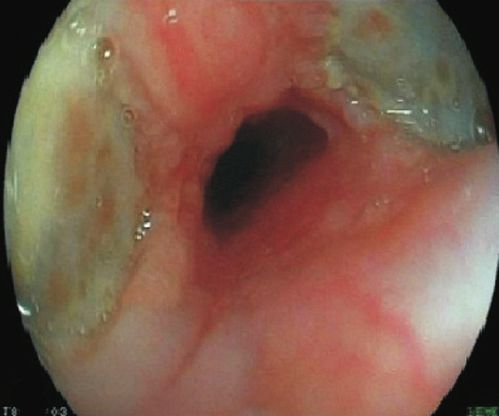
Gastrointestinal Foreign Bodies

                                    A                                                                              D

                                                                                                                   E

                                    B
                                                                                                                   F
                                                                                          C

                                  Fig. 3. (A, B) X-ray and post-exit images of ingested glass beads. (C) Superabsorbent water balls. (D) A large
                                  superabsorbent object at endoscopy and (E, F) after surgery.

                                  personal experience include the following. If available examining a sample of the ingested
                                  object provides information about the potential risk and can help design the endoscopy
                                  approach. Endoscopy would be appropriate for objects that can reach a size that could cause
                                  obstruction in the GI tract and have hard tissues. Fig. 3C shows the samples of the water balls
                                  ingested by the patient. Although they remained for hours, they were 3.42 mm.

                                  CONCLUSION
                                  In conclusion, timely endoscopic procedure for GI FBs can save lives and prevent difficult-
                                  to-manage morbidities. Therefore, if a patient presents with blunt or sharp objects, such
                                  as batteries, multiple magnets or one magnet together with a metal object, and FBs that
                                  cause obstruct the esophagus, and present with increased secretion, the objects should be
                                  removed emergently. Other FBs in the esophagus, sharp or pointed objects in the stomach,
                                  two magnets, or one magnet together with a metal object in the stomach should be urgently
                                  removed. The removal of the remaining FBs should be considered optional. However,
                                  symptomatic FB should be removed immediately.

https://pghn.org                  https://doi.org/10.5223/pghn.2023.26.1.1                                                                         10
Gastrointestinal Foreign Bodies
REFERENCES
1. Kramer RE, Lerner DG, Lin T, Manfredi M, Shah M, Stephen TC, et al. Management of ingested foreign
bodies in children: a clinical report of the NASPGHAN Endoscopy Committee. J Pediatr Gastroenterol
Nutr 2015;60:562-74.
PUBMED | CROSSREF
2. Thomson M, Tringali A, Dumonceau JM, Tavares M, Tabbers MM, Furlano R, et al. Paediatric
gastrointestinal endoscopy: European Society for Paediatric Gastroenterology Hepatology and Nutrition and
European Society of Gastrointestinal Endoscopy guidelines. J Pediatr Gastroenterol Nutr 2017;64:133-53.
PUBMED | CROSSREF
3. Birk M, Bauerfeind P, Deprez PH, Häfner M, Hartmann D, Hassan C, et al. Removal of foreign bodies in
the upper gastrointestinal tract in adults: European Society of Gastrointestinal Endoscopy (ESGE) Clinical
Guideline. Endoscopy 2016;48:489-96.
PUBMED | CROSSREF
4. ASGE Standards of Practice Committee, Ikenberry SO, Jue TL, Anderson MA, Appalaneni V, Banerjee S, et
al. Management of ingested foreign bodies and food impactions. Gastrointest Endosc 2011;73:1085-91.
PUBMED | CROSSREF
5. Oliva S, Romano C, De Angelis P, Isoldi S, Mantegazza C, Felici E, et al. Foreign body and caustic
ingestions in children: a clinical practice guideline. Dig Liver Dis 2020;52:1266-81.
PUBMED | CROSSREF
6. Kurowski JA, Kay M. Caustic ingestions and foreign bodies ingestions in pediatric patients. Pediatr Clin
North Am 2017;64:507-24.
PUBMED | CROSSREF
7. Gregori D, Scarinzi C, Morra B, Salerni L, Berchialla P, Snidero S, et al. Ingested foreign bodies causing
complications and requiring hospitalization in European children: results from the ESFBI study. Pediatr
Int 2010;52:26-32.
PUBMED | CROSSREF
8. Aydoğdu S, Arikan C, Cakir M, Baran M, Yüksekkaya HA, Saz UE, et al. Foreign body ingestion in Turkish
children. Turk J Pediatr 2009;51:127-32.
PUBMED
9. Sink JR, Kitsko DJ, Mehta DK, Georg MW, Simons JP. Diagnosis of pediatric foreign body ingestion:
clinical presentation, physical examination, and radiologic findings. Ann Otol Rhinol Laryngol
2016;125:342-50.
PUBMED | CROSSREF
10. Chotigavanich C, Ballali S, Foltran F, Passali D, Bellussi L, Gregori D; ESFBI Study Group. Foreign bodies
injuries in children: analysis of Thailand data. Int J Pediatr Otorhinolaryngol 2012;76 Suppl 1:S80-3.
PUBMED | CROSSREF
11. Gummin DD, Mowry JB, Beuhler MC, Spyker DA, Brooks DE, Dibert KW, et al. 2019 Annual report of
the American Association of Poison Control Centers’ National Poison Data System (NPDS): 37th annual
report. Clin Toxicol (Phila) 2020;58:1360-541.
PUBMED | CROSSREF
12. Li ZS, Sun ZX, Zou DW, Xu GM, Wu RP, Liao Z. Endoscopic management of foreign bodies in the upper-
GI tract: experience with 1088 cases in China. Gastrointest Endosc 2006;64:485-92.
PUBMED | CROSSREF
13. Antoniou D, Christopoulos-Geroulanos G. Management of foreign body ingestion and food bolus
impaction in children: a retrospective analysis of 675 cases. Turk J Pediatr 2011;53:381-7.
PUBMED
14. Limpias Kamiya KJ, Hosoe N, Takabayashi K, Hayashi Y, Sun X, Miyanaga R, et al. Endoscopic removal of
foreign bodies: a retrospective study in Japan. World J Gastrointest Endosc 2020;12:33-41.
PUBMED | CROSSREF
15. Houston R, Powell S, Jaffray B, Ball S. Clinical guideline for retained button batteries. Arch Dis Child
2021;106:192-4.
PUBMED | CROSSREF
16. Horowitz R, Cico SJ, Bailitz J. Point-of-care ultrasound: a new tool for the identification of gastric foreign
bodies in children? J Emerg Med 2016;50:99-103.
PUBMED | CROSSREF
17. van Wassenaer EA, Daams JG, Benninga MA, Rosendahl K, Koot BGP, Stafrace S, et al. Non-radiologist-
performed abdominal point-of-care ultrasonography in paediatrics - a scoping review. Pediatr Radiol
2021;51:1386-99.
PUBMED | CROSSREF
https://pghn.org https://doi.org/10.5223/pghn.2023.26.1.1 11Gastrointestinal Foreign Bodies
18. Le Coz J, Orlandini S, Titomanlio L, Rinaldi VE. Point of care ultrasonography in the pediatric emergency
department. Ital J Pediatr 2018;44:87.
PUBMED | CROSSREF
19. Little DC, Shah SR, St Peter SD, Calkins CM, Morrow SE, Murphy JP, et al. Esophageal foreign bodies in
the pediatric population: our first 500 cases. J Pediatr Surg 2006;41:914-8.
PUBMED | CROSSREF
20. Celik S, Aydemir B, Tanrıkulu H, Okay T, Doğusoy I. [Esophageal foreign bodies in children and adults:
20 years experience]. Ulus Travma Acil Cerrahi Derg 2013;19:229-34. Turkish.
PUBMED | CROSSREF
21. Fang Y, Qin Z. Comparison of endoscopy alone with surgery converted from endoscopy for the removal
of esophageal foreign bodies in adults: a retrospective study from a single center. Med Sci Monit
2021;27:e929142.
PUBMED | CROSSREF
22. Oncel M, Sunam GS, Elsurer C, Yildiran H. Use of Magill forceps to remove foreign bodies in children.
Surg J (N Y) 2017;3:e91-5.
PUBMED | CROSSREF
23. Baral BK, Joshi RR, Bhattarai BK, Sewal RB. Removal of coin from upper esophageal tract in children with
Magill’s forceps under propofol sedation. Nepal Med Coll J 2010;12:38-41.
PUBMED
24. Harned RK 2nd, Strain JD, Hay TC, Douglas MR. Esophageal foreign bodies: safety and efficacy of Foley
catheter extraction of coins. AJR Am J Roentgenol 1997;168:443-6.
PUBMED | CROSSREF
25. Litovitz T, Whitaker N, Clark L, White NC, Marsolek M. Emerging battery-ingestion hazard: clinical
implications. Pediatrics 2010;125:1168-77.
PUBMED | CROSSREF
26. Litovitz T, Whitaker N, Clark L. Preventing battery ingestions: an analysis of 8648 cases. Pediatrics
2010;125:1178-83.
PUBMED | CROSSREF
27. Leinwand K, Brumbaugh DE, Kramer RE. Button battery ingestion in children: a paradigm for
management of severe pediatric foreign body ingestions. Gastrointest Endosc Clin N Am 2016;26:99-118.
PUBMED | CROSSREF
28. Al Lawati TT, Al Marhoobi RM. Timing of button battery removal from the upper gastrointestinal system
in children. Pediatr Emerg Care 2021;37:e461-3.
PUBMED | CROSSREF
29. Lee JH, Lee JH, Shim JO, Lee JH, Eun BL, Yoo KH. Foreign body ingestion in children: should button
batteries in the stomach be urgently removed? Pediatr Gastroenterol Hepatol Nutr 2016;19:20-8.
PUBMED | CROSSREF
30. Mubarak A, Benninga MA, Broekaert I, Dolinsek J, Homan M, Mas E, et al. Diagnosis, management, and
prevention of button battery ingestion in childhood: a European Society for Paediatric Gastroenterology
Hepatology and Nutrition position paper. J Pediatr Gastroenterol Nutr 2021;73:129-36.
PUBMED | CROSSREF
31. Akilov KA, Asadullaev DR, Yuldashev RZ, Shokhaydarov SI. Cylindrical and button battery ingestion in
children: a single-center experience. Pediatr Surg Int 2021;37:1461-6.
PUBMED | CROSSREF
32. Cyrany J, Melek J, Dedek P, Toms J, Rejchrt S. Cylindrical battery ingested by a 1-year-old baby - does the
voltage matter? Endoscopy 2014;46 Suppl 1 UCTN:E543-4.
PUBMED | CROSSREF
33. Middelberg LK, Funk AR, Hays HL, McKenzie LB, Rudolph B, Spiller HA. Magnet injuries in children: an
analysis of the National Poison Data System from 2008 to 2019. J Pediatr 2021;232:251-6.e2.
PUBMED | CROSSREF
34. Rosenfield D, Strickland M, Hepburn CM. After the recall: reexamining multiple magnet ingestion at a
large pediatric hospital. J Pediatr 2017;186:78-81.
PUBMED | CROSSREF
35. Miyamoto R, Okuda M, Kikuchi S, Iwayama H, Hataya H, Okumura A. A nationwide questionnaire survey
on accidental magnet ingestion in children in Japan. Acta Paediatr 2021;110:314-25.
PUBMED | CROSSREF
36. Sola R Jr, Rosenfeld EH, Yu YR, St Peter SD, Shah SR. Magnet foreign body ingestion: rare occurrence but
big consequences. J Pediatr Surg 2018;53:1815-9.
PUBMED | CROSSREF
https://pghn.org https://doi.org/10.5223/pghn.2023.26.1.1 12Gastrointestinal Foreign Bodies
37. Deveci U, Bakal Ü, Doğan Y. Foreign body in liver: sewing needle. Turk J Gastroenterol 2014;25:737-8.
PUBMED | CROSSREF
38. Tee WQ, Chang YL, Kuo PJ, Kang CH. Necrotizing fasciitis from an iliopsoas muscle abscess caused by a
toothpick: a case report and literature review. Int J Surg Case Rep 2020;76:463-7.
PUBMED | CROSSREF
39. Harhar M, Jabi R, El Harroudi T, Bouziane M. Fishbone-induced appendicitis: a case report. Cureus
2021;13:e15003.
PUBMED | CROSSREF
40. Enes Silva J, Pinelas S, Pacheco M, Patacho M, Almeida J. Gastric perforation by fish bone with hepatic
abscess formation presenting as prolonged fever. IDCases 2021;24:e01159.
PUBMED | CROSSREF
41. Elbakouri A, Yaqine K, Bouali M, Elhattabi K, Bensardi F, Fadil A. Fish bone perforation of the small
bowel: a case report. Ann Med Surg (Lond) 2021;65:102348.
PUBMED | CROSSREF
42. Ojio H, Tanaka Y, Sato Y, Imai T, Okumura N, Matsuhashi N, et al. A case of submucosal abscess of the
esophagus mimicking a mediastinal abscess. Clin J Gastroenterol 2021;14:402-6.
PUBMED | CROSSREF
43. Liu YH, Lv ZB, Liu JB, Sheng QF. Perianorectal abscesses and fistula due to ingested jujube pit in infant:
two case reports. World J Clin Cases 2020;8:4930-7.
PUBMED | CROSSREF
44. Wang S, Liu J, Chen Y, Yang X, Xie D, Li S. Diagnosis and treatment of nine cases with carotid
artery rupture due to hypopharyngeal and cervical esophageal foreign body ingestion. Eur Arch
Otorhinolaryngol 2013;270:1125-30.
PUBMED | CROSSREF
45. Zhang X, Liu J, Li J, Hu J, Yu F, Li S, et al. Diagnosis and treatment of 32 cases with aortoesophageal fistula
due to esophageal foreign body. Laryngoscope 2011;121:267-72.
PUBMED | CROSSREF
46. Cho MK, Lee MS, Han HY, Woo SH. Fish bone migration to the urinary bladder after rectosigmoid colon
perforation. World J Gastroenterol 2014;20:7075-8.
PUBMED | CROSSREF
47. Udemgba C, Missov E, Percy R, Sattiraju S. A case report of an unusual left atrial mass. Eur Heart J Case
Rep 2021;5:ytaa500.
PUBMED | CROSSREF
48. Pan S, Chai Y, Shen G. Recurrent pneumonia caused by a migrated esophageal foreign body. Thorac
Cardiovasc Surg 2013;61:513-5.
PUBMED | CROSSREF
49. Mulita F, Papadopoulos G, Tsochatzis S, Kehagias I. Laparoscopic removal of an ingested fish bone from
the head of the pancreas: case report and review of literature. Pan Afr Med J 2020;36:123.
PUBMED | CROSSREF
50. Bass RM, Hurshman LF, Winkler LF. Rupture of the carotid artery from a hypopharyngeal foreign body.
Arch Otolaryngol 1978;104:471-3.
PUBMED | CROSSREF
51. Sugunan S, Ajith Krishnan AS, Devakumar VK, Arif AK. Safety-pin induced hemopericardium and cardiac
tamponade in an infant. Indian Pediatr 2018;55:521-2.
PUBMED | CROSSREF
52. Cay A, Imamoğlu M, Sarihan H, Sayil O. Duodenocolic fistula due to safety pin ingestion. Turk J Pediatr
2004;46:186-8.
PUBMED
53. Mirza B, Sheikh A. Open safety pin ingestion presenting as incarcerated umbilical hernia. APSP J Case
Rep 2011;2:25.
PUBMED
54. Gün F, Salman T, Abbasoglu L, Celik R, Celik A. Safety-pin ingestion in children: a cultural fact. Pediatr
Surg Int 2003;19:482-4.
PUBMED | CROSSREF
55. Kalayci A, Tander B, Kocak S, Rizalar R, Bernay F. Removal of open safety pins in infants by flexible
endoscopy is effective and safe. J Laparoendosc Adv Surg Tech A 2007;17:242-5.
PUBMED | CROSSREF
56. Demiroren K. A case series of ingested open safety pin removal using a proposed endoscopic removal
technique algorithm. Pediatr Gastroenterol Hepatol Nutr 2019;22:441-6.
PUBMED | CROSSREF
https://pghn.org https://doi.org/10.5223/pghn.2023.26.1.1 13Gastrointestinal Foreign Bodies
57. Lim CW, Park MH, Do HJ, Yeom JS, Park JS, Park ES, et al. Factors associated with removal of impacted
fishbone in children, suspected ingestion. Pediatr Gastroenterol Hepatol Nutr 2016;19:168-74.
PUBMED | CROSSREF
58. Mirza B, Sheikh A. Mortality in a case of crystal gel ball ingestion: an alert for parents. APSP J Case Rep
2012;3:6.
PUBMED
59. Moon JS, Bliss D, Hunter CJ. An unusual case of small bowel obstruction in a child caused by ingestion of
water-storing gel beads. J Pediatr Surg 2012;47:E19-22.
PUBMED | CROSSREF
60. Zamora IJ, Vu LT, Larimer EL, Olutoye OO. Water-absorbing balls: a “growing” problem. Pediatrics
2012;130:e1011-4.
PUBMED | CROSSREF
61. Kim HB, Kim YB, Ko Y, Choi YJ, Lee J, Kim JH. A case of ingested water beads diagnosed with point-of-
care ultrasound. Clin Exp Emerg Med 2020;7:330-3.
PUBMED | CROSSREF
62. Fuger M, Desmoulins C, Khen Dunlop N, Gobbo F, Blakime P, Chéron G. Bowel obstruction due to
ingestion of a water-absorbing bead. Arch Pediatr 2018;25:136-8.
PUBMED | CROSSREF
63. Mehmetoğlu F. A retrospective 10-year analysis of water absorbent bead ingestion in children. Emerg Med
Int 2018;2018:5910527.
PUBMED | CROSSREF
https://pghn.org https://doi.org/10.5223/pghn.2023.26.1.1 14You can also read