Measurement of reactive oxygen species - Institut für Pharmakologie - PD Dr. med. Huige Li huigeli@uni mainz.de
←
→
Page content transcription
If your browser does not render page correctly, please read the page content below
Institut für Pharmakologie

   Measurement of
reactive oxygen species

             PD Dr. med. Huige Li
            huigeli@uni‐mainz.de
Reactive oxygen species
SOD C t l
Catalase
H2O2 H2O
NADPH GPx
oxidase Fe2+
Xanthine
oxidase
Mito
Mito- O2- OH
chondria
Uncoupled NO H+
eNOS GPx
ONOO- NO2-
Institut für Pharmakologie
Huige LiMeasurement of reactive oxygen species
Colorimetric assays
y
Chemiluminescence‐based assays
y
Fluorescence‐based
Electron spin resonance
Institut für Pharmakologie
Huige LiNitro Blue Tetrazolium
One of the oldest and most established methods to detect
i t
intracellular
ll l superoxide
id
Institut für Pharmakologie
Huige LiExamples of tetrazolium salts
MTT assay used to determine cytotoxicity
3‐(4,5‐Dimethylthiazol‐2‐ Mitochondrial Purple
yl)‐2,5‐diphenyl‐
reductases formazan
tetrazolium bromide
TTC assay to indicate cellular respiration
1,3,5‐
2,3,5‐Triphenyl
2 3 5 Triphenyl Dehydrogenases
triphenyl‐
tetrazolium chloride
formazan
Institut für Pharmakologie
Huige LiNitro Blue Tetrazolium
Reduction of NBT to formazan, a dark blue precipitate.
As NBT needs to be reduced, only superoxide (which may act as electron donor or
acceptor), but not hydrogen peroxide (which is exclusively an oxidizing agent) is
capable of reacting with NBT.
NOX4-negative NOX4-expressing
Spectrophotometer
(absorbance at 560 nm)
imaging
Serrander L et al. Biochem J 2007; 406: 105-114 Institut für Pharmakologie
Huige LiNitro Blue Tetrazolium
NBT detects intracellular superoxide; O2‐ >> H2O2
NBT is susceptible to reduction by several tissue reductases.
NBT has been shown to artificially generate superoxide by
auto‐oxidation.
The specificity for superoxide should be confirmed by inhibition
of NBT staining by polyethylene‐glycolated (PEG)‐SOD.
Detection of superoxide in biological samples should not rely
exclusively on NBT reduction.
Dikalov S, Griendling KK, Harrison DG. Hypertension 2007; 49: 717 - 727. Institut für Pharmakologie
Huige LiCytochrome c reduction – the theory
· · Fe3+
:O:O: e-
·· ·· Xanthine
oxidase
id
e-
·· ·
:O:O: Fe2+
·· ··
Dikalov S, Griendling KK, Harrison DG. Hypertension 2007; 49: 717 - 727. Institut für Pharmakologie
Huige LiCytochrome c reduction – the assay
Acetylated ferricytochrome c Acetylated ferricytochrome c
+ Tissue + Tissue
+ catalase + catalase
+ SOD
37°C 30 min in 96‐well plate
Remove tissue
Absorbances at 540,
540 550,
550 and 560 nm
Dikalov S, Griendling KK, Harrison DG. Hypertension 2007; 49: 717 - 727. Institut für Pharmakologie
Huige LiCytochrome c reduction – the assay
Acetylated ferricytochrome c Acetylated ferricytochrome c
+ Tissue + Tissue
+ catalase + catalase
+ SOD
37°C 30 min in 96‐well plate
Remove tissue
without SOD Absorbances at 540,
540 550,
550 and 560 nm with SOD
Dikalov S, Griendling KK, Harrison DG. Hypertension 2007; 49: 717 - 727. Institut für Pharmakologie
Huige LiCytochrome c reduction assay
Strengths
• th
the “gold
“ ld standard”
t d d” (by
(b some researchers)
h ) for
f superoxide
id d
detection
t ti with
ith
phagocytes, isolated enzymes like xanthine oxidase.
• It allows q
quantification of superoxide
p without addition of a standard,,
because the extinction coefficient of reduced cytochrome c is
known.
W k
Weaknesses
• Low sensitivity: for vascular tissues one is working at the lower limit of
the range
g of superoxide
p detection.
• Identifcal tissues in samples ± SOD.
• Cytochrome c reduction only detects extracellular superoxide.
superoxide
Dikalov S, Griendling KK, Harrison DG. Hypertension 2007; 49: 717 - 727. Institut für Pharmakologie
Huige LiCytochrome c reduction – the verdict
Cytochrome c reduction is a time‐honored approach for
measuringg superoxide.
p
It is difficult, but not impossible, to apply to vascular and
myocardial tissues because one is working at the lower limit
of the assay’s sensitivity.
The results are more variable than other methods
methods, making it
necessary to use a large “n.”
Given these
Gi th caveats,
t it is
i almost
l t always
l accepted
t d by
b reviewers
i
without question.
Dikalov S, Griendling KK, Harrison DG. Hypertension 2007; 49: 717 - 727. Institut für Pharmakologie
Huige LiChemiluminescence‐based assays
On exposure to superoxide, chemiluminescent probes release a photon,
which in turn can be detected by a scintillation counter or a luminometer.
Because most of these compounds are cell permeable, the superoxide
measured reflects extracellular as well as intracellular superoxide production
Lucigenin: bis-N-methylacridinium nitrate
Cypridina luciferin analogues, such as
• Coelenterazine:
C l i 2-(4-hydroxybenzyl)-6-(4-hydroxyphenyl) 8-benzyl-3,7-
dihydroimidazol[1,2-α]pyrazin-3-one
• CLA: 22-methyl-6-phenyl-3,7-dihydroimidazo
methyl 6 phenyl 3,7 dihydroimidazo (1,2
(1,2-α)-pyrazin-3-one
α) pyrazin 3 one
• MCLA : 2-methyl-6-(p-methoxyphenyl)-3,7-dihydroimidazo(1,2-α)pyrazin-3-one
Luminol: 5-amino-2,3-dihydroxy-1,4-phthalayineidone
y y p y
L-012: 8-amino-5-chloro-7-phenylpyrido[3,4-d]pyridazine-1,4-(2H,3H) dione
Institut für Pharmakologie
Huige LiLucigenin chemiluminescence
O2·- + LC2+ LC·+ + O2·
(lucigenin) (cation radical)
O2·- + LC·+ LCO2
(dioxetane)
LCO2 2N-methylacridone + h
Institut für Pharmakologie
Huige LiLucigenin chemiluminescence
Strengths
• Specific for superoxide ‐ no need to prepare a second sample with SOD
to prove that the signal is derived from superoxide.
• Intracellular and extracellular superoxide, because
lucigenin
g penetrates cells
p
Weaknesses
• Redox cycling
• Low sensitivity: Lucigenin signal is usually only slightly above
background, normal chemiluminescence plate readers or
luminometers typically used for luciferase assay are not sensitive
enough to detect the low counts yielded by superoxide reaction with 5
µM lucigenin. (Münzel: Scintillation counters switched to the out‐of‐
coincidence mode are optimal for this purpose).
Institut für Pharmakologie
Huige LiLucigenin chemiluminescence
O2·- + LC2+ LC·+ + O2·
(lucigenin) (cation radical)
O2·- + LC·+ LCO2
(dioxetane)
LCO2 2N-methylacridone + h
flavin
Redox cycling containing
enzymes
LC·+ O2·- + LC2+
O2
Institut für Pharmakologie
Huige LiLucigenin chemiluminescence
Institut für Pharmakologie
Munzel, T. et al. Arterioscler Thromb Vasc Biol 2002;22:1761‐1768 Huige LiCypridina luciferin analogs
Coelenterazine: 2‐(4‐hydroxybenzyl)‐6‐(4‐hydroxyphenyl) 8‐benzyl‐3,7‐dihydroimidazol[1,2‐α]pyrazin‐3‐one
Coelenterazine is the molecule responsible for the fluorescence of
various bioluminescent marine organisms in the genus cypridina and
is the light‐emitting component of the fluorescent protein aeqourin.
Coelenterazine does not undergo redox cycling and was found to
be useful as a probe for the detection of superoxide.
Institut für Pharmakologie
Huige LiCypridina luciferin analogs
Coelenterazine: 2‐(4‐hydroxybenzyl)‐6‐(4‐hydroxyphenyl) 8‐benzyl‐3,7‐dihydroimidazol[1,2‐α]pyrazin‐3‐one
Cypridina luciferin analog (CLA): 2‐methyl‐6‐phenyl‐3,7‐dihydroimidazo (1,2‐α)‐pyrazin‐3‐one
Methylated‐modified CLA (MCLA): 2‐methyl‐6‐(p‐methoxyphenyl)‐3,7‐dihydroimidazo(1,2‐α)pyrazin‐3‐one
Of these, MCLA seems to have the highest signal:background ratio and
emits 100
100‐fold
fold more light for the same stimulus as coelenterazine.
coelenterazine
Institut für Pharmakologie
Huige LiLuminol & L‐012
Luminol (5‐amino‐2,3‐dihydro‐1,4‐phthalazinedione) is
one of the oldest chemiluminescent probes used to detect ROS.
ROS
Luminol is oxidized by a variety of ROS, including O2‐, H2O2, HO, and ONOO‐.
LL‐012:
012: 8
8‐amino‐5‐chloro‐7‐phenylpyrido[3,4‐d]pyridazine‐1,4‐(2H,3H)
amino 5 chloro 7 phenylpyrido[3,4 d]pyridazine 1,4 (2H,3H) dione
• a modified form of luminol
• detects O2‐, ONOO‐, and probably other ROS
Luminol and L‐012 don’t undergo redox cycling
Institut für Pharmakologie
Huige LiL‐012
8000 cells/ml + PDBu
8000 cells/ml,
non-stimulated
ti l t d
Daiber A, et al. and Munzel T. Free Radic Biol Med. 2004; 36:101-111. Institut für Pharmakologie
Huige LiL‐012
Coelenterazine
100 µM 100 µM 100 µM 1 µM
Daiber A, et al. and Munzel T. Free Radic Biol Med. 2004; 36:101-111. Institut für Pharmakologie
Huige LiL‐012
EoL‐1: human eosinophilic leukemia cell line; fMLP: N‐Formylmethionyl‐leucyl‐phenylalanine
Nishinaka Y et al. (BBRC. 1993; 193: 554-559) Institut für Pharmakologie
Huige LiFluorescence‐Based Assays
Fluorescent probes:
• Dihydroethidium
Dih d thidi (DHE)
• H2DCF‐DA
• Amplex Red
Detection:
• Fluorescence plate reader
• Fluorescence microscope
• Fluorescence‐activated cell sorter (FACS)
Institut für Pharmakologie
Huige LiDihydroethidium ‐ HPLC
Institut für Pharmakologie
Huige LiResveratrol decreases superoxide production
p ‐/‐ mice
in the heart of apoE
(Detection of dihydroethidium products with HPLC)
750
Ethidium Control O2-
se (mV)
Res 30
2-HE ((nM/g)
Res 100 500
*
Respons
2-HE **
250
R
0
Control Res 30 Res 100
Time ((min))
2-HE = 2-hydroxyethidium = superoxide-specific product from dihydroethidium (DHE)
Ethidium = non-specific oxidation product from DHE Institut für Pharmakologie
Huige LiDihydroethidium ‐ HPLC
Strengths
• Sensitive enough
g for vascular tissues.
• It can be used to detect intracellular superoxide.
• Moreover, samples can be stored after incubation and the HPLC
assays performed at later time (stable at -20°C stable for several
months). This allows frozen samples to be sent to another laboratory for
analysis.
Weaknesses
• DHE is a light-sensitive dye.
dye For this reason,
reason all of the procedures
should be performed in dim light.
• DHE can react with oxygen in solution. DHE should be prepared as a
stock solution in argon-purged buffers using dark tubes.
Dikalov S, Griendling KK, Harrison DG. Hypertension 2007; 49: 717 - 727. Institut für Pharmakologie
Huige LiDihydroethidium ‐ HPLC
Verdicht
• The detection of 2‐hydroxyethidium formation from DHE is
a simple and accurate method for estimating intracellular
superoxide.
id
• Given that 2‐hydroxyethidium
y y is not formed byy other
common oxidants, this assay is as close to being a
“gold standard” for detecting superoxide in intact tissues or
cells as anything currently available.
available
Dikalov S, Griendling KK, Harrison DG. Hypertension 2007; 49: 717 - 727. Institut für Pharmakologie
Huige LiDihydroethidium – fluorescence microscope
Vessels are harvested from experimental
animals and 30‐µm frozen sections are
animals,
obtained. The sections are then allowed to
thaw and are incubated with DHE.
DHE is cell permeable and reacts with ROS to
SHR form 2‐hydroxyethidium and ethidium, which
in turn intercalate with DNA, providing
nuclear fluorescence (excitation 520 nm /
emission 610 nm).
• Semiquantitative
• Not specific for superoxide
• Topographical location of ROS
SHR + LL‐NAME
NAME
Li H et al. J Am Coll Cardiol 2006; 47:2536-2544 Institut für Pharmakologie
Huige LiDCF‐DA and its derivates
H2DCF-DA: 2',7'-dichlorodihydrofluorescein diacetate, cell-permeable,
also known as dichlorofluorescin diacetate,
Chloromethyl-H2DCFDA
Carboxy-H2DCFDA
Esterases
H2DCF‐DA H2DCF‐DA H2DCF
cell‐permeable cell‐permeable cell‐impermeable
nonfluorescent nonfluorescent nonfluorescent
H2O2, ONOO-, O2-
DCF
cell‐impermeable
fluorescent
Dikalov S, Griendling KK, Harrison DG. Hypertension 2007; 49: 717 - 727. Institut für Pharmakologie
Huige LiDCF‐DA and its derivates
Strengths
• Intracellular ROS.
• It can be detected by imaging; plate reader or FACS.
• Localization within tissue
• Sensitive: possible to detect ROS within a single cell.
Weaknesses
• Not
N t specific.
ifi “a“ generall iindicator
di t off ROS”
ROS”.
• High potential of artifacts.
• Photoreduction of DCF results in artificial production of a semiquinone radical
that in turn can reduce O2 to O2-
• Redox reaction with H2DCF and DCF generates O2-
• Oxidation of H2DCF to DCF can be self-catalyzed by peroxidases
• Oxidation of H2DCF by H2O2 is a indirect reaction and requires peroxidase
activity transitionmetals,
activity, transitionmetals and heme enzymes
Institut für Pharmakologie
Huige LiAmplex Red
when excited at
colorless and 530 nm
nm, strongly
nonfluorescent emits light at
590 nm
Institut für Pharmakologie
Huige LiAmplex Red
Strengths
• Highly sensitive and specific,
• detection limit: 5 pmol H2O2.
• Signal abolished by exogenous catalase.
• Resorufin is a veryy stable product
p that allows detection of H2O2 both in
oxidative and reductive conditions.
• Simple; no special equipments required
Weaknesses
• Auto‐oxidation: At high concentrations (50 µM) the Amplex Red dye
can be auto oxidized and produce O2‐ and H2O2. Low concentrations
auto‐oxidized
of Amplex Red (10 µM) minimize this problem.
• Amplex Red detects extracellular H2O2.
• H2O2 is
i diffusible,
diff ibl so that
th t values
l measured
d in
i the
th buffer
b ff should
h ld provide
id
an index of what was originally produced by the tissue.
Institut für Pharmakologie
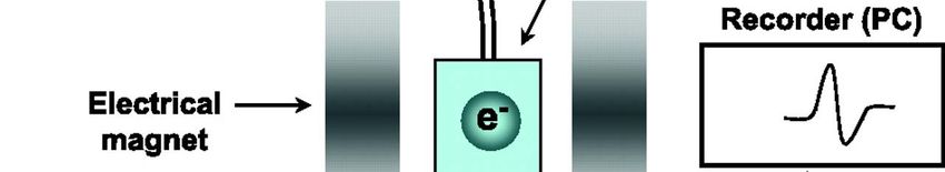
Huige LiElectron Spin Resonance
ESR signal is
proportional to
the number of
the unpaired
electrons
present in the
sample.
Institut für Pharmakologie
Huige LiESR – the use of “spin traps”
NO
colloid Fe(DETC)2 (diethyldithiocarbamate)
Lipid radicals
• N‐t‐butyl‐α‐phenyl‐nitrone
• α‐[4‐pyridyl
py y 1‐oxide]‐N‐tert‐butyl‐nitrone
y
O2‐, OH
• DMPO: 5,5‐dimethyl‐pyrroline‐N‐oxide
• DEPMPO: 5‐(diethoxyphosphoryl)‐5‐methyl‐1‐pyrroline‐N‐oxide
Institut für Pharmakologie
Huige LiESR – the use of “spin traps”
OH
Institut für Pharmakologie
Huige LiESR – the use of “spin traps”
74 mol/L/s
O2-
Fe3+‐cyt c Fe2+‐cyt c
3 x 10⁶ mol/L/s
Institut für Pharmakologie
Huige LiESR – cyclic hydroxylamine probes
CPH: 1‐hydroxy‐3‐carboxy‐2,2,5,‐tetramethyl‐pyrrolidine hydrochloride
• Reacts rapidly with O2‐ (3.2 x 10³ mol/L/s) and ONOO‐
• The
Th resultant
lt t 3
3‐carboxy‐proxyl
b l radical
di l is
i very stable,
t bl undergoing
d i minimal
i i l
bioreduction
CMH: 1‐hydroxy‐3‐methoxycarbonyl‐2,2,5,5‐tetramethylpyrrolidine
• Cell‐permeable
• Detection of intracellular O2‐
Institut für Pharmakologie
Huige LiElectron Spin Resonance
Strengths
• ESR is an excellent approach
pp for the detection of radical.
• When studying isolated enzymes or chemical reactions, the nitrone spin
traps are very useful.
• For studying intact tissues, cells, or homogenates, cyclic hydroxylamines
are better.
• Sensitivityy of ESR can reach 1 nmol/L,
/ , wenn cyclic
y hydroxylamines
y y are
used.
Weaknesses
• Expensive.
• Extensive trainingg is needed to operate
p the spectometer
p correctly.
y
Institut für Pharmakologie
Huige LiSummary
Intra‐/extra‐
ROS Sensitivity Practice
cellular
Colorimetric assays
Nitro Blut Tetrazolium intracellular O2‐ low easy
Cytochrome c extracellular O2‐ low easy
Chemiluminescence
Lucigenin intra & extra O2‐ low difficult
MCLA intra & extra O2‐ low easyy
L‐012 extracelluar O2‐, ONOO‐ high easy
Fluorescent
Dihydroethidium (DHE) intracelluar O2‐ high moderate
H2DCF‐DA intracellular ROS high easy
Amplex Red extracellular H2O2 high easy
Electron Spin Resonance intra / extra O2‐, OH moderate difficult
Institut für Pharmakologie
Huige LiReferences
Dikalov S, Griendling KK, Harrison DG. Measurement of reactive oxygen species in
cardiovascular studies. Hypertension 2007; 49: 717 ‐ 727.
Münzel T, Afanas’ev IB, Kleschyov AL, Harrison DG. Detection of superoxide in vascular tissue.
Arterioscler Thromb Vasc Biol, 2002; 22: 1761 ‐ 1768.
Daiber A,
A et al.
al & Münzel T.
T Measurement of NAD(P)H oxidase‐derived
oxidase derived superoxide with the
luminol analogue L‐012. Free Radic Biol Med. 2004; 36:101‐111.
Daiber A, et al. & Münzel T. Detection of superoxide and peroxynitrite in model systems and
mitochondria
it h d i by b th
the luminol
l i l analogue
l L‐012.
L 012 Free
F R di Res.
Radic R 2004;
2004 38:38 259
259‐269.
269
Sohn HY, et al. & Pohl U. Sensitive superoxide detection in vascular cells by the new
chemiluminescence dye L‐012. J Vasc Res. 1999; 36: 456‐464.
http://www.invitrogen.com/site/us/en/home/References/Molecular‐Probes‐The‐
Handbook.html
Institut für Pharmakologie
Huige LiYou can also read