Mesocorticolimbic Dopamine Pathways Across Adolescence: Diversity in Development
←
→
Page content transcription
If your browser does not render page correctly, please read the page content below
REVIEW
                                                                                                                                       published: 08 September 2021
                                                                                                                                       doi: 10.3389/fncir.2021.735625

                                            Mesocorticolimbic Dopamine
                                            Pathways Across Adolescence:
                                            Diversity in Development
                                            Lauren M. Reynolds 1,2 * and Cecilia Flores 3 *
                                            1
                                             Plasticité du Cerveau CNRS UMR8249, École supérieure de physique et de chimie industrielles de la Ville de Paris (ESPCI
                                            Paris), Paris, France, 2 Neuroscience Paris Seine CNRS UMR 8246 INSERM U1130, Institut de Biologie Paris Seine,
                                            Sorbonne Université, Paris, France, 3 Department of Psychiatry and Department of Neurology and Neurosurgery, McGill
                                            University, Douglas Mental Health University Institute, Montréal, QC, Canada

                                            Mesocorticolimbic dopamine circuity undergoes a protracted maturation during
                                            adolescent life. Stable adult levels of behavioral functioning in reward, motivational,
                                            and cognitive domains are established as these pathways are refined, however,
                                            their extended developmental window also leaves them vulnerable to perturbation by
                                            environmental factors. In this review, we highlight recent advances in understanding
                                            the mechanisms underlying dopamine pathway development in the adolescent
                                            brain, and how the environment influences these processes to establish or disrupt
                                            neurocircuit diversity. We further integrate these recent studies into the larger historical
                                            framework of anatomical and neurochemical changes occurring during adolescence
                                            in the mesocorticolimbic dopamine system. While dopamine neuron heterogeneity is
                            Edited by:
                                            increasingly appreciated at molecular, physiological, and anatomical levels, we suggest
                         Mark Howe,
     Boston University, United States       that a developmental facet may play a key role in establishing vulnerability or resilience to
                       Reviewed by:         environmental stimuli and experience in distinct dopamine circuits, shifting the balance
                   Anthony A. Grace,        between healthy brain development and susceptibility to psychiatric disease.
              University of Pittsburgh,
                         United States      Keywords: dopamine, adolescence, development, mesocorticolimbic dopamine system, microglia, guidance
                   Bita Moghaddam,          cues, miRNA, puberty
              University of Pittsburgh,
                         United States

                  *Correspondence:
                  Lauren M. Reynolds        INTRODUCTION
             lauren.reynolds@espci.fr
                           Cecilia Flores   Dopamine (DA) neurotransmission contributes to a multitude of behaviors, including motor
               cecilia.flores@mcgill.ca     control, reward learning, cognitive control, decision making, motivation, and salience attribution
                                            (Wise, 2004; Schultz, 2007; Bromberg-Martin et al., 2010; Cools and D’Esposito, 2011; Orsini
            Received: 02 July 2021          et al., 2015; Coddington and Dudman, 2019). The ability of this modulatory neurotransmitter
        Accepted: 17 August 2021
                                            system to simultaneously direct different types of behavior may result from the heterogeneity
     Published: 08 September 2021
                                            of DA neurons originating in ventral midbrain nuclei and projecting to limbic or cortical
                             Citation:      regions. Limbic and cortical DA pathways have been shown to differ in their molecular markers,
            Reynolds LM and Flores C
                                            anatomical organization, and response to stimuli (Roeper, 2013; Lammel et al., 2014; Morales
 (2021) Mesocorticolimbic Dopamine
      Pathways Across Adolescence:
                                            and Margolis, 2017; Poulin et al., 2018; Nguyen et al., 2021). However, less emphasis has been
             Diversity in Development.      placed on the distinct developmental trajectories of dopamine projections. Mesocorticolimbic DA
    Front. Neural Circuits 15:735625.       pathways continue to develop across postnatal life, throughout what is considered adolescence and,
      doi: 10.3389/fncir.2021.735625        often, into early adulthood. Incorporating this knowledge in our understanding of DA diversity is

Frontiers in Neural Circuits | www.frontiersin.org                                 1                                   September 2021 | Volume 15 | Article 735625
Reynolds and Flores Mesocorticolimbic Dopamine Development in Adolescence
particularly important since the DA system is increasingly adulthood at 2 months of age. When considering the age range
considered as a ‘‘plasticity system,’’ whose development can be of adolescence in rodents, it is important to keep in mind that
shaped by positive or negative experiences, allowing organisms while puberty and adolescence necessarily coincide temporally,
to adapt to their surrounding environmental conditions (Barth these terms are not interchangeable (Spear, 2000; Schneider,
et al., 2019; Reynolds and Flores, 2021). 2013). Puberty can be clearly defined by the onset of sexual
Adolescence is a time when organisms undergo dramatic maturity, while adolescence is a more diffuse period representing
physical, hormonal, and behavioral changes as they transition the gradual transition from a juvenile state to independence.
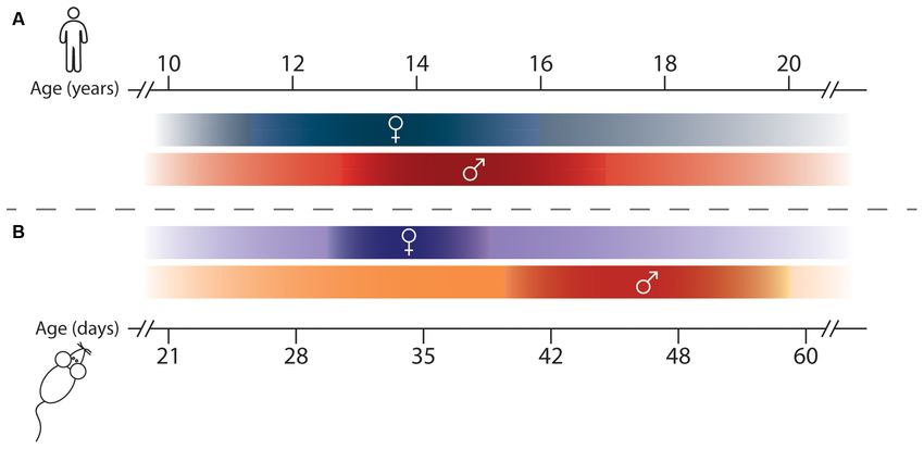
from juveniles to adults. While in humans this period has been While studies in rodents consider a range of PND 28–42 to be
historically framed as ranging from 12–20 years of age, the peri-pubertal in male animals, adolescence is instead suggested
boundaries of adolescence are increasingly recognized as difficult to extend from the age of weaning (PND 21) until adulthood
to define precisely, with modern definitions extending between (PND 60; Spear, 2000; Tirelli et al., 2003; Burke and Miczek,
10 and 24 years of age (Figure 1A; Hollenstein and Lougheed, 2013; Schneider, 2013; Figure 1B). This timeline encompasses
2013; Sawyer et al., 2018). The brain undergoes exuberant the entire post-weaning period when rodents exhibit distinct
development during this time, with cortical gray matter neurobiological and behavioral changes and are navigating their
thickness, notably in the prefrontal cortex (PFC), decreasing environment independently for the first time. This demarcation
before stabilizing at adult levels, and with white matter volume is more aligned with the modern, extended definition of human
increasing until early adulthood (Blakemore, 2012; Paquola et al., adolescence (Sawyer et al., 2018).
2019). These macroscale changes likely result from cellular, In this review, we provide an overview of preclinical findings
molecular, and connectivity neuroadaptations in adolescence, as regarding the adolescent development of mesocorticolimbic DA
postmortem studies show dramatic age-dependent changes in pathways, by both situating it within a historical context and
myelination, neuronal structure, and synapse density during this by emphasizing novel advances in understanding its cellular
time (Petanjek et al., 2011; Miller et al., 2012; Catts et al., 2013). and molecular underpinnings. While the majority of preclinical
A particular interest in the postnatal development of work on DA development has been performed exclusively
DAergic systems has emerged because of evidence showing in males, we highlight throughout the review studies that
that performance in DA-dependent cognitive tasks improves include both males and female subjects, since sex differences
gradually across adolescence (Wahlstrom et al., 2010; Luciana have been noted in adult DA circuitry architecture and
et al., 2012; Galvan, 2017; Larsen and Luna, 2018). However, function. We outline proposed mechanisms by which adolescent
direct evidence of cellular and molecular maturational changes experiences interact with developmental programming to shape
in the human DA system across adolescence remains scarce, adult mesocorticolimbic DA connectivity and function. Finally,
and it is limited to post-mortem studies which suggest that we identify important gaps in our knowledge which present
DA signaling remains dynamic across postnatal life (Weickert promising avenues for future research.
et al., 2007; Rothmond et al., 2012). Studies in adult volunteers
can estimate DA release in subcortical regions by measuring MESOCORTICOLIMBIC DOPAMINE
the binding of radioactive ligands to DA receptors using CIRCUIT ORGANIZATION
positron emission tomography (PET). However, because of
its radioactive nature, PET is contraindicated for imaging in Since its initial characterization in the 1960s, the anatomical
minors without medical necessity (Ernst and Luciana, 2015). To organization of the rodent DA circuitry has been
overcome this impasse, recent studies have introduced tissue iron comprehensively described and reviewed on several occasions
concentration as a proxy measure for DA concentration, as it (a non-exhaustive list of reviews: Björklund and Lindvall, 1984;
is easily distinguishable using non-invasive functional magnetic Björklund and Dunnett, 2007; Sesack and Grace, 2010; Yetnikoff
resonance imaging (fMRI), can be extracted from existing et al., 2014a; Morales and Margolis, 2017). This review focuses
fMRI datasets (Peterson et al., 2019), and is correlated with specifically on the mesocorticolimbic DA circuitry, which
radioligand binding for the vesicular monoamine transporter consists of cell bodies located in the ventral tegmental area
(VMAT2) in adult subjects (Larsen et al., 2020). Tissue iron (VTA) that send ascending fibers rostrally toward limbic and
levels in the human striatum increase throughout adolescence, cortical regions through the tightly fasciculated medial forebrain
before stabilizing in adulthood (Larsen and Luna, 2014; Larsen bundle (Figure 2A; Nieuwenhuys et al., 1982). At the level of
et al., 2020), and are associated with the ongoing maturation of the nucleus accumbens (NAc) these fibers diverge to reach
DA-dependent behaviors (Parr et al., 2021). While changes in their terminal target, with the densest innervation comprising
tissue iron levels have not yet been assessed in the developing mesolimbic DA axons that remain in the NAc or that extend
PFC, these results suggest that striatal DA regions are undergoing to more dorsal regions of the striatum (STR). Mesocortical DA
dynamic maturation in adolescence, mirroring previous findings fibers course along the medial forebrain bundle with mesolimbic
from preclinical studies. DA axons, but split off toward the PFC by either passing through
Most of our knowledge about adolescent mesocorticolimbic the NAc, STR, and external capsule; or by extending ventrally
DA development comes from preclinical research, and from to bypass the NAc before curving dorsally, just caudal to the
rodent studies in particular. One practical advantage of using olfactory bulb (Figure 2B; Kalsbeek et al., 1988, 1992; Voorn
rodents in developmental research is their compressed lifespan et al., 1988; Kolk et al., 2009; Manitt et al., 2011; Brignani and
since they are born after about 3 weeks of gestation and reach Pasterkamp, 2017). Interestingly, despite the close proximity
Frontiers in Neural Circuits | www.frontiersin.org 2 September 2021 | Volume 15 | Article 735625
Reynolds and Flores                                                                                          Mesocorticolimbic Dopamine Development in Adolescence

  FIGURE 1 | Adolescence and Puberty timing in humans and rodents. (A) Timeline of adolescence (shaded lines) and puberty (darker portions of the lines) in girls (♀)
  and boys (♂), adapted from Hollenstein and Lougheed (2013), Sawyer et al. (2018), and Brix et al. (2019). (B) Adolescence (shaded lines) and puberty (darker
  portions of the lines) timing in female (♀) and male (♂) rodents, adapted from Vetter-O’Hagen and Spear (2012) and Schneider (2013).

of mesolimbic and mesocortical DA axons throughout their
trajectory to forebrain regions, there is little or no overlap in the
targets they innervate. Unlike other neuromodulatory systems,
VTA DA neurons rarely send axon collaterals between different
forebrain regions (Fallon, 1981; Fallon and Loughlin, 1982;
Swanson, 1982; Lammel et al., 2008; Beier et al., 2015; Reynolds
et al., 2018). Since mesocortical DA axons pass through the
striatum en route to the PFC, the lack of mesocorticolimbic
DA collaterals suggests that the striatum functions as a ‘‘choice
point’’ (Stoeckli and Landmesser, 1998), where DA axons
segregate into their cortical and limbic projections.
    While these pathways begin to be established in embryonic
or early postnatal development (see Riddle and Pollock, 2003;
Prakash and Wurst, 2006; Heuvel and Pasterkamp, 2008; Money
and Stanwood, 2013; Brignani and Pasterkamp, 2017 for detailed
reviews), significant alterations in mesocorticolimbic DA wiring
are increasingly observed in late postnatal development.

GROWTH AND ORGANIZATION OF THE
MESOCORTICAL DOPAMINE PATHWAY IN
                                                                                        FIGURE 2 | Mesocorticolimbic dopamine system organization. (A)
ADOLESCENCE                                                                             Two-dimensional rendering of the mesocorticolimbic DA pathway (black) and
                                                                                        the major nuclei of interest in the horizontal plane. (B) Sagittal view of the
Reports that the density of mesocortical DA innervation                                 mesocorticolimbic DA pathway (black) and the major nuclei, with the STR
continues to increase during adolescence were first published in                        semi-transparent in the foreground. Two-dimensional representations were
the 1980s, shortly after the introduction of antibodies against                         adapted from the three-dimensional regions of interest in the Allen brain atlas
                                                                                        (Wang et al., 2020). PFC, prefrontal cortex; NAc, nucleus accumbens; STR,
tyrosine hydroxylase (TH, the rate-limiting enzyme of dopamine
                                                                                        dorsal striatum; MFB, medial forebrain bundle; VTA, ventral tegmental area.
synthesis), which allowed clear detection of DA axons in the PFC
(Verney et al., 1982). The earliest studies used light microscopy
and TH immunofluorescence to show that DA axons in the                                 DA fibers in these regions, however, continues to increase until
supragenual anterior cingulate and prelimbic cortices of rats                          PND 60 (Berger et al., 1985; Kalsbeek et al., 1988), with no
already show their typical thin morphology with irregularly                            further changes between PND 60 and PND 90 (Figure 3A). This
spaced varicosities by early-to-mid adolescence. The density of                        adolescent increase in PFC DA innervation density remains a

Frontiers in Neural Circuits | www.frontiersin.org                                 3                                    September 2021 | Volume 15 | Article 735625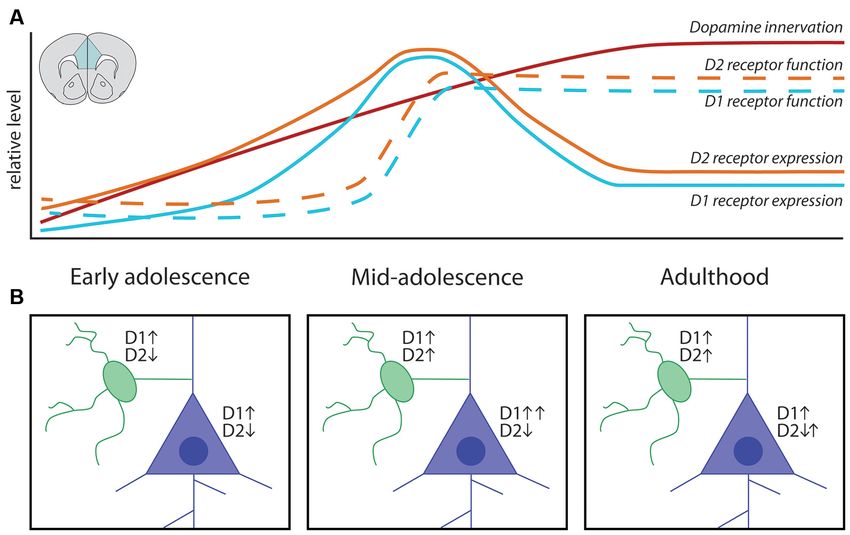
Reynolds and Flores                                                                           Mesocorticolimbic Dopamine Development in Adolescence

robust finding, as these initial qualitative descriptions have since       PFC (Figure 4B), in line with the lack of collaterals observed
been replicated (Benes et al., 1996) and extended by quantitative          in previous studies (Fallon, 1981; Fallon and Loughlin, 1982;
analysis in the PFC of rats and mice by several research teams             Lammel et al., 2008; Beier et al., 2015). This discovery is the first
(Manitt et al., 2011; Naneix et al., 2012; Willing et al., 2017;           proof of long-range growth of axons in adolescence and explains
Hoops et al., 2018). Although the large majority of studies on             why the mesocorticolimbic DA system is particularly vulnerable
mesocortical DA development have been performed only in male               to adolescent experiences.
rodents, female rats have been shown to exhibit a similar pattern              The size of DAergic varicosities in the PFC increases from
of innervation across postnatal ages (Willing et al., 2017).               ∼1.2 µm at PND 20 to ∼2.4 µm by PND 60 (Benes et al.,
    The extended postnatal increase in PFC DA fiber density                1996), and PFC DA concentration increases significantly during
contrasts to other neuromodulatory systems, such as                        this time (Nomura et al., 1976; Leslie et al., 1991; Naneix et al.,
norepinephrine (NE) and serotonin, which reach adult PFC                   2012). Varicosities are sites of DA synthesis, release, and re-
innervation density levels in rodents within the first 2–3 weeks of        uptake, and in the PFC at least 93% of them form functional
life (Levitt and Moore, 1979; Lidov et al., 1980). Distinguishing          synaptic contacts with local neurons (Séguéla et al., 1988). DA
PFC DA axons from NE axons using immunolabeling for TH,                    axons form synapses onto PFC glutamatergic pyramidal neurons;
which is required for the synthesis of both catecholamines, is             which represent the primary projection neurons from the PFC
often cited as a methodological concern for anatomical studies.            to other regions of the brain (Goldman-Rakic and Brown, 1982;
Despite the fact that TH is also present in NE neurons, visually           Goldman-Rakic et al., 1992; Krimer et al., 1997; Carr et al.,
distinguishing DA and NE axons in the PFC is feasible. NE                  1999; Carr and Sesack, 2000; Lambe et al., 2000), a phenomenon
axons are thick, with regularly spaced rounded varicosities;               that is conserved in humans, non-human primates, and rodents.
while DA axons are thin and sinuous, with irregularly spaced               GABAergic interneurons, and in particular those that express
varicosities (Berger et al., 1974; Miner et al., 2003). The two            parvalbumin (e.g., fast-spiking interneurons), also receive inputs
axonal populations differ further in their distribution, with DA           from DA axons and express high levels of DA receptors (Verney
fibers densely concentrated in the inner layers of the pregrenual          et al., 1990; Benes et al., 1993, 2000; Sesack et al., 1995, 1998;
and supragenual medial PFC, while NE fibers are spread across              Le Moine and Gaspar, 1998; Seamans and Yang, 2004; Glausier
all layers (Berger et al., 1976; Levitt and Moore, 1979; Miner             et al., 2009; Tritsch and Sabatini, 2012). Both DAergic synapses
et al., 2003). Immunostaining for TH in the PFC labels PFC                 onto PFC pyramidal neurons and GABAergic interneurons
DA axons nearly exclusively because it only rarely overlaps with           have been reported to increase during adolescence. A study
NE-specific markers, such as dopamine-β-hydroxylase (DBH) or               in rats shows that the number of DA appositions onto PFC
the NE transporter (Pickel et al., 1975; Berger et al., 1983; Miner        GABAergic interneurons increases in adolescence (Benes et al.,
et al., 2003; Naneix et al., 2012). When compared directly within          1996). A study in non-human primates shows that the number
the same study, DA fiber density in the cingulate, prelimbic,              of DAergic, but not serotonergic, appositions onto pyramidal
and infralimbic subregions of the pregenual medial PFC has                 neurons proliferates during adolescence, however, no changes
been shown to increase up to three-fold across adolescence,                in the number of DA oppositions onto GABA neurons were
whereas DBH-stained NE fiber density remains stable across this            detected in this study (Lambe et al., 2000). These results suggest
time (Naneix et al., 2012). The seminal work of Rosenberg and              that the release of DA in the PFC evolves during adolescence in
Lewis shows that the same pattern of adolescent increase in PFC            parallel to the establishment of mature pre-synaptic connectivity.
DA innervation is found in non-human primates, suggesting                  Indeed, disruption of PFC DA innervation in adolescence results
that this pattern is indeed conserved across mammalian species             in altered dendritic arborization and dendritic spine density of
(Rosenberg and Lewis, 1994, 1995; Lewis, 1997).                            layer V pyramidal neurons, indicating that PFC DA development
    For many years this increase in PFC DA fiber density across            in adolescence drives the structural maturation of local PFC
adolescence was thought to represent the progressive increase              circuits (Manitt et al., 2011, 2013; Reynolds et al., 2018).
in the sprouting of new branches from DA axons already
innervating the PFC early in life, as long-range axon growth
was assumed to be complete before adolescence. However,                    POSTSYNAPTIC CHANGES ACROSS
studies using anterograde or retrograde labeling have challenged           ADOLESCENCE IN THE MESOCORTICAL
this notion by suggesting that axons are still growing during              DOPAMINE SYSTEM
postnatal development to connect from PFC to the amygdala
(Arruda-Carvalho et al., 2017), or from the forebrain to the               Both pyramidal and GABAergic interneurons in the PFC express
VTA (Yetnikoff et al., 2014c). By harnessing an intersectional             DA receptors of the D1 (D1 and D5) and D2 (D2, D3, D4)
viral labeling technique (Figure 4), we were able to restrict              families (Gaspar et al., 1995; Vincent et al., 1995; Knable and
fluorescent labeling only to DA neurons with axons present in              Weinberger, 1997; Lu et al., 1997; Davidoff and Benes, 1998; Le
the NAc at PND 21. When the mice reached adulthood, we                     Moine and Gaspar, 1998; Mitrano et al., 2014). DA receptors
found that a subset of these DA axons in fact grew through                 are seven transmembrane G-protein coupled receptors that
the NAc to reach the medial or orbital PFC during adolescence              initiate intracellular cascades by increasing cAMP (D1-type) or
(Figure 4A; Hoops et al., 2018; Reynolds et al., 2018). When               decreasing cAMP (D2-type; Seamans and Yang, 2004; Tritsch
we performed these same intersectional viral injections in adult           and Sabatini, 2012). DA receptors are localized to apical and
mice, we observed very few, if any, labeled DA axons in the                basilar dendritic arbors of pyramidal neurons and to dendrites

Frontiers in Neural Circuits | www.frontiersin.org                     4                                September 2021 | Volume 15 | Article 735625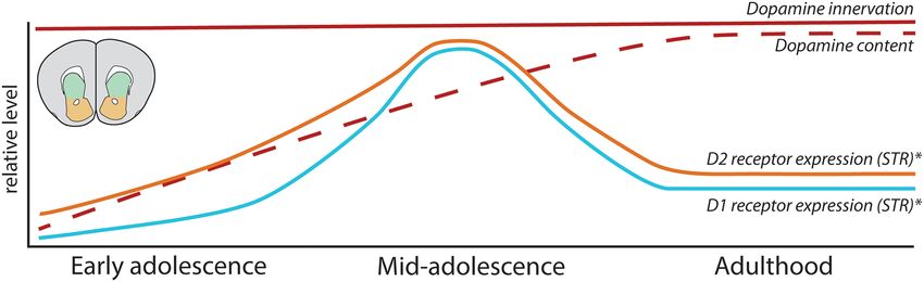
Reynolds and Flores                                                                                       Mesocorticolimbic Dopamine Development in Adolescence

  FIGURE 3 | Adolescent maturation of dopamine connectivity and function in the prefrontal cortex. (A) Summary of maturational changes in DA connectivity in the
  PFC across adolescence. (B) Early in adolescence DA signaling through D1 receptors is excitatory onto both classes of neurons, with DA signaling through
  D2 receptors inhibiting pyramidal neurons and weakly inhibiting GABAergic interneurons. In mid-adolescence DA signaling through D2 receptors becomes excitatory
  onto GABAergic interneurons. DA signaling through D1 receptors is now able to interact with glutamatergic NMDA receptors on pyramidal neurons, increasing the
  activating effect of DA. In adulthood D1 and D2 receptor populations attain their mature function.

  FIGURE 4 | Mesocortical axon growth in adolescence revealed by an intersectional viral labeling technique. (A) Spatiotemporally specific labeling of DA neurons
  with axons present in the NAc at PND21 reveals that DA axons continue to grow to the PFC in adolescence. (B) Labeled DA axons are not found in the PFC of mice
  when subjected to the same manipulation in adulthood, supporting previous findings that DA neurons do not commonly send collaterals between these regions.

and cell bodies of GABAergic interneurons that synapse onto                          DA receptor expression is more complex (Figure 3A).
pyramidal neurons. Signaling through PFC postsynaptic DA                             Autoradiography studies in rats using radio-labeled DA
receptors thus directly and indirectly modulates PFC output                          receptor ligands ([3 H]SCH-23390 for D1-like receptors,
(Gulledge and Jaffe, 2001; Seamans et al., 2001; Dong and                            [3 H]nemonapride (YM-09151–2) or [3 H]-raclopride for D2-like
White, 2003; Trantham-Davidson et al., 2004; Santana et al.,                         receptors) show discrepant results regarding the patterns of
2009; Tritsch and Sabatini, 2012) and calibrates the balance                         receptor expression across postnatal life. While some studies
of excitation/inhibition in the PFC, which also matures in                           indicate a steady increase in PFC D1-like receptor density until
adolescence (O’Donnell, 2011).                                                       PND 60 in rats (Tarazi et al., 1999; Tarazi and Baldessarini,
   While the density of DA fibers in the PFC shows a linear                          2000), others show that D1-like expression in the PFC peaks
increase across adolescence, the trajectory of postsynaptic                          in adolescence, before being pruned back in adulthood (Leslie

Frontiers in Neural Circuits | www.frontiersin.org                               5                                  September 2021 | Volume 15 | Article 735625
Reynolds and Flores                                                                        Mesocorticolimbic Dopamine Development in Adolescence

et al., 1991; Andersen et al., 2000; Brenhouse et al., 2008).            through D2 receptors has either a weak inhibitory effect or
These seemingly disparate findings may be reconciled by results          no effect (Gorelova et al., 2002; Tseng and O’Donnell, 2007).
from immunohistochemical tracing experiments showing that                However, after adolescence an excitatory effect of D2 receptor
the adolescent peak in D1 receptor expression observed in                stimulation emerges in PFC GABAergic interneurons, creating
adolescence occurs only in corticolimbic pyramidal projection            the inhibitory tone characteristic of the mature PFC and
neurons (Brenhouse et al., 2008; Brenhouse and Andersen,                 balancing the enhanced DA-driven excitation of pyramidal
2011), a level of nuance which may be difficult to capture               neurons (Tseng and O’Donnell, 2007; O’Donnell, 2011). The
with radioligand binding assays. Another consideration is that           adolescent shift of DAergic regulation over excitatory and
different subtypes of DA receptors may not follow the same               inhibitory transmission in the PFC is thought to be a critical
developmental pattern of expression, but most radioligands do            step in the developmental calibration of cognitive control (Klune
not differentiate between different receptors of the same family         et al., 2021), and to be dysregulated in psychiatric disorders
[e.g., [3 H]SCH-23390 will bind both D1 and D5 DA receptors,             of adolescent onset (O’Donnell, 2011; Caballero et al., 2016;
which are expressed in the rodent PFC (Lidow et al., 2003)].             Caballero et al., 2021).
Recent results from quantitative real-time PCR (qPCR) studies
in rats bolster the idea that PFC D1 and D5 receptor subtypes
show a peak in mRNA levels during adolescence (Naneix et al.,
                                                                         COMING OF AGE IN THE STRIATUM:
2012; Zbukvic et al., 2017).                                             MESOCORTICOLIMBIC DOPAMINE
    Regarding D2-like receptors, early radioligand studies in rats       PATHWAY SEGREGATION AND
also indicate that their expression in the PFC increases steadily        FUNCTIONAL MATURATION
in adolescence (Tarazi et al., 1998; Tarazi and Baldessarini,
2000), but later findings show peak expression in adolescence            As our understanding of mesocorticolimbic DA system
(Andersen et al., 2000; Brenhouse and Andersen, 2011). This              development progresses, it is clear that both mesolimbic and
inconsistency may also stem from the non-specific nature                 mesocortical pathways are still developing throughout the
of radioligand binding, as Naneix et al. (2012) show an                  adolescent period. DA signaling in the striatum continues to
adolescent peak in mRNA expression for the long isoform                  mature across adolescence: both TH protein and DA content
of the D2 receptor and the D4 receptor, but not for the                  increase until adulthood (Pardo et al., 1977; Giorgi et al., 1987;
short isoform of the D2 receptor, in the PFC using qPCR.                 Broaddus and Bennett, 1990; Rao et al., 1991; Naneix et al., 2012;
Another consideration when assessing apparent discrepancies              Matthews et al., 2013; Lieberman et al., 2018), and are essential
in the literature is that radioligand binding assays indirectly          for the construction of postsynaptic circuits. Medium spiny
determine receptor protein levels and/or functional capacity,            neurons (MSNs) in the NAc and STR achieve their namesake
whereas qPCR determines mRNA expression. Overall, evidence               spiny appearance during early adolescence, with marked
from studies in rats indicates that DA receptor expression in            increases in dendritic spine density occurring between PND
the PFC is dynamic in adolescence, most likely with a period             15 and 30 (Tepper and Trent, 1993; Tepper et al., 1998). The
of overexpression followed by pruning (Figure 3A). However,              proportion of MSNs showing their characteristic DA-sensitive
this adolescent peak is less apparent at least in C57BL6 mice,           inward rectification potassium currents also continues to
according to an autoradiography study (Pokinko et al., 2017),            increase until early adulthood (Tepper et al., 1998; Zhao et al.,
suggesting differences across species. It should be noted that           2016), and the effect of D2 receptor signaling of MSNs switches
the aforementioned receptor expression studies were performed            from inhibitory to facilitatory (Benoit-Marand and O’Donnell,
exclusively in male rodents and whether sex differences exist            2008). During this same timeline, NAc DA varicosities shift
in the trajectory of PFC DA receptor expression remains an               their synaptic contacts from the soma of MSNs to their dendritic
open question.                                                           spines (Antonopoulos et al., 2002), and the intrinsic excitability
    In addition to the dynamic changes in DA receptor                    of MSN changes from a juvenile hyper-excitable state to a
expression, postsynaptic responses to extracellular DA have              reduced, mature level of responsivity. This process is triggered
also been shown to evolve in PFC pyramidal and GABAergic                 by the gradual increase in striatal DA concentration (Lieberman
neurons during adolescence (Figure 3B; O’Donnell, 2010,                  et al., 2018).
2011). Slice electrophysiology experiments have shown that                   In contrast to the PFC, the changes in DA function in the
both pyramidal and GABAergic PFC neurons respond to DA                   striatum are not associated with changes in the density of DA
application (Seamans and Yang, 2004). Studies in adult rats              innervation (Figure 5), as STR and NAc DA input achieves its
show that DA signaling through D1 receptors in PFC pyramidal             adult density by PND 20 in rodents (Voorn et al., 1988; Kalsbeek
neurons interacts with glutamatergic NMDA receptor signaling,            et al., 1992), and the optical density of TH-positive DA fibers
producing activity levels that resemble those observed in vivo           does not change during adolescence in these regions (Naneix
during information processing. Notably, this process is absent           et al., 2012). Nevertheless, these results should be interpreted
before mid-adolescence (Tseng and O’Donnell, 2005), emerging             with care, considering that the DA innervation to the STR and
only around PND 45, when D1 mRNA expression peaks in                     NAc is up to 40-fold denser than in the PFC. It is possible that the
the rat PFC (Tseng and O’Donnell, 2005; Naneix et al., 2012).            margin of error, even using precise stereological methods, masks
In fact, before PND 36, DA signaling through D1 receptors in             potential anatomical differences between ages (Bérubé-Carrière
GABAergic interneurons potentiates their firing, but signaling           et al., 2012; Manitt et al., 2013; Reynolds et al., 2015).

Frontiers in Neural Circuits | www.frontiersin.org                   6                               September 2021 | Volume 15 | Article 735625
Reynolds and Flores Mesocorticolimbic Dopamine Development in Adolescence
FIGURE 5 | Development of mesolimbic dopamine connectivity and function in the adolescent striatum. Summary of maturational changes in DA connectivity in the
STR across adolescence. ∗ Indicates that these findings are sex-dependent, with this pattern of receptor overexpression and pruning only seen in male rodents.
Our developmental studies using intersectional viral tracing D2 receptors in striatopallidal MSNs (Ince et al., 1997; Smith
techniques indeed demonstrate that the fine organization of et al., 1998; Kreitzer, 2009; Bamford et al., 2018). Unlike the
mesolimbic DA connectivity is much more malleable than PFC, where ∼25% of non-pyramidal neurons co-express D1 and
previously thought. The striatum represents a major choice point D2 DA receptors (Vincent et al., 1995), D1 and D2 receptor
for DA axons: while the large majority of DA axons already expression is almost completely segregated between these two
innervating the striatum by early adolescence are destined to MSN populations (Hersch et al., 1995; Ince et al., 1997; Bertran-
remain there, mesocortical axons must pass through this densely Gonzalez et al., 2010; Frederick et al., 2015). Interestingly, D1 and
innervated DA region and continue to grow into the PFC during D2 colocalization in MSNs is apparent in embryos and neonates,
adolescence (Reynolds et al., 2018). Notably, the level of DA but it decreases between E18 and PND 14 (Thibault et al., 2013;
innervation in these two pathways is inversely correlated: the Biezonski et al., 2015). It remains to be determined whether the
more DA axons that keep growing to the PFC in adolescence, segregation of DA receptors in striatonigral and striatopallidal
the fewer that remain behind and form connections in the NAc. MSNs is dynamic at other postanal periods, as well as the age
Mesolimbic DA axon targeting is not a passive process [i.e., they when the segregated pattern of MSN DA receptor expression
are actively undergoing target recognition processes within the is stabilized.
NAc in adolescence (Reynolds et al., 2018; Cuesta et al., 2020)], Rodent studies have shown that striatal DA receptor
and it profoundly influences the structural organization of expression changes across adolescence, although their exact
postsynaptic MSN neurons. maturational pattern is controversial. Early autoradiography
studies using the radioligand [3 H]SCH-23390 to assess
DEVELOPMENTAL PATTERNS OF D1 receptor binding in Sprague–Dawley rats found that
DOPAMINE RECEPTORS IN THE striatal D1 receptors either increase until achieving stable adult
ADOLESCENT STRIATUM levels before or during early adolescence (Murrin and Zeng,
1990; Leslie et al., 1991; Schambra et al., 1994) or exhibit no
In contrast to the high synaptic incidence of DA varicosities in developmental changes (Broaddus and Bennett, 1990). Instead,
the PFC (Séguéla et al., 1988), the majority of DA varicosities in more recent studies using the same radioligand and rat strain
the NAc and STR (60–70%) do not form direct synaptic contacts show that, similarly to the PFC, DA D1-like receptors are
with postsynaptic neurons (Descarries et al., 1996; Descarries and overexpressed during adolescence before being pruned back in
Mechawar, 2000; Bérubé-Carrière et al., 2012). Recent reports adulthood in the NAc and STR (Gelbard et al., 1989; Teicher
further indicate that only ∼30% of DA varicosities in the STR et al., 1995; Andersen et al., 1997; Tarazi et al., 1999; Tarazi and
contain the necessary active zone sites to release DA, and that Baldessarini, 2000). Similar findings using autoradiography have
many varicosities are in fact ‘‘silent’’ (Pereira et al., 2016; Liu et al., been reported in C57BL/6 mice (Pokinko et al., 2017).
2018, 2021; Liu and Kaeser, 2019). Up to 90% of local striatal Striatal expression of D2 receptors has also been reported
neurons are GABAergic projection neurons, usually referred to change during adolescence. Early autoradiography studies
to as MSNs or as spiny projection neurons (Kreitzer, 2009; showed increased D2 receptor expression across early postnatal
Collins and Saunders, 2020), and they can be segregated into two life, reaching adult levels by early adolescence (Pardo et al., 1977;
main populations based on their projection target. In rodents, Hartley and Seeman, 1983; Murrin and Wanyun, 1986; Rao et al.,
striatonigral MSNs project mainly to the substantia nigra pars 1991; Schambra et al., 1994). This evidence seems to be consistent
reticulata and entopeduncular nucleus, while striatopallidal despite the use of different radioligands and strains of rats, with
MSNs instead project primarily to the globus pallidus (Smith changes in expression in the NAc showing a less pronounced
et al., 1998). While striatonigral and striatopallidal MSNs are peak than in the STR (Teicher et al., 1995; Andersen et al., 1997;
morphologically indistinguishable at the somatic level, they can Tarazi et al., 1998), and mRNA expression of D2 receptors peaks
be differentiated by a number of molecular markers, notably by in the adolescent STR (Naneix et al., 2012). These D2 expression
prominent expression of D1 receptors in striatonigral MSNs and changes have not been detected in mice (Pokinko et al., 2017).
Frontiers in Neural Circuits | www.frontiersin.org 7 September 2021 | Volume 15 | Article 735625Reynolds and Flores Mesocorticolimbic Dopamine Development in Adolescence
While the majority of these studies have been performed et al., 2016); and blunted stimulant-induced DA release in the
only in male rodents, evidence suggests that the adolescent NAc (Grant et al., 2007). This protective Dcc haploinsufficient
overexpression and pruning of D1 receptors in striatal regions phenotype, which is also displayed by mice haploinsufficient for
is not seen in female rats (Andersen et al., 1997). Similarly, the Netrin-1, is driven by increased DA innervation and content
adolescent overexpression and subsequent pruning in striatal in the PFC, and by augmented stimulant drug-induced DA
D2 receptors observed in male rats is absent in females (Andersen release in this region (Grant et al., 2007; Pokinko et al.,
et al., 1997), with no apparent sex differences in D2 receptor 2015), indicating increased mesocortical inhibitory control over
expression levels in adulthood. Males and females may therefore striatal DA function. Notably, these DAergic changes are only
have distinct developmental trajectories for DA receptors in apparent in adult Dcc haploinsufficient mice, with no observable
striatal regions, which could lead to different sensitive periods differences in mesocorticolimbic DA structure or function in
of development. juveniles, and occur in both males and females (Grant et al.,
2009).
MECHANISMS UNDERLYING Netrin-1 and DCC are expressed across the lifetime
in mesocorticolimbic DA circuits, and their spatiotemporal
MESOCORTICOLIMBIC DOPAMINE distribution is pathway-specific. DA neurons express high levels
CIRCUIT ORGANIZATION IN of DCC receptors across species, including humans (Osborne
ADOLESCENCE et al., 2005; Manitt et al., 2010; Reyes et al., 2013). Netrin-1 is
expressed in forebrain terminal regions of DA axons, including
DA circuitry is increasingly recognized as a ‘‘plasticity system’’ the NAc, STR, and PFC (Shatzmiller et al., 2008; Manitt et al.,
(Barth et al., 2019; Reynolds and Flores, 2021), where the 2011). In male rodents, the expression of DCC in the VTA and
environment can alter its development and induce long-term of Netrin-1 in the NAc is highest during embryonic and early
repercussions for adult behavioral functioning. The protracted postnatal development, waning gradually during adolescence,
maturational timeline of mesocortical DA circuitry, therefore, and stabilizing to low levels in adulthood (Manitt et al., 2010;
results in a prolonged period of vulnerability, when experiences Cuesta et al., 2018, 2020). While all mesolimbic DA axons are rich
such as exposure to stress or drugs of abuse can disrupt its in DCC receptor levels, DA axons in the PFC only rarely express
development and induce susceptibility to psychiatric disease DCC (Manitt et al., 2011). The localization of DCC receptors in
later in life (Meaney et al., 2002; Gulley and Juraska, 2013; PFC DA axons is increased in adult Dcc haploinsufficent mice,
Jordan and Andersen, 2017; Areal and Blakely, 2020). The suggesting that the greater PFC DA innervation and content
studies discussed in the preceding sections provide a holistic results from ectopic growth of DCC-expressing mesolimbic DA
understanding of the developmental changes occurring in the fibers (Manitt et al., 2011). Conditional reduction of Dcc in
mesocorticolimbic DA circuitry during adolescence but only DA neurons in adolescence recapitulates completely this ectopic
recently have the cellular and molecular mechanisms underlying DCC-positive DA axon phenotype in the PFC (Manitt et al.,
these processes begun to be unraveled. Below, we outline three 2013).
main mechanisms identified to date which orchestrate adolescent Using the same intersectional viral labeling technique we used
mesocorticolimbic DA development and show examples of how to demonstrate that mesocortical DA axons continue to grow to
they can be impacted by ongoing experiences. the PFC in adolescence, we also showed that the complementary
action of Netrin-1 and DCC mediates the targeting of mesolimbic
Role of Guidance Cues in Dopamine Axon DA neurons at the NAc choice point. Reduced Dcc expression
Growth and Targeting in DA axons innervating the NAc in adolescence, results in
Guidance cues are secreted proteins, either diffusible or bound their ectopic growth in the PFC and a concomitant reduction in
to cellular membranes, that act as a signal to direct growing NAc DA varicosities (Reynolds et al., 2018). High levels of DCC
axons to their appropriate targets (Battum et al., 2015). Their in mesolimbic DA axons are necessary for them to recognize
role in early DA development has long been appreciated, with the NAc as their final target in adolescence. This phenotype is
a number of guidance cues shown to be implicated in the replicated when silencing Netrin-1 in the NAc (Cuesta et al.,
differentiation, migration, and early axonal pathfinding of DA 2020).
neurons in embryonic and early postnatal life (see Heuvel Experience-induced regulation of Netrin-1 and/or DCC
and Pasterkamp, 2008; Bodea and Blaess, 2015; Brignani and expression robustly shapes the adolescent brain. Social defeat
Pasterkamp, 2017 for exhaustive reviews on the role of guidance stress in adolescence, but not in adulthood, downregulates
cues in early DA development). The guidance cue Netrin-1 and Dcc expression in the VTA of male mice, disrupts PFC
its receptor DCC (deleted in colorectal cancer) have emerged as DA innervation, and leads to cognitive control deficits in
critical players in establishing mesocorticolimbic DA circuitry adulthood (Vassilev et al., 2021). Mild traumatic brain injury
and are highly linked to psychiatric disorders of adolescent in mid-adolescent male mice reduces Netrin-1 expression in
onset (Vosberg et al., 2019; Torres-Berrío et al., 2020a). Mice the NAc and alters mesocorticolimbic DA organization (Kaukas
with Dcc haploinsufficiency show marked functional changes et al., 2020). Both Netrin-1 and DCC levels expression can encode
in DA systems, including blunted behavioral responses to the effects of experience on DA circuitry. Notably, repeated
amphetamine, methamphetamine, and cocaine (Flores et al., exposure to amphetamine downregulates DCC in the VTA and
2005; Grant et al., 2007; Flores, 2011; Kim et al., 2013; Reynolds Netrin-1 in the NAc, in early adolescence (Yetnikoff et al.,
Frontiers in Neural Circuits | www.frontiersin.org 8 September 2021 | Volume 15 | Article 735625Reynolds and Flores Mesocorticolimbic Dopamine Development in Adolescence
2007, 2011, 2014b; Cuesta et al., 2018, 2019), overlapping with onto these pyramidal neurons (Parkhurst et al., 2013). In the
the period that mesolimbic DA axons are undergoing targeting NAc, microglia play an important role in the elimination of
events. Drug-induced DCC downregulation requires D2 receptor DA receptors as Kopec et al. show that the peak in DA
signaling (Cuesta et al., 2018), reinforcing the link between DCC D1 receptor levels observed in the NAc around PND 30 in male
function and mesolimbic DA axon targeting in adolescence, as rats declines afterward due to microglia pruning. In agreement
mesocortical DA neurons lack D2 receptors (Lammel et al., with earlier autoradiography results (Andersen et al., 1997), this
2008). Exposure to recreational-like doses of amphetamine in event occurs only in males and aligns with a peak in their
early adolescence, but not later in life, leads to a dramatic increase social behavior. Females show an earlier peak (∼PND20) in
in the volume of PFC DA innervation, altered DA function, D1 receptor levels, which is followed by a microglia-independent
and long-term impairments in cognitive control in male mice decline in expression (Kopec et al., 2018). Indeed, a growing
(Reynolds et al., 2015, 2019; Hoops et al., 2018; Reynolds and body of work shows that microglial processes are sex-dependent
Flores, 2019). These effects are not observed following exposure (Schwarz and Bilbo, 2012; VanRyzin et al., 2018, 2020; Bordt
to therapeutic-like amphetamine doses, which instead increases et al., 2020).
DCC protein expression in the VTA and leads to the overall Several studies have linked experiences in adolescence to
improvement in cognitive performance in adulthood (Cuesta microglial changes within the mesocorticolimbic DA circuitry.
et al., 2019), in line with reports in non-human primates (Soto Adolescent food restriction increases the ramification of
et al., 2012). Studies of how experience regulates Netrin-1/DCC microglia in the PFC of male and female rats (Ganguly
expression in female mice are ongoing, but their bidirectional et al., 2018), while social defeat stress decreases the number
regulation already observed in male mice indicates that the of PFC microglia in male mice and induced deficits in
Netrin-1/DCC system can be viewed more as a molecular target DA-dependent cognitive behavior (Reynolds et al., 2018; Zhang
of plasticity rather than a target of vulnerability. Experiences that et al., 2019). Drugs of abuse in adolescence have been shown
upregulate DCC expression in adolescence may in fact promote to induce noticeable changes in microglia expression in the
healthy brain development. NAc: nicotine exposure increases microglia ramification in the
NAc of male and female mice in a DA D2 receptor-mediated
process, leading to excessive synaptic pruning and increased
Pruning of Connections in Adolescence: cocaine self-administration in adulthood (Linker et al., 2020).
Microglia as Sculptors of Dopamine Adolescent morphine exposure induces long-term changes in
Circuitry NAc microglial function in male rats, which are associated with
Neuro-immune interactions in adolescence are increasingly increased conditioned place preference reinstatement to this
recognized as critical to the refinement of neural networks, drug in adulthood (Schwarz and Bilbo, 2013). Traumatic brain
and early immune challenges are a potential risk factor injury in adolescent, but not adult, mice increased microglia
for DA-dependent neuropsychiatric disorders (Brenhouse and specifically in the NAc during early adulthood (Cannella et al.,
Schwarz, 2016). Microglia, in particular, have emerged as 2020), with a concomitant decrease in DA receptor expression.
potent regulators of maturational processes, including activity- All of these findings poise microglial-mediated processes as
dependent synaptic pruning (Paolicelli et al., 2011; Schafer a critical mechanism by which adolescent experiences shape
et al., 2012), the establishment of synaptic transmission and mesocorticolimbic DA development.
correlated brain activity (Zhan et al., 2014), and myelination
in adolescence (Hughes and Appel, 2020). The neuroimmune Puberty as a Driver of Dopamine Circuitry
system is tightly linked to the development of DA circuitry. Development
Altered microglia function has been linked to DA system Sex differences have been described regarding the structure and
impairments, for example, DA damage in Parkinson’s disease function of adult pre- and postsynaptic components of DA
patients (Ouchi et al., 2005) and D1 receptor deficiency in the circuitries, including differences in structural organization, DA
PFC of adult ADHD patients (Yokokura et al., 2020). Changes content, and regulation of local DA release (Becker et al., 2001,
in the expression of complement cascade proteins, important 2012; Becker, 2009; Gillies et al., 2014; Walker et al., 2017; Becker
markers for immune-mediated phagocytosis and elimination, and Chartoff, 2018; Kokane and Perrotti, 2020; Zachry et al.,
have been observed in schizophrenia patients (Sekar et al., 2016; 2020). Ovarian hormones are key regulators of some of these
Rey et al., 2020). Functional studies suggest these complement observed sex differences, notably DA neuron firing rates (Zhang
cascade alterations result in exaggerated synapse pruning in the et al., 2008; Calipari et al., 2017) and striatal DA release (Xiao
adolescent PFC by overactive microglia (Sellgren et al., 2019) and and Becker, 1994; Castner et al., 2005; Calipari et al., 2017; Yoest
in impaired social behavior in adulthood (Comer et al., 2020; et al., 2019). A greater number of DA neurons have been shown
Yilmaz et al., 2021). to project to the PFC in adult female rats in comparison to
Preclinical studies have further elaborated the role of adult males, with ∼50% of retrogradely labeled VTA neurons
microglia in normative mesocorticolimbic DA adolescent expressing TH in females compared to only ∼30% in males
development. In the PFC, microglia transiently prune dendritic (Kritzer and Creutz, 2008).
spines of the densely DA-innervated layer V PFC neurons in Because gonadectomy in adult animals alters PFC DA fiber
mid-adolescence (Mallya et al., 2018) and microglial depletion in distribution (Kritzer and Kohama, 1998; Kritzer, 1998, 2003;
adolescence impairs the formation and elimination of synapses Adler et al., 1999; Kritzer et al., 1999), the pubertal spike in
Frontiers in Neural Circuits | www.frontiersin.org 9 September 2021 | Volume 15 | Article 735625Reynolds and Flores Mesocorticolimbic Dopamine Development in Adolescence
sex hormones has been long posited to drive sex differences in expression and DA content during adolescence, including a peak
adult PFC DA innervation. However, contrary to the maturation in DA D1 receptor expression in the PFC (Weickert et al.,
of other PFC neurotransmitter systems (Drzewiecki et al., 2016, 2007; Rothmond et al., 2012), a marked decline in striatal DA
2020; Piekarski et al., 2017; Delevich et al., 2020, 2021), evidence receptors (Seeman et al., 1987), and a dramatic increase in striatal
regarding a role for puberty in the development of mesocortical DA content (Haycock et al., 2003). A PET neuroimaging study
DA circuitry remains elusive. DA innervation to the PFC has in 18–32-year-old subjects shows that the decline in striatal
been shown to increase along a similar timescale throughout D2/3 receptor expression also occurs in humans (Larsen et al.,
adolescence in both male and female rats, with no apparent effect 2020), indicating that findings from preclinical studies on DA
of puberty onset in this trajectory (Willing et al., 2017). However, system development have strong translational implications.
puberty may drive subtle changes in PFC DA synthesis and Here, we integrate evidence regarding mechanistic processes
release which would still profoundly impact the developing PFC, underlying mesocorticolimbic DA development while situating
even without discernible alterations in DA axon architecture. them within the historical context. We chose to highlight three
For example, sex differences in PFC TH expression and in PFC mechanisms involved in DA maturation: axon guidance and
neuronal organization emerge after puberty in mice with genetic targeting, microglial-dependent pruning, and puberty. By no
reduction of the catechol-o-methyltransferase (COMT) enzyme means do we intend to imply that these are the only ongoing
(Sannino et al., 2017). processes involved in mesocorticolimbic DA development. For
In the striatum, there are sex differences in the distribution of example, macroautophagy has been suggested to play a role
DA receptors, with female rats generally showing approximately in the synaptic pruning occurring in the adolescent striatum
10% less D1 receptor density than males, and with D1 receptor (Hernandez et al., 2012; Lieberman et al., 2020). DA neurons
density in females varying during the estrous cycle (Lévesque and projecting to the PFC have been shown to have different
Paolo, 1989, 1990; Lévesque et al., 1989). However, peripubertal molecular properties than those projecting to the NAc (Lammel
sex hormones do not seem to play a role in establishing these et al., 2008), suggesting that intrinsic differences may also
sex-specific DA receptor patterns (Andersen et al., 2002). Sex contribute to their divergent development. Inputs to the VTA
differences in striatal DAergic structure and function have are also still developing in adolescence (Yetnikoff et al., 2014c),
recently been suggested to be strain-dependent, with some of the which may influence the maturation of DA neurons themselves.
sex-specific characteristics commonly seen in Sprague-Dawley Indeed, in vivo electrophysiology experiments indicate that
rats not observable in Long-Evans rats (Rivera-Garcia et al., DA neuron firing rates vary across adolescence in male rats
2020), highlighting the need for the consideration not only of sex (McCutcheon and Marinelli, 2009; McCutcheon et al., 2012). We
but also species and strain in experimental design. More evidence propose that all these cellular and molecular processes converge
is needed to determine whether the sex differences observed in and interact, and that experience may impact DA development
adult DA circuitry result from puberty-dependent or puberty- through any one - or multiple - pathways (Figure 6). Indeed, mild
independent developmental processes. This issue will become traumatic brain injury in adolescent rats induces sex-specific
clearer as SABV (sex as a biological variable) is increasingly
included in study designs (Shansky and Murphy, 2021).
DISCUSSION
Aberrant mesocorticolimbic DA function is a prominent
characteristic of psychiatric disorders that have an adolescent
onset. Identification of the mechanisms underlying the
normative maturation of this system during adolescence
is essential to understand the developmental origins of
mental health. The structure and function of pre- and
postsynaptic components of mesocorticolimbic DA circuits
differ significantly across terminal regions, between sexes, and
as a function of experience. These differences include structural
divergence, fluctuations in DA release, and/or variation in
DA-induced modulation of postsynaptic neuron signaling
pathways. Adolescence is a particularly sensitive time for
the establishment of these properties; the discussion of DA
projection heterogeneity is thus incomplete without considering
the developmental programming of these systems. FIGURE 6 | Dopamine as a “plasticity system”: convergent mechanistic
While this review mainly focuses on advances in preclinical processes shape adolescent mesocorticolimbic dopamine maturation in
research, it is important to note that similar protracted response to environmental cues. In this review, we identify three mechanistic
developmental patterns in mesocorticolimbic DA circuitries have processes that contribute to the adolescent establishment of DA pathways.
We comment on how each of these processes may link experiential factors to
been observed in humans. As seen in rodents, post-mortem alterations in DA development.
human brain studies have shown changes in DA receptor
Frontiers in Neural Circuits | www.frontiersin.org 10 September 2021 | Volume 15 | Article 735625Reynolds and Flores Mesocorticolimbic Dopamine Development in Adolescence
changes in Netrin-1 levels in the NAc (Kaukas et al., 2020), and in peripheral fluids (Torres-Berrío et al., 2020b; Morgunova and
also increases microglia-mediated pruning of DA receptors in Flores, 2021). Finally, a critical question that remains open is
this region (Cannella et al., 2020). whether and how ‘‘positive’’ experiences can promote healthy
We are only at the beginning of understanding the complex DA system development and improve mental health outcomes
interplay of genes and environmental factors that build DA in emerging adults.
circuitry in adolescence. Many interesting and important lines
of inquiry remain to be addressed. The field will move forward AUTHOR CONTRIBUTIONS
by placing special emphasis on identifying molecular drivers
of sex differences in mesocorticolimbic DA maturation and LMR and CF wrote the manuscript. All authors contributed to
making the inclusion of male and female subjects obligatory in the article and approved the submitted version.
neurodevelopmental research. As work on these topics advances,
a major focus should also be placed on unraveling epigenetic FUNDING
mechanisms linking adolescent experiences to changes in DA
development, and on discovering non-invasive longitudinal LMR was supported by a NIDA–Inserm Postdoctoral Drug
biomarkers for evaluating the state of DA system development. Abuse Research Fellowship from the National Institute on Drug
This work would eventually allow for preventive and therapeutic Abuse (NIDA, USA) and Institut national de la santé et de la
interventions precisely targeted in time. MicroRNAs, for recherche médicale (Inserm, France). CF was supported by the
example, show promise to serve as such markers, as they NIDA (R01DA037911) and the Canadian Institute for Health
regulate guidance cue genes in adolescence and are detectable Research (MOP-74709; MOP-119543).
REFERENCES Becker, J. B., Perry, A. N., and Westenbroek, C. (2012). Sex differences in the
neural mechanisms mediating addiction: a new synthesis and hypothesis. Biol.
Adler, A., Vescovo, P., Robinson, J. K., and Kritzer, M. F. (1999). Gonadectomy Sex Differ. 3:14. doi: 10.1186/2042-6410-3-14
in adult life increases tyrosine hydroxylase immunoreactivity in the prefrontal Beier, K. T., Steinberg, E. E., DeLoach, K. E., Xie, S., Miyamichi, K., Schwarz, L.,
cortex and decreases open field activity in male rats. Neuroscience 89, 939–954. et al. (2015). Circuit architecture of VTA dopamine neurons revealed by
doi: 10.1016/s0306-4522(98)00341-8 systematic input-output mapping. Cell 162, 622–634. doi: 10.1016/j.cell.2015.
Andersen, S. L., Rutstein, M., Benzo, J. M., Hostetter, J. C., and Teicher, M. H. 07.015
(1997). Sex differences in dopamine receptor overproduction and elimination. Benes, F. M., Taylor, J. B., and Cunningham, M. C. (2000). Convergence and
Neuroreport 8, 1495–1498. doi: 10.1097/00001756-199704140-00034 plasticity of monoaminergic systems in the medial prefrontal cortex during the
Andersen, S. L., Thompson, A. P., Krenzel, E., and Teicher, M. H. (2002). Pubertal postnatal period: implications for the development of psychopathology. Cereb.
changes in gonadal hormones do not underlie adolescent dopamine receptor Cortex 10, 1014–1027. doi: 10.1093/cercor/10.10.1014
overproduction. Psychoneuroendocrinology 27, 683–691. doi: 10.1016/s0306- Benes, F. M., Vincent, S. L., and Molloy, R. (1993). Dopamnine-Immunoreactive
4530(01)00069-5 axon varicosities form nonrandom contacts with GABA-immunoreactive
Andersen, S. L., Thompson, A. T., Rutstein, M., Hostetter, J. C., and neurons of rat medial prefrontal cortex. Synapse 15, 285–295. doi: 10.1002/syn.
Teicher, M. H. (2000). Dopamine receptor pruning in prefrontal cortex during 890150405
the periadolescent period in rats. Synapse 37, 167–169. doi: 10.1002/1098- Benes, F. M., Vincent, S. L., Molloy, R., and Khan, Y. (1996). Increased interaction
2396(200008)37:23.0.CO;2-B of dopamine-immunoreactive varicosities with GABA neurons of rat medial
Antonopoulos, J., Dori, I., Dinopoulos, A., Chiotelli, M., and Parnavelas, J. G. prefrontal cortex occurs during the postweanling period. Synapse 23, 237–245.
(2002). Postnatal development of the dopaminergic system of the striatum in doi: 10.1002/(SICI)1098-2396(199608)23:43.0.CO;2-8
the rat. Neuroscience 110, 245–256. doi: 10.1016/s0306-4522(01)00575-9 Benoit-Marand, M., and O’Donnell, P. (2008). D2 dopamine modulation
Areal, L. B., and Blakely, R. D. (2020). Neurobehavioral changes arising from of corticoaccumbens synaptic responses changes during adolescence. Eur.
early life dopamine signaling perturbations. Neurochem. Int. 137:104747. J. Neurosci. 27, 1364–1372. doi: 10.1016/j.jinsphys.2021.104295
doi: 10.1016/j.neuint.2020.104747 Berger, B., Tassin, J. P., Blanc, G., Moyne, M. A., and Thierry, A. M. (1974).
Arruda-Carvalho, M., Wu, W.-C., Cummings, K. A., and Clem, R. L. (2017). Histochemical confirmation for dopaminergic innervation of the rat cerebral
Optogenetic examination of prefrontal-amygdala synaptic development. cortex after destruction of the noradrenergic ascending pathways. Brain Res.
J. Neurosci. 37, 2976–2985. doi: 10.1523/JNEUROSCI.3097-16.2017 81, 332–337. doi: 10.1016/0006-8993(74)90948-2
Bamford, N. S., Wightman, R. M., and Sulzer, D. (2018). Dopamine’s effects on Berger, B., Thierry, A. M., Tassin, J. P., and Moyne, M. A. (1976).
corticostriatal synapses during reward-based behaviors. Neuron 97, 494–510. Dopaminergic innervation of the rat prefrontal cortex: a fluorescence
doi: 10.1016/j.neuron.2018.01.006 histochemical study. Brain Res. 106, 133–145. doi: 10.1016/0006-8993(76)
Barth, B., Portella, A. K., Dubé, L., Meaney, M. J., and Silveira, P. P. (2019). ‘‘The 90078-0
interplay between dopamine and environment as the biological basis for the Berger, B., Verney, C., Febvret, A., Vigny, A., and Helle, K. B. (1985). Postnatal
early origins of mental health,’’ in Early Life Origins of Ageing and Longevity ontogenesis of the dopaminergic innervation in the rat anterior cingulate
(Cham: Springer), 121–140. cortex (Area 24). immunocytochemical and catecholamine fluorescence
Battum, E. Y. V., Brignani, S., and Pasterkamp, R. J. (2015). Axon histochemical analysis. Dev. Brain Res. 21, 31–47. doi: 10.1016/0165-
guidance proteins in neurological disorders. Lancet Neurol. 14, 532–546. 3806(85)90021-5
doi: 10.1016/S1474-4422(14)70257-1 Berger, B., Verney, C., Gay, M., and Vigny, A. (1983). Immunocytochemical
Becker, J. B. (2009). Sexual differentiation of motivation: a novel mechanism. characterization of the dopaminergic and noradrenergic innervation of
Horm. Behav. 55, 646–654. doi: 10.1016/j.yhbeh.2009.03.014 the rat neocortex during early ontogeny. Prog. Brain Res. 58, 263–267.
Becker, J. B., and Chartoff, E. (2018). Sex differences in neural mechanisms doi: 10.1016/S0079-6123(08)60028-X
mediating reward and addiction. Neuropsychopharmacology 44, 166–183. Bertran-Gonzalez, J., Hervé, D., Girault, J.-A., and Valjent, E. (2010). What is
doi: 10.1038/s41386-018-0125-6 the degree of segregation between striatonigral and striatopallidal projections.
Becker, J. B., Molenda, H., and Hummer, D. L. (2001). Gender differences in the Front. Neuroanat. 4:136. doi: 10.3389/fnana.2010.00136
behavioral responses to cocaine and amphetamine. Ann. N Y Acad. Sci. 937, Bérubé-Carrière, N., Guay, G., Fortin, G. M., Kullander, K., Olson, L., Wallén-
172–187. doi: 10.1111/j.1749-6632.2001.tb03564.x Mackenzie, Å., et al. (2012). Ultrastructural characterization of the mesostriatal
Frontiers in Neural Circuits | www.frontiersin.org 11 September 2021 | Volume 15 | Article 735625You can also read