Lassa virus glycoprotein complex review: insights into its unique fusion machinery
←
→
Page content transcription
If your browser does not render page correctly, please read the page content below
Bioscience Reports (2022) 42 BSR20211930
https://doi.org/10.1042/BSR20211930

Review Article

Lassa virus glycoprotein complex review: insights
into its unique fusion machinery
Hallie N. Pennington and                         Jinwoo Lee
Department of Chemistry and Biochemistry, College of Computer, Mathematics, and Natural Science, University of Maryland College Park, College Park, MD 20740, U.S.A.
Correspondence: Jinwoo Lee (jinwoo@umd.edu)

                                                                                                                                                                                           Downloaded from http://portlandpress.com/bioscirep/article-pdf/42/2/BSR20211930/928951/bsr-2021-1930c.pdf by guest on 02 March 2022
                                           Lassa virus (LASV), an arenavirus endemic to West Africa, causes Lassa fever—a lethal
                                           hemorrhagic fever. Entry of LASV into the host cell is mediated by the glycoprotein com-
                                           plex (GPC), which is the only protein located on the viral surface and comprises three sub-
                                           units: glycoprotein 1 (GP1), glycoprotein 2 (GP2), and a stable signal peptide (SSP). The
                                           LASV GPC is a class one viral fusion protein, akin to those found in viruses such as human
                                           immunodeficiency virus (HIV), influenza, Ebola virus (EBOV), and severe acute respiratory
                                           syndrome coronavirus 2 (SARS-CoV-2). These viruses are enveloped and utilize membrane
                                           fusion to deliver their genetic material to the host cell. Like other class one fusion proteins,
                                           LASV-mediated membrane fusion occurs through an orchestrated sequence of conforma-
                                           tional changes in its GPC. The receptor-binding subunit, GP1, first engages with a host cell
                                           receptor then undergoes a unique receptor switch upon delivery to the late endosome. The
                                           acidic pH and change in receptor result in the dissociation of GP1, exposing the fusion sub-
                                           unit, GP2, such that fusion can occur. These events ultimately lead to the formation of a
                                           fusion pore so that the LASV genetic material is released into the host cell. Interestingly,
                                           the mature GPC retains its SSP as a third subunit—a feature that is unique to arenaviruses.
                                           Additionally, the fusion domain contains two separate fusion peptides, instead of a standard
                                           singular fusion peptide. Here, we give a comprehensive review of the LASV GPC compo-
                                           nents and their unusual features.

                                         Introduction
                                         The Mammarenavirus genus of the Arenaviridae family comprises a diverse group of enveloped,
                                         negative-sense single-stranded RNA viruses and can be divided into two categories, Old-World (OW)
                                         or New-World (NW), based on phylogenetic differences and geographical distribution [1,2]. OW are-
                                         naviruses are those that circulate in Africa, Asia and Europe, whereas those classified as NW circulate in
                                         the Americas [3]. Various species of rodents are the natural reservoirs for these viruses with endemicity
                                         dependent on the species’ indigenous location. Several viruses in this family are pathogenic in humans
                                         and are causative agents of viral hemorrhagic fevers [4]—one of which, Lassa virus (LASV), an OW are-
                                         navirus, will be the focus of this review article.
                                            LASV is endemic to West Africa and is the most prevalent arenavirus afflicting humans [5,6]. It is
                                         primarily spread by direct contact with excrement from infected Mastomys natalensis rodents and, rarely,
Received: 09 December 2021
                                         between humans by bodily fluids [7–12]. Infection with LASV causes Lassa fever, a hemorrhagic fever with
Revised: 24 January 2022                 high morbidity and mortality that affects an estimated 100000–300000 individuals annually [10–15]. The
Accepted: 26 January 2022                case fatality rate (CFR) of LASV historically averages ∼20% [16–19]. For perspective, the severe acute
                                         respiratory syndrome coronavirus 2 (SARS-CoV-2) pandemic CFR averaged between 1 and 3% [20–25].
Accepted Manuscript online:
28 January 2022                          Despite the high pathogenicity of LASV, there are currently no FDA-approved vaccines or antivirals for
Version of Record published:             the explicit treatment of Lassa fever [13,26–29]. Therapeutic options are limited to off-label usage of the
14 February 2022                         antiviral ribavirin, which is only effective in the early stages of infection and has severe side effects, and

© 2022 The Author(s). This is an open access article published by Portland Press Limited on behalf of the Biochemical Society and distributed under the Creative Commons Attribution   1
License 4.0 (CC BY).
Bioscience Reports (2022) 42 BSR20211930
                                                                                                                                         https://doi.org/10.1042/BSR20211930

           induction of passive immunity with survivors’ antibodies [11–13,29–32]. Several small-molecule compounds that
           inhibit arenavirus entry have recently been described and show promise in vitro but have not yet advanced past
           laboratory studies [28,33–42]. Recently, the attenuated Candid#1 vaccine was shown to be effective against Junı́n virus
           (JUNV), an NW arenavirus that is the causative agent of Argentine hemorrhagic fever [43,44]. However, the vaccine
           is ineffective against LASV [45–49]. This has largely been attributed to ample glycosylation of the LASV glycoprotein
           complex (GPC) that provides immunological resistance [50–54]. Given the relatively high CFR of LASV infections
           combined with its high infectivity and minimal treatment options, this virus has the potential to be a great risk to
           public health following the completion of the zoonotic jump. Accordingly, LASV has been categorized as a category
           A, biosafety level 4 agent by the Centers for Disease Control and Prevention [55]. The World Health Organization
           also lists LASV as one of the top five infectious diseases requiring prioritized research due to its threat to public health
           [56,57].
               Delivery of LASV’s genetic material into the host cell is an integral component of its lifecycle. LASV contains a class

                                                                                                                                                                                           Downloaded from http://portlandpress.com/bioscirep/article-pdf/42/2/BSR20211930/928951/bsr-2021-1930c.pdf by guest on 02 March 2022
           one fusion protein, akin to those found in human immunodeficiency virus (HIV), Ebola virus (EBOV), SARS-CoV-2,
           and influenza. These viruses are enveloped and must utilize membrane fusion to deliver their genetic material. The
           generally accepted model for this process involves the formation of a fusion pore between the host and viral mem-
           branes [58–65]. This process is achieved by a glycoprotein located on the virion surface that undergoes a dramatic,
           energetically favorable conformational change [66–70]. LASV fusion is mediated by the surface GPC, which exists
           on the virion surface as a trimer of tripartite GPC monomers [71]. GPC is initially expressed as a single polypeptide
           and undergoes proteolytic cleavage to yield three subunits: glycoprotein 1 (GP1), glycoprotein 2 (GP2), and the stable
           signal peptide (SSP) (Figure 1A,B) [62,72]. GP1 interacts with host cell receptors, GP2 facilitates membrane fusion,
           and SSP has various roles, including sensing pH changes to prompt fusion under appropriate conditions, stabilizing
           GP2’s intermediate conformation, and intracellular trafficking of the GPC precursor [73–75].
               LASV follows the general Arenaviridae lifecycle (Figure 1C) where host cell entry is mediated by engagement with
           a host cell receptor, followed by endocytosis [76–80]. During viral entry, GP1 is responsible for binding at least two
           different cellular receptors: α-dystroglycan (αDG) followed by lysosomal-associated membrane protein 1 (LAMP1).
           This receptor switch is pH-dependent and unique to LASV as it is not observed in any other arenaviruses [79]. GP1
           first engages with the host cell receptor αDG that is located on the plasma membrane. The virion is then internalized
           and delivered to the late endosome, which is where the membrane fusion event ultimately transpires. The low-pH
           environment of this compartment triggers GP1 to undergo conformational changes that cause it to now engage with
           the intracellular receptor LAMP1. This leads to the dissociation of GP1 from the GPC, which results in the exposure
           of the fusion peptide at the N-terminus of GP2 [77,80–82]. The fusion peptide inserts into the lysosomal membrane,
           resulting in a metastable pre-hairpin structure, which spontaneously folds back on itself to form a six-helix bundle
           (6HB), overcoming the energy barrier associated with the fusion of the host and viral membranes. The formation of
           the 6HB brings the virion and lysosomal membranes in proximity leading to the opening of a fusion pore and delivery
           of the viral genome into the host cell cytoplasm (Figure 2) [60,82].
               GPC and its components prove an interesting target for potential therapeutics owing to its location on the surface
           of the viral envelope and its central role in membrane fusion, a required step in the viral life cycle. Interestingly,
           compared with other class one fusion proteins, LASV has several unique structural features including the receptor
           switch from αDG to LAMP1 as mediated by GP1 and the resulting low pH for optimal fusion. Moreover, another
           unique feature of LASV is that the fusion peptide contains two distinct components: an N-terminal fusion peptide
           (NFP), similar to HIV and influenza, and an internal fusion loop (IFL) with a disulfide bond, similar to EBOV. The
           only other class one fusion protein shown to have both an NFP and IFL are coronaviruses [83–88]. The unusual
           retention of SSP also has intriguing roles in GPC synthesis and maturation as well as fusion activity [74,75,89–91].
           Due to the existence of several excellent review articles on LASV epidemiology, life cycle, and genomic organization
           [92–98], we will be focusing our discussion on the GPC components and their distinctive properties as related to
           membrane fusion. All arenaviruses have strong sequence conservation and use a similar mechanism to enter the host
           cell. As a result, viruses within the same category are often cross-analyzed. Therefore, this review will use evidence
           from LASV and other arenaviruses, such as lymphocytic choriomeningitis virus (LCMV), another OW arenavirus
           that is genetically and serologically similar to LASV, and JUNV. For clarity purposes, any non-LASV residues will be
           followed by the corresponding LASV residue in parentheses.

2   © 2022 The Author(s). This is an open access article published by Portland Press Limited on behalf of the Biochemical Society and distributed under the Creative Commons Attribution
    License 4.0 (CC BY).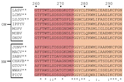
Bioscience Reports (2022) 42 BSR20211930
https://doi.org/10.1042/BSR20211930

(A)
  A                     SSP                               GP1                                                              GP2
     N                                                                                                                                                                 C
                               58                                                         259                                                                   491
                              SPase                                                    SKI-1/S1P
            2
           myr

   B                                                                                      C
                                                                                         (C)                            2
 (B)                                                                                                                                                6
                                                                                                 1

                                                                                                                                                                                           Downloaded from http://portlandpress.com/bioscirep/article-pdf/42/2/BSR20211930/928951/bsr-2021-1930c.pdf by guest on 02 March 2022
                                                                                                                                           5

                                                                                                                   3                                              4
                                             GP2

      Viral membrane
                               SSP

Figure 1. Schematic representation of the LASVGPC
(A) The GPC open-reading frame is diagrammed with the cleavage sites of SPase and SKI-1/S1P indicated. In SSP (pink), the
myristoylation at glycine 2 is marked. The eleven N-glycosylation motifs are denoted at their respective locations in the mature
GP1 (dark blue) and GP2 (gray) subunits. (B) Cartoon illustration of the GPC trimer as it would appear embedded with in the vi-
ral membrane (tan). (C) An illustration of GPC’s involvement in the LASV viral lifecycle. GP1 binds to the host receptor (1) before
being endocytosed (2). Upon internalization, the virion is delivered to the late endosome where GP1 engages with the intracellular
receptor LAMP1 (3). GP1 dissociates so that GP2 is exposed and can mediate membrane fusion leading to the delivery of the
viral genome into the host cell cytoplasm (4). The genome is directed to the endoplasmic reticulum where it is transcribed, trans-
lated, and replicated (5) before being trafficked to the plasma membrane for assembly and budding (6). Abbreviations: LAMP1,
lysosomal-associated membrane protein 1; SPase, cellular signal peptidase.

            1. Pre-fusion State                     2. Prehairpin Intermediate              3. Six Helix Bundle Formation                     4. Post-fusion State

 Host cell membrane
                                                                      Fusion peptide
                        GP2

 Viral membrane
                  SSP

Figure 2. Membrane fusion illustration
(1.) The pre-fusion structure of GPC in the viral membrane (tan) is shown. (2.) Upon delivery to the acidic lysosome, GP1 (dark blue)
engages with LAMP1 resulting in its dissociation from GPC as triggered by the low pH. GP2 (light gray) becomes unclamped such
that the fusion peptide and internal fusion loop at the N-terminus of GP2 (red) are now exposed and inserted into the lysosomal
membrane (light blue), forming a metastable pre-fusion hairpin. (3.) GP2 then folds back on itself in an energetically favorable
rearrangement. This forms the 6HB and brings the host cell and viral membranes in proximity. (4.) The two membranes then fuse
(green), which forms a complete fusion pore and final post-fusion state that exists as a trimer of GP2-SSP. From here the viral
genome is released into the cytosol.

© 2022 The Author(s). This is an open access article published by Portland Press Limited on behalf of the Biochemical Society and distributed under the Creative Commons Attribution   3
License 4.0 (CC BY).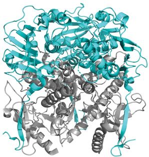
Bioscience Reports (2022) 42 BSR20211930
                                                                                                                                         https://doi.org/10.1042/BSR20211930

           Discussion
           Maturation of the GPC yields three components
           To fully comprehend the uniqueness of the LASV GPC components, one must first understand the intricacies leading
           to their development. Like other class one glycoproteins, GPC is first synthesized as a precursor polypeptide that is
           subsequently cleaved by host cell peptidases to yield the mature GPC. For LASV, GPC is synthesized from the RNA
           genome as a precursor that is directed to the endoplasmic reticulum. Here, the mature SSP is created via cellular
           signal peptidase (SPase) cleavage but remains noncovalently associated with the GPC [89]. At the same time, the
           GPC precursor is translated and undergoes N-linked glycosylation. There are 11 N-glycosylation motifs (N–X–S/T
           where X is any amino acid, except proline) within the LASV GPC with seven located in GP1 and four in GP2 (Figure
           1A) [71,99]. The four glycosylation sites within GP2 are highly conserved across most arenaviruses, whereas the
           number of glycosylation sites in GP1 varies considerably. This extensive amount of N-linked glycosylation is often

                                                                                                                                                                                           Downloaded from http://portlandpress.com/bioscirep/article-pdf/42/2/BSR20211930/928951/bsr-2021-1930c.pdf by guest on 02 March 2022
           referred to as a glycan shield and leaves few regions open to antibody binding. Glycosylation therefore has implications
           in the arenavirus’ ability to evade antibody-mediated immune responses as the epitope is masked [50–54,62]. These
           glycosylation motifs are also involved in GP1 receptor recognition, which will be examined in detail shortly [100–102].
              For all class one glycoproteins, the precursor fusion protein must be primed via proteolytic cleavage before it can be
           triggered to induce fusion. For LASV, this priming event occurs after glycosylation and SSP cleavage when the GPC
           is translocated to the Golgi apparatus as directed by the SSP. Here, the precursor GPC undergoes post-translational
           proteolytic cleavage by subtilase/kexin isozyme-1 (SKI-1/S1P) to yield the mature GPC containing the GP1 and GP2
           components [75,103–106]. For other class one glycoproteins, the signal peptide’s function has been served following
           final protein localization, so it is degraded and not retained in the mature glycoprotein [60]. Interestingly, the LASV
           SSP remains associated with the mature GPC—a feature that is unique to arenaviruses and will thus be covered in
           depth later in this review article. The SSP, GP1, and GP2 are noncovalently bound to each other and form a novel
           structure not observed in other class one fusion proteins (Figure 1B) [107]. The GPC is then trafficked to the cellular
           plasma membrane for virion assembly and budding, where it is incorporated on to the surface of the viral envelope
           (Figure 1C) [108,109]. It is this mature GPC and its components that are responsible for LASV entry.

           Receptor binding occurs via GP1
           LASV entry is a multistep process that is first facilitated by GP1, which is 200 amino acids in length and spans from T59
           to L259 of the mature GPC. Initially, GP1 binds with a host cell receptor located on the plasma membrane, specifically
           the α subunit of dystroglycan (Figure 3A) [110,111]. After binding to αDG, LASV is endocytosed into the host cell,
           then delivered to the late endosome where it engages with the intracellular receptor LAMP1. This receptor switch is
           unique to LASV and not observed in other arenaviruses.

           LASV first engages with host cell receptors at the plasma membrane to enter the host cell
           Dystroglycan is a highly conserved cell-surface glycoprotein that is synthesized as a single protein before undergoing
           post-translational cleavage to yield αDG, the peripheral protein, and βDG, the transmembrane protein [112–115].
           It is involved in extracellular matrix adhesion and is a critical component for membrane integrity in numerous cell
           types, including nerve and muscle cells. In the host cell, αDG is subjected to extensive post-translational modifica-
           tions, including glycosylation that is critical for its function as an extracellular matrix receptor and arenavirus-binding
           site [111,116–124]. Regardless, the expression of functional αDG has been demonstrated to be insufficient for
           productive entry of LASV, but the exact factors that influence entry remain unclear [125]. In the absence of
           αDG, it has been suggested that GP1 can interact with heparan sulfate, dendritic cell-specific intercellular adhe-
           sion molecule-3-grabbing nonintegrin (DC-SIGN), liver and lymph node sinusoidal endothelial cell C-type lectin
           (LSECtin), and Tyro3/Axl/Mer (TAM) family members to gain entry—albeit with reduced infectivity [78,126–130].
           Interestingly, these receptors have been shown to play a role in the entry of several other viruses, including EBOV,
           dengue virus, ZIKA virus, and SARS coronaviruses [131–138]. This highlights the remarkable adaptability of LASV
           in the entry process.
              Mutational analysis of GP1 has highlighted a potential receptor-binding domain (RBD1) at the top central core of
           the GPC trimer that is important in αDG binding [139]. Residues H141, N146, F147, and Y150 cluster in this core
           interface and were implicated in the αDG-binding site (Figure 3B, purple) [139]. Coincidentally, the glycosylation
           motif at N119 is located near this binding site but does not occlude it (Figure 3B, pink) [50,51]. An N119Q muta-
           tion revealed that this glycan protects the αDG-binding site from antibody-mediated neutralization by shielding the
           epitope [50,53]. The necessity of glycosylation for efficient αDG binding by GP1 has also been validated by inhibi-
           tion experiments [140]. Additionally, residues located in the same region at the top central core of the LCMV GP1

4   © 2022 The Author(s). This is an open access article published by Portland Press Limited on behalf of the Biochemical Society and distributed under the Creative Commons Attribution
    License 4.0 (CC BY).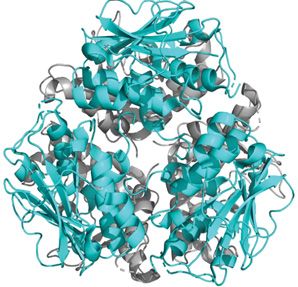
Bioscience Reports (2022) 42 BSR20211930
https://doi.org/10.1042/BSR20211930

  (A)                                                        (B)
  A                                                           B                                                           C(C)

                                                                                                                                                                                           Downloaded from http://portlandpress.com/bioscirep/article-pdf/42/2/BSR20211930/928951/bsr-2021-1930c.pdf by guest on 02 March 2022
                          90c                                                        90c                                                        90c

Figure 3. Crystal structure (PDB: 5VK2) of LASV GP1 trimer from the top (upper) and side (lower) views
(A) Cartoon representation of GP1 (cyan) as it appears when associated with GP2 (gray). (B) A surface model of GP1 is presented
with the RBD1 highlighted. Residues implicated in αDG-binding cluster in the top central core of GP1, forming the αDG-binding
site (purple). The glycosylation motif at N119 (pink) is located near this site but does not occlude it. It is important to note that
the crystal structure lacks a glycan in the third GP1 subunit, which would be present in vivo. (C) The RBD2 of GP1 is represented
via a surface model. A histidine triad (yellow) forms the LAMP1-binding site. Protonation of this triad leads to a conformational
change that exposes the RBD2. However, this triad is shielded by the glycosylation motif at N89 (black) in the pre-fusion state,
which particularly packs against H92. At low pH, this glycan is shifted away to allow for LAMP1 binding. Abbreviation: RBD1/2,
receptor-binding domain 1/2.

were implicated in αDG binding [141–143]. This includes S153 (N148), Y155 (Y150), R190 (G186), and F260 (I254),
further strengthening the importance of this region for function as RBD1 (Figure 3B, purple).

A unique receptor switch by LASV occurs after endocytosis
Initially, it was suggested that LASV cell entry occurred via clathrin-mediated endocytosis [144], but subsequent
studies described the usage of a clathrin- and dynamin-independent pathway [6,76,145,146]. Genome-wide RNA
interference silencing screens identified sodium hydrogen exchangers (NHEs), which have previously been implicated
in macropinocytosis, as host factors involved in the multiplication of LCMV [147,148]. In a successive study, NHE
was validated to be the entry factor and was thus indicative that LASV enters the host cell through macropinocytosis
[146,149]. This type of endocytosis involves the nonspecific uptake of extracellular molecules via large vacuoles [150].
Intriguingly, macropinocytosis is the entry pathway for over 20 viruses, including HIV, herpes simplex virus 1, and
EBOV [151–156].
   Upon macropinocytosis, the virion is internalized and delivered to the late endosome where fusion ultimately takes
place. Studies have shown that LASV-mediated fusion occurs at an unusually acidic pH of below 5.0 with optimal fu-
sion suggested to occur at a pH of ∼4.0 [157–159]. While extremely low pH is likely sufficient enough to trigger fusion,
successful and efficient LASV entry requires GP1 to undergo a unique receptor switch from αDG to LAMP1 [78].
Like αDG, LAMP1 must be glycosylated for this engagement to occur as previous studies showed that an N-linked

© 2022 The Author(s). This is an open access article published by Portland Press Limited on behalf of the Biochemical Society and distributed under the Creative Commons Attribution   5
License 4.0 (CC BY).
Bioscience Reports (2022) 42 BSR20211930
                                                                                                                                         https://doi.org/10.1042/BSR20211930

           glycan in this receptor was a critical determinant in LASV binding [78,160]. Binding to LAMP1, which is localized
           to the inner membrane of the late endosome/lysosome, shifts the pH of optimal fusion to be less acidic [159,161].
           Engagement with LAMP1 thus increases the efficiency of LASV fusion, but the exact role remains to be clarified. No
           other OW arenavirus has been shown to interact with LAMP1, regardless of full conservation of the LAMP1-binding
           site in GP1. This receptor switch is thus unique to LASV [160]. Thus far, the only other virus shown to utilize late
           endosome receptor-assisted fusion is EBOV, but again the exact mechanism remains unclear.
              It has been shown that in the low pH environment of the late endosomal compartment, GP1 undergoes irreversible
           conformational changes that decrease its affinity for αDG while increasing its affinity for the acidic LAMP1 receptor
           [78,161,162]. Structural and mutational analysis has revealed a highly conserved histidine triad (H92/93/230) in GP1
           that forms the LAMP1-binding site and second receptor-binding domain (RBD2) (Figure 3C, yellow) [139,163–165].
           Protonation in this triad, specifically H230, has been proposed to facilitate conformational changes that expose the
           LAMP1-binding site [164–166]. Curiously, GP1 glycosylation motif at N89 was reported to shield the histidine triad

                                                                                                                                                                                           Downloaded from http://portlandpress.com/bioscirep/article-pdf/42/2/BSR20211930/928951/bsr-2021-1930c.pdf by guest on 02 March 2022
           in the pre-fusion conformation before being redirected after endocytosis to allow for LAMP1 binding (Figure 3C,
           black) [51,71,167]. More specifically, H92 packs against N89 at physiological pH, but N89 is shifted ∼10 Å away at
           low pH [101]. This further supports the notion that glycosylation is involved in receptor recognition and binding by
           GP1. Moreover, at neutral pH, H93 makes a hydrogen bond with the main chain oxygen of N90 [71]. When the pH is
           reduced, H93 rotates such that it is no longer capable of making this hydrogen bond. It thus appears that modifications
           in the location of the glycosylation motif at N89 and hydrogen bonding result in the opening of the binding site so that
           the histidine triad may engage with LAMP1. Additionally, four other residues (L84N, K88E, L107F, and H170S) that
           neighbor the histidine triad were identified that decrease LAMP1-binding affinity when mutated and thus may be
           involved in the binding site as well [168]. It is interesting that another histidine may be important in LAMP1 binding
           that exists outside RBD2.
              To summarize, GP1 serves as the R for arenaviruses. GP1 will first engage with the host cell receptor on the plasma
           membrane, primarily αDG, then be endocytosed via macropinocytosis. Fusion occurs upon the virion’s delivery to
           the late endosome. In LASV, this is a multistep process that involves a receptor switch from the primary receptor,
           αDG, to the intracellular receptor, LAMP1. We anticipate future experimentation will likely delve into why LASV
           is the only arenavirus to utilize assisted fusion and a receptor switch. Previous work has already suggested that en-
           gagement with LAMP1 increases fusion efficiency, but whether this has any implications in LASV morbidity remains
           unclear. Investigation into the underlying molecular mechanisms of immune evasion and the residues involved in this
           interaction may answer this question. Additionally, given GP1’s critical role in viral entry, deducing these mechanisms
           would have serious impacts on the development of therapeutic interventions.

           Membrane fusion is mediated by GP2
           GP2 is 231 amino acids in length and spans from G260 to R491 of the GPC (Figure 4A,B). Structurally, GP2 has the prin-
           ciple features of a class one fusion protein and is categorized as such due to its largely α-helical secondary structure
           and two heptad repeat (HR) regions that are preceded by a fusion peptide [59,81,169–171]. There are eight known
           domains of the GP2 including the NFP (G260 –P275 ), IFL (G276 –N295 ), heptad repeat 1 (HR1) (E308 –S358 ), T-loop
           (C364 –C385 ), heptad repeat 2 (HR2) (N395 –I411 ), membrane proximal external region (MPER, T412 –P427 ) transmem-
           brane domain (TM, L428 –L447 ), and C-terminal cytoplasmic tail (H448 –R491 ) [9,157]. For this review article, we will
           be grouping these domains into three sections: fusion domain (NFP and IFL), coiled-coil (HR1, T-loop, and HR2),
           and C-terminus region (MPER, TM, and cytoplasmic tail).

           Unique properties of the LASV fusion domain
           LASV GP2 follows the generally accepted model for the function of class one viral fusion proteins where the fusion
           mechanism is mediated by its refolding to form a 6HB. For LASV, this occurs after GP1 dissociation where GP2
           is uncovered so that the fusion peptide is now exposed. Class one viral fusion proteins all contain a hydrophobic
           sequence that is responsible for engaging the target membrane and thus bridging the host and viral membranes.
           Typically, this is either an NFP or IFL located within the fusion subunit of the surface glycoprotein. Most often, class
           one fusion proteins have been found to contain an NFP, except for EBOV which has an IFL [172]. However, in the
           case of LASV and the arenavirus family, both an NFP and IFL are present and create a fusion domain that initiates
           fusion by targeting the host cell membrane [157,173,174]. Thus far, the only other class one fusion protein with this
           characteristic are coronaviruses, potentially contributing to their remarkable infectivity [83–88].
              The LASV fusion domain is moderately conserved among all OW and/or pathogenic arenaviruses (Figure 4C).
           Furthermore, the canonical fusion motif Gly-X-Phe located at the N-terminus of the fusion domain is also highly
           conserved among OW arenaviruses where X is either T or L [175]. This motif has been established in fusion peptides

6   © 2022 The Author(s). This is an open access article published by Portland Press Limited on behalf of the Biochemical Society and distributed under the Creative Commons Attribution
    License 4.0 (CC BY).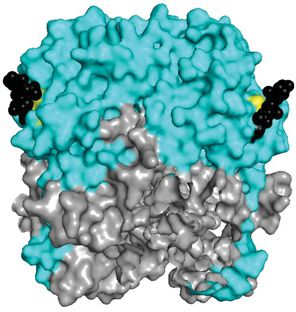
Bioscience Reports (2022) 42 BSR20211930
https://doi.org/10.1042/BSR20211930

(A)
 A
                                                                                                                                                 Cytoplasmic
             NFP           IFL                            HR1                        T-loop          HR2           MPER           TM
                                                                                                                                                     tail

   260 GTFTWTLSDS                       EGKDTPGGYC                 LTRWMLIEAE                  LKCFGNTAVA                  KCNEKHDEEF                 CDMLRLFDFN
   320 KQAIQRLKAE                       AQMSIQLINK                 AVNALINDQL                  IMKNHLRDIM                  GIPYCNYSKY                 WYLNHTTTGR
   380 TSLPKCWLVS                       NGSYLNETHF                 SDDIEQQADN                  MITEMLQKEY                  MERQGKTPLG                 LVDLFVFSTS
   440 FYLISIFLHL                       VKIPTHRHIV                 GKSCPKPHRL                  NHMGICSCGL                  YKQPGVPVKW                 KR
  B                                                                         C
                                                                           (C)

                                                                                                                                                                                           Downloaded from http://portlandpress.com/bioscirep/article-pdf/42/2/BSR20211930/928951/bsr-2021-1930c.pdf by guest on 02 March 2022
(B)

Figure 4. Sequence and structure of the LASV GP2
(A) The known domains of GP2 are color-coded: NFP (red); IFL (orange); HR1 (yellow); T-loop (green); HR2 (blue); MPER (purple);
TM (black); cytoplasmic tail (gray). (B) Cartoon representation of the pre-fusion crystal structure of a GP2 monomer (PDB: 5VK2).
(C) Sequence alignment of the NFP and IFL of pathogenic and/or OW arenaviruses with numbering according to the LASV se-
quence. Pathogenic arenaviruses are denoted via **. The prototypic Pichinde virus is also shown. Residues with full conservation
are indicated by an asterisk (*), whereas those with strongly and weakly similar properties are shown via a semicolon (:) and period
(.), respectively. Abbreviations: CHAVB, Chapare virus; DNDV, Dandenong virus; GTOV, Guanarito virus; IPPYV, Ippy virus; LUJOV,
Lujo virus; MACV, Machupo virus; MOBV, Mobala virus; MOPV, Mopeia virus; PICV, Pichinde virus; SABV, Sabia virus; WWAV,
Whitewater Arroyo virus.

of other viruses, like HIV and influenza [176]. Single-point mutations to alanine for each hydrophobic amino acid
in the fusion domain resulted in a significant reduction or abolishment of fusogenicity, validating the involvement of
both the NFP and IFL in fusion [157]. Multiple residues were shown to impact fusogenicity through in vitro studies
including G260A/R, F262A, W264A, G271A, which are all located in the NFP, and G277A, Y278A, L280A, W283A,
L290A, and G294A located in the IFL. Four of these residues are well conserved across all arenaviruses and were
indicated as critical for efficient fusion: W264, G277, Y278, and L280. Mutations in this region likely reduce the
hydrophobicity of the fusion domain, which inhibits adequate insertion into the target membrane to facilitate fusion.
This further strengthens the postulation that both the NFP and IFL are required for efficient fusion.
   Nonetheless, the necessity for both fusion peptides in LASV and their exact mechanism of action remains poorly
understood. The pre-fusion structure of these two proteins has been characterized as a random coil with the only
discernible secondary structural element being a disulfide bond [71]. On the other hand, these proteins have been
omitted from post-fusion structures of GP2 and thus remains unsolved [169,170]. Thus, future experiments will likely
be centered around solving the post-fusion structure of the fusion domain to fill this knowledge gap. We anticipate
that subsequent studies will investigate the residue-specific interactions that are critical to the function of the LASV
fusion domain.

Coiled-coil structure further classifies LASV as a class one fusion protein
Now that we have finished discussing the LASV fusion domain, we must highlight another major defining charac-
teristic of class one fusion proteins—the formation of the 6HB. In both their pre- and post-fusion states, class one

© 2022 The Author(s). This is an open access article published by Portland Press Limited on behalf of the Biochemical Society and distributed under the Creative Commons Attribution   7
License 4.0 (CC BY).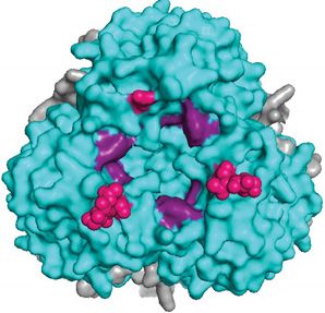
Bioscience Reports (2022) 42 BSR20211930
                                                                                                                                         https://doi.org/10.1042/BSR20211930

           (A)A                                          (B) B
                                                                                                          C
                                                                                                          (C)                              D(D)

                                                                                                                                     D401

                                                                                                                        K320        E404

                                                                                                             R325                  D408
                                                                                                                        K327
                                                                                                             E329

                                                                                                                                                                                           Downloaded from http://portlandpress.com/bioscirep/article-pdf/42/2/BSR20211930/928951/bsr-2021-1930c.pdf by guest on 02 March 2022
           (E)E
               300 KCNEKHDEEF                      CDMLRLFDFN                  KQAIQRLKAE                 AQMSIQLINK                 AVNALINDQL                  IMKNHLRDIM

               360 GIPYCNYSKY                      WYLNHTTTGR                  TSLPKCWLVS                 NGSYLNETHF                 SDDIEQQADN                  MITEMLQ

           Figure 5. Representation of the LASV coiled-coil in different states
           (A) The pre-fusion state of the LASV HR1 (yellow) and HR2 (blue) trimers that exist independently but are connected by the T-loop
           (green). No helical core is present with the trimers oriented nearly perpendicular to the threefold axis (PDB: 5VK2). (B) In the
           post-fusion state, the HR1, HR2, and T-loop unravel and reorient to form the characteristic coiled-coil/6HB. A central trimer of HR1
           is surrounded by three HR2. Each monomer is connected by a T-loop (PDB: 5OMI). (C) A monomer of the coiled-coil is presented
           with the residues that form salt bridges indicated in black. (D) A surface model of the coiled-coil (PDB: 5OMI). The hydrophobic
           interactions (red) between adjacent HR1 and HR2 monomers of the 6HB form a core that is further stabilized by salt bridges (black).
           (E) The sequence of the LASV HRs as connected by the T-loop. Residues that form salt bridges are shown in black and linked to
           their corresponding ionic partner, while residues involved in the hydrophobic core are shown in red.

           glycoproteins exist as trimers. In the pre-fusion state, the HR1 and HR2 trimers exist independently as α helices
           that are connected by the T-loop (Figure 5A). The T-loop itself does not appear to have a function aside from con-
           necting the HR1 and HR2 [177]. Following the initiation of membrane fusion, a prehairpin intermediate structure is
           formed where the HR1 sits atop the HR2 in an antiparallel manner. HR1 and HR2 coordinate with each other to form
           the coiled-coil where a central HR1 trimer is surrounded by three HR2. The intermediate structure spontaneously
           collapses on itself where the three HR2 pack on to the outside of the central trimeric HR1 core to create the 6HB
           (Figure 5B). This formation of the 6HB is energetically favorable and the major driving force for fusion as it brings
           the target and virion membranes nearby such that they may fuse. Ultimately, the final post-fusion conformation of
           the trimer-of-hairpins is achieved, and a complete fusion pore is formed.
              Typically, in class one fusion proteins the trimer is formed about a stabilizing central three-helical core with a
           threefold axis that remains largely unaffected between the pre- and post-fusion conformations [95]. Nonetheless,
           a pre-fusion LASV crystal structure was recently generated that demonstrated this core is nonexistent and GP2 is
           oriented nearly perpendicular to the threefold axis in the pre-fusion conformation (Figure 5A) [71]. To achieve the
           post-fusion conformation, every section of GP2 must thus unravel and reorient. The structure presented here aligned
           well with a previously determined structure of the LCMV GPC monomer. This reorientation is thus unique to are-
           naviruses [95]. Furthermore, alanine substitutions in JUNV at I333 (V341), L336 (L344), I347 (L355), and L350
           (I358) in HR1 and R392 (S400) and W395 (I403) in HR2 resulted in reduced fusogenicity of the GP2 [178]. This
           infers that these buried residues are important in promoting fusion. In the pre-fusion conformation, these residues
           are positioned such that they have interhelical interactions that likely result in the stabilization of the coiled coil. In
           particular, R392 (S400) may impart specificity to the process of coiled-coil folding at the expense of protein stability
           [179].

8   © 2022 The Author(s). This is an open access article published by Portland Press Limited on behalf of the Biochemical Society and distributed under the Creative Commons Attribution
    License 4.0 (CC BY).
Bioscience Reports (2022) 42 BSR20211930
https://doi.org/10.1042/BSR20211930

   In the post-fusion crystal structure, there are three HR1 helices in a coiled-coil orientation with three HR2 helices
entwined in an antiparallel manner forming the 6HB [169,170]. The three HR1 helices were shown to be closely
packed against each other by hydrophobic force in a parallel manner and create a central core [169]. The C-terminal
HR2 adopts such an orientation that places its helix close to the N-terminal HR1 helices of the adjacent monomers.
This positioning ultimately creates a hydrophobic core involving A407 and I411 of the HR2 and L313 and F316 of
the adjacent HR1 (Figure 5C,D, red) [170]. The T-loop region and HR2 are packed against the hydrophobic grooves
of the central coiled-coil. W386, V388, L394, and F399 of HR2 are deeply buried in these grooves of HR1 with many
other long-chained, hydrophobic residues, such as I403 and M410, involved as well (Figure 5C,D, red). Granted, for
stability purposes, only the HR1, T-loop, and HR2 domains of the LASV GP2 were included in this crystal structure
[169] but aligned well with a similar reported LASV 6HB structure [170]. A separate study validated the necessity of
L394 and I403 for GP2 function [9]. Additionally, using alanine scanning mutagenesis, I323 was identified as being
critical for fusion and optimal GP2 function. This trend can likely be justified by the loss of hydrophobicity in this

                                                                                                                                                                                           Downloaded from http://portlandpress.com/bioscirep/article-pdf/42/2/BSR20211930/928951/bsr-2021-1930c.pdf by guest on 02 March 2022
region, which does not allow for the efficient formation of the 6HB.
   In addition to the hydrophobic interactions, several electrostatic interactions stabilize the 6HB. D347 and K352
of one HR1 domain bind to residues of opposite charges in the other two HR1 domains near the T-loop [169]. This
interchain salt bridge was also found in the corresponding residues of LCMV and seemed to stabilize the end of
the inner core [81]. Additional work on LCMV elucidated other stabilizing interactions, particularly multiple ionic
interactions, which seem to also exist in LASV based on sequence homology and structural alignment. Two separate
salt bridges exist between lysine side chains in the N-terminal helix and aspartate and/or glutamate side chains in
the C-terminal helix. These are K326 (K320) with D414 (D408) and K333 (K327) with both D407 (D401) and E410
(E404) (Figure 5C–E, black). K331 (R325) and D335 (E329) form a salt bridge to connect two consecutive turns in
the N-terminus helix (Figure 5C–E, black). These salt bridges are thus well conserved and aid in stabilization between
GP2 monomers of the post-fusion conformation.

C-terminus region has multiple domains that are key to LASV fusogenicity
Our review of GP2 will conclude with a discussion of the C-terminus domains and their roles in fusogenicity. This in-
cludes the TM, cytoplasmic tail, and MPER, which is located between HR2 and TM from residues T412 to P427 (Figure
4A) [180]. In class one fusion proteins, namely EBOV and HIV, the MPER has been shown to have important func-
tions in fusion and glycoprotein stabilization [181–186]. The MPER in HIV is the target of the broadest neutralizing
antibodies and has implications in membrane fusion due to destabilizing effects on the lipid membrane [187–192].
Additionally, in EBOV, the MPER was shown to interact with its IFL and membrane lipids during fusion—potentially
contributing to the driving force of 6HB formation to promote fusion [181,193]. Following this logic, the MPER in
GP2 has been investigated for any impacts on LASV fusogenicity.
   In LASV, the formation of the 6HB brings the fusion domain near the TM region, including the MPER, which
may contribute to the driving force of fusion. This was investigated using alanine scanning mutagenesis of all 16
residues in the MPER [180]. It was revealed that M414 and L415 in the C-terminus of HR2 and K417 and Y419 in the
N-terminus of the MPER were critical for membrane fusion (Figure 6A). Even though mature GPC was present with
these mutants, it was nonfunctional and incapable of facilitating fusion. Mutation to a more conserved residue, such
as M414L, L415I, K417R, and Y419F, resulted in partial recovery of fusogenicity. The finding that K417 is critical for
GP2 function comes as no surprise since it is highly conserved and the equivalent lysine in Guanarito virus (GTOV),
an NW arenavirus with pathogenicity in humans, forms a salt bridge with a conserved aspartate and glutamate in the
adjacent HR1 [194]. Moreover, E418, R422, T426, and P427 all exhibited reduced fusogenic activity when mutated to
alanine. R422 was validated as a critical residue for GP2 function in a separate study, but it is unclear if this residue
forms a salt bridge, and if so, with what partner [9]. Altogether, this is indicative that the MPER is critical to the
stability of GP2 with implications in efficient LASV-mediated fusion.
   Directly following the MPER lies the TM. Unfortunately, structural characterization of the TM has been
challenging—largely due to its hydrophobic nature. Instead, critical residues in the TM have been identified through
inhibitor screens. The TM has been shown to have an interaction with the first helix of the SSP, which forms an
interface that is often the target of inhibitors and has been proposed to have a role in the pH-sensing mechanism
[27–29,39,195]. The mechanism of action behind these inhibitors seems to be the stabilization of the pre-fusion GPC
complex via the TM–SSP interface as revealed by inhibition studies [195–197]. The dietary supplement capsaicin [28]
and the antifungal isavuconazole [27] were both revealed to target the same three residues in the TM: V431, F434,
and V435. Capsaicin and isavuconazole targeted the adjacent region within the N-terminal transmembrane region
of the SSP at A25 and S27, respectively. Curiously, the mutation F427I (V435) in the TM of the JUNV GP2 confers
immunological resistance to the live-attenuated Candid#1 vaccine [198]. This mutation has an epistatic interaction

© 2022 The Author(s). This is an open access article published by Portland Press Limited on behalf of the Biochemical Society and distributed under the Creative Commons Attribution   9
License 4.0 (CC BY).
Bioscience Reports (2022) 42 BSR20211930
                                                                                                                                         https://doi.org/10.1042/BSR20211930

            A (A)

                                                                                                                                                                                            Downloaded from http://portlandpress.com/bioscirep/article-pdf/42/2/BSR20211930/928951/bsr-2021-1930c.pdf by guest on 02 March 2022
            B(B)
                                                                                                                          90

            C (C)

         Figure 6. Characterization of the SSP and cytoplasmic tail of GP2
         (A) Sequence alignment of the LASV GP2 C-terminus of pathogenic and/or OW arenaviruses. Residues in the MPER (purple), TM
         (shaded black), and cytoplasmic tail implicated in fusion are bolded. The ZBD1 (orange) and ZBD2 (red) are shown. (B) Illustration
         of the SSP (pink) and its association with the TM (black) of GP2 from the top and side view. Helix 1 (H1) creates an interface with the
         TM, whereas helix 2 (H2) contains C57, the fourth ligand in ZBD2 (red). ZBD1 (orange) is shown as well. Each ZBD coordinates with
         a zinc ion (purple) (C) Sequence alignment of the SSP with the myristoylation motif outlined in green. Both helical transmembrane
         regions are shaded in tan. K33 and C57 are shown in red. Pathogenic arenaviruses are denoted via **. The prototypic Pichinde virus
         (PICV) is also shown. Residues with full conservation are indicated by an asterisk (*), whereas those with strongly and weakly similar
         properties are shown via a semicolon (:) and period (.), respectively. Abbreviations: CHAVB, Chapare virus; DNDV, Dandenong virus;
         GTOV, Guanarito virus; IPPYV, Ippy virus; LUJOV, Lujo virus; MACV, Machupo virus; MOBV, Mobala virus; MOPV, Mopeia virus;
         PICV, Pichinde virus; SABV, Sabia virus; WWAV, Whitewater Arroyo virus; ZBD, zinc-binding domain.

10   © 2022 The Author(s). This is an open access article published by Portland Press Limited on behalf of the Biochemical Society and distributed under the Creative Commons Attribution
     License 4.0 (CC BY).
Bioscience Reports (2022) 42 BSR20211930
https://doi.org/10.1042/BSR20211930
with a lysine-to-serine mutated residue in the SSP (K33), a residue that is critical for the SSP’s role in pH-sensing
[198,199]. These mutations are the sole basis for the vaccine’s attenuation and have thus raised concerns for rever-
sion to the pathogenic phenotype. Regardless, more structural information is necessary to validate this hypothesis.
Like the fusion domain, the pre-fusion and post-fusion state of the TM remains to be clarified. Elucidation of this
structural information will shed some light on the interactions within the TM-SSP interface and their roles in the
functionality of the GP2.
The retention of SSP is dependent not only on its interaction with the TM but the cytoplasmic tail as well. This
was inferred as replacement of the TM and cytoplasmic tail with an unrelated sequence impacted SSP association,
whereas substitutions elsewhere in the GPC did not [75]. Furthermore, the cytoplasmic tail of JUNV contains six cys-
teine and histidine residues that are essential for SSP association and form two different zinc-binding domain (ZBD)
motifs [200–202]. The first ZBD (ZBD1) is formed by H447 (H455), H449 (H457), and C455 (C463), whereas the
second ZBD (ZBD2) is created by H459 (H467), C467 (C475), and C469 (C477) (Figure 6A). These two ZBD motifs
Downloaded from http://portlandpress.com/bioscirep/article-pdf/42/2/BSR20211930/928951/bsr-2021-1930c.pdf by guest on 02 March 2022
are completely conserved across all NW and OW arenaviruses (Figure 6B). Using isothermal titration calorimetry,
ZBD1 and a nonconserved H485 were first identified to bind zinc with a dissociation constant (K d ) in the nanomolar
range [200,201]. Oddly, H485 is absent from all NW and OW arenaviruses that are not within the same group as
JUNV, including LASV (Figure 6B). This lack of H485 raises the postulation of what might complete the tetrahedral
structure. Is it water like ZBD2 before the SSP association? Could it be another residue within the SSP, such as C41
which is moderately conserved? Probably not considering mutational analysis revealed H485 was important in the
coordination of the JUNV ZBD but was not necessary for the retention of SSP or membrane fusion activity. What
about other conserved residues within GP2? Nonetheless, there is no obvious answer to these questions as no other
histidine or cysteine residues are strictly conserved across all arenaviruses.
To follow-up, York and colleagues hypothesized that ZBD2 was formed with C57 of the SSP as the fourth ligand
(Figure 6C) [201]. Before SSP association, a water molecule acts as a fourth ligand to complete the tetrahedral coor-
dination of zinc. The necessity of these zinc-binding residues was further validated in a separate study on Pichinde
virus, a nonpathogenic NW prototypic arenavirus [202]. In the same study, it was found that the fully conserved W503
(W488) was also indispensable for viral replication. Individual mutational studies on all seven of these residues (H447,
H449, C455, H459, C467, C469, and W503) resulted in the complete abolishment of SSP association and rendered the
GPC inactive [200–202]. It was thus speculated that these residues either facilitate the proper folding of the cytoplas-
mic tail and its association with the SSP or merely that zinc coordination is required for SSP retention. The rationale
for the former will be discussed more in-depth later in this review article.
Altogether, this affirms the characterization of the LASV GP2 as a class one fusion protein as well as highlights
some interesting features. Unlike other class one fusion glycoproteins, LASV GP2 contains a noncanonical NFP and
IFL that must be characterized further to completely understand their necessity. The TM also requires further experi-
mentation, including structural investigation and functional analysis. Interestingly, multiple inhibitors seem to target
the same region (residues 434 to 437) in the TM of various OW and NW arenaviruses. This could potentially be a re-
gion of crucial hydrophobic interactions between the TM-SSP interface. Moreover, an investigation is also necessary
to fully probe the novel ZBDs, specifically the fourth ligand of ZBD1 in LASV and other arenaviruses. Other future
directions will likely include characterizing the exact role of the MPER, how the GP2 coordinates with the SSP, and
any conformational changes that are undergone during the fusion process.
Unusual retention of the SSP in the mature GPC
Although the structural changes undergone by the LASV GPC during membrane fusion follow the same framework
as other class one fusion proteins, the pH-induced activation in the late endosome is distinct—in part due to the
unusual retention of the SSP in the mature GPC and its involvement in pH sensing. Conventional signal peptides
are 18–30-amino acid peptides and are responsible for directing proteins to the proper intra- and/or extracellular
location [203–207]. After successful translocation, the signaling peptide is cleaved and degraded. They usually contain
a positively charged N-terminus, central hydrophobic region, and polar C-terminus [208,209]. On the contrary, the
LASV SSP is abnormally long at 58 amino acids and spans from M1 to T58 of the mature GPC (Figure 6C). It is
proposed to traverse the membrane twice with two distinct hydrophobic domains: helix 1 (H1, E17 –L32 ) and helix 2
(H2, C41 –G54 ). These helices have an antiparallel orientation and are connected by an eight amino acid ectodomain
[89–91,199,210–213]. The N- and C-termini extend from M1 –E16 and R55 –T58 , respectively, and reside in the cytosol
[91,199].
Co-immunoprecipitation experiments have also demonstrated that the LASV SSP was retained in the mature
GPC—a feature that is unique to arenaviruses [73,107,199]. After SPase cleavage, the SSP remains noncovalently
© 2022 The Author(s). This is an open access article published by Portland Press Limited on behalf of the Biochemical Society and distributed under the Creative Commons Attribution 11
License 4.0 (CC BY).Bioscience Reports (2022) 42 BSR20211930
https://doi.org/10.1042/BSR20211930
associated with the GPC complex via the TM and ZBD2 [90,91,196,200,201]. Confocal microscopy studies on JUNV
have demonstrated that the SSP is required for translocation of the GP1 and GP2 to the Golgi apparatus [212]. When
the SSP was replaced with a traditional signal peptide or altogether absent, the GPC precursor was retained within
the ER and was not processed or functional [73–75,89,214,215]. Therefore, not only is the SSP unusually retained in
the mature form of the GPC, but it is also an integral third subunit required for the proper maturation of GPC.
While the LASV SSP maintains the conventional role of a signal peptide for intracellular trafficking, it also has a role
in the LASV pH-dependent fusion process. The association of the SSP with GP2 throughout membrane fusion has
been shown to influence the pH-sensing mechanism and stabilize the intermediate conformation of GP2 [197]. More
specifically, the highly conserved K33 within the SSP has been shown to be required for fusion activity of the GPC.
When this residue was substituted for alanine, fusion was not observed, likely due to a complete loss of fusogenicity.
Substitutions that reduced positive polarity, such as K33R, K33H, and K33Q, increased the threshold pH for fusion
such that optimal pH was less acidic [196]. Intriguingly, K33 is located within the short ectodomain loop that links
Downloaded from http://portlandpress.com/bioscirep/article-pdf/42/2/BSR20211930/928951/bsr-2021-1930c.pdf by guest on 02 March 2022
the two transmembrane helices of the SSP (Figure 6B,C) [75]. We postulate that this location positions K33 at a key
location in the extracellular environment for sensing pH alterations and/or forming an intersubunit salt bridge with
GP2.
The pH-sensing mechanism also seems to be influenced by ZBD2. As mentioned previously, four residues comprise
the novel ZBD2 (C57, H459, C467, and C469)—one of which, C57, is located within the SSP [201]. This cysteine is
invariable across all arenaviruses whereas residues flanking it are variable (Figure 6C) [90,201]. The variable residues
are important for SPase cleavage and GPC maturation but are not responsible for SSP retention, whereas C57 has
been shown to have roles in SSP retention [90,199]. Mutation at C57 resulted in the complete abolishment of SSP
association with GP2 [200]. It could be speculated that zinc is required for SSP retention and the loss of the intersub-
unit ZBD hinders this coordination. However, other studies have shown that proper C57 placement is promoted by
the N-terminal region of the SSP or transmembrane regions of the GP2 [212]. Thus, it is more so likely that ZBD2
coordinates with C57 to retain and position the ectodomain loop of the SSP appropriately [200–202].
Another unusual property is that the SSP is myristoylated at a conserved N-terminal myristoylation motif, specifi-
cally G2 –T6 [73]. Myristoylation has been demonstrated to be an important modification for the viral lifecycle, mem-
brane targeting, and conformational stability [216–225]. In the case of the LASV SSP, a point mutation to alanine at
G2 resulted in a significant hindrance of membrane fusion activity while GPC assembly was unaffected [91,226,227].
Previous studies have also demonstrated that myristoylation does not appear to affect SSP retention, either. The mech-
anism of action behind this motif is unclear but has been suggested to function late in the fusion process to promote
the merging of the membranes [227].
Overall, these studies have suggested the LASV SSP has multiple unique properties when compared with other class
one fusion proteins and signal peptides. Based on the lack of GPC processing in the absence of SSP, it is evident that
SSP retention is required for proper GPC maturation. Aside from this, however, there is an extensive knowledge gap
surrounding the LASV SSP. Given its unusual retention in the mature form, it is difficult to draw direct comparisons
to other class one fusion proteins. As a result, the structural and functional characteristics remain poorly understood.
The identification of an intersubunit ZBD that is coordinated with C57 of the SSP and the ability of K33 to sense
pH, both by coordinating with GP2, provides an interesting starting point for future experimentation. Based on the
location of these residues, it seems a detailed understanding of the localized interactions between the SSP and GP2
will provide key insights into the unique function of SSP. Specific investigation into the role of myristoylation is also
required. Together, these insights will improve our knowledge surrounding the unusual retention of the SSP, which
may be helpful in the development of new therapeutics as multiple inhibitors seem to target the GP2–SSP interface.
Conclusions
Despite LASV being the most prevalent arenavirus afflicting humans, there are limited treatment options due to a
lack of structural and functional information. The GPC is the only protein located on the virion surface of the LASV
particle and has a vital role in the virus’ lifecycle, which makes it an exciting target for drug development. In other
class one fusion proteins, the incorporation of the viral particle is dependent on an interplay of interactions between
the matrix protein and glycoprotein. However, before this can be fully understood in LASV, fundamental questions
about the LASV GPC and the unique properties of its components must be answered. First, the molecular mecha-
nism by which the glycosylated GP1 manages to evade antibody-mediated immune responses must be understood.
The receptor switch by GP1 is also unique; thus, a deeper understanding of the residues involved in the receptor
switch mechanism should be completed. The most extensive knowledge gap involves the LASV GP2 and SSP. Fur-
ther experimentation must be completed on the fusion domain to dissect the requirement of two fusion proteins by
12 © 2022 The Author(s). This is an open access article published by Portland Press Limited on behalf of the Biochemical Society and distributed under the Creative Commons Attribution
License 4.0 (CC BY).Bioscience Reports (2022) 42 BSR20211930
https://doi.org/10.1042/BSR20211930
LASV to facilitate fusion efficiently. Moreover, the structural features of the GP2 and SSP interaction, specifically the
TM–SSP interface and novel ZBDs, must be addressed. Following this, the molecular mechanism by which the GP2
and SSP sense pH changes to promote membrane fusion should be elucidated. In terms of the SSP alone, the role of
its myristoylation requires further investigation. Ultimately, these experiments will provide fundamental information
for novel targets that will be instrumental in drug design against LASV.
Competing Interests
The authors declare that there are no competing interests associated with the manuscript.
Acknowledgements
We would like to thank the entire Lee lab for their continual advice, support, and encouragement in constructing this manuscript.
Downloaded from http://portlandpress.com/bioscirep/article-pdf/42/2/BSR20211930/928951/bsr-2021-1930c.pdf by guest on 02 March 2022
Abbreviations
αDG, α-dystroglycan; 6HB, six-helix bundle; CFR, case fatality rate; CHAVB, Chapare virus; DNDV, Dandenong virus; EBOV,
Ebola virus; GP1, glycoprotein 1; GP2, glycoprotein 2; GPC, glycoprotein complex; GTOV, Guanarito virus; HIV, human im-
munodeficiency virus; HR1, heptad repeat 1; HR2, heptad repeat 2; IFL, internal fusion loop; IPPYV, Ippy virus; JUNV, Junı́n
virus; LAMP1, lysosomal-associated membrane protein 1; LASV, Lassa virus; LCMV, lymphocytic choriomeningitis virus; LUJOV,
Lujo virus; MACV, Machupo virus; MOBV, Mobala virus; MOPV, Mopeia virus; MPER, membrane proximal external region; NFP,
N-terminal fusion peptide; NHE, sodium hydrogen exchanger; NW, New-World arenavirus; OW, Old-World arenavirus; PICV,
Pichinde virus; RBD1, receptor-binding domain 1; RBD2, receptor-binding domain 2; SABV, Sabia virus; SARS-CoV-2, severe
acute respiratory syndrome coronavirus 2; SPase, cellular signal peptidase; SSP, stable signal peptide; TAM, Tyro3/Axl/Mer; TM,
transmembrane domain; WWAV, Whitewater Arroyo virus; ZBD, zinc-binding domain.
References
1 Radoshitzky, S.R., Bào, Y., Buchmeier, M.J., Charrel, R.N., Clawson, A.N., Clegg, C.S. et al. (2015) Past, present, and future of arenavirus taxonomy.
Arch. Virol. 160, 1851–1874, https://doi.org/10.1007/s00705-015-2418-y
2 McLay, L., Liang, Y. and Ly, H. (2014) Comparative analysis of disease pathogenesis and molecular mechanisms of New World and Old World
arenavirus infections. J. Gen. Virol. 95, 1–15, https://doi.org/10.1099/vir.0.057000-0
3 Yun, N.E. and Walker, D.H. (2012) Pathogenesis of Lassa fever. Viruses 4, 2031–2048, https://doi.org/10.3390/v4102031
4 Charrel, R.N. and de Lamballerie, X. (2010) Zoonotic aspects of arenavirus infections. Vet. Microbiol. 140, 213–220,
https://doi.org/10.1016/j.vetmic.2009.08.027
5 McCormick, J.B. and Fisher-Hoch, S.P. (2002) Lassa fever. Curr. Top. Microbiol. Immunol. 262, 75–109,
https://doi.org/10.1007/978-3-642-56029-3˙4
6 Rojek, J.M., Sanchez, A.B., Nguyen, N.T., de la Torre, J.-C. and Kunz, S. (2008) Different mechanisms of cell entry by human-pathogenic old world
and new world arenaviruses. J. Virol. 82, 7677–7687, https://doi.org/10.1128/JVI.00560-08
7 Lecompte, E., Fichet-Calvet, E., Daffis, S., Koulémou, K., Sylla, O., Kourouma, F. et al. (2006) Mastomys natalensis and Lassa fever, West Africa.
Emerg. Infect. Dis. 12, 1971–1974, https://doi.org/10.3201/eid1212.060812
8 Woyessa, A.B., Maximore, L., Keller, D., Dogba, J., Pajibo, M., Johnson, K. et al. (2019) Lesson learned from the investigation and response of Lassa
fever outbreak, Margibi County, Liberia, 2018: case report. BioMed Central Infect. Dis. 19, 610, https://doi.org/10.1186/s12879-019-4257-z
9 Willard, K.A., Alston, J.T., Acciani, M. and Brindley, M.A. (2019) Identification of residues in Lassa virus glycoprotein subunit 2 that are critical for
protein function. Pathogens 8, 1–13, https://doi.org/10.3390/pathogens8010001
10 McCormick, J.B., Webb, P.A., Krebs, J.W., Johnson, K.M. and Smith, E.S. (1987) A prospective study of the epidemiology and ecology of Lassa fever.
J. Infect. Dis. 135, 437–444, https://doi.org/10.1093/infdis/155.3.437
11 Centers for Disease Control and Prevention (2019) Lassa Fever [Fact Sheet]. https://www.cdc.gov/vhf/lassa/pdf/factsheet.pdf
12 World Health Organization (2017) Lassa fever fact sheet [press release]. https://www.who.int/news-room/fact-sheets/detail/lassa-fever
13 Herring, S., Oda, J.M., Wagoner, J., Kirchmeier, D., O’Connor, A., Nelson, E.A. et al. (2021) Inhibition of arenaviruses by combinations of orally
available approved drugs. Antimicrob. Agents Chemother. 65, 1–17, https://doi.org/10.1128/AAC.01146-20
14 Richmond, J.K. and Baglole, D.J. (2003) Lassa fever: epidemiology, clinical features, and social consequences. Br. Med. J. 327, 1271–1275,
https://doi.org/10.1136/bmj.327.7426.1271
15 Safronetz, D., Lopez, J.E., Sogoba, N., Traore, S.F., Raffel, S.J., Fischer, E.R. et al. (2010) Detection of Lassa virus, Mali. Emerg. Infect. Dis. 16,
1123–1126, https://doi.org/10.3201/eid1607.100146
16 Nigeria Centre for Disease Control (2021) Lassa Fever Situation Report [press release].
https://ncdc.gov.ng/diseases/sitreps/?cat=5&name=An%20update%20of%20Lassa%20fever%20outbreak%20in%20Nigeria
17 Yaro, C.A., Kogi, E., Opara, K.N., Batiha, G.E., Baty, R.S., Albrakati, A. et al. (2021) Infection pattern, case fatality rate and spread of Lassa virus in
Nigeria. BMC Infect. Dis. 21, 149, https://doi.org/10.1186/s12879-021-05837-x
18 Bagcchi, S. (2020) Lassa fever outbreak continues across Nigeria. Lancet Infect. Dis. 20, 543, https://doi.org/10.1016/S1473-3099(20)30299-1
© 2022 The Author(s). This is an open access article published by Portland Press Limited on behalf of the Biochemical Society and distributed under the Creative Commons 13
Attribution License 4.0 (CC BY).You can also read