NETWORK MEDICINE LINKS SARS-COV-2/COVID- 19 INFECTION TO BRAIN MICROVASCULAR INJURY AND NEUROINFLAMMATION IN DEMENTIA-LIKE COGNITIVE IMPAIRMENT
←
→
Page content transcription
If your browser does not render page correctly, please read the page content below
Zhou et al. Alzheimer's Research & Therapy            (2021) 13:110
https://doi.org/10.1186/s13195-021-00850-3

 RESEARCH                                                                                                                                            Open Access

Network medicine links SARS-CoV-2/COVID-
19 infection to brain microvascular injury
and neuroinflammation in dementia-like
cognitive impairment
Yadi Zhou1†, Jielin Xu1†, Yuan Hou1†, James B. Leverenz2,3, Asha Kallianpur1,2, Reena Mehra2,4, Yunlong Liu5,
Haiyuan Yu6,7,8, Andrew A. Pieper9,10,11,12,13,14, Lara Jehi2,3 and Feixiong Cheng1,2,15*

  Abstract
  Background: Dementia-like cognitive impairment is an increasingly reported complication of SARS-CoV-2 infection.
  However, the underlying mechanisms responsible for this complication remain unclear. A better understanding of
  causative processes by which COVID-19 may lead to cognitive impairment is essential for developing preventive
  and therapeutic interventions.
  Methods: In this study, we conducted a network-based, multimodal omics comparison of COVID-19 and neurologic
  complications. We constructed the SARS-CoV-2 virus-host interactome from protein-protein interaction assay and CRISPR-
  Cas9-based genetic assay results and compared network-based relationships therein with those of known neurological
  manifestations using network proximity measures. We also investigated the transcriptomic profiles (including single-cell/
  nuclei RNA-sequencing) of Alzheimer’s disease (AD) marker genes from patients infected with COVID-19, as well as the
  prevalence of SARS-CoV-2 entry factors in the brains of AD patients not infected with SARS-CoV-2.
  Results: We found significant network-based relationships between COVID-19 and neuroinflammation and brain
  microvascular injury pathways and processes which are implicated in AD. We also detected aberrant expression of AD
  biomarkers in the cerebrospinal fluid and blood of patients with COVID-19. While transcriptomic analyses showed
  relatively low expression of SARS-CoV-2 entry factors in human brain, neuroinflammatory changes were pronounced. In
  addition, single-nucleus transcriptomic analyses showed that expression of SARS-CoV-2 host factors (BSG and FURIN) and
  antiviral defense genes (LY6E, IFITM2, IFITM3, and IFNAR1) was elevated in brain endothelial cells of AD patients and
  healthy controls relative to neurons and other cell types, suggesting a possible role for brain microvascular injury in
  COVID-19-mediated cognitive impairment. Overall, individuals with the AD risk allele APOE E4/E4 displayed reduced
  expression of antiviral defense genes compared to APOE E3/E3 individuals.

* Correspondence: chengf@ccf.org
†
 Yadi Zhou, Jielin Xu and Yuan Hou contributed equally to this work.
1
 Genomic Medicine Institute, Lerner Research Institute, Cleveland Clinic,
Cleveland, OH 44195, USA
2
 Department of Molecular Medicine, Cleveland Clinic Lerner College of
Medicine, Case Western Reserve University, Cleveland, OH 44195, USA
Full list of author information is available at the end of the article

                                         © The Author(s). 2021 Open Access This article is licensed under a Creative Commons Attribution 4.0 International License,
                                         which permits use, sharing, adaptation, distribution and reproduction in any medium or format, as long as you give
                                         appropriate credit to the original author(s) and the source, provide a link to the Creative Commons licence, and indicate if
                                         changes were made. The images or other third party material in this article are included in the article's Creative Commons
                                         licence, unless indicated otherwise in a credit line to the material. If material is not included in the article's Creative Commons
                                         licence and your intended use is not permitted by statutory regulation or exceeds the permitted use, you will need to obtain
                                         permission directly from the copyright holder. To view a copy of this licence, visit http://creativecommons.org/licenses/by/4.0/.
                                         The Creative Commons Public Domain Dedication waiver (http://creativecommons.org/publicdomain/zero/1.0/) applies to the
                                         data made available in this article, unless otherwise stated in a credit line to the data.
Zhou et al. Alzheimer's Research & Therapy   (2021) 13:110                                                      Page 2 of 19

 Conclusion: Our results suggest significant mechanistic overlap between AD and COVID-19, centered on
 neuroinflammation and microvascular injury. These results help improve our understanding of COVID-19-associated
 neurological manifestations and provide guidance for future development of preventive or treatment interventions,
 although causal relationship and mechanistic pathways between COVID-19 and AD need future investigations.
 Keywords: Alzheimer’s disease, Brain microvasculature, Cognitive impairment, COVID-19, Dementia, Network medicine,
 Neuroinflammation, SARS-CoV-2, Single-cell/nucleus

Introduction                                                   CoV-2 may directly infect the brain [19–21], potentially
Patients with COVID-19 commonly develop neurologic             through the olfactory bulb [19], others have shown that
symptoms and/or complications, such as a loss of taste         SARS-CoV-2 is absent from the brain [22] and cerebro-
or smell, stroke, delirium, and rarely new onset seizures      spinal fluid (CSF) [13]. COVID-19 has also been suggested
[1, 2]. Based on the experience with other coronaviruses,      to cause inflammation within the central nervous system
it was predicted early on that COVID-19 patients might         (CNS) [18, 22, 23], as well as microvascular injury [22].
also be at risk for cognitive dysfunction. For example,        For example, the SARS-CoV-2 spike protein, which read-
after the severe acute respiratory syndrome (SARS-CoV-1)       ily crosses the blood-brain barrier (BBB) [24, 25], induces
outbreak in 2002 and the Middle East respiratory syndrome      an inflammatory response within microvascular endothe-
(MERS) outbreak in 2012, both caused by human corona-          lial cells, leading to BBB dysfunction [25].
viruses (HCoVs), 20% of recovered patients reported on-           Multi-omics datasets for patients with COVID-19,
going memory impairment [3]. Evidence now supports             such as bulk and single-cell/nucleus transcriptomic [26],
similar complications after COVID-19, which due to the         proteomic [27], and interactomic (protein-protein inter-
global pandemic, is poised to potentially lead to a surge in   actions [PPIs]) datasets [28–32], have been generated in
cases of Alzheimer’s-like dementia or other forms of neuro-    order to conduct unbiased investigation of the patho-
cognitive impairment in the near future [4–8].                 physiological pathways. We reasoned that network-based
   On the one hand, individuals with dementia (vascular        drug-disease and disease-disease proximity approaches
dementia, presenile dementia, and Alzheimer's disease,         [33–36], which shed light on the relationship between
etc.) were shown to have elevated risks for COVID-19           drugs (and drug targets) and diseases (gene and protein
compared to those without dementia [9]. COVID-19               determinants of disease mechanisms in the human PPI
patients with dementia have elevated mortality rate            network), would provide mechanistic insights into the
[10, 11], and the most frequent symptoms included              pathobiology of cognitive dysfunction after SARS-CoV-2
hypoactive delirium and functional status worsening            infection, potentially suggesting novel targets for further
[11]. On the other hand, COVID-19 may lead to cognitive        therapeutic investigation. Thus, we investigated Alzheimer’s
impairments, such as shown by poor neuropsychological          disease (AD)-like pathobiology associated with SARS-CoV-
assessments [4, 12] or shown by behaviors or symptoms          2 infection by using a network-based multimodal omics
such as agitation, confusion, inattention, and disorienta-     analytic methodology (Fig. 1). Specifically, we leveraged
tion [13]. COVID-19 patients admitted to intensive care        bulk and single-cell/nuclei RNA-sequencing, proteomics,
unit (ICU) have elevated frequency of delirium [14]. In a      and interactomics (SARS-CoV-2 virus-host PPIs from mass
recent study of a large cohort of more than 236,000            spectrometry assays and genetic interactions from CRISPR-
COVID-19 survivors, it was shown that the survivors who        Cas9 assays) from COVID-19 and AD patients. We hypoth-
required hospitalization, ICU admission, or had encephal-      esized that SARS-CoV-2 host factors would be localized in
opathy during COVID-19 had elevated risks of neuro-            a subnetwork within the comprehensive PPI network and
logical and psychiatric disorders [8]. Another study using     that proteins associated with certain neurologic function
73,000 non-hospitalized COVID-19 survivors shows vari-         would be targeted by the virus either directly, or indirectly
ous incident sequalae, such as mental health disorders and     through PPIs with virus host factors. As detailed below, our
neurocognitive disorders [15]. Jaywant et al. reported that    comprehensive analyses show scant evidence of direct brain
of 57 recovering COVID-19 patients referred for neuro-         and neuron damage by COVID-19, but robust evidence for
psychological evaluation before hospital discharge, 81%        involvement of pathways of neuroinflammation and brain
had cognitive impairment, including mild, moderate, and        microvascular injury in COVID-19.
severe cognitive impairment [16].
   Clarification of the underlying molecular mechanisms        Materials and methods
of COVID-19-induced cognitive impairment is mandatory          SARS-CoV-2 host factor profiles
for developing effective therapeutic strategies for patients   In total, we have gathered ten datasets of SARS-CoV-2
[9, 17, 18]. While some studies have shown that SARS-          (and other HCoVs) target host genes/proteins from
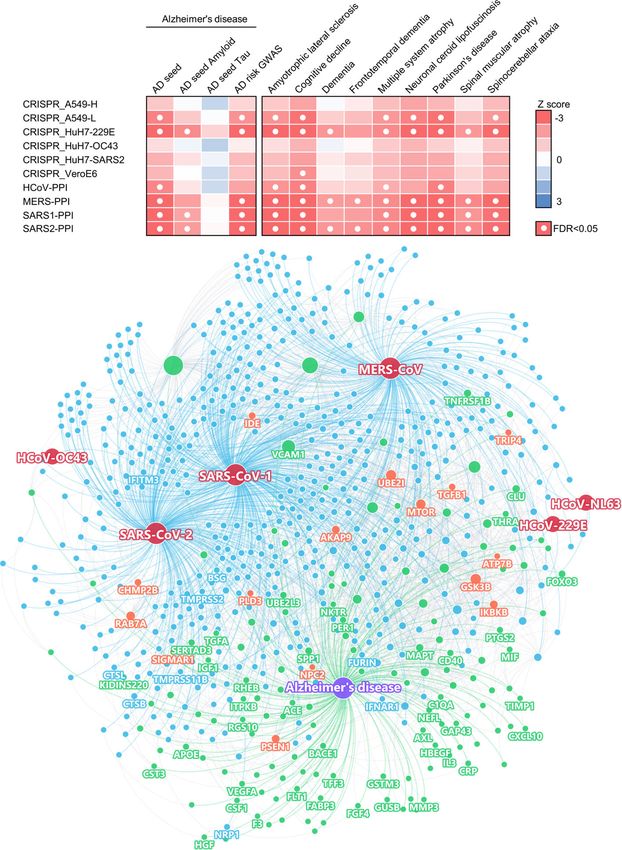
Zhou et al. Alzheimer's Research & Therapy     (2021) 13:110                                                                          Page 3 of 19

 Fig. 1 A diagram illustrating a network-based, multimodal omics analytic framework. We examined the transcriptomes (both bulk and single-cell
 or single-nucleus) of patients with COVID-19 (blood and cerebrospinal fluid [CSF] samples) or Alzheimer’s disease (AD) (brain samples). We also
 compiled ten SARS-CoV-2 host (human) factor datasets based on CRISPR-Cas9 assays or protein-protein interaction assays, AD blood and CSF
 markers, and neurological disease-associated genes/proteins. Using network proximity analysis in the human protein-protein interactome, we
 investigated network-based associations between SARS-CoV-2 host factors and several selected neurological diseases. To understand the potential
 mechanisms through which SARS-CoV-2 affect the brain, including direct brain invasion, neuroinflammation, and microvascular injury, we
 examined (1) the expression changes of AD markers in COVID-19 patients, (2) the expression of SARS-CoV-2 host factors in AD patients and
 healthy individuals at tissues, brain regions, and single-cell/nucleus levels. These transcriptomic analyses were accompanied by network analysis
 to uncover the potential mechanisms (key genes or pathways) involved in protein-protein interactions. We also compared the susceptibility of
 SARS-CoV-2 infection among AD patients with different APOE genotypes using the single-nucleus transcriptomic datasets

various data sources (Table S1). Specifically, six of these                CRISPR-Cas9-based datasets, we considered the top-100
datasets were based on CRISPR-Cas9 assay results, in-                      host factors using the ranking methods described in the re-
cluding (1–2) CRISPR_A549-H and CRISPR_A549-L,                             spective original publications [30–32]. We also examined
based on high (-H) and low (-L) multiplicity of infection                  the effect of using top-50, -150, and -200 genes. In addition
of SARS-CoV-2 in A549 cells [30]; (3–5) CRISPR_HuH7-                       to the CRISPR datasets, we collected three mass
SARS2, CRISPR_HuH7-229E, CRISPR_HuH7-OC43, based                           spectrometry-based virus-host PPI datasets [28, 29] for
on HuH7 cells infected by SARS-CoV-2, HCoV-229E, and                       SARS-CoV-2, SARS-CoV-1, and MERS-CoV, named as
HCoV-OC43, respectively [31]; and (6) CRISPR_VeroE6,                       SARS2-PPI, SARS1-PPI, and MERS-PPI. The last dataset,
based on SARS-CoV-2-infected VeroE6 cells [32]. For the                    HCoV-PPI, was from our recent studies [37, 38] containing

Zhou et al. Alzheimer's Research & Therapy   (2021) 13:110                                                     Page 4 of 19

HCoVs target host proteins supported by literature-based      10 males with varying stages of AD [50] was used to
evidence. Functional enrichment analyses, including Kyoto     examine the expression of the four key SARS-CoV-2 entry
Encyclopedia of Genes and Genomes (KEGG) and Gene             factors: ACE2, TMPRSS2, FURIN, and NRP1, in neurons.
Ontology (GO) biological process enrichment analyses,
were performed using Enrichr [39] for the CRISPR datasets.    GSE157827
A list of main SARS-CoV-2 entry factors and proteins in-      This single-nuclei RNA-sequencing dataset from the
volved in antiviral defense was assembled [18], including     prefrontal cortex region of 12 AD patients and 9 normal
ACE2, BSG, NRP1, TMPRSS2, TMPRSS11A, TMPRSS11B,               controls [51] was used to test the susceptibility of brain
FURIN, CTSB, CTSL, LY6E, IFITM1, IFITM2, IFITM3,              endothelial cells to SARS-CoV-2 infection and damage.
IFNAR1, and IFNAR2.                                           Six cell types were included: astrocytes, endothelial cells,
                                                              excitatory neurons, inhibitory neurons, microglia, and
Neurological disease gene profiles                            oligodendrocytes. The APOE genotypes of these individ-
We extracted neurologic disease-associated genes/proteins     uals are also available in this dataset.
from the Human Gene Mutation Database (HGMD) [40]
and defined a gene to be disease-associated, if it had at     GSE138852
least one disease-associated mutation from HGMD re-           This single-nuclei RNA-sequencing dataset from the en-
ported in the literature. The details of these neurological   torhinal cortex of individuals with AD (n = 6) and
disease genes can be found in Table S2, including the         healthy controls (n = 6) [52] was used to validate the
reported mutations, disease terms used to identify the        findings of the expression of SARS-CoV-2 entry factors
neurological diseases [41], and original references. For      in brain endothelial cells. Six cell types were included:
AD, we assembled four datasets from AlzGPS [42], based        astrocytes, endothelial cells, neurons, microglia, oligo-
on our previous work [43] (Table S2). These datasets con-     dendrocytes, and oligodendrocyte progenitor cells.
tain experimentally validated genes (denoted as “seed”
genes) in amyloid pathology (amyloid) or tauopathy (tau),     GSE157103
as well as high-confidence AD risk genes identified by        This bulk RNA-sequencing dataset of 125 peripheral
genome-wide association study (GWAS) [44].                    blood mononuclear cell (PBMC) samples [53] was used
                                                              to examine the expression spectrum of AD blood
Alzheimer's disease blood and CSF markers                     biomarkers. DEGs were analyzed by disease severity
We compiled a list of AD blood and CSF protein markers        conditions: 66 intensive care unit (ICU) patients (n = 50
from previous studies [45–47], which included 29 blood        COVID-19 patients vs. n = 16 non-COVID-19 patients),
markers and 31 CSF markers. The expression alteration of      59 non-ICU patients (n = 49 COVID-19 patients vs. n = 10
these markers in AD or AD-related pathologies, such as        non-COVID-19 patients), and all 125 patients. Adjustments
tauopathy, were extracted from these studies. The details     for the effects of age and sex were made.
of these markers can be found in Table S3.
                                                              GSE149689
Transcriptomic data analyses                                  This single-cell RNA-sequencing PBMC dataset of 6
Two categories of transcriptomic datasets, including three    samples from severe COVID-19 patients, 4 samples from
from AD patients and three from COVID-19 patients,            mild COVID-19 patients, and 4 samples from healthy
were used (Table S4). These datasets are described below.     controls [54] was used to examine the expression
All single-cell analyses were performed using Seurat v3.1.5   spectrum of AD blood markers. 13 cell types were in-
[48] following the processing steps from the original pub-    cluded in this dataset: lgG− B cells, lgG+ B cells, CD4+ T
lication of each dataset. Cell types were identified using    cell effector memory (EM)-like cells, CD4+ T cell non-
markers based on the original publications, unless already    EM-like cells, CD8+ T cell EM-like cells, CD8+ T cell
annotated in the metadata. Differential expression analysis   non-EM-like cells, dendritic cells, monocytes, intermedi-
was performed using the “FindMarkers” function from           ate monocytes, nonclassical monocytes, natural killer
Seurat for the single-cell/nuclei datasets. For the bulk      cells, platelets, and red blood cells.
RNA-sequencing dataset, differential expression analysis
was performed using edgeR v3.12 [49]. Differentially          GSE163005
expressed genes (DEGs) were determined by false discov-       This single-cell RNA-sequencing CSF dataset [55] was
ery rate (FDR) < 0.05 and |log2foldchange| > 0.5.             used to examine the expression spectrum of AD CSF
                                                              markers. This neuro-COVID-19 dataset contains 8
GSE147528                                                     COVID-19 patients, 9 multiple sclerosis (MS) patients, 9
This single-nuclei RNA-sequencing dataset from the            idiopathic intracranial hypertension (IIH) patients, and 5
superior frontal gyrus and entorhinal cortex regions of       viral encephalitis (VE) patients. Based on the original
Zhou et al. Alzheimer's Research & Therapy (2021) 13:110 Page 5 of 19
publication, the cells were categorized into three major d AB −d r
cell groups of T cells, dendritic cells, and monocytes. Four Z dAB ¼ ð3Þ
σr
comparisons were performed for each major cell group:
COVID-19 vs. MS, COVID-19 vs. IIH, COVID-19 vs. VE, where d r and σr are the mean and standard deviation
and COVID-19 vs. non-COVID-19 (MS, IIH, and VE). of the proximities, respectively. In each degree-
controlled permutation test, two protein sets were ran-
Human protein-protein interactome domly selected which had similar degree distribution to
The human protein-protein interactome was from our that of the original two protein sets to reduce the effect
previous studies [33, 34, 56, 57], and contains 17,706 of degree biases based on our previous studies [33, 38,
protein nodes and 351,444 unique PPI edges. Each PPI 56]. A P value was computed using the permutation test
edge has one or more source information of five categor- accordingly. P values were corrected for FDR at 0.05.
ies of evidence from publicly available databases and Gene set pairs with FDR < 0.05 and Z < − 1.5 were con-
datasets: protein complexes identified by robust affinity sidered significantly proximal.
purification-mass spectrometry from BioPlex V2.016 The largest connect component (LCC) was computed
[58]; binary PPIs discovered by high-throughput yeast by NetworkX [74]. Significance of LCC was computed in
two-hybrid systems in three datasets [33, 59, 60]; signaling the same way as the network proximity using permuta-
networks revealed by low-throughput experiments from tion test repeated 1000 times. Eigenvector centrality [75]
SignaLink2.0 [61]; low-throughput or high-throughput ex- of the nodes in the networks was computed using Gephi
periments uncovered kinase-substrate interactions from 0.9.2 [76] to evaluate the influence of the nodes consid-
KinomeNetworkX [62], Human Protein Resource Database ering the importance of their neighbors.
(HPRD) [63], PhosphoNetworks [64], PhosphositePlus [65],
DbPTM 3.0 [66], and Phospho.ELM [67]; and PPIs curated Tissue and brain region expression specificity
from literatures identified by yeast two-hybrid studies, affin- We retrieved the transcriptomic data in raw count and
ity purification-mass spectrometry, low-throughput experi- transcripts per million (TPM) from the GTEx v8 release
ments, or protein three-dimensional structures from [77] for 33 human tissues and 13 brain regions, and
BioGRID [68], PINA [69], Instruct [70], MINT [71], IntAct examined expression across different tissues and brain
[72], and InnateDB [73]. Inferred PPIs derived from evolu- regions. At the tissue level, the brain regions were
tionary analysis, gene expression data, and metabolic associ- combined as one “brain” tissue. We first defined a gene
ations were excluded. to be tissue- or brain region-expressed if it had a count
per million (CPM) ≥ 0.5 in over 90% samples. Then, to
Network analyses quantify the significance of the expression of a gene in a
We used network proximity metrics to quantify the net- tissue or brain region, we normalized its expression
work associations of two gene/protein modules. The “short- using the z score method.
est” proximity measure was used to evaluate the overall
average distance among all genes in the neurological dis- Innate immune genes
ease gene sets and the SARS-CoV-2 host factor profiles: We retrieved a list of 1031 human innate immunity
genes from InnateDB [73], which were associated in the
1 X
d SAB ¼ d ða; bÞ ð1Þ published literature with roles in innate immunity.
jjAjj kBk a∈A;b∈B
Statistical analysis and network visualization
where d(a, b) represents the shortest path length be- Python package SciPy v1.3.0 [78] was used for the
tween gene a from module A and b from module B in statistical tests unless specified otherwise. P < 0.05 (or
the human protein-protein interactome. “closest” prox- FDR < 0.05 when applicable) was considered statistically
imity measure was used to quantify the distance among significant throughout the study. Networks were visualized
the AD markers and the DEGs from the COVID-19 with Gephi 0.9.2 [76] and Cytoscape 3.8.0 [79].
omics datasets focusing on the genes that are closest to
the genes in the other module: Results
! A network-based, multimodal omics analytic framework
1 X X In this study, we present a network-based, multimodal
d CAB ¼ minb∈B d ða; bÞ þ mina∈A d ða; bÞ
jjAjj þ kBk a∈A b∈B omics (including bulk and single-cell/nuclei RNA-
ð2Þ sequencing, proteomics, and interactomics) analysis
method for investigating the underlying mechanisms of
All network proximities were converted to Z scores COVID-19-associated cognitive dysfunction or impairment.
based on permutation tests of 1000 repeats: We hypothesized that for COVID-19 to have neurologicalZhou et al. Alzheimer's Research & Therapy (2021) 13:110 Page 6 of 19
impacts in the host CNS, its host factors (genes/proteins) art network proximity measures (see the “Materials and
should be localized in the corresponding subnetwork within methods” section), we evaluated the network-based
the human PPI network, and either directly target the relationship for the gene/protein sets between virus-host
neurological disease-associated genes/proteins or indirectly factors and each disease/condition under the human inter-
affect them through PPIs (Fig. 1). We utilized single-cell/ actome network model (Fig. 2a and Fig. S2). We found sig-
nuclei RNA-sequencing data from both COVID-19 patients nificant proximities between the SARS-CoV-2 virus-host
with neurological manifestations (neuro-COVID-19) and interactome (including PPIs and genetic interactions) and
brains of AD patients not infected by SARS-CoV-2, brain- genes associated with neurological diseases in the human
region-specific gene expression data from the GTEx interactome network (average Z = − 1.82). The SARS-CoV-
database [77], SARS-CoV-2 virus-host PPIs from mass 2 virus-host PPIs (average Z = − 2.54) showed more signifi-
spectrometry assays, genetic interactions from CRISPR- cant network proximities (white circles, Fig. 2a) compared
Cas9 assays (Table S1), and disease-related genetic data to CRISPR-Cas9-derived host factors (average Z = − 1.34).
(Table S2). The top three neurological diseases or conditions with the
We compiled ten virus-host interaction datasets across smallest network proximities to SARS-CoV-2 were AD
SARS-CoV-2, SARS-CoV-1 and MERS-CoV, and other (average Z = − 2.75) [9, 17], cognitive decline (average Z =
common HCoVs, including six datasets from CRISPR- − 2.77), and PD (average Z = − 2.94). Recent case reports of
Cas9 assays and four datasets for virus-human PPIs COVID-19 patients developing parkinsonism suggest that
(Table S1). Functional enrichment analyses of each COVID-19 patients may have an increased risk of PD later
dataset revealed that virus-host PPIs and host factors are in life [80]. We noticed that amyloid pathology has signifi-
significantly enriched in pathways well-known to be cant network proximity (average Z = -1.55) with the PPI
involved in SARS-CoV-2 infection and related immune datasets. However, there are no significant network-based
responses (Supplementary Results, Fig. S1). Using these relations between tauopathy-related genes and the SARS-
datasets, we computed their network associations with CoV-2 interactome. One possible explanation is the
ten neurological diseases or conditions. To determine incompleteness of genes/proteins related to tauopathy in
whether brain damage was caused by SARS-CoV-2 dir- the datasets. In addition to SARS-CoV-2, HCoV-229E also
ect infection of the brain, we evaluated expression levels showed a significant network proximity to neurological
of SARS-CoV-2 entry genes at brain region and brain diseases, suggesting a common association between corona-
single-cell levels. Neuroinflammation was evaluated by viruses and cognitive dysfunction [81].
identifying alterations in expression of AD blood and
CSF biomarkers in COVID-19 patients using data from A network-based relationship between COVID-19 and
peripheral blood mononuclear cell (PBMC) and CSF Alzheimer’s disease
samples (neuro-COVID-19 dataset). Lastly, microvascu- To examine further why cognitive impairment has such
lar injury was evaluated by examining the expression of significant network-based association with the SARS-
SARS-CoV-2 entry factors and antiviral defense genes in CoV-2 interactome, we focused on AD and visualized
brain endothelial cells of AD and healthy control sam- the PPIs among AD seed genes/proteins (Fig. 2b, green
ples. We also compared the expression of SARS-CoV-2 nodes) and host genes/proteins illustrated by the four
entry factors and antiviral defense genes in individuals SARS-CoV-2 virus-human PPI datasets (Fig. 2b, blue
with different APOE genotypes. nodes). We found a large number of PPIs among these
proteins, including multiple blood and CSF biomarkers
Strong network-based relationships of COVID-19 to and SARS-CoV-2 entry factors (nodes with gene sym-
neurological manifestations bols). Here, we discuss several markers that may have
We assembled experimentally validated gene/protein important roles in COVID-19-associated AD (Table S5)
profiles for ten neurological diseases or conditions, according to network measures (connectivity and eigen-
including AD, amyotrophic lateral sclerosis, cognitive vector centrality [EC]), including vascular cell adhesion
decline, dementia, frontotemporal dementia, multiple protein 1 (VCAM1) (connectivity K = 73), ras-related
system atrophy, neuronal ceroid lipofuscinosis, Parkinson’s protein Rab-7a (RAB7A) (K = 30), and transforming
disease (PD), spinal muscular atrophy, and spinocerebellar growth factor beta 1 (TGFB1) (K = 10). These proteins
ataxia (Table S2). First, we quantified the network distance have high EC values, a measure of potential node (gene/
of the SARS-CoV-2 host factor datasets and neurological protein) influence on the network that considers the in-
diseases in the human protein-protein interactome. A close fluence of its neighbors: VCAM1 EC = 0.59 (rank 6 out
network distance between SARS-CoV-2 host factors and of 153 AD genes/proteins), RAB7A EC = 0.17 (rank 25),
neurological disease-associated genes/proteins suggests re- and TGFB1 EC = 0.19 (rank 22).
lated or shared mechanistic pathways between COVID-19 VCAM1 is located at the endothelial cell surface and
and a specific neurological disease [38]. Using state-of-the- is activated by cytokines [82]. It is also an AD biomarkerZhou et al. Alzheimer's Research & Therapy (2021) 13:110 Page 7 of 19 Fig. 2 A network landscape of COVID-19 and neurological diseases. a Network proximity analysis shows strong network associations between COVID-19 and neurological diseases. Heatmap shows the “shortest” network proximities in Z score (see the “Materials and methods” section). Smaller Z scores indicate smaller network proximities between the two gene sets. b Protein-protein interaction network of the SARS-CoV-2 and other human coronaviruses host factors and the Alzheimer’s disease-associated genes/proteins. SARS-CoV-2 entry factors, antiviral defense genes, and AD biomarkers are highlighted by their gene symbols with elevated expression in the blood [83, 84] and CSF controls, and significantly decreased in the convalescence [45, 46] of AD patients. VCAM1 levels were also signifi- phase compared to severe patients [86]. Notably, VCAM1 cantly associated with the severity of dementia and struc- also plays an important role in COVID-19-induced vascu- ture changes of white matter [84], and brain endothelial litis [87]. RAB7A is a direct target of non-structural pro- VCAM1 at the blood-brain barrier has been proposed as a tein 7 (nsp7) of SARS-CoV-2 [29], and also one of the top target for treating age-related neurodegeneration [85]. host factors in CRISPR-Cas9-based SARS-CoV-2 datasets. Serum VCAM1 levels were also significantly elevated in RAB7A knockout reduces cell surface angiotensin- severe COVID-19 patients compared to mild patients and converting enzyme 2 (ACE2) levels, which thereby reduces
Zhou et al. Alzheimer's Research & Therapy   (2021) 13:110                                                   Page 8 of 19

SARS-CoV-2 entry into cells [30]. RAB7A is also a poten-     exact test FDR = 0.035; network proximity Z = − 4.39, FDR
tial AD biomarker whose blood expression level is posi-      < 0.001) compared to blood protein markers and blood
tively associated with high memory test performance [47].    DEGs (Fisher’s exact test FDR = 1.00, proximity Z = − 2.16,
TGFB1 is a cytokine that controls cell growth and differ-    proximity FDR = 0.020). Altogether, we found a more sig-
entiation [88, 89] and a potential AD marker with            nificant network-based relationship between COVID-19
decreased expression in the blood of AD patients [47].       and AD in CSF (including monocytes) compared to
The anti-inflammatory and neuroprotective role of            PBMCs from COVID-19 patients. We next examined the
TGFB1 against AD has already been demonstrated in ani-       overall expression spectrum of these markers in both
mal models [90, 91]. Using bulk RNA-sequencing data          PBMCs and CSF (Fig. 3a, b).
from PBMC samples of COVID-19 patients, we also found           In PBMCs, the expression of several AD markers was
that TGFB1 expression was significantly decreased in both    altered by SARS-CoV-2 infection, such as TGFB1,
mild COVID-19 patients and those requiring ICU level         SERTA domain-containing protein 3 (SERTAD3), gluta-
care, as compared to non-COVID-19 patients (Table S3).       thione S-transferase M3 (GSTM3), kinase D-interacting
   Altogether, these results encouraged us to explore fur-   substrate of 220 kDa (KIDINS220), natural killer tumor
ther the pathological relationships between COVID-19         recognition sequence (NKTR), arylsulfatse B (ARSB), and
and AD and to identify potential pathological pathways       insulin-like growth factor 1 (IGF1) (Fig. 3a). Some of the
by which SARS-CoV-2 infection could lead to AD-like          markers have expression changes in the same direction
dementia.                                                    in COVID-19 and AD or AD-related pathologies, includ-
                                                             ing TGFB1, GSTM3, and NKTR. Using the PBMC sin-
Neuroinflammation-mediated association between neuro-        gle-cell RNA-sequencing data, we found that
COVID-19 and AD                                              prostaglandin-endoperoxide synthase 2 (PTGS2) and period
We next turned to investigate whether neuroinflamma-         circadian regulator 1 (PER1) were significantly elevated in
tion was a shared mechanism between COVID-19 and             monocytes (Fig. S3) of severe COVID-19 patients. PTGS2
AD by investigating the expression levels of well-known      expression was also elevated in the bulk PBMC dataset,
AD blood and CSF marker genes in COVID-19 patients           although not significantly. PER1 is a circadian clock gene
with neurological manifestations (neuro-COVID-19). To        involved in AD [92]. In the CSF, several AD markers were
this end, we compiled a list of blood and CSF protein        also altered, such as secreted phosphoprotein 1 (SPP1), C-
markers for AD from previous studies [45–47] (Table S3)      X-C motif chemokine ligand 10 (CXCL10), and TNF recep-
with their expression alterations in AD or AD-related        tor superfamily member 1B (TNFRSF1B) (Fig. 3b). TNFR
pathologies. We then examined their expression in            SF1B showed consistent expression changes in AD or AD-
COVID-19 patient PBMC [53, 54] and CSF [55] samples.         related pathologies, as well as in COVID-19 patient CSF
We performed differential expression analyses for the        samples. We also found that CXCL10 protein level was
PBMC bulk RNA-sequencing dataset [53] of COVID-19            increased in CSF of COVID-19 patients [93] (Fig. 3b).
patients vs. non-COVID-19 patients. For the other single-       To understand the potential pathological conse-
cell level PBMC dataset [54], we compared mild / severe      quences of these alterations by SARS-CoV-2 infection,
COVID-19 patients to healthy controls. We used an            we interrogated the human protein-protein interactome,
additional single-cell RNA-sequencing dataset generated      the ten HCoVs host factor datasets, and the transcrip-
from CSF samples of neuro-COVID-19 patients with well-       tome data from PBMCs (Fig. 3c) of COVID-19 patients
defined neurological manifestations [55].                    and CSF samples of neuro-COVID-19 patients (Fig. 3d).
   We first examined the degree of overlap between AD        We selected three AD blood markers (TGFB1, GSTM3,
markers and differentially expressed genes (DEGs) in         and NKTR) and three CSF markers (SPP1, CXCL10, and
PBMCs or CSF from COVID-19 patients and found sig-           TNFRSF1B) as examples. Figure 3c, d shows the PPIs
nificant overlap in CSF monocytes (FDR = 0.015, Fisher’s     among these markers (centered nodes) and their neigh-
exact test, Table S3), but not in PBMCs (FDR = 1.00,         bors, which interact with many DEGs or SARS-CoV-2
Table S3). We further computed the network proxim-           host factors. For example, NKTR interacts with zinc fin-
ities of the AD markers and DEGs and found that blood        ger CCH-type containing 18 (ZC3H18) (SARS-CoV-2
markers and DEGs from PBMCs do not show significant          host factor), small nuclear interacting protein 1 (SNIP1)
network proximities, whereas CSF markers and DEGs            (SARS-CoV-1 and SARS-CoV-2 host factor), and casein
from CSF monocytes were significantly proximal (Table S3,    kinase II subunit alpha (CSNK2A2) (SARS-CoV-1,
Z = − 3.69, FDR = 0.009). We also examined the overlaps      SARS-CoV-2, and MERS-CoV host factor). NKTR and
of the immune genes in the protein markers and the DEGs      its PPI partners transcription initiation factor TFIID sub-
and found that the CSF markers (immune genes) still have     unit 1 (TAF1), 40S ribosomal protein S14 (RPS14), and
strong overlap and close network proximity to the CSF        arrestin beta 2 (ARRB2) are differentially expressed in
monocyte DEGs (immune genes) in COVID-19 (Fisher’s           the PBMCs of COVID-19 patients. ARRB2 inhibits toll-
Zhou et al. Alzheimer's Research & Therapy (2021) 13:110 Page 9 of 19 Fig. 3 Neuroinflammation-mediated association between COVID-19 and Alzheimer's disease (AD). The expression of AD a blood and b cerebrospinal fluids (CSF) protein markers in COVID-19 patients. Heatmaps show the fold change (FC) of the comparisons indicated above. c, d Network analyses of the AD markers that are differentially expressed in COVID-19 vs. non-COVID-19. Neighbors of these markers that are the SARS-CoV-2 host factors (non-circle nodes) or are DEGs (denoted by “+”) in the COVID-19 datasets are shown. Node shape indicates the number of SARS-CoV-2 host factor datasets that contain the node. Edge colors indicate the protein-protein interaction source type. PBMC, peripheral blood mononuclear cells. DEG, differentially expressed genes. ICU, intensive care unit like receptor 4 (TLR4)-mediated inflammatory signaling factor 3 (TRAF3), which controls type I interferon (IFN- [94], which is activated by the SARS-CoV-2 spike pro- I) production [97]. Integrins may function as an alterna- tein [95]. In CSF, innate immune genes SPP1, CXCL10, tive docking receptor for SARS-CoV-2 [98], and ITGB1 and TNFRSF1B are differentially expressed in COVID- is also essential for the migration of monocytes across 19 vs. non-COVID-19 patients. Many of their PPI part- the endothelium [99]. ners are also SARS-CoV-2 host factors, among which In summary, the expression of these selected AD some are innate immune gene products, such as integrin markers was significantly altered by SARS-CoV-2 subunit beta 1 (ITGB1), which is highly expressed in infection. Using network and multi-omics data analysis, airway epithelial cells [96], and TNF receptor-associated we found that SARS-CoV-2 infection impacts several
Zhou et al. Alzheimer's Research & Therapy   (2021) 13:110                                                    Page 10 of 19

immune-related genes/pathways that could lead to AD-          other cell types of the brain. The JAK-STAT signaling
like neurologic impairment.                                   pathway mediates the biological functions of several cy-
                                                              tokines involved in cytokine release syndrome (CRS)
Elevated expression of SARS-CoV-2 host factors in brain       [108], which is common in COVID-19 [109]. Notably,
endothelial cells                                             JAK inhibition reduces SARS-CoV-2 infection in the
We next evaluated the susceptibility of brain endothelial     liver and reduces overall morbidity and mortality in
cells to SARS-CoV-2 infection and potential microvascu-       COVID-19 patients in a pilot clinical trial [110]. Inhib-
lar injury. For this, we analyzed the single-nuclei RNA-      ition of JAK-STAT signaling has therefore been pro-
sequencing dataset from the prefrontal cortex region of       posed as a treatment strategy for COVID-19 [111].
12 AD patients and 9 cognitively healthy controls [51]
(Fig. 4a). We examined expression of SARS-CoV-2 entry         Reduced expression of antiviral defense genes in APOE
factors across the six cell types: astrocytes, endothelial    E4/E4 individuals
cells, excitatory neurons, inhibitory neurons, microglia,     It has been suggested that SARS-CoV-2 neurotropism in
and oligodendrocytes (Fig. 4b). We observed low expres-       neurons and astrocytes may be affected by the APOE
sion levels of ACE2, transmembrane serine protease 2          genotype [112]. Individuals carrying APOE E2 have de-
(TMPRSS2), furin (FURIN), and neuropilin 1 (NRP1) in          creased AD risk [113, 114], and those carrying APOE E4
neurons in both AD patients and healthy controls. For         have increased risk [114], relative to carriers of the nor-
example, ACE2 and TMPRSS2 are mostly absent across            mal APOE E2 allele. Therefore, we examined the expres-
all six cell types. However, NRP1 is expressed in endo-       sion of these genes in endothelial cells (Fig. 4d) and
thelial cells, astrocytes, and microglia, and expression is   other cell types (Fig. S5). We found that the expression
elevated in these cell types than in neurons. NRP1 was        of some of these genes varies by APOE genotype. NRP1
reported to mediate SARS-CoV-2 cell entry in addition         (log2FC = 0.52, FDR = 1.00) and BSG (log2FC = 0.34,
to ACE2 and TMPRSS2 [100, 101]. Basigin (BSG) is              FDR = 1.00) have slightly elevated expression (lack of
much more strongly expressed in endothelial cells than        statistical significance) in E3/E3 AD patients than in E4/
other cell types, and has been reported as a docking          E4 AD patients in endothelial cells (Table S7). The ex-
receptor for SARS-CoV-2 [102], in addition to ACE2            pression of FURIN and CTSB are similar between APOE
and NRP1. Among the proteases, FURIN has an elevated          E3/E3 and E4/E4 AD patients (|log2FC|< 0.1, FDR >
expression in endothelial cells compared to other cell        0.05). Yet, several antiviral defense genes, including
types, and cystatin B (CSTB) is highly expressed in           LY6E, IFITM2, IFITM3, and IFNAR1, have overall ele-
microglia. Differential gene expression analysis con-         vated expression in E3/E3 AD patients compared to E4/
firmed that BSG and FURIN have significantly higher           E4 AD patients (Fig. 4d). These results suggest that AD
expression in the brain endothelial cells than in other       patients with APOE E4/E4 genotype may have less active
cell types (Table S6). In addition to these SARS-CoV-2        antiviral defense gene expression activities, which could
entry factors, we also found elevated expression of           render them at increased risk for SARS-CoV-2 infection.
antiviral defense system genes in brain endothelial cells,
including lymphocyte antigen 6 family member E                Overall low expression of SARS-CoV-2 host factors in
(LY6E), interferon-induced transmembrane protein 2            human brain
(IFITM2) and 3 (IFITM3), and interferon alpha and beta        As SARS-CoV-2 infection depends on key entry factors,
receptor subunit 1 (IFNAR1). These findings are further       including ACE2, TMPRSS2, FURIN, and NRP1, we first
confirmed in a second single-nuclei RNA-sequencing            examined expression of these entry factors in healthy tis-
dataset [52] (Fig. S4). LY6E impairs entry of coronavirus     sues using GTEx data [77]. We found overall low ex-
by inhibiting spike protein-mediated membrane fusion          pression of SARS-CoV-2 entry factors (ACE2, TMPRSS2,
[103]. IFN-I receptors (IFNAR) play important roles in        FURIN, and NRP1) in the human brain (Fig. S6). Brain-
IFN-I-mediated antiviral immunity [104], and IFN-             specific expression of the four SARS-CoV-2 entry factors
induced transmembrane protein 3 (IFITM3) inhibits             (blue bars in the highlighted yellow column of Fig. 5a)
SARS-CoV-2 cell entry [105, 106]. IFITM3 is also associ-      are lower than in other tissues.
ated with AD through its ability to bind and upregulate         It is possible that these entry factors express in certain
γ-secretase, which leads to increased Aβ production           brain regions, such as thalamus, brain stem, and hippo-
[107]. Network analysis also revealed several important       campus, which may be targeted by SARS-CoV-2 from
PPI partners of these antiviral defense genes (Fig. 4c),      the olfactory bulb [115, 116]. Therefore, we further ex-
such as signal transducer and activator of transcription 3    amined expression of these entry factors across different
(STAT3) and janus kinase 1 (JAK1). These immune               brain regions. Among the 13 brain regions, no region
genes are the HCoVs host factors and have significantly       showed high specificity for ACE2, TMPRSS2, FURIN, or
elevated expression in endothelial cells compared to          NRP1 (Fig. 5b and Fig. S7). The Spearman's rank
Zhou et al. Alzheimer's Research & Therapy (2021) 13:110 Page 11 of 19 Fig. 4 Elevated expression of SARS-CoV-2 host factors in human brain endothelial cells.a UMAP visualization of the single-nuclei RNA-sequencing dataset from the prefrontal cortex region of Alzheimer’s disease (AD, n = 12) patients and healthy controls (CT, n = 9). b Expression of the entry factors and antiviral defense proteins in different cell types in AD and CT groups. c Network analyses of the antiviral defense genes that are differentially expressed in brain endothelial cells vs. other cell types. Node shape indicates the number of SARS-CoV-2 host factor datasets that contain the node. Edge colors indicate the protein-protein interaction source type. d Expression of the entry factors and antiviral defense proteins in individuals with different APOE genotypes (AD-E3/E3 n = 4, AD-E4/E4 n = 2, AD-E3/E4 n = 5, AD-E2/E4 n = 1, CT-E2/E3 n = 2, CT-E3/E3 n = 5, CT-E3/E4 n = 2). Excit neuron, excitatory neuron. Inhibit neuron, Inhibitory neuron correlation coefficient (ρ) for TMPRSS2, FURIN, and It has been reported that ACE2 has an overall low ex- NRP1 with ACE2 does not show a co-expression (|ρ|max pression in lung [117, 118], as also shown in Fig. 5a, but = 0.42 for ACE2 and FURIN in nucleus accumbens) in higher expression in certain cell types such as lung al- any of the 13 brain regions (Fig. 5C). veolar type II (AT2) epithelial cells [117], bronchial
Zhou et al. Alzheimer's Research & Therapy      (2021) 13:110                                                                           Page 12 of 19

 Fig. 5 Expression of key SARS-CoV-2 entry factors across 33 human tissues, 13 brain regions, and brain cell types/subpopulations. a Expression
 specificity of key SARS-CoV-2 entry factors in 33 tissues and b expression specificity of these genes in 13 brain regions using data from the GTEx
 database (see the “Materials and methods” section). c Co-expression of TMPRSS2, FURIN, and NRP1 vs. ACE2 in the brain regions. d Expression of
 key SARS-CoV-2 entry factors in the neuron cells. e Co-expression of TMPRSS2, FURIN, and NRP1 vs. ACE2 in the neuron. SCC, Spearman’s rank
 correlation coefficient. EC, entorhinal cortex. SFG, superior frontal gyrus. Excit neuron, excitatory neuron. Inhibit neuron, Inhibitory neuron

secretory cells [119], nasal mucosa [118], and absorptive                    cells (Fig. 5d). Notably, we found very low expression of
enterocytes in the ileum [120]. This prompted us to                          SARS-CoV-2 entry factors as well, consistent with our
investigate the brain expression of the entry factors at                     findings shown in Fig. 4b. In addition, co-expression of
the single-cell/nuclei level. Using single-nuclei RNA-                       TMPRSS2, FURIN, or NRP1 with ACE2 is low (Fig. 5e,
sequencing data of the caudal entorhinal cortex and the                      |ρ|max = 0.03 for ACE2 and FURIN in inhibitory neurons
superior frontal gyrus from AD patients [50], we exam-                       in the entorhinal cortex region). These results suggest
ined the expression of the four key SARS-CoV-2 entry                         that neurons are unlikely to be a direct target for SARS-
factors in the excitatory neuron and inhibitory neuron                       CoV-2 infection. However, we should note that even
Zhou et al. Alzheimer's Research & Therapy   (2021) 13:110                                                     Page 13 of 19

though its expression is low overall, NRP1 has a rela-        in PBMCs and CSF of COVID-19 patients. We identified
tively higher expression than the other three genes. To-      several AD marker genes (e.g., NKTR, GSTM3, TGFB1,
gether, these expression results at the tissue, brain         TNFRSF1B, SPP1, and CXCL10) which may provide
region, and single-nuclei levels suggest that SARS-CoV-       insights into the shared pathobiology of cognitive
2 is unlikely to directly invade brain and that cognitive     dysfunction in COVID-19 and AD. These genes were
impairment with COVID-19 is more likely caused by             significantly altered in PBMCs or CSF of COVID-19
neuroinflammation (Fig. 3) and microvascular injury           patients. Network analysis showed that these genes are
(Fig. 4).                                                     enriched in PPIs of immune-related gene products, such
                                                              as ITGB1 and ARRB2. Moreover, many of the PPI
Discussion                                                    partners of these genes are either the host factors of
The negative effects of COVID-19 on the CNS may have          SARS-CoV-2, or are significantly altered in COVID-19
a long-term impact that could possibly increase the like-     patients, or both. In addition, the endothelial cells also
lihood of developing AD-like dementia [1, 2, 4, 5, 121].      have elevated expression of antiviral defense genes
Here, we investigated the potential mechanisms for this       (LY6E, IFITM2, IFITM3, and IFNAR1) (Fig. 4b). We
effect. Using network proximity measure in the human          identified important PPI partners (STAT3 and JAK1) of
PPI, we found strong network-based relationship be-           these genes using network analysis combined with
tween SARS-CoV-2 host factors (based on PPI assays            SARS-CoV-2 host factor datasets and differential expres-
and CRISPR-Cas9 genetic assays) and disease-associated        sion analyses. Due to the inflammation role of the JAK-
genes/proteins of dementia-like cognitive impairment.         STAT signaling pathway in COVID-19, its inhibition by
Network analysis of the SARS-CoV-2 host factors and           baricitinib has been studied as a potential treatment
AD-associated genes/proteins reveals that these two sets      [111] in several clinical trials (NCT04320277 and
have significant network proximities in the human             NCT04321993). We also found that individuals with
interactome. Several AD-associated proteins were              APOE E4/E4 have overall lower expression of antiviral
highlighted, including RAB7A, TGFB1, and VCAM1,               defense genes compared to individuals with APOE E3/
with potentially high impact on the network according         E3, suggesting a lack of expression of these genes and
to their degrees and eigenvector centralities. In addition,   potentially an elevated risk of SARS-CoV-2 infection.
the expression of these genes is also altered in COVID-       Human-induced pluripotent stem cell models showed an
19 patients based on the results of transcriptomic            elevated susceptibility to SARS-CoV-2 infection in APOE
analyses.                                                     E4/E4 brain cells [112]. Further observations of APOE-
   Previous studies have shown that SARS-CoV-2 is             related susceptibility to SARS-CoV-2 infection are
absent from the brain [22] and CSF [13]. However, evi-        warranted.
dence also exists that SARS-CoV-2 may directly infect           In summary, our observations provide mechanistic in-
the brain [19–21]. To test the possibility of direct brain    sights into two questions: (a) whether SARS-CoV-2 in-
invasion by SARS-CoV-2, we investigated the expression        fection could potentially increase the risk of AD and
of key entry factors of SARS-CoV-2 at three levels: tis-      AD-like dementia; and (b) whether individuals with AD
sue, brain regions, and brain cell types. We found very       and AD-like dementia have increased risk of SARS-
low expression of ACE2 and TMPRSS2 in the brain and           CoV-2 infection. Our analyses show a low possibility of
neurons. ACE2 is the main known SARS-CoV-2 docking            direct brain invasion by SARS-CoV-2 (Fig. 5). However,
receptor [117–119]; yet, it has little to no expression in    we found significant mechanistic overlap between AD
neurons (Figs. 4b and 5d). Recent studies found two           and COVID-19 (Fig. 2) centered on neuroinflammation
additional SARS-CoV-2 docking receptors, NRP1 [100,           and microvascular injury pathways or processes (Figs. 3
101] and BSG [102]. BSG, NRP1, and FURIN have ele-            and 4). It was found that dementia patients had twice
vated expression in the endothelial cells in the prefrontal   the risk of COVID-19 compared to those without de-
cortex region of both AD patients and healthy controls        mentia [9]. Although nursing home stays were adjusted
compared to other brain cell types (Fig. 4b). Our results     in this study [9], it could still potentially explain the high
suggest that it is unlikely for SARS-CoV-2 to target neu-     risk in dementia patients, due to a higher nursing home
rons directly via ACE2. However, we cannot rule out the       stay tendency in these patients. Other factors, such as
possibility that SARS-CoV-2 may enter the brain               aging, a major risk factor of SARS-CoV-2 infection, may
through the cerebral endothelium using receptors such         also confound the results. We found that the SARS-
as BSG and NRP1 or other unknown entry factors. In            CoV-2 entry factors and the antiviral defense genes have
addition, other HCoVs, including HCoV-229E and                similar transcriptomic expression in the brain cells be-
HCoV-OC43, have been detected in human brains [122].          tween AD patients and control individuals (Fig. 4b and
   Neuroinflammation is a major hallmark of AD, and we        Fig. S4, Table S8). These observations do not suggest an
analyzed the expression of AD blood and CSF markers           elevated risk of COVID-19 in AD patients by differential
Zhou et al. Alzheimer's Research & Therapy (2021) 13:110 Page 14 of 19
expression profiles of SARS-CoV-2 entry factors or anti- explanations, such as small sample size during differ-
viral defense genes. However, there may be yet unknown ential expression analysis, patient heterogeneity in the
SARS-CoV-2 entry factors and antiviral defense systems omics profiling studies, and discrepancy between
involving other genes that may have altered expression mRNA and protein expression levels [126]. Possible
in AD patients, which could lead to an elevated risk of pathways of neuroinflammation and microvascular in-
COVID-19. Therefore, longitudinal clinical and functional jury were tested using data of either individuals with
studies are warranted to inspect the causal relationship of AD or COVID-19, but not both. Future studies using
dementia and an elevated risk of SARS-CoV-2 infection in genetics and multi-omics data from individuals with
the near future. both AD and COVID-19 will be needed to confirm
Due to the shared pathways and network-based rela- and extend these network-based findings. In addition,
tionships between COVID-19 and other diseases such as microvasculature components other than endothelial
AD, repurposing COVID-19 treatments may help indi- cells, such as pericytes, are not investigated in this
viduals with other diseases (including AD) as well. For study. Fourth, the significance of our findings in the
example, we recently identified melatonin as a repurpo- context of the general population of COVID-19 fre-
sable drug for COVID-19 [38]. Multiple preclinical quently suffering from “brain fog” without a formal
studies showed that melatonin was a potential treatment diagnosis of AD needs further investigation. Last, the
for AD as well [123, 124]. The methodologies utilizing potential mechanisms of key genes and pathways
omics data and unbiased network-based analysis in this discovered in this study help understand the relations
study can be applied to other infectious diseases based between COVID-19 and its neurological manifesta-
on the high generalizability of network proximity meas- tions. However, further clinical and functional obser-
ure [33, 38, 56]. Our future works include investigating vations are needed to determine the causal
the causal relationships using techniques such as relationships, such as through the use of Mendelian
Mendelian randomization analysis and providing randomization.
visualization and analyses tools in a web server similar
to our recent work [42]. Conclusions
In this study, we investigated COVID-19-assoicated
Limitations neurological manifestations using both network medi-
We acknowledge several limitations. First, our human cine methodologies and bulk/single-cell/single-nuclei
protein-protein interactome was built using high-quality transcriptomic data analyses. We identified strong
data from multiple sources; yet it is still incomplete. The shared neuroinflammatory responses between COVID-
PPIs in our interactome are undirected. However, it has 19 and AD. Several AD markers (CXCL10, TNFRSF1B,
been shown that incorporating the directionality of the SPP1, TGFB1, GSTM3, and NKTR) have significantly al-
human PPI does not change network proximity results tered expression in COVID-19 patients. Low expression
[125]. The network associations could be either positive levels of SARS-CoV-2 entry factors were found in hu-
or negative and require further investigation. In addition, man brains, indicating low possibility of direct brain
as our network proximity analysis relies on disease- damage by the virus. Transcriptomic analyses showed el-
associated genes, literature bias could affect the results evated expression levels of SARS-CoV-2 host factors
because more highly-studied genes are more likely to ap- (BSG and FURIN) and antiviral defense genes (LY6E,
pear in the dataset. Highly studied genes, such as innate IFITM2, IFITM3, and IFNAR1) in brain endothelial cells
immune genes, tend to have higher degrees in the hu- compared to other cell types, suggesting possible brain
man interactome (Fig. S8). Therefore, degree-controlled microvascular injury by SARS-CoV-2 infection. In
permutation tests were based on gene sets that had simi- addition, individuals with APOE E4/E4 may have in-
lar degree distributions to the gene sets-of-interest to re- creased risk of SARS-CoV-2 infection by an overall
duce the effect of literature-based degree biases based lower expression of antiviral defense genes (LY6E, IFIT
on our previous studies [33, 38, 56]. Second, we analyzed M2, IFITM3, and IFNAR1) compared to individuals with
expression levels of the key SARS-CoV-2 entry factors APOE E3/E3. Altogether, these results can improve our
and found low expression levels for ACE2 and TMPR understanding of COVID-19-associated neurological
SS2. However, we cannot rule out the possibility of manifestations and provide guidance for future risk
SARS-CoV-2 directly targeting the brain via as-yet un- management of potential cognitive impairment by
identified mechanisms. Third, although we found several SARS-CoV-2 infection. Our findings could lay the
AD protein markers that have similar alterations in foundation for future research that ultimately leads to
COVID-19 and AD, there are also protein markers that testable and measurable serum biomarkers that could
have opposite directions in the gene expression change in identify patients at highest risk of neurological complica-
AD and COVID-19. There are several potential tions with COVID-19.Zhou et al. Alzheimer's Research & Therapy (2021) 13:110 Page 15 of 19
Supplementary Information Declarations
The online version contains supplementary material available at https://doi.
org/10.1186/s13195-021-00850-3. Ethics approval and consent to participate
Not applicable
Additional file 1: Figure S1. Functional enrichment analysis and largest
connected component of the six CRISPR-Cas9-based SARS-CoV-2 host Consent for publication
factor datasets. Figure S2. Network proximity results using different Not applicable
numbers of top genes from the CRISPR-Cas9-based SARS-CoV-2 host fac-
tor datasets. Figure S3. Single-cell level expression of AD blood markers Competing interests
in the PBMC samples of COVID-19 patients. Figure S4. Expression Dr. Leverenz has received consulting fees from Acadia, Biogen, Eisai, GE
spectrum of the SARS-CoV-2 entry factors in the entorhinal cortex from Healthcare, and Sunovion. Dr. Cheng has received consulting fees from the
Alzheimer’s disease patients and controls. Figure S5. Expression National Institute of Aging. The other authors declare that they have no
spectrum of the SARS-CoV-2 entry factors in individuals with different competing interests.
APOE genotypes. Figure S6. Expression of the key SARS-CoV-2 entry fac-
tors in different tissues. Figure S7. Expression of the key SARS-CoV-2 Author details
1
entry factors in different brain regions. Figure S8. Cumulative degree dis- Genomic Medicine Institute, Lerner Research Institute, Cleveland Clinic,
tribution of 964 innate immune genes, 14267 brain expressed genes, and Cleveland, OH 44195, USA. 2Department of Molecular Medicine, Cleveland
3383 brain specific genes. Clinic Lerner College of Medicine, Case Western Reserve University,
Additional file 2: Table S1. SARS-CoV-2 host factor datasets. Cleveland, OH 44195, USA. 3Lou Ruvo Center for Brain Health, Neurological
Institute, Cleveland Clinic, Cleveland, OH 44195, USA. 4Neurological Institute,
Additional file 3: Table S2. Neurological diseases-associated genes/ Cleveland Clinic, Cleveland, OH 44195, USA. 5Department of Medical and
proteins. Molecular Genetics, Indiana University School of Medicine, Indianapolis, IN
Additional file 4: Table S3. Alzheimer’s disease markers and their 46202, USA. 6Weill Institute for Cell and Molecular Biology, Cornell University,
expressions. Ithaca, NY 14850, USA. 7Department of Computational Biology, Cornell
Additional file 5: Table S4. Transcriptomic datasets used in this study. University, Ithaca, NY 14850, USA. 8Tri-Institutional Training Program in
Computational Biology and Medicine, Cornell University, Ithaca, NY 14850,
Additional file 6: Table S5. Raw data and network analysis results of USA. 9Harrington Discovery Institute, University Hospitals Cleveland Medical
the nodes in Fig. 2b. Center, Cleveland, OH 44106, USA. 10Department of Psychiatry, Case Western
Additional file 7: Table S6. Differentially expressed genes in brain Reserve University, Cleveland, OH 44106, USA. 11Geriatric Psychiatry, GRECC,
endothelial cells vs. other cell types. Louis Stokes Cleveland VA Medical Center, Cleveland, OH 44106, USA.
12
Additional file 8: Table S7. Differentially expressed genes in brain Institute for Transformative Molecular Medicine, School of Medicine, Case
endothelial cells by comparing APOE genotype E3/E3 and E4/E4 in Western Reserve University, Cleveland, OH 44106, USA. 13Weill Cornell Autism
Alzheimer’s disease patients. Research Program, Weill Cornell Medicine of Cornell University, New York, NY
10065, USA. 14Department of Neuroscience, School of Medicine, Case
Additional file 9: Table S8. Differentially expressed genes in Western Reserve University, Cleveland, OH 44106, USA. 15Case
Alzheimer’s disease patients vs. normal controls for each cell type. Comprehensive Cancer Center, School of Medicine, Case Western Reserve
University, Cleveland, OH 44106, USA.
Received: 13 March 2021 Accepted: 28 May 2021
Acknowledgements
We thank all helpful discussions and critical comments regarding this
manuscript from the COVID-19 Research Intervention Advisory Committee
members at the Cleveland Clinic. References
1. Mao L, Jin H, Wang M, Hu Y, Chen S, He Q, et al. Neurologic Manifestations
of Hospitalized Patients With Coronavirus Disease 2019 in Wuhan, China.
JAMA Neurol. 2020;77(6):683–90. https://doi.org/10.1001/jamaneurol.202
Authors’ contributions
0.1127 pmid: 32275288.
F.C. conceived the study. Y.Z., J.X., and Y.H. performed data processing and
2. Li YC, Bai WZ, Hashikawa T. The neuroinvasive potential of SARS-CoV2 may
analyses. A.K., R.M., H.Y., Y.L., J.B.L., A.A.P., and L.J. discussed and interpreted all
play a role in the respiratory failure of COVID-19 patients. J Med Virol. 2020;
results. Y.Z. and F.C. wrote and critically revised the manuscript and all
92(6):552–5. https://doi.org/10.1002/jmv.25728 pmid: 32104915.
authors gave final approval.
3. Rogers JP, Chesney E, Oliver D, Pollak TA, McGuire P, Fusar-Poli P, et al.
Psychiatric and neuropsychiatric presentations associated with severe
coronavirus infections: a systematic review and meta-analysis with
Funding comparison to the COVID-19 pandemic. Lancet Psychiatry. 2020;7(7):611–27.
This work was supported by the National Institute of Aging (R01AG066707 https://doi.org/10.1016/S2215-0366(20)30203-0 pmid: 32437679.
and 3R01AG066707-01S1) and the National Heart, Lung, and Blood Institute 4. Zhou H, Lu S, Chen J, Wei N, Wang D, Lyu H, et al. The landscape of
(R00HL138272) to F.C. This work has also been supported by the National cognitive function in recovered COVID-19 patients. J Psychiatr Res. 2020;129:
Institute of Neurological Disorders and Stroke (3R01NS097719-04S1) to F.C. 98–102. https://doi.org/10.1016/j.jpsychires.2020.06.022 pmid: 32912598.
and L.J. This work has also been supported in part by the VeloSano Pilot 5. Miners S, Kehoe PG, Love S. Cognitive impact of COVID-19: looking beyond
Program (Cleveland Clinic Taussig Cancer Institute) to F.C. the short term. Alzheimers Res Ther. 2020;12(1):170. https://doi.org/10.1186/
s13195-020-00744-w pmid: 33380345.
6. Romero-Sanchez CM, Diaz-Maroto I, Fernandez-Diaz E, Sanchez-Larsen A,
Availability of data and materials Layos-Romero A, Garcia-Garcia J, et al. Neurologic manifestations in
The transcriptomic datasets used in this study (GSE147528, GSE157827, hospitalized patients with COVID-19: The ALBACOVID registry. Neurology.
GSE138852, GSE157103, GSE149689, and GSE163005) were downloaded from 2020;95(8):e1060–e70. https://doi.org/10.1212/WNL.0000000000009937
the NCBI GEO database (https://www.ncbi.nlm.nih.gov/geo/). The GTEx v8 pmid: 32482845.
dataset was downloaded from https://gtexportal.org/home/. The human 7. Meppiel E, Peiffer-Smadja N, Maury A, Bekri I, Delorme C, Desestret V, et al.
protein-protein interactome and the network proximity code can be found Neurologic manifestations associated with COVID-19: a multicentre registry.
in https://github.com/ChengF-Lab/COVID-19_Map. Interactive version of Fig. Clin Microbiol Infect. 2021;27(3):458–66. https://doi.org/10.1016/j.cmi.202
2b and all original network files can be found in https://github.com/ChengF- 0.11.005 pmid: 33189873.
Lab/COVID-19_Brain. The AD multi-omics datasets can be found in https:// 8. Taquet M, Geddes JR, Husain M, Luciano S, Harrison PJ. 6-month
alzgps.lerner.ccf.org/. neurological and psychiatric outcomes in 236 379 survivors of COVID-19: aYou can also read