Modifications of Rect-Spring to Enhance the Engagement of Ectopically Entrapped Molars with 2 Case Reports
←
→
Page content transcription
If your browser does not render page correctly, please read the page content below
children
Case Report
Modifications of Rect-Spring to Enhance the Engagement of
Ectopically Entrapped Molars with 2 Case Reports
Min Sun Song                , Chung-Min Kang          , Je Seon Song       , Hyung-Jun Choi, Jaeho Lee and Seong-Oh Kim *

                                          Department of Pediatric Dentistry, College of Dentistry, Yonsei University, Seoul 03722, Korea;
                                          sms202@yuhs.ac (M.S.S.); kangcm@yuhs.ac (C.-M.K.); songjs@yuhs.ac (J.S.S.); choihj88@yuhs.ac (H.-J.C.);
                                          leejh@yuhs.ac (J.L.)
                                          * Correspondence: ksodds@yuhs.ac; Tel.: +82-2-22283171; Fax: +82-2-3657420

                                          Abstract: The Rect-spring appliance, used for the management of ectopically erupting molars, shows
                                          weak retention on mesially tilted molars. We present three modifications of the appliance for better
                                          engagement and their advantages. We describe cases of two 7-year-old patients with ectopically
                                          erupting maxillary first molars with a 2.2 mm and 2.5 mm depth of entrapment, respectively. The
                                          modified Rect-spring (mRS) was inserted between the ectopically erupting first molar and adjacent
                                          primary second molar, and exerted a distalization force with an interproximal wedging effect at
                                          the same time. After 3 months, the ectopically erupting first molars were successfully brought into
                                          proper occlusion. No discomfort was reported. The mRS is suitable for various locking cases except
                                          for severely tilted molars without requiring any laboratory procedures. We suggest it as the first
                                          choice for unlocking the first molars.


                                          Keywords: ectopic eruption; first molar; tilted molar; distalization; obtuse angle
Citation: Song, M.S.; Kang, C.-M.;
Song, J.S.; Choi, H.-J.; Lee, J.; Kim,
S.-O. Modifications of Rect-Spring to
Enhance the Engagement of                 1. Introduction
Ectopically Entrapped Molars with 2
                                                Ectopic eruption of the permanent molars is a common eruption disturbance with
Case Reports. Children 2021, 8, 823.
                                          an incidence of 4% [1,2]. It is classified into two types; irreversible and reversible [3]. Ac-
https://doi.org/10.3390/
                                          cording to a previous study, approximately 66% of ectopically erupting permanent molars
children8090823
                                          showed self-improvement by the age of seven [4]. Left uncorrected ectopically erupting mo-
Academic Editor: Loc Do
                                          lars between the ages of seven and eight are considered irreversible ectopic eruptions [5,6].
                                          Advanced root resorption resulting in premature exfoliation of the adjacent primary molars,
Received: 31 August 2021                  space problems in the dental arches, and disturbed eruption of succedaneous teeth can be
Accepted: 17 September 2021               possible consequences of untreated, irreversible ectopic eruptions [7,8].
Published: 19 September 2021                    Rect-spring (RS) was introduced in 2014 for the effective treatment of ectopically
                                          erupting molars. It was made of a standard 0.9 mm stainless steel wire by chairside,
Publisher’s Note: MDPI stays neutral      manually, not ready-made, comprising two engaging mesial arms and a single occlusal
with regard to jurisdictional claims in   helical spring [9]. Despite its simplicity, the device sometimes showed unstable engagement,
published maps and institutional affil-   mainly in mesially tilted molars. This limitation was due to the innate nature of the
iations.                                  perpendicular angles between the arms and the occlusal spring (Figure 1A,C,E). Enlogating
                                          the vertical components to place the tip of the engaging arms underneath the undercut area
                                          of the ectopic molar was first suggested in order to circumvent this limitation; however,
                                          the extended arms caused significant discomfort.
Copyright: © 2021 by the authors.               This report presents a modified approach for deep and stable engagement without
Licensee MDPI, Basel, Switzerland.        increasing the actual vertical arm length, describes two cases of ectopic eruption managed
This article is an open access article    using a modified Rect-spring (mRS), and demonstrates its advantages and limitations.
distributed under the terms and
conditions of the Creative Commons
Attribution (CC BY) license (https://
creativecommons.org/licenses/by/
4.0/).

Children 2021, 8, 823. https://doi.org/10.3390/children8090823                                              https://www.mdpi.com/journal/children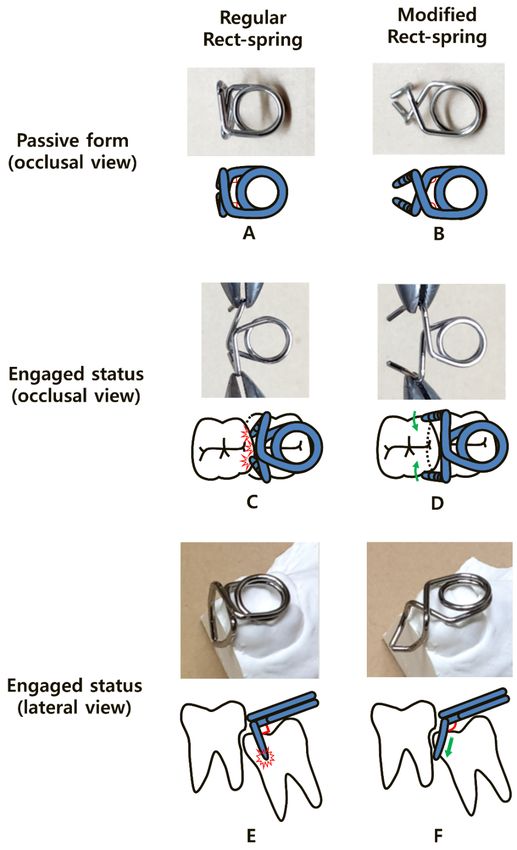
Children 2021,8,8,823
Children2021, x FOR PEER REVIEW 22of
of99
Figure 1. Comparison of different schematic views between
Figure between the
the regular
regular Rect-spring
Rect-spring(RS)
(RS)and
andmodified
modified
Rect-spring (mRS),
Rect-spring (mRS), and
and the
thepractical
practicalapplications
applicationsofofeach.
each.(A)
(A)Perpendicular
Perpendicular angles (red
angles marks)
(red of the
marks) of
RS. RS.
the (B) Obtuse angles
(B) Obtuse (red marks)
angles of the of
(red marks) mRS.
the(C) Unwanted
mRS. retracting
(C) Unwanted effect of the
retracting vertical
effect of thearms dur-
vertical
ing regular
arms duringRS engagement,
regular interferinginterfering
RS engagement, with retention.
with(D) Wide opening
retention. (D) Wideof the vertical
opening of components
the vertical
of the mRS reduces the tension of the helical spring and enhances the distal movement of the locked
components of the mRS reduces the tension of the helical spring and enhances the distal movement
molar on engagement. (E) The perpendicular angles (red angles) between the vertical and horizontal
of the locked molar on engagement. (E) The perpendicular angles (red angles) between the vertical
components cause unstable retention during engagement, with a risk of premature contact (red collision
and
mark).horizontal components
(F) The obtuse cause
angle (red unstable
angle) retention
and curved armsduring
engageengagement,
the undercut with
of thealocked
risk ofmolar
premature
(green
contact
arrow). (red collision mark). (F) The obtuse angle (red angle) and curved arms engage the undercut
of the locked molar (green arrow).
2. Material and Methods
2.1. Participants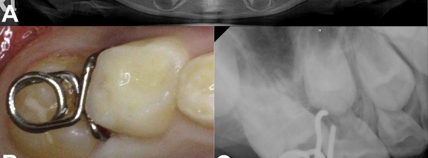
Children 2021, 8, 823                                                                                           3 of 9

                        2. Material and Methods
                        2.1. Participants
                             Two Korean children aged seven years who had visited the Yonsei University Dental
                        Hospital participated in this study. Inclusion criteria comprised participants having ec-
                        topically erupting permanent first molar with mesial angulation of which the amount of
                        entrapment was less than 4 mm, and those in general good health. Participants showing se-
                        rious tooth mobility of primary second molar expecting an early loss and who had severely
                        angulated ectopic molar were excluded. The consent of a child participant and parental
                        permission was obtained from all participants to use the diagnostic records, including
                        photographs and X-rays. All participants were fully aware of the precautions during
                        treatment. Any management done to the participants was not harmful, and there was no
                        serious complication. Since mRS is made of stainless steel wire, it is not toxic and hardly
                        causes allergic reactions.

                        2.2. Modification Design
                             First, we changed the perpendicular angles of the horizontal part (Figure 1A) to
                        obtuse angles (Figure 1B). This modification reduced the unwanted retracting effect of the
                        vertical arms that interfered with safety and firm engagement (Figure 1C). The obtuse angle
                        widened the limited distance between the two vertical arms when activated (Figure 1C)
                        and reduced the tension on the helical spring (Figure 1D).
                             Second, we increased the perpendicular angles between the horizontal and vertical
                        parts of the appliance in the lateral views (Figure 1E,F). As the increased angulation
                        enabled the vertical arms to move more mesially, their tips could reach the mesial undercut
                        of the locked molar with greater ease without an increase in the actual length. In addition,
                        the swinging motion of the arms increased the distal tipping force on the locked molar
                        (Figure 1F).
                             Third, we added an anterior curvature to the end of the vertical components to
                        shorten the length of the arms (Figure 1F). Without this curvature, the ends of the two
                        vertical arms could be entrapped in the height of contour, causing premature contact
                        (Figure 1E). The additional anterior curvature also accelerated the swinging effect and
                        increased stabilization during engagement by placing the ends of the arms more mesially
                        (Figure 1D). A detailed explanation of the above-mentioned modifications is available via
                        video online.

                        2.3. Clinical Procedure
                             The clinical procedure of RS or mRS is simple and only requires wiring and inser-
                        tion between the ectopic erupting molar and adjacent deciduous molar under infiltrative
                        anesthesia. Since the device produces a wedging effect, infiltrative local anesthesia on
                        interdental papilla is required for patient discomfort control.
                             The distance between two vertical arms needs to be wide opened during insertion,
                        and this can be accomplished by two utility pliers pulling each vertical arm or flattening
                        the inner loop of the occlusal spring, which resembles the activation process. Re-activation
                        of mRS is required every three to four weeks as the distalizing force decreases. Since mesial
                        angulation of ectopic molar decreases as the locking improves, it is desirable to change
                        the obtuse angle between the vertical component and horizontal component to an original
                        perpendicular angle.
                             A soft food diet is recommended during treatment to prevent mRS from falling out,
                        and patients should be careful not to swallow it.
                             After aligning the ectopic molar, the device is removed, and then its spontaneous
                        eruption is awaited. Careful radiographic examination is required until the further eruption
                        of the treated tooth to its occlusal level. We suggest a 6–12-month interval period before
                        the follow-up to evaluate the success of treatment and complications, such as the early loss
                        of the deciduous molar.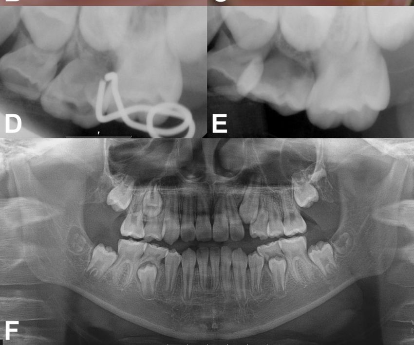
Children 2021, 8, 823                                                                                            4 of 9

                        3. Case Report
                              Case 1: A 7-year-old boy without a history of systemic disease presented with a chief
                        complaint of delayed eruption of the left maxillary permanent first molar. A panoramic
                        radiograph showed 2.2 mm deep entrapment of the left maxillary first molar with consid-
                        erable distal root resorption of the adjacent primary second molar; early loss of the right
                        maxillary primary second molar and, left maxillary and mandibular primary canines, and
                        congenitally missing right mandibular lateral incisor (Figure 2A).
                              We decided to unlock the left maxillary first molar using a mRS based on these clinical
                        and radiographic evaluations. We planned the space regaining treatment of the right
                        maxillary second premolar and left maxillary canine to take place several years later in
                        the late mixed dentition period. The mRS was inserted after the infiltrative anesthesia
                        (Figure 2B,C). The horizontal spring was placed on the distal cusp of the first molar without
                        any discomfort or mucosal irritation (Figure 2C). One month after delivery, the mesial
                        entrapment of the first molar was relieved (Figure 2D). Three months later, the maxillary left
                        first molar was successfully unlocked and reached the same occlusal level as the adjacent
                        primary second molar (Figure 2E). Resorption of the mesiobuccal root of the left maxillary
                        primary second molar was observed with slightly increased mobility (Figure 2E); however,
                        we maintained the primary second molar until its spontaneous exfoliation. The patient
                        did not report any discomfort during this treatment. The two-year follow-up panoramic
                        radiograph showed undisturbed full eruption of the left maxillary premolars (Figure 2F).
                        As the patient’s left maxillary second premolar and right maxillary canine were fully
                        erupted compared to their counterparts, we plan to initiate space regaining treatment by
                        shifting the molars posteriorly.
                              Case 2: A 7-year-old girl without any relevant medical history was referred from a
                        local clinic for her mesially tilted right maxillary first molar locked under the adjacent
                        primary second molar. Clinical and radiographic examination revealed an approximately
                        2.5 mm deep entrapment of the right maxillary first molar with considerable root resorption
                        of the right maxillary primary second molar (Figure 3A). There were no other dental
                        abnormalities. We inserted a mRS to align the first molar after infiltrative anesthesia. The
                        horizontal spring was placed directly above the occlusal surface of the right maxillary first
                        molar without premature contact. The two engaging vertical arms completely embraced
                        the mesial surface of the first molar (Figure 3B,C). One month after the insertion, a distal
                        movement of approximately 1 mm was observed on the periapical radiograph (Figure 3D).
                        Within 3 months, the mesially tilted right maxillary first molar escaped the entrapment,
                        moving to its normal position with no sign of increased mobility of the right primary
                        second molar after removal of the mRS (Figure 3E).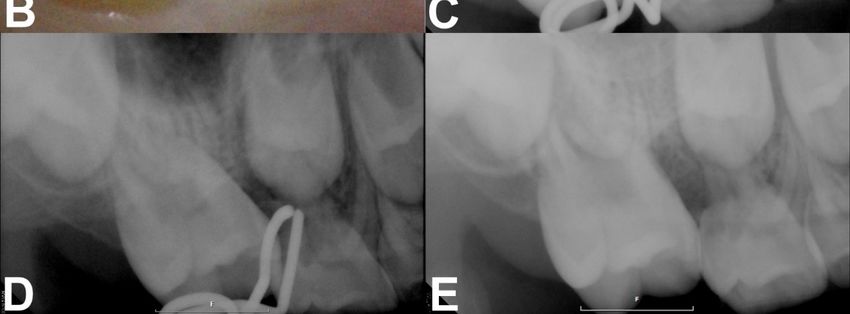
Children 2021, 8, x FOR PEER REVIEW 4 of 9
radiograph showed 2.2 mm deep entrapment of the left maxillary first molar with consid-
Children 2021, 8, 823 erable distal root resorption of the adjacent primary second molar; early loss of the right
5 of 9
maxillary primary second molar and, left maxillary and mandibular primary canines, and
congenitally missing right mandibular lateral incisor (Figure 2A).
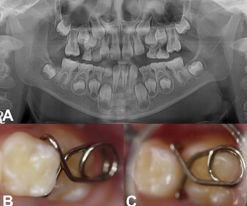
Figure 2. Pre-treatment and treatment progress images of case 1. (A) Pre-treatment panoramic radio-
Figure 2. Pre-treatment and treatment progress images of case 1. (A) Pre-treatment panoramic
graph at 7 years of age. (B,C) Clinical photograghs after the insertion of mRS. (D) Periapical radiograph
radiograph at 7 years of age. (B,C) Clinical photograghs after the insertion of mRS. (D) Periapical
radiograph obtained 1 month after delivery, showing relieved locking. (E) Post-treatment radiograph
showing the successfully corrected left maxillary first molar with proper occlusion after 3 months.
(F) Post-treatment panoramic radiograph taken at 9 years of age. Left maxillary premolars fully
erupted without space deficiency.Children 2021, 8, 823 6 of 9
Children 2021, 8, x FOR PEER REVIEW 6 of 9
Figure 3.
Figure 3. Pre-treatment
Pre-treatment and
andtreatment
treatmentprogress
progressimages
imagesofofcase
case2.2.(A)
(A)Panoramic
Panoramicradiograph revealed
radiograph revealedthethe
ectopic eruption
ectopic of
eruption
the right maxillary first molar. (B,C) Intraoral photograph and periapical radiograph after insertion of the mRS. (D) Periapical
of the right maxillary first molar. (B,C) Intraoral photograph and periapical radiograph after insertion of the mRS. (D)
radiograph obtained at the 1-month checkup. (E) 3 months after treatment showing a corrected right maxillary first molar.
Periapical radiograph obtained at the 1-month checkup. (E) 3 months after treatment showing a corrected right maxillary
first molar.
4. Discussion
Ectopic eruption of the permanent first molar presents as an irregular eruption pat-
4. Discussion
tern of the permanent
Ectopic eruptionmaxillary first molar,
of the permanent resulting
first in a partial
molar presents as impaction
an irregular under the distal
eruption pat-
surface of the primary second molar [5,10,11]. As ectopic eruption is asymptomatic,
tern of the permanent maxillary first molar, resulting in a partial impaction under patients
the
usually noticeof
distal surface it the
afterprimary
significant tooth
second mobility
molar of the
[5,10,11]. Asprimary
ectopic second
eruptionmolar occurs, or it
is asymptomatic,
could be usually
patients accidentally found
notice on regular
it after radiographic
significant examination
tooth mobility [12]. The second
of the primary magnitude
molarof
impaction, severity of locking, and increased resorption of primary second
occurs, or it could be accidentally found on regular radiographic examination [12]. The molar are relia-
ble predictive parameters of irreversible ectopic eruption [13]. In our first case, the 7-year-Children 2021, 8, 823 7 of 9
magnitude of impaction, severity of locking, and increased resorption of primary second
molar are reliable predictive parameters of irreversible ectopic eruption [13]. In our first
case, the 7-year-old boy had already lost the right maxillary primary second molar due to
ectopic eruption of the right maxillary first molar when he first visited our clinic, requiring
additional space regaining treatment. The outcome of ectopic eruption usually appears
during age seven [6]. Routine panoramic radiographic examination at age six would be
helpful for the early detection of this problem, preventing such early loss. The best space
maintaining device is the deciduous tooth. According to a longitudinal study, partially
resorbed primary second molars show no significant root resorption changes or repair with
secondary dentin after mesial locking is relieved [14]. Resorbed deciduous molars can
survive for a long time and function well regarding occlusion and space maintenance [7].
Therefore, early intervention during an asymptomatic period is critical for a favorable
prognosis [2].
The treatment option for early intervention depends on the severity of impaction [2],
grade of root resorption [2,10,15], degree of mesial angulation [15], and mobility of the
primary second molar with the presence of subjective discomfort [10]. In mild grade with an
entrapment depth of 1 mm, the interproximal wedging method using self-locking springs,
such as a kesling spring, NiTi separating spring, or triangular wedging spring, could be an
acceptable treatment technique [16,17]. Separators gain approximately 0.3–0.4 mm space at
once, and their force generally maintains up to 1 week requiring frequent clinical visits [18].
If the depth of entrapment is over 2 mm, as in our cases, the separation method alone is
insufficient, and distal tipping methods using active fixed appliances, such as Humphrey
or Halterman appliance, have been traditionally recommended [19,20]. However, these
appliances require additional laboratory procedures and apply an excessive anchorage
burden on the adjacent primary second molar. In these two cases, we chose the mRS for
unlocking the permanent first molars. It requires monthly re-activation, which is easily
achieved by widening the diameter of the horizontal spring or gradually reducing the
angles of the engaging arms. The unlocking procedure was completed within three months
on average.
The biomechanism of the mRS is highly effective due to its scientific design. The
two engaging arms exert distalizing and interproximal wedging force simultaneously.
Unlike conventional fixed appliances, mRS can prevent re-locking after the termination
of active distalization by separating the contact. The mesial arms also provide rigid and
safe retention. As the eruption angle of the locked molar becomes steeper, the curved
arms with obtuse angles enable smooth insertion following the contour of the crown and
firm engagement.
However, the mRS is not universal and has several limitations. First, it is not rec-
ommended for severely tilted molars. A remarkable anterior open bite may occur after
insertion due to unfavorable distal protrusion, resulting in occlusal interference. The
greater the inclination of the impacted tooth, the more is the distal protrusion of the device
(Figure 4). Clinicians should evaluate the radiographic view and be conscious of its use
on a severely tilted molar. A self-locking spring type that has a similar function to mRS
also cannot avoid occlusal interference [16]. Other methods, such as K loop or bonded NiTi
wire, that are mainly placed on the buccal/lingual side of teeth, not on the occlusal surface,
should be considered in such cases [21,22].
Second, it can fall out into the oral cavity and pose a risk of swallowing or aspiration.
As it does not directly attach to the tooth, it can disengage, especially in an unstable
mRS. If swallowed, it may be excreted after several days, and if aspirated, patients would
experience frequent coughs, requiring medical consultations. To prevent aspiration, tying
dental floss to the appliance during the insertion procedure is recommended, and using a
mRS in patients with uncontrolled swallowing reflex should be avoided.due to unfavorable distal protrusion, resulting in occlusal interference. The greater the in-
clination of the impacted tooth, the more is the distal protrusion of the device (Figure 4).
Clinicians should evaluate the radiographic view and be conscious of its use on a severely
tilted molar. A self-locking spring type that has a similar function to mRS also cannot avoid
Children 2021, 8, 823 occlusal interference [16]. Other methods, such as K loop or bonded NiTi wire, that are mainly 8 of 9
placed on the buccal/lingual side of teeth, not on the occlusal surface, should be consid-
ered in such cases [21,22].
Figure 4. Favorable state of the mRS engaged on a slightly tilted molar (A), and unfavorable state of the mRS on a severely
tilted molar (B). A severe mesial angulation of the locked molar leads to occlusal interference (red collision mark, (B)). RS
and mRS are not recommended in such cases.
Third, the mRS needs to be activated frequently, every three to four weeks, owing to
the high-short acting force produced by stainless steel wire. We made the helical spring as
wide as possible to cover the total occlusal surface to reduce this limitation. Using beta-
titanium wires with good formability and average stiffness for RS can be an alternative
choice [23].
The mRS could be used as a simple and effective method to correct ectopic eruption
before attempting other complicated techniques. It may succeed even in locking condi-
tions with severe root resorption of the primary molar. The contraindications include
unstable engagement and a prominent open bite due to extreme inclination. Clinicians
should consider the indications and select the best appliance for the management of an
ectopic eruption.
5. Conclusions
Irreversible ectopic eruption of the permanent first molar could cause severe root
resorption and early loss of the adjacent primary second molar. Treating ectopic eruption as
soon as possible before premature exfoliation of the primary second molar is essential. This
clinical report describes modifications of RS and presents their advantages and limitations.
The mRS does not require any laboratory procedures and can be successfully applied to
various locking conditions with easy modifications. Unlocking ectopically erupting molars
with mRS can be considered as the first choice of treatment.
Author Contributions: Writing—original draft preparation, M.S.S.; writing—review and editing,
C.-M.K., J.S.S., H.-J.C., J.L., S.-O.K.; orthodontic treatment, S.-O.K. All authors have read and agreed
to the published version of the manuscript.
Funding: This research received no external funding.
Institutional Review Board Statement: Not applicable.
Informed Consent Statement: Informed consent was obtained from all subjects involved in the study.
Conflicts of Interest: The authors declare no conflict of interest.
References
1. Grover, P.S.; Lorton, L. The incidence of unerupted permanent teeth and related clinical cases. Oral Surg. Oral Med. Oral Pathol.
1985, 59, 420–425. [CrossRef]
2. Barberia-Leache, E.; Suarez-Clúa, M.C.; Saavedra-Ontiveros, D. Ectopic eruption of the maxillary first permanent molar:
Characteristics and occurrence in growing children. Angle Orthod. 2005, 75, 610–615. [PubMed]
3. Young, D. Ectopic eruption of the first permanent molar. J. Dent. Child 1980, 24, 153–162.
4. Harrison, L., Jr.; Michal, B. Treatment of ectopically erupting permanent molars. Dent. Clin. N. Am. 1984, 28, 57–67.
5. Bjerklin, K.; Kurol, J. Ectopic eruption of the maxillary first permanent molar: Etiologic factors. Am. J. Orthod. 1983, 84, 147–155.
[CrossRef]Children 2021, 8, 823 9 of 9
6. Bjerklin, K.; Kurol, J. Prevalence of ectopic eruption of the maxillary first permanent molar. Swed. Dent. J. 1981, 5, 29–34.
7. Kurol, J.; Bjerklin, K. Ectopic eruption of maxillary first permanent molars: A review. ASDC J. Dent. Child. 1986, 53, 209–214.
8. Yaseen, S.M.; Naik, S.; Uloopi, K. Ectopic eruption—A review and case report. Contemp. Clin. Dent. 2011, 2, 3. [CrossRef]
9. Jun, H.; Lee, H.S.; Song, J.S.; Lee, J.-H.; Choi, B.-J.; Kim, S.O. The Rect-spring: A new device for treating ectopically erupting
permanent molars. Pediatr. Dent. 2014, 36, 143E–146E.
10. Chintakanon, K.; Boonpinon, P. Ectopic eruption of the first permanent molars: Prevalence and etiologic factors. Angle Orthod.
1998, 68, 153–160.
11. Mucedero, M.; Rozzi, M.; Cardoni, G.; Ricchiuti, M.R.; Cozza, P. Dentoskeletal features in individuals with ectopic eruption of the
permanent maxillary first molar. Korean J. Orthod. 2015, 45, 190–197. [CrossRef] [PubMed]
12. Kurol, J. Early treatment of tooth-eruption disturbances. Am. J. Orthod. Dentofac. Orthop. 2002, 121, 588–591. [CrossRef] [PubMed]
13. Dabbagh, B.; Sigal, M.J.; Tompson, B.D.; Titley, K.; Andrews, P. Ectopic eruption of the permanent maxillary first molar: Predictive
factors for irreversible outcome. Pediatr. Dent. 2017, 39, 215–218.
14. Kurol, J.; Bjerklin, K. Resorption of maxillary second primary molars caused by ectopic eruption of the maxillary first permanent
molar: A longitudinal and histological study. ASDC J. Dent. Child. 1982, 49, 273–279.
15. Kennedy, D.B.; Turley, P.K. The clinical management of ectopically erupting first permanent molars. Am. J. Orthod. Dentofac.
Orthop. 1987, 92, 336–345. [CrossRef]
16. Vallakati, A.; Jyothikiran, H.; Ravi, S.; Patel, P. Orthodontic separators—A systemic review. J. Orofac. Health Sci. 2014, 5, 118–122.
[CrossRef]
17. Kim, Y.H.; Park, K.T. Simple treatment of ectopic eruption with a triangular wedging spring. Pediatr. Dent. 2005, 27, 143–145.
18. Shamsuddin, S.V.; Alshahrani, S.; Sam, G.; Jeri, S.Y.; Bhuyan, S.K.R.; Mahanta, D. Assessment of separation impact and perception
of discomfort on various orthodontic separators: A comparative clinical study. J. Pharm. Bioallied Sci. 2021, 13, 642. [CrossRef]
19. Humphrey, W. A simple technique for correcting an ectopically erupting first permanent molar. J. Dent. Child. 1962, 29, 176–178.
20. Halterman, C.W. A simple technique for the treatment of ectopically erupting permanent first molars. J. Am. Dent. Assoc. 1982,
105, 1031–1033. [CrossRef]
21. Nam, O.H.; Ahn, H.J.; Kim, M.S.; Park, J.-H. Treatment of ectopic permanent maxillary first molar using a K-loop. J. Clin. Pediatr.
Dent. 2015, 39, 387–391. [CrossRef] [PubMed]
22. Kim, W.-S.; Kim, Y.; Cho, J.-H.; Oh, H.; Hwang, H.-S. Unlocking ectopically erupting permanent first molars using light wires. J.
Am. Dent. Assoc. 2020, 151, 857–862. [CrossRef] [PubMed]
23. Kusy, R.P. A review of contemporary archwires: Their properties and characteristics. Angle Orthod. 1997, 67, 197–207. [PubMed]You can also read