Molecular Phylogeny of Endophytic Fungi from Rattan (Calamus castaneus Griff.) Spines and Their Antagonistic Activities against Plant Pathogenic ...
←
→
Page content transcription
If your browser does not render page correctly, please read the page content below
Journal of
              Fungi
Article
Molecular Phylogeny of Endophytic Fungi from Rattan
(Calamus castaneus Griff.) Spines and Their Antagonistic
Activities against Plant Pathogenic Fungi
Nurul Farizah Azuddin                     , Masratul Hawa Mohd, Nik Fadzly N. Rosely, Asyraf Mansor and Latiffah Zakaria *

                                            School of Biological Sciences, Universiti Sains Malaysia, Penang USM 11800, Malaysia;
                                            farizah0610@gmail.com (N.F.A.); masratulhawa@usm.my (M.H.M.); nfadzly@usm.my (N.F.N.R.);
                                            asyrafm@usm.my (A.M.)
                                            * Correspondence: Lfah@usm.my; Tel.: +60-134-238-090

                                            Abstract: Calamus castaneus is a common rattan palm species in the tropical forests of Peninsular
                                            Malaysia and is noticeable by the yellow-based spines that cover the stems. This study aimed
                                            to determine the prevalence of fungal endophytes within C. castaneus spines and whether they
                                            inhibit the growth of fungal pathogens. Twenty-one genera with 40 species of fungal endophytes
                                            were isolated and identified from rattan palm spines. Based on molecular identification, the most
                                            common isolates recovered from the spines were Colletotrichum (n = 19) and Diaporthe spp. (n = 18),
                                            followed by Phyllosticta spp., Xylaria sp., Trichoderma spp., Helminthosporium spp., Penicillium spp.,
                                            Fusarium spp., Neopestalotiopsis spp., Arthrinium sp., Cyphellophora sp., Cladosporium spp., Curvularia

                                     sp., Bionectria sp., and Acremonium spp. Non-sporulating fungi were also identified, namely Nemania
                                            primolutea, Pidoplitchkoviella terricola, Muyocopron laterale, Acrocalymma fici, Acrocalymma medicaginis,
Citation: Azuddin, N.F.; Mohd,
                                            and Endomelanconiopsis endophytica. The isolation of these endophytes showed that the spines harbor
M.H.; Rosely, N.F.N.; Mansor, A.;
                                            endophytic fungi. Most of the fungal endophytes inhibited the growth of several plant pathogenic
Zakaria, L. Molecular Phylogeny of
Endophytic Fungi from Rattan
                                            fungi, with 68% of the interactions resulting in mutual inhibition, producing a clear inhibition zone
(Calamus castaneus Griff.) Spines and       of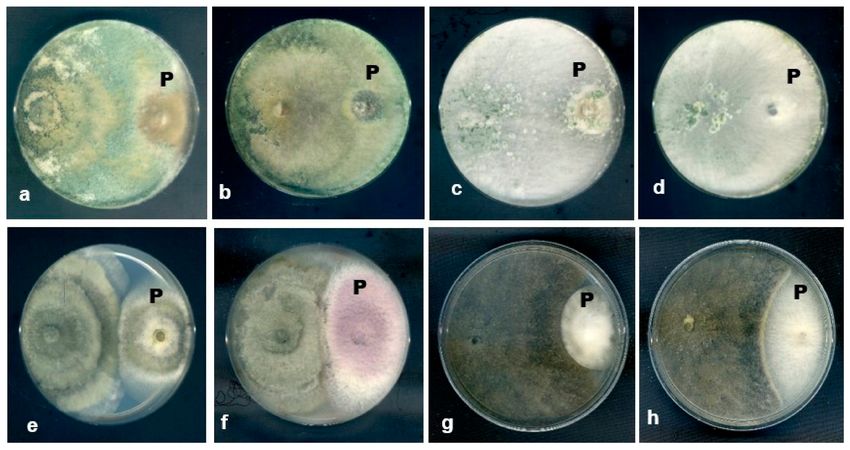
J. Fungi 2021, 7, 301 2 of 23
spines, which cover the stems and the middle part of the upper leaves. The spines are
arranged as a single line on the stem, while at the bottom of the leaves, the spines are
arranged in two parallel lines [8]. These sharp structures may harbor various types of
fungi as the presence of endophytic fungi, particularly dermatophytes in spines, thorns,
and prickles, has been reported by Halpern et al. (2011) [9]. As C. castaneus is common
and relatively easy to find in the forests, studying the presence of endophytic fungi in
the spines of this rattan species is of interest. Novel endophytic fungal isolates that have
the potential to be developed as biocontrol agents against several plant pathogenic fungi
might also be recovered from spines of C. castaneus. As there is a lack of information on
the fungal endophytes from spines, the objectives of this study were to determine the
occurrence of endophytic fungi in the spines of C. castaneus and identify the endophytic
fungi through molecular methods. The antagonistic activity of the fungal endophytes from
the spines to inhibit growth of several plant pathogenic fungi was also tested using a dual
culture method. Knowledge on the endophytic fungal community in spines of C. castaneus
contributes to in-depth information on the occurrence of fungal endophytes in various
plant parts as well as identifying potential biocontrol agents against plant pathogens.
2. Materials and Methods
2.1. Sample Collection and Isolation of Endophytic Fungi
The spines of C. castaneus were randomly collected from rattan trees found in three
rainforests, in two states of the Peninsula Malaysia, namely in Bukit Panchor State Park,
Penang (5.1602◦ N, 100.5480◦ E); Segari Melintang Forest Reserve, Perak (4◦ 18–200 N,
100◦ 34–360 E); and Belum Rainforest, Gerik, Perak (5◦ 34 58.340 N, 101◦ 15 30.70 E). The
spines were kept in an envelope and transported to the laboratory. The spines were placed
in a beaker, covered with a net cloth, and placed under running tap water overnight to
remove any debris, dirt, and epiphytes adhered to the surface. Thereafter, the spines were
surface sterilized by soaking in 70% ethanol for 5 min, followed by 5% sodium hypochlorite
(NaOCl) for 5 min. Then, the samples were washed with sterile distilled water three times
for 2 min and blotted dry using sterile filter papers to remove excess water. The sterilized
spines were plated onto potato dextrose agar (PDA, HiMedia Laboratory, Maharashta,
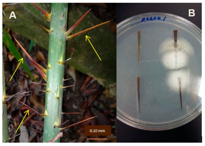
India) plates and incubated at room temperature (27 ± 1 ◦ C) until there was visible mycelial
growth from the spine tissues (Figure 1). Sixty spine samples were used for isolation.
Figure 1. Calamus castaneus spines (yellow arrow) and isolation of endophytic fungi. (A) Spines on
stem of rattan palm (C. castaneus). (B) Mycelia growth from the spines.
The efficiency of the surface sterilization technique was determined using an imprint
method [1]. The surface sterilized spines were imprinted or dabbed on the surface of a PDA
plate and the plate was incubated at room temperature. Surface sterilization is considered
effective if no fungal colony grows on the imprint plate. Mycelia growing from the spine
tissue were sub cultured onto new PDA plates. A pure culture of the isolate was obtained
using the spore suspension method and the plates were incubated at room temperature for
seven days.
The fungal isolates were sorted into their respective groups or genera based on the
appearance of the colonies and microscopic characteristics.J. Fungi 2021, 7, 301 3 of 23
2.2. DNA Extraction and PCR Amplification
The fungal isolates were grown in potato dextrose broth and incubated at room
temperature for six days. Mycelia were harvested and ground with liquid nitrogen in a
sterile mortar and pestle to a fine powder. The DNeasy® Plant Mini kit (Qiagen, Hilden,
Germany) was used to extract genomic DNA, according to the manufacturer’s instructions.
The internal transcribed spacer (ITS) region was used to identify all endophytic
fungal isolates recovered from the spines except Xylaria. The primers used were ITS1 and
ITS4 [10]. After amplification of the ITS, species identity was obtained based on the basic
local alignment search (BLAST) and a combination of at least two genes/regions was used
for further confirmation of the species (Table 1). However, for several fungal genera, the
analysis of the ITS region was not sufficient to differentiate closely related species.
Table 1. Gene/regions used for the identification of endophytic fungi from C. castaneus spines.
Region/Gene Primers Sequence (50 -30 ) Fungal Genera References
ITS 1 TCC GTA GGT GAA CCT GCG G
ITS All fungal genera White et al. (1990) [10]
ITS 4 TCC GCT TAT TGA TAT GC
GDF1 GCC GTC AAC GAC CCC TTC ATT GA Templeton et al. (1992) [11]
GAPDH Colletotrichum spp.
GDR2 GGG TGG AGT CGT ACT TGA GCA TGT
EF1 ATG GGT AAG GAG GAC AAG AC
Fusarium spp.
EF2 GGA AGT ACC AGT GAT CAT GTT
EF1-728F CAT CGA GAA GTT CGA GAA GG O’Donnell et al. (1998) [12]
Diaporthe spp.
EF1-986R TAC TTG AAG GAA CCC TTA CC
TEF-1α EF1-728F CAT CGA GAA GTT CGA GAA GG Phyllosticta spp. Carbone and Kohn (1999) [13]
EF2 GGA AGT ACC AGT GAT CAT GTT Arthrinium sp.
Pestalotiopsis spp.
EF1-728F CAT CGA GAA GTT CGA GAA GG
Trichoderma spp.
TEF1-rev GCC ATC CTT GGA GAT ACC AGC
T1 AAC ATG CGT GAG ATT GTA AGT
Xylaria sp.
T22 TCT GGA TGT TGG GAA TCC
T1 AAC ATG CGT GAG ATT GTA AGT O’Donnell and Cigelnik (1997) [14]
Fusarium spp.
T2 TAG TGA CCC TTG GCC CAG TTG
β-tubulin
Bt2a GGT AAC CAA ATC GGT GCT TTC Glass and Donaldson (1995) [15]
Penicillium spp.
Bt2b ACC CTC AGT GTA GTG ACC CTT GGC
T1 AAC ATG CGT GAG ATT GTA AGT Cyphellophora sp.
Bt2b ACC CTC AGT GTA GTG ACC CTT GGC Diaporthe spp.
ACT-512F ATG TGC AAG GCC GGT TTC G Xylaria sp.
ACT Carbone and Kohn (1999) [13]
ACT-783R TAC GAG TCC TTC TGG CCC AT Cladosporium sp.
LROR ACC CGC TGA ACT TAA GC Non-sporulating
Vilgalys and Hester (1990) [16]
LR5 TCC TGA GGG AAA CTT CG fungi
LSU De Hoog and Gerrits Van Den Ende
V9G TTA CGT CCC TGC CCT TTG TA
Corynespora spp. (1998) [17]
LR5 TCC TGA GGG AAA CTT CG Vilgalys and Hester (1990) [16]
PCR reactions were prepared in a total volume of 50 µL containing 8 µL of 5X Green
GoTaq® Flexi Buffer, 8 µL of 25 mM MgCl2 , 1 µL of 10 mM dNTP mix, 8 µL each of 5 µM
forward and reverse primers, deionized distilled water, 0.3 µL of 5 U/µL GoTaq® DNA
Polymerase (Promega, Madison, WI, USA), and 0.6 µL of DNA template. EconoTaq® Plus
Green 2× Master Mix reagent (Middleton, WI, USA) was used to amplify β-tubulin and
ACT. The PCR reaction was prepared in a total volume of 50 µL containing 25 µL EconoTaq®
Plus Green 2× Master Mix, 0.5 µL each of the forward and reverse primers (100 µM), 1 µLJ. Fungi 2021, 7, 301 4 of 23
of DNA template, and deionized distilled water. The amplification was performed in a
thermal cycler (Bio-Rad MyCycler PCR System version 1.065) programmed to 85 s at 94 ◦ C,
35 s at 95 ◦ C for 35 cycles, 55 s at 59 ◦ C, 90 s at 72 ◦ C, and a final 10 min extension at 72 ◦ C.
A 1% agarose gel (Promega, Middleton, WI, USA) was used to detect the PCR products
in 1 × Tris-Borate-EDTA (TBE) buffer stained with FloroSafe DNA stain (Axil Scientific,
Singapore). PCR products were sent to a service provider for Sanger DNA sequencing.
2.3. Molecular Identification and Phylogenetic Analysis
The DNA sequences were aligned manually and edited using the Molecular Evolution
Genetic Analysis version 7 (MEGA7 version 7) [18]. Forward and reverse sequences
were aligned with ClustalW using pairwise alignments. The aligned forward and reverse
sequences were edited when necessary to form a consensus sequence. For species identity,
a BLAST search was used to analyze the number of bases and determine the maximum
identity of the consensus sequences from the GenBank database.
A phylogenetic analysis was also conducted, particularly for species that are known
to belong to a species complex or for isolates whose ITS sequences cannot be used to
confidently identify the isolates to the species levels. Multiple sequence alignments were
generated and used to construct phylogenetic trees based on combined sequences. A
maximum likelihood (ML) tree was constructed with 1000 bootstraps replicates. The
heuristic method used in ML was the nearest neighbor interchange (NNI) and the initial
tree for ML was generated automatically. The best model for ML tree was determined
from the model search with number of discrete gamma categories 5. The results show that
the Kimura 2 parameter model was the best model. Missing data or gaps were treated as
complete deletion.
2.4. Antagonistic Activity
The ability of the fungal endophytes to inhibit the mycelial growth of several plant
pathogenic fungi was determined with a dual culture method using PDA. Several endo-
phytic fungi from C. castaneus spines were selected to assess their antagonistic activity
against several plant pathogenic fungi. The endophytic fungi were chosen based on fungal
genera or species that have been reported as antagonists against plant pathogens, such as
Xylaria cubensis, Penicillium indicum, Penicillium oxalicum, Trichoderma harzianum, and Tricho-
derma koningiopsis. Endophytic fungal species that have not been reported as antagonists
were also tested, namely Endomelanconiopsis endophytica, Neopestalotiopsis saprophytica, Col-
letotrichum endophytica, Colletotrichum siamense, Colletotrichum boninense, Diaporthe arengae,
Diaporthe tectonae, Diaporthe cf. nobilis, and Diaporthe cf. heveae.
Selected plant pathogenic fungi were obtained from the culture collection at the Plant
Pathology Laboratory, School of Biological Sciences, Universiti Sains Malaysia, Penang,
Malaysia. The pathogenic fungi included two anthracnose chili pathogens, C. truncatum
and C. scovellei; two pathogens that cause dragon fruit stem rot, Fusarium proliferatum and
F. fujikuroi; and F. solani and F. oxysporum, which are associated with crown disease in oil
palm. Four pathogens associated with mango diseases were also included: Lasiodiplodia
theobromae and Pestalotiopsis mangiferae, which are the causal pathogens of the mango leaf
spot, and L. pseudotheobromae and D. pascoei, which cause mango stem-end rot.
A combination of the endophytic fungi and plant pathogenic fungi tested in dual
culture test is shown in Table 2. A control plate harbored only plant pathogenic fungi
without the endophytes. Mycelial plugs (5 mm) of the pathogen and endophyte were
cultured 6 cm apart. The plates and three replications were incubated at room temperature
for seven days. The experiment was repeated twice.J. Fungi 2021, 7, 301 5 of 23
Table 2. Combination of endophytic fungi and plant pathogenic fungi tested in dual culture test.
Endophytic Fungi
C. endophytica C. siamense C. boninense X. cubensis X. cubensis D. arengae D. tectonae D. cf. nobilis D. cf. heveae
Plant Pathogenic Fungi
(BP9) (BP14) (SM21) (SM22) (BR90) (SM45) (BR62) (BR67) (BR74)
√ √ √ √ √ √ √ √ √
C.truncatum
√ √ √ √ √ √ √ √ √
C. scovellei
√ √ √ √ √ √ √ √ √
F. solani
√ √ √ √ √ √ √ √ √
F. oxysporum
√ √ √ √ √ √ √ √ √
F. proliferatum
√ √ √ √ √ √ √ √ √
F. fujikuroi
√ √ √ √ √ √ √ √ √
L. theobromae
√ √ √ √ √ √ √ √ √
P. mangiferae
√ √ √ √ √ √ √ √ √
L. pseudotheobromae
√ √ √ √ √ √ √ √ √
D. pascoei
Endophytic Fungi
Plant Pathogenic Fungi N. saprophytica T. harzianum End. endophytica Pen.oxalicum
Pen. indicum (BR91) T. koningiopsis (BR96)
(BP1) (BR94) (BR98) (BR102)
√ √ √ √ √ √
C.truncatum
√ √ √ √ √ √
C. scovellei
√ √ √ √ √ √
F. solani
√ √ √ √ √ √
F. oxysporum
√ √ √ √ √ √
F. proliferatum
√ √ √ √ √ √
F. fujikuroi
√ √ √ √ √ √
L. theobromae
√ √ √ √ √ √
P. mangiferae
√ √ √ √ √ √
L. pseudotheobromae
√ √ √ √ √ √
D. pascoeiJ. Fungi 2021, 7, 301 6 of 23
After seven days, the percentage of the pathogen growth inhibition (PGI) was calcu-
lated according to the method described by Skidmore and Dickinson (1976) [19]:
PGI (%) = (R1 − RI2/R) × 100.
R1—radial growth of plant pathogenic fungi in control plate.
R2—radial growth of plant pathogenic fungi in dual culture plate.
R1 was measured from the point of inoculation to the pathogen colony margin on the
control plate and R2 was measured from the point of inoculation to the colony margin on
the dual culture plate in the direction of the endophytes.
Statistical analysis of the PGI value was performed using ANOVA in SPSS statistical
software version 24. Interactions between plant pathogens and endophytic fungi were
assigned in a range of interactions from types A to E, according to the interactions described
by Skidmore and Dickinson (1976) [19]. Type A interactions occurred when the pathogens
and endophytic fungi displayed intermingling growth; type B interactions represented
the overgrowth of pathogens by endophytic fungi; type C interactions represented the
overgrowth of endophytic fungi by pathogens; type D interactions represented mutual
inhibition with a clear inhibition zone at small distance (2 mm).
3. Results
3.1. Molecular Identification
A total of 108 isolates of endophytic fungi comprising 21 genera with 40 species were
recovered from the C. castaneus spines (Table 3). Fungi isolated from the spines were
confirmed as endophytes as no fungal growth on the imprinted plates was observed. The
imprint method was used as an indication that the epiphytes from the surface of the spines
had been removed. A successful and correct procedure of surface sterilization removes
epiphytes from the surface of the spines, which results in no fungal growth and must be
used in all studies concerning endophytes [20,21].
Table 3. Molecular identification of endophytic fungi isolated from C. castaneus spines.
Genbank Accession Number
%
Isolates ITS GAPDH β-Tubulin TEF-1α ACT LSU
Similarity
Colletotrichum spp.
C. siamense BP4 MN635697 MT077122 - - - - 99
C. siamense BP8 MN635698 MT077123 - - - - 99
C. siamense BP14 MN635699 MT077124 - - - - 99
C. fructicola BP5 MN635702 MT077113 - - - - 99
C. fructicola SM40 MN635702 MT077114 - - - - 99
C. endophytica BP9 MN635726 MT077115 - - - - 99
C. endophytica BP10 MN635727 MT077116 - - - - 99
C. endophytica BP11 MN635728 MT077117 - - - - 99
C. endophytica SM31 MN635729 MT077118 - - - - 99
C. endophytica SM33 MN635730 MT077119 - - - - 99–100
C. endophytica SM43 MN635731 MT077120 - - - - 99
C. endophytica SM44 MN635732 MT077121 - - - - 99
C. horii BP3 MN635649 MT077107 - - - - 99
C. horii BP7 MN635650 MT077108 - - - - 99
C. horii BP12 MN635651 MT077109 - - - - 99
C. horii BP13 MN635652 MT077110 - - - - 99
C. cliviae SM25 MN652631 MT077111 - - - - 99
C. cliviae SM26 MN652632 MT077112 - - - - 99
C. boninense SM21 MN635733 MT077106 - - - - 99J. Fungi 2021, 7, 301 7 of 23
Table 3. Cont.
Genbank Accession Number
%
Isolates ITS GAPDH β-Tubulin TEF-1α ACT LSU
Similarity
Diaporthe spp.
D. arengae SM28 MN651480 - MT077062 MT077093 - - 98–99
D. arengae SM41 MN651481 - MT077064 MT077095 - - 98–99
D. arengae SM35 MN651483 - MT077068 MT077099 - - 98–99
D. arengae SM49 MN651487 - MT077069 MT077089 - - 98–99
D. arengae SM38 MN651484 - MT077066 MT077097 - - 98–99
D. arengae SM39 MN651485 - MT077067 MT077098 - - 98–99
D. arengae SM45 MN635732 - MT077065 MT077096 - - 97–98
D. arengae SM29 MN651486 - MT077063 MT077094 - - 98–99
D. arecae SM30 MN651482 - MT077061 MT077090 - - 99
D. hongkongensis SM42 MN651488 - MT077085 MT077103 - - 97–99
D. cf. heveae SM36 MN651489 - MT077080 MT077092 - - 96–99
D. cf. heveae BR74 MN636282 - MT077079 MT077091 - - 96–99
D. cf. nobilis BR67 MN651491 - MT077084 MT077088 - - 96–98
Diaporthe sp.SM46 MN651495 - MT077083 MT077100 - - 98–99
Diaporthe sp. SM59 MN651496 - MT077081 MT077101 - - 95–99
Diaporthe sp. BR103 MN651497 - MT077082 MT077102 - - 98–99
D. tectonae SM62 MN651493 - MT077086 MT077104 - - 95–97
D. tectonae SM63 MN651494 - MT077087 MT077105 - - 95–98
Phyllosticta spp.
P. capitalensis SM20 MN635748 - - MT118281 - - 99
P. capitalensis SM23 MN635749 - - MT118282 - - 99
P. capitalensis SM32 MN635750 - - MT118283 - - 99–100
P. capitalensis SM37 MN635751 - - MT118284 - - 99–100
P. capitalensis SM48 MN635752 - - MT118285 - - 99
P. capitalensis SM53 MN635753 - - MT118286 - - 99
P. capitalensis SM58 MN635754 - - MT118287 - - 99
P. carochlae SM27 MN652663 - - MT118272 - - 99
P. carochlae SM34 MN652664 - - MT118269 - - 95–99
P. carochlae SM51 MN652665 - - MT118270 - - 97–99
P. carochlae SM52 MN652666 - - MT118271 - - 97–99
Neopestalatiopsis spp.
N. saprophytica BP1 MN635619 - - MT264943 - - 99
N. formicarum BP2 MN635621 - - MT264929 - - 99
N. formicarum BP6 MN635622 - - MT264930 - - 99
Trichoderma spp.
T. harzianum BR93 MN636262 - - MT264931 - - 99–100
T. harzianum BR94 MN636263 - - MT264932 - - 99
T. harzianum BR95 MN636264 - - MT264933 - - 98–99
T. harzianum BR93 MN636262 - - MT264931 - - 99–100
T. koningiospsis BR96 MN636269 - - MT264934 - - 99
T. koningiospsis BR97 MN636270 - - MT264935 - - 99
T. koningiospsis BR99 MN636271 - - MT264936 - - 99
T.koningiospsis BR100 MN636272 - - MT264937 - - 99
Xylaria cubensis
X. cubensis SM22 - - MT118273 - MT077070 - 99
X. cubensis BR84 - - MT118274 - MT077071 - 99
X. cubensis BR85 - - MT118275 - MT077072 - 99
X. cubensis BR88 - - MT118276 - MT077073 - 99
X. cubensis BR89 - - MT118277 - MT077074 - 99
X. cubensis BR90 - - MT118278 - MT077075 - 99
X. cubensis BR101 - - MT118279 - MT077076 - 99J. Fungi 2021, 7, 301 8 of 23
Table 3. Cont.
Genbank Accession Number
%
Isolates ITS GAPDH β-Tubulin TEF-1α ACT LSU
Similarity
X. cubensis BR105 - - MT118280 - MT077077 - 99
X. cubensis BR106 - - - - MT077078 - 95–99
Pidoplitchkoviella terricola
Pid. terricola SM17 MN652667 - - - - MW338725 96
Pid. terricola SM18 MN652668 - - - - MW338726 96
Pid. terricola SM19 MN652669 - - - - MW338727 96
Pid. terricola SM24 MN652670 - - - - MW338728 96
Pid. terricola SM57 MN652671 - - - - MW338729 96
Pid. terricola BR79 MN652672 - - - - MW338730 96
Helminthosporium spp.
H. endiandrea SM61 MT279339 - - - - MW338667 99
H. endiandrea SM64 MT279340 - - - - MW338668 99
H. livistonae BR76 MN652658 - - - - MW338703 93–97
H. livistonae BR78 MN652659 - - - - MW338704 93–98
H. livistonae BR80 MN652660 - - - - MW338705 93–99
H. livistonae BR83 MN652673 - - - - MW338706 93–99
H. livistonae BR87 MT279326 - - - - MW338669 99–100
Cladosporium halotolerans
Cla. halotolerans SM50 MN636281 - - - MT264919 - 99
Cla. halotolerans BR75 MN636282 - - - MT264920 - 99
Penicillium spp.
Pen. indicum SM65 MN635766 - MT264923 - - - 99
Pen. indicum BR91 MN635767 - MT264924 - - - 99
Pen. oxalicum BR102 MN636265 - MT264925 - - - 99
Pen. oxalicum BR104 MN636266 - MT264926 - - - 99
Pen. oxalicum BR107 MN636267 - MT264927 - - - 99
Pen. oxalicum BR108 MN636268 - MT264928 - - - 99
Fusarium spp.
F. lateritium BR66 - - MT296784 MT264940 - - 99–100
F. decemcellulare BR72 - - MT296782 MT264938 - - 99
F. decemcellulare BR77 - - MT296783 MT264939 - - 99
F. lateritium BR82 - - MT296785 MT264941 - - 99
F. oxysporum BR86 - - MT296786 MT264942 - - 99
F. solani BR92 - - MT296787 MT264944 - - 99
Cyphellophora guyanensis
Cyp. guyanensis BR71 MN636279 - MT264921 - - - 99–100
Cyp. guyanensis BR73 MN636280 - MT264922 - - - 99
Arthrinium urticae
Art. urticae SM47 MN636276 - - - - - 98–99
Art. urticae SM55 MN636277 - - - - - 98–99
Art. urticae SM56 MN636278 - - - - - 99
Nemania primolutea
Nem.primolutea BP15 MN652661 - - - - - 99
Nem.primolutea BP16 MN652662 - - - - - 99J. Fungi 2021, 7, 301 9 of 23
Table 3. Cont.
Genbank Accession Number
%
Isolates ITS GAPDH β-Tubulin TEF-1α ACT LSU
Similarity
Cuvularia. lunata SM54 MN637803 - - - - - 99
Muyocopron laterale SM60 MN637806 - - - - - 96
Endomelanconiopsis endophytica BR98 MN637809 - - - - - 99
Acrocalymma fici BR68 MN637807 - - - - - 96
Acrocalymma medicaginis BR81 MN637808 - - - - - 96
Acremonium hennebertii BR70 MN637805 - - - - - 99
Bionectria pityrodes BR69 MN637804 - - - - - 99
Note: Colletotrichum endophytica is synonymous with Colletotrichum endophyticum.
Endophytic fungal species recovered from C. castaneus spines identified using ITS
and other additional markers are shown in Table 2. Most of the isolates were successfully
identified to the species levels except for three isolates of Diaporthe. The most common
isolates recovered from the spines were Colletotrichum spp. (n = 19) and Diaporthe spp.
(n = 18), followed by Phyllosticta spp. (n = 11), Xylaria sp. (n = 9), Trichoderma spp. (n = 7),
Helminthosporium spp. (n = 7), Penicillium spp. (n = 6), Fusarium spp. (n = 6), Neopestalotiopsis
spp. (n = 3), Arthrinium sp. (n = 3), Cyphellophora sp. (n = 2), Cladosporium spp. (n = 2),
Curvularia sp. (n = 1), Bionectria sp. (n = 1), Acremonium sp. (n = 1), and six species of
non-sporulating fungi.
Six species of Colletotrichum were identified using ITS and GAPDH sequences, namely
C. horii (n = 4), C. siamense (n = 3), C. fructicola (n = 2), C. cliviae (n = 2), C. endophytica
(n = 7), and C. boninense (n = 1) (Table 3). All the species identified are members of the
C. gloeosporioides species complex. In addition to ITS, the GAPDH gene was included as an
additional marker as the gene is among the most effective secondary markers to distinguish
species in the genus Colletotrichum. Moreover, GAPDH is the easiest gene to amplify and
sequence [22,23]. The phylogenetic analysis showed that isolates from the same species
were grouped in the same clade as their epitype strains (Figure 2), which confirmed the
identity of the endophytic Colletotrichum species obtained from C. castaneus spines.
Based on phylogenetic analysis of the combined ITS, TEF-1α, and β-tubulin se-
quences, 18 isolates of Diaporthe spp. were phylogenetically identified as D. arengae (n = 8),
D. hongkongensis (n = 1), Diaporthe cf. heveae 2 (n = 2), D. cf. nobilis (n = 1), D. arecae (n = 1),
D. tectonae (n = 2), and Diaporthe spp. (n = 3). In the ML tree, isolates of the same species
were grouped together with their epitype strains (Table 2, Figure 3).
Endophytic isolates of Phyllosticta, Trichoderma, and Neopestalotiopsis were identified
through molecular methods using ITS and TEF-1α sequences (Table 3, Figure 4A–C).
Isolates of Phyllosticta were identified as P. capitalensis (n = 7) and P. carochlae (n = 4). Seven
isolates of endophytic Trichoderma were identified as T. harzianum (n = 3) and T. koningiopsis
(n = 4). Two species of endophytic Neopestalotiopsis, N. saprophytica (n = 1) and N. formicarum
(n = 2) were also isolated from C. castaneus spines.J. Fungi 2021, 7, 301 10 of 23
Figure 2. Maximum likelihood tree inferred from combined sequences of internal transcribed spacer
(ITS) and GAPDH of Colletotrichum isolates from C. castaneus spines with bootstrap values higher
than 50% are shown next to the branches.
Figure 3. Maximum likelihood tree inferred from combined sequences of ITS, TEF-1α, and β-tubulin
of Diaporthe isolates from C. castaneus spines with bootstrap values higher than 50% are shown next
to the branches.J. Fungi 2021, 7, 301 11 of 23
Figure 4. Maximum likelihood tree inferred from combined sequences of ITS and TEF-1α for
(A) Phyllosticta spp., (B) Trichoderma spp., and (C) Neopestalotiopsis spp. from C. castaneus spines with
bootstrap values higher than 50% are shown next to the branches.J. Fungi 2021, 7, 301 12 of 23
Nine isolates of the endophytic X. cubensis were identified using β-tubulin and ACT
sequences (Table 3, Figure 5).
Figure 5. Maximum likelihood tree inferred from combined sequences of β-tubulin and ACT of
X. cubensis from C. castaneus spines with bootstrap values higher than 50% are shown next to
the branches.
Based on ITS and LSU sequences, endophytic isolates of Helmintosporium were iden-
tified as H. livistonae (n = 5) and H. endiandrae (n = 2) (Table 2, Figure 6A). Isolates of
Pidoplitchkoviella terricola (n = 6) were identified using ITS and LSU sequences. The endo-
phytic P. terricola isolates were clustered in the same main clade as the reference strain
(CBS 180.77) but the isolates formed a separate sub-clade (Figure 6B), which might indicate
that the isolates represent different phylogenetic strains of the species.
Figure 6. Maximum likelihood tree inferred from combined sequences of ITS and LSU for
(A) Helminthosporium spp. and (B) Pidoplitchkoviella terricola from C. castaneus spines with boot-
strap values higher than 50% are shown next to the branches.J. Fungi 2021, 7, 301 13 of 23
Based on ITS and β-tubulin sequences, isolates of endophytic Arthrinium urticae
(n = 3), Cyphellophora guyanensis (n = 2), and two species of Penicillium, P. indicum (n = 2)
and P. oxalicum (n = 4) were identified (Table 3, Figure 7A–C).
Figure 7. Maximum likelihood tree inferred from combined sequences of ITS and β-tubulin for
(A) Arthrinium urticae, (B) Cyphellophora guyanensis, and (C) Penicillium spp. from C. castaneus spines
with bootstrap values higher than 50% are shown next to the branches.
Four species of endophytic Fusarium, F. lateritium (n = 2), F. decemcellulare (n = 2),
F. oxysporum (n = 1), and F. solani (n = 1) were identified using TEF-1α and β-tubulinJ. Fungi 2021, 7, 301 14 of 23
(Table 3, Figure 8). Two isolates of Cladosporium halotolerans were identified using ITS and
ACT sequences (Table 3, Figure 9).
Figure 8. Maximum likelihood tree inferred from combined sequences of TEF-1α and β-tubulin of
Fusarium spp. from C. castaneus spines with bootstrap values higher than 50% are shown next to
the branches.
Figure 9. Maximum likelihood tree inferred from combined sequences of ITS and ACT of
C. halotolerans isolates from C. castaneus spines with bootstrap values higher than 50% are shown next
to the branches.
Several species of the endophytic fungi were identified using ITS sequences (Table 3,
Figure 10A–G), namely Curvularia lunata (n = 1), Bionectria pityrodes (n = 1), Acremonium
hennebertii (n = 1), Nemania primolutea (n = 2), Muyocopron laterale (n = 1), Acrocalymma fici
(n = 1), Acrocalymma medicaginis (n = 1), and Endomelanconiopsis endophytica (n = 1).J. Fungi 2021, 7, 301 15 of 23
Figure 10. Cont.J. Fungi 2021, 7, 301 16 of 23
Figure 10. (A–G) Maximum likelihood tree inferred from combined sequences of ITS for (A) Curvu-
laria lunata, (B) Bionectria pityrodes (C) Acremonium hennebertii, (D) Nemania primolutea, (E) Muyocopron
laterale, (F) Acrocalymma spp., and (G) Endomelanconiopsis endophytica from C. castaneus spines of with
bootstrap values higher than 50% are shown next to the branches.
3.2. Antagonistic Activity
In general, most of the endophytic fungi from C. castaneus spines inhibited mycelial
growth of the plant pathogenic fungi tested (Table 4). Only three species of Diaporthe,
D. cf. nobilis, D. cf. heveae, and D. tectonae, as well as two isolates of X. cubensis did not show
antagonistic activity against L. theobromae and L. pseudotheobromae (Table 4). Both pathogens
overgrew the endophytic fungi as L. theobromae and L. pseudotheobromae are fast growing
fungi able to compete for space and nutrients.J. Fungi 2021, 7, 301 17 of 23
Table 4. Antagonistic activity of endophytic fungi against plant pathogenic fungi in dual culture test.
Endophytic Fungi and PGI Value
C. endophytica C. siamense C. boninense X. cubensis X. cubensis D. arengae D. tectonae D. cf. nobilis D. cf.
Plant Pathogenic Fungi
(BP9) (BP14) (SM21) (SM22) (BR90) (SM45) (BR62) (BR67) heveae(BR74)
C. truncatum 33.33 ± 6.03 cd 13.33 ± 5.58 ab 20.46 1.38 bc 0 ± 0.00 a 1.11 ± 1.72 a 15.24 ± 14.53 ab 45.49 ± 4.04 d 19.57 ± 0.70 bc 38.34 ± 2.40 d
C. scovellei 19.52 ± 0.56 abc 55.85 ± 3.27 cd 28.10 ± 6.24 bcd 0.57 ± 1.73 a 1.33 ± 2.37 a 57.73 ± 4.05 cd 70.59 ± 3.51 f 30.55 ± 0.15 bcd 60.50 ± 5.47 de
F. solani 35.96 ± 2.15 de 31.58 ± 1.66 d 20.18 ± 2.72 bc 13.16 ± 0.66 a 13.16 ± 2.35 a 16.23 ± 1.98 ab 41.23 ± 2.72 e 17.54 ± 2.15 ab 35.53 ± 2.20 de
F. oxysporum 28.47 ± 0.69 abc 49.65 ± 1.57 ef 33.33 ± 1.32 bc 26.39 ± 1.70 ab 20.49 ± 2.77 a 60.76 ± 2.05 fg 61.35 ± 1.66 g 34.72 ± 2.85 bc 57.99 ± 4.04 ef
F. proliferatum 28.58 ± 4.01 bc 16.10 ± 0.86 abc 4.45 ± 3.01 a 4.80 ± 3.93 a 6.85 ± 3.88 ab 17.11 ± 15.18 abc 40.45 ± 17.79 cd 14.97 ± 8.87 abc 19.50 4.35 abc
F. fujikuroi 41.90 ± 2.76 cd 33.64 ± 8.95 abc 28.44 ± 1.64 a 27.83 ± 1.50 a 25.99 ± 3.96 a 48.93 ± 1.80 def 55.35 ± 6.10 ef 40.06 ± 2.76 bcd 45.8 ± 1.53 de
L. theobromae 58.20 ± 5.22 ef 40.23 ± 2.50 bc 50.10 ± 1.00 bcde 55.85 ± 11.90 def 57.40 ± 4.55 ef 38.49 ± 2.86 b 0 ± 0.00 a 0 ± 0.00 a 0 ± 0.00 a
Pes. mangiferae 27.78 ± 2.33 de 31.48 ± 1.67 ef 22.22 ± 1.99 c 22.59 ± 1.67 c 22.22 ± 1.99 c 27.04 ± 1.67 d 44.07 ± 1.67 g 29.63 ± 2.30 def 33.33 ± 1.99 f
L. pseudotheobromae 43.56 ± 1.38 cd 42.89 ± 1.00 cd 40.22 ± 1.00 b 0.00 ± 0.00 a 0.00 ± 0.00 a 43.56 ± 1.38 cd 56.44 ± 1.38 g 44.44 ± 1.38 d 47.78 ± 1.00 e
D. pascoei 39.42 ± 31 abc 38.00 ± 4.88 abc 31.30 ± 2.64 ab 27.82 ± 2.40 a 29.56 ± 1.46 ab 32.75 ± 4.35 abc 38.55 ± 2.38 abc 36.23 ± 2.38 abc 39.13 ± 3.65 abc
Endophytic Fungi and PGI Value
Plant Pathogenic Fungi N. saprophytica Pen. indicum Pen. oxalicum
T. harzianum (BR94) T. koningiopsis (BR96) End. endophytica (BR98)
(BP1) (BR91) (BR102)
C. truncatum 19.44 ± 2.51 bc 7.22 ± 2.51 ab 89.33 ± 2.99 e 80.05 ± 5.75 e 53.65 ± 10.85 d 1.34 ± 2.33 a
C. scovellei 48.46 ± 8.00 cd 3.20 ± 4.66 a 85.80 ± 5.47 e 89.45 ± 2.55 e 45.70 ± 7.39 bcd 8.09 ± 2.13 ab
F. solani 35.96 ± 2.15 de 16.67 ± 11.39 ab 62.28 ± 2.15 f 74.56 ± 2.72 g 24.56 ± 2.72 c 25.44 ± 2.15 c
F. oxysporum 46.88 ± 1.14 de 30.56 ± 7.65 abc 76.74 ± 4.45 h 76.04 ± 1.74 h 59.03 ± 5.38 fg 37.85 ± 1.57 cd
F. proliferatum 30.18 ± 8.98 bcd 7.94 ± 7.11 ab 57.38 ± 17.22 e 51.63 ± 13.52 de 23.52 ± 8.66 abc 11.36 ± 6.34 abc
F. fujikuroi 43.43 ± 6.19 cd 32.42 ± 5.37 abc 71.25 ± 1.50 g 59.94 ± 11.16 fg 46.18 ± 1.50 de 30.28 ± 1.64 ab
L. theobromae 43.07 ± 2.89 abc 46.83 ± 0.89 abcd 82.86 ± 1.28 f 77.62 ± 6.30 f 63.59 ± 4.83 e 48.85 ± 3.89 abcd
Pes. mangiferae 27.41 ± 2.30 cd 7.41 ± 3.04 a 88.89 ± 1.41 g 60.00 ± 1.99 f 32.52 ± 1.89 de 14.07 ± 2.69 b
L. pseudotheobromae 52.44 ± 1.09 f 41.56 ± 1.00 bc 73.78 ± 1.09 h 93.56 ± 1.00 i 53.56 ± 1.00 f 44.44 ± 1.09 d
D. pascoei 53.04 ± 6.22 d 39.71 ± 1.809 bc 66.96 ± 1.56 e 66.67 ± 9.30 e 44.35 ± 1.10 cd 39.71 ± 1.809 bc
Superscript letters mean of six replicates, value followed by the same letter are not significantly different (p < 0.05) according to Tukey’s test.J. Fungi 2021, 7, 301 18 of 23
Based on the observation of the dual culture plates, the most common interactions
between the fungal endophytes and plant pathogenic fungi were type D interaction, which
is mutual inhibition with a clear inhibition zone (J. Fungi 2021, 7, 301 19 of 23
of plants, including a medicinal plant (Carapa guianensis) [26], palms (Livistona chinensis
and Ptychosperma macarthuri) [27,28], coffee berries (Coffea arabica) [29] and mangrove
(Rhizophora stylosa) [30].
The endophytic fungal species from genera Colletotrichum, Trichoderma, Penicillium,
Phomopsis, Phyllosticta, and Xylaria are among common fast-growing culturable fungi, which
might be one of the reasons these genera were mostly recovered as endophytic fungi from
the spines. Moreover, the methods used in this study were culture-dependent methods
of which only culturable isolates were recovered from the spines. In culture-dependent
methods, several growth parameters including temperature, light, nutrient, and aeration
contribute to the growth of the endophytic fungi [31]. By using culture-dependent methods,
fast-growing fungal isolates commonly inhibit the growth of slow-growing isolates and
thus many fast-growing fungi were recovered [32]. Unculturable endophytic fungi could
not grow or were difficult to grow on culture media. Thus, unculturable endophytic fungi
are commonly analyzed using culture-independent methods such as denaturing gradient
gel electrophoresis and high-throughput sequencing methods [33,34]. These methods can
directly amplify endophytic fungi residing in the plant tissues.
Colletotrichum spp. (n = 19) and Diaporthe spp. (n = 18) were the most common
endophytes isolated from C. castaneus spines. Species from both genera have been reported
as endophytes in the roots, leaves, and stem of several plants, including mangrove tree
leaves (Acanthus ebracteatus and Phoenix paludosa) [35], leaves of Sapindus saponaria [36], and
twigs of a woody tree (Acer truncatum) [37]. Therefore, the endophytic fungal species from
both genera isolated from C. castaneus spines are similar to those previously reported from
other types of plants that harbor fungal endophytes [35–37].
Although numerous endophytic species from C. castaneus spines are common en-
dophytes, several species have not been reported as endophytes from any plant. These
endophytes are P. carochlae, P. indicum, Arthrinium urticae, C. guyanensis, A. hennebertiien-
nebertii, and P. terricola. Among these endophytic fungi, P. terricola is a rare species and
was only reported in the rhizosphere of Quercus rubra in Ukraine [38] and from earthworm
casts in Domica Cave, Slovakia [39].
Dermatophytes of animals and humans have been reported from spines, thorns, and
prickles [40]. Dermatophytes causing subcutaneous mycosis and infection may occur by
inoculation of the dermatophytes into subcutaneous tissues by penetration of spines and
thorns [41,42]. Among the dermatophytes from plants, Fonsecaea pedrosoi was reported in
thorns of Mimosa pudica isolated from the site of infection [43]. Cladophialophora carrionii has
also been isolated from plants. Another dermatophyte, Sporothrix schenckii, is commonly
transmitted through a prick from roses [44,45]. However, in the present study, dermato-
phytes were not recovered from C. castaneus spines, which might be due to different host
plants, environmental conditions, and geographical location. These factors may contribute
to the endophytic fungi occurrence and diversity in the host plant [46,47].
An antagonistic activity assay was conducted to assess the ability of the fungal endo-
phytes from C. castaneus spines to be used as antagonists that inhibit the growth of plant
pathogens. Among the endophytic fungi recovered from C. castaneus spines, T. harzianum,
and T. koningiospsis highly inhibited growth of all tested plant pathogens. Other endo-
phytic fungi tested produced low to moderate inhibition. The results of the present study
indicated endophytic T. harzianum and T. koningiopsis showed strong antagonistic effects
against all the pathogens tested and successfully inhibited the growth of the pathogens.
Trichoderma harzianum has been reported to inhibit growth of C. truncatum, causal pathogen
of strawberry anthracnose [48], and mango anthracnose [49]. So far, there are no reports on
antagonistic activity of T. koningiopsis against anthracnose pathogens, but this species has
strong antagonistic activity against F. oxysporum, Rhizoctonia solani, and Botrytis cinerea that
infected tomato and cucumber seedlings [50]. Trichoderma koningiopsis was also reported as
strong antagonistic fungus, showing 85% growth inhibition of Calonectria pseudonaviculata
causing blight of boxwood plant [51].J. Fungi 2021, 7, 301 20 of 23
Several reports are available on the antagonistic activity of T. harzianum against plant
pathogenic Fusarium spp. Trichoderma harzianum inhibited growth of F. proliferatum, causing
basal rot of onion bulb [52] and stalk rot of maize [53] as well as inhibiting growth of
F. solani, causal pathogen of root rot of olive tree [54]. As for T. koningiopsis, this fungus
exhibited strong antagonistic activity against F. proliferatum, causal pathogen of soybean
damping-off [55].
As one of the effective antagonistic fungi, Trichoderma spp. have several mechanisms
of inhibition, which include competition for space and nutrients, antibiosis by secretion of
antifungal compounds, mycoparasitism, and induced resistance [56]. These mechanisms
may occur with T. harzianum and T. koningiospsis as both grew faster than the pathogens.
Endomelanconiopsis endophytica and D. tectonae may also be considered as effective
antagonistic fungi. Both endophytic fungi moderately inhibited the mycelial growth of
all tested plant pathogens except for L. theobromae and L. pseudotheobromae, whereby both
pathogens grew faster than the endophytes. The inhibition mechanisms might be similar to
that of Trichoderma spp., in which the mycelial growth of the tested pathogens was inhibited
by competition, antibiosis, or mycoparasitism.
Antagonistic activity of E. endophytica against other plant pathogenic fungi has not
been reported, but in a study by Ferreira et al. (2015) [26], the extract of this endophytic
fungus displayed trypanocidal activity against amastigote forms of Trypanosoma cruzi. For
endophytic D. tectonae, this fungus moderately inhibited growth of Phytopthora palamivora,
pathogen of cocoa black pod [57].
Endophytic fungi residing in the spines exhibited antagonistic activity, indicating their
ability to produce bioactive compounds. These bioactive compounds may be involved in
defense mechanisms against pathogen infections, chemical defense [6,58], and adaption
and survival in the host plant [26].
Various groups of chemical compounds were produced by endophytic fungi in-
cluding alkaloids, chinones, cytochalasins, depsipeptides, flavanoids, furandiones, iso-
coumarins, peptides, phenols, perylene derivatives, quinines, steroids, terpenoids, and
xanthones [59–62]. Several of these bioactive compounds exhibited antifungal activity
against plant pathogenic fungi. For example, koninginins recovered from T. koningiopsis
have been reported to inhibit growth of F. solani, F. oxysporum, and Alternaria panax [63].
Trichoderma harzianum ability to reduce pathogens of stored kiwi fruits, and Fusarium
wilt of cucumber was due to a compound identified as pyrone 6-pentyl-2H-pyran-2-one
(6-PP) [63,64]. There are in fact various types of compounds identified from endophytic
fungi that exhibited antifungal activity against fungal pathogens [65–68].
As a conclusion, a total of 108 isolates of endophytic fungi were isolated from C. casta-
neus spines and 40 species were identified. The results demonstrate that C. castaneus spines
harbor diverse groups of endophytic fungi with an antagonistic activity against several
plant pathogenic fungi. Among the endophytic fungi, T. harzianum and T. koningiopsis
inhibited all plant pathogens tested with a high percentage of inhibition. The antagonis-
tic activity against plant pathogenic fungi indicated that the endophytic fungi have the
potential to be developed for use as biocontrol agents. Therefore, further studies should
be performed to detect and identify bioactive compounds produced by the endophytic
fungi as well as to understand the mechanism the endophytes used to inhibit the pathogen
growth. To the best of our knowledge, the present study is the first to determine the
occurrence and diversity of filamentous fungi in spines of rattan palm.
Author Contributions: Conceptualization: L.Z. and N.F.N.R.; methodology, data curation, original
draft; L.Z. and N.F.A.; writing—review and editing, funding acquisition: L.Z.; supervision: L.Z.,
M.H.M., N.F.N.R. and A.M.; methodology, investigation, formal analysis, N.F.A., L.Z., M.H.M.,
N.F.N.R., A.M. All authors have read and agreed to the published version of the manuscript.
Funding: This work was supported by the Fundamental Research Grant Scheme (FRGS) from the
Ministry of Education, Malaysia (203/PBIOLOGY/6711776).J. Fungi 2021, 7, 301 21 of 23
Acknowledgments: We thank Rahmad Zakaria and postgraduate students from Plant Biology for
their assistance in collecting the spine samples from the rain forests.
Conflicts of Interest: The authors declare no conflict of interest.
References
1. Schulz, B.; Boyle, C. The endophytic continuum. Mycol. Res. 2005, 109, 661–686. [CrossRef] [PubMed]
2. Backman, P.A.; Sikora, R.A. Endophytes: An emerging tool for biological control. Biol. Control 2008, 46, 1–3. [CrossRef]
3. Bilal, L.; Asaf, S.; Hamayun, M.; Gul, H.; Iqbal, A.; Ullah, I.; Lee, I.-J.; Hussaim, A. Plant growth promoting endophytic fungi
Aspergillus fumigatus TS1 and Fusarium proliferatum BRL1 produce gibberellins and regulates plant endogenous hormones.
Symbiosis 2018, 76, 117–127. [CrossRef]
4. Arnold, A.E.; Mejía, L.C.; Kyllo, D.; Rojas, E.I.; Maynard, Z.; Robbins, N.; Herre, E.A. Fungal endophytes limit pathogen damage
in a tropical tree. Proc. Natl. Acad. Sci. USA 2003, 100, 15649–15654. [CrossRef] [PubMed]
5. Mejia, L.C.; Rojas, E.I.; Maynard, Z.; Van Bael, S.; Arnold, A.E.; Hebbar, P.; Samuels, G.J.; Robbins, N.; Herre, E.A. Endophytic
fungi as biocontrol agents of Theobroma cacao pathogens. Biol. Control 2008, 46, 4–14. [CrossRef]
6. Gao, F.K.; Dai, C.C.; Liu, X.Z. Mechanisms of fungal endophytes in plant protection against pathogens. Afr. J. Microbiol. Res. 2010,
4, 1346–1351.
7. Halpern, M.; Raats, D.; Lev-Yadun, S. Plant biological warfare: Thorns inject pathogenic bacteria into herbivores. Environ.
Microbiol. 2007, 9, 584–592. [CrossRef]
8. Dransfield, J.A. Manual of the rattans of the Malay Peninsula. In Malayan Forest Records 29; Forest Department, Ministry of
Primary Industries: Kuala Lumpur, Malaysia, 1979.
9. Halpern, M.; Waissler, A.; Dror, A.; Lev-Yadun, S. Biological warfare of the spiny plant: Introducing pathogenic microorganisms
into herbivore’s tissues. In Advances in Applied Microbiology; Laskin, A.I., Bennett, J.W., Gadd, G.M., Eds.; Academic Press:
Cambridge, MA, USA, 2011; Volume 74, pp. 97–116.
10. White, T.; Bruns, T.; Lee, S.; Taylor, J. Amplification and direct sequencing of fungal ribosomal RNA genes for phylogenetics. In
PCR Protocols: A Guide to Methods and Applications; Innis, M.A., Gelfand, D.H., Sninsky, J.J., White, T.J., Eds.; Academic Press: San
Diego, CA, USA, 1990; pp. 315–322.
11. Templeton, M.D.; Rikkerink, E.H.A.; Solon, S.L.; Crowhurst, R.N. Cloning and molecular characterization of the glyceraldehyde-
3-phosphate dehydrogenase encoding gene and cDNA from the plant pathogenic fungus Glomerella cingulata. Gene 1992, 122,
225–230. [CrossRef]
12. O’Donnell, K.; Kistlerr, H.C.; Cigelnik, E.; Ploetz, R.C. Multiple evolutionary origins of the fungus causing panama disease of
banana: Concordant evidence from nuclear and mitochondrial gene genealogies. Proc. Natl. Acad. Sci. USA 1998, 95, 2044–2049.
[CrossRef]
13. Carbone, I.; Kohn, L.M. A method for designing primer sets for speciation studies infilamentous ascomycetes. Mycologia 1999, 91,
553–556. [CrossRef]
14. O’Donnell, K.; Cigelnik, E. Two divergent intragenomic rDNA ITS2 types withina monophyletic lineage of the fungus Fusarium
are nonorthologous. MoI. Phylogenet. Evol. 1997, 7, 103–116. [CrossRef]
15. Glass, N.L.; Donaldson, G.C. Development of primer sets designed for use with the PCR to amplify conserved genes from
filamentous ascomycetes. Appl. Environ. Microbiol. 1995, 61, 1323–1330. [CrossRef]
16. Vilgalys, R.; Hester, M. Rapid genetic identification and mapping of enzymatically amplified ribosomal DNA from several
Cryptococcus species. J. Bacteriol. 1990, 172, 4238–4246. [CrossRef] [PubMed]
17. De Hoog, G.S.; Gerrits Van Den Ende, A.H.G. Molecular diagnostics of clinical strains of filamentous Basidiomycetes. Mycoses
1998, 189, 183–189. [CrossRef] [PubMed]
18. Kumar, S.; Stecher, G.; Tamura, K. MEGA7: Molecular evolutionary genetics analysis version 7.0 for bigger datasets. Mol. Biol.
Evol. 2016, 33, 1870–1874. [CrossRef]
19. Skidmore, A.M.; Dickinson, C.H. Colony interactions and hyphal interference between Septoria nodorum and phylloplane fungi.
Trans. Brit. Mycol. Soc. 1976, 66, 57–64. [CrossRef]
20. Schulz, B.; Wanke, U.; Draeger, S.; Aust, H.-J. Endophytes from herbaceous plants and shrubs: Effectiveness of surface sterilization
methods. Mycol. Res. 1993, 97, 1447–1450. [CrossRef]
21. Sánchez Márquez, S.; Bills, G.F.; Zabalgogeazcoa, I. The endophytic mycobiota of the grass Dactylis glomerata. Fungal Diver. 2007,
27, 171–195.
22. Cai, L.; Hyde, K.D.; Taylor, P.W.; Weir, B.S.; Waller, J.M.; Abang, M.M.; Zhang, J.Z.; Yang, Y.L.; Phoulivong, S.; Liu, Z.Y.; et al. A
polyphasic approach for studying. Colletotrichum Fungal Divers 2009, 39, 183–204.
23. Weir, B.S.; Johnston, P.R.; Damm, U. The Colletotrichum gloeosporioides species complex. Stud Mycol. 2012, 73, 115–180. [CrossRef]
24. Schoch, C.L.; Seifert, K.A.; Huhndorf, S.; Robert, V.; Spouge, J.L.; Levesque, C.A.; Chen, W. Fungal Barcoding Consor-
tium. 2012. Nuclear ribosomal internal transcribed spacer (ITS) region as a universal DNA barcode marker for Fungi.
Proc. Natl. Acad. Sci. USA 2012, 109, 6241–6246. [CrossRef] [PubMed]
25. Stielow, J.B.; Lévesque, C.A.; Seifert, K.A.; Meyer, W.; Iriny, L.; Smits, D.; Renfurm, R.; Verkley, G.J.; Groenewald, M.; Chaduli,
D.; et al. One fungus, which genes? Development and assessment of universal primers for potential secondary fungal DNA
barcodes. Persoonia 2015, 35, 242–263. [CrossRef] [PubMed]J. Fungi 2021, 7, 301 22 of 23
26. Ferreira, M.C.; de Vieira, M.L.A.; Zani, C.L.; de Alves, T.M.A.; Junior PA, S.; Murta SM, F.; Rosa, L.H. Molecular phylogeny,
diversity, symbiosis and discover of bioactive compounds of endophytic fungi associated with the medicinal Amazonian plant
Carapa guianensis Aublet (Meliaceae). Biochem. Syst. Ecol. 2015, 59, 36–44. [CrossRef]
27. Guo, L.D.; Hyde, K.D.; Liew, E.C.Y. Identification of endophytic fungi from Livistona chinensis (Palmae) using morphological and
molecular techniques. New Phytol. 2000, 147, 617–630. [CrossRef]
28. Song, J.; Pongnak, W.; Soytong, K. Isolation and identification of endophytic fungi from 10 species palm trees. J. Agric. Technol.
2016, 12, 349–363.
29. Prihastuti, H.; Cai, L.; Chen, H.; McKenzie, E.H.C.; Hyde, K.D. Characterization of Colletotrichum species associated with coffee
berries in northern Thailand. Fungal Divers. 2009, 39, 89–109.
30. Zhang, H.W.; Song, Y.C.; Tan, R.X. Biology and chemistry of endophytes. Nat. Prod. Rep. 2006, 23, 753–771. [CrossRef] [PubMed]
31. Carraro, L.; Maifreni, M.; Bartolomeoli, I.; Martino, M.E.; Novelli, E.; Frigo, F.; Marino, M.; Cardazzo, B. Comparison of culture-
dependent and -independent methods for bacterial community monitoring during Montasio cheese manufacturing. Res. Microbiol.
2011, 162, 231–239. [CrossRef]
32. Nocker, A.; Burr, M.; Camper, K. Genotypic microbial community profiling: A critical technical review. Microb. Ecol. 2007, 54,
276–289. [CrossRef]
33. Vaz-Moreira, I.; Egas, C.; Nunes, O.C.; Manaia, C.M. Culture-dependent and culture-independent diversity surveys target
different bacteria: A case study in a freshwater sample. Anton Leeuw. 2011, 100, 245–257. [CrossRef]
34. Shokralla, S.; Spall, J.L.; Gibson, J.F.; Hajibabaei, M. Next-generation sequencing technologies for environmental DNA research.
Mol. Ecol. 2012, 21, 1794–1805. [CrossRef]
35. Rajamani, T.; Suryanarayanan, T.S.; Murali, T.S.; Thirunavukkarasu, N. Distribution and diversity of foliar endophytic fungi in
the mangroves of Andaman Islands, India. Fungal Ecol. 2018, 36, 109–116. [CrossRef]
36. Santos, C.M.; Ribeiro, A.S.; Garcia, A.; Polli, A.D.; Polonio, J.C.; Azevedo, J.L.; Pamphile, J.A. Enzymatic and antagonist activity
of endophytic fungi from Sapindus saponaria L. (Sapindaceae). Acta Biol. Colomb. 2019, 24, 322–330. [CrossRef]
37. Sun, X.; Guo, L.D.; Hyde, K.D. Community composition of endophytic fungi in Acer truncatum and their role in decomposition.
Fungal Divers. 2011, 47, 85–95. [CrossRef]
38. Kirilenko, S.T. Pidoplitchkoviella terricola–a new ascomycete. Mikrobiol. Zhurnal 1975, 37, 603–605, [in Ukrainian with English
summary].
39. Nováková, A. Pidoplitchkoviella terricola–an interesting fungus from the Domica Cave (Slovakia). Int. J. Speleology 2009, 38, 23–26.
[CrossRef]
40. Halpern, M.; Raats, D.; Lev-Yadun, S. The potential anti-herbivory role of microorganisms on plant thorns. Plant Signal. Behav.
2007, 2, 503–504. [CrossRef] [PubMed]
41. Lo’pez-Martı´nez, R.; Me´ndez-Tovar, L.J. Chromoblastomycosis. Clin. Dermatol. 2007, 25, 188–194. [CrossRef]
42. Son, Y.-M.; Kang, H.-K.; Na, S.-Y.; Lee, H.-Y.; Baek, J.-O.; Lee, J.-R.; Roh, J.-Y.; Seo, Y.-H. Chromoblastomycosis caused by
Phialophora Richardsiae. Ann. Dermatol. 2010, 22, 362–366. [CrossRef]
43. Salgado, C.G.; da Silva, J.P.; Diniz, J.A.; da Silva, M.B.; da Costa, P.F.; Teixeira, C.; Salgado, U.I. Isolation of Fonsecaea pedrosoi
from thorns of Mimosa pudica, aprobable natural source of chromoblastomycosis. Rev. Inst. Med. Trop. São Paulo 2004, 46, 33–36.
[CrossRef]
44. Engle, J.; Desir, J.; Bernstein, J.M. A rose by any other name. Skinmed 2007, 6, 139–141. [CrossRef]
45. Haldar, N.; Sharma, M.K.; Gugnani, H.C. Sporotrichosis in north-east India. Mycoses 2007, 50, 201–204. [CrossRef]
46. Arnold, A.E. Understanding the diversity of foliar fungal endophytes: Progress, challenges, and frontiers. Fungal Biol. Rev. 2007,
21, 51–66. [CrossRef]
47. Vega, F.E.; Simpkins, A.; Aime, M.C.; Posada, F.; Peterson, S.W.; Rehner, S.A.; Infante, F.; Castillo, A.; Arnold, A.E. Fungal
endophyte diversity in coffee plants from Colombia, Hawai’i, Mexico and Puerto Rico. Fungal Ecol. 2010, 3, 122–138. [CrossRef]
48. Porras, M.; Barrau, C.; Romero, F. Biological control of anthracnose with Trichoderma in strawberry fields. Acta Hortic. 2009, 842,
351–354. [CrossRef]
49. Alvindia, D.G. The antagonistic action of Trichoderma harzianum strain DGA01 against anthracnose-causing pathogen in mango
cv.‘Carabao’. Biocontrol Sci. Technol. 2018, 28, 591–602. [CrossRef]
50. Tsegaye Redda, E.; Ma, J.; Mei, J.; Li, M.; Wu, B.; Jiang, X. Antagonistic potential of different Isolates of Trichoderma against
Fusarium oxysporum, Rhizoctonia solani, and Botrytis cinerea. Eur. J. Exp. Biol. 2018, 8, 1–8. [CrossRef]
51. Kong, P.; Hong, C. Biocontrol of boxwood blight by Trichoderma koningiopsis Mb2. Crop Prot. 2017, 98, 124–127. [CrossRef]
52. Ghanbarzadeh, B.; Safaie, N.; Goltapeh, E.M. Antagonistic activity and hyphal interactions of Trichoderma spp. against Fusarium
proliferatum and F. oxysporum in vitro. Arch. Phytopathol. Pflanzenschutz 2014, 47, 1979–1987. [CrossRef]
53. Taha Yassin, M.; Abdel-Fattah Mostafa, A.; Al-Askar, A.A.; Sayed, S.R.M.; Mostafa Rady, A. Antagonistic activity of Trichoderma
harzianum and Trichoderma viride strains against some fusarial pathogens causing stalk rot disease of maize, in vitro. J. King Saud
Univ. Sci. 2021, 33, 101363.
54. Ben Amira, M.; Lopez, D.; Triki Mohamed, A.; Khouaja, A.; Chaar, H.; Fumanal, B.; Venisse, J.S. Beneficial effect of Trichoderma
harzianum strain Ths97 in biocontrolling Fusarium solani causal agent of root rot disease in olive trees. Biol. Control 2017, 110,
70–78. [CrossRef]J. Fungi 2021, 7, 301 23 of 23
55. Milanesi, P.M.; Blume, E.; Antonioli, Z.I.; Muniz, M.F.B.; Santos, R.F.; dos Finger, G.; Durigon, M.R. Biocontrol of Fusarium spp.
with Trichoderma spp. and growth promotion in soybean seedlings. Rev. Ciênc. Agrár. 2013, 36, 347–356.
56. Verma, M.; Brar, S.K.; Tyagi, R.D.; Surampalli, R.Y.; Valéro, J.R. Antagonistic fungi, Trichoderma spp.: Panoply of biological control.
Biochem. Eng. J. 2007, 37, 1–20. [CrossRef]
57. Sudarma, I.M.; Puspawati, N.M.; Suada, I.K. The potency of endofit fungi in cocoa as biological agent to control cocoa pod disease
caused by Phytophthota palmivora (Butler) Butler. Adv. Trop. Biodivers. Environ. Sci. 2017, 1, 6–10. [CrossRef]
58. Khare, E.; Mishra, J.; Arora, N.K. Multifaceted interactions between endophytes and plant: Developments and prospects. Front.
Microbiol. 2018, 9, 1–12. [CrossRef] [PubMed]
59. Tan, R.X.; Zau, W.X. Endophytes: A rich source of functional metabolites. Nat. Prod. Rep. 2001, 18, 448–459. [CrossRef]
60. Strobel, G.A.; Daisy, B.; Castillo, U.; Harper, J. Natural products from endophytic fungi. J. Nat. Prod. 2004, 67, 257–268. [CrossRef]
61. Zhang, P.; Li, X.M.; Liu, H.; Li, X.; Wang, B.G. Two new alkaloids from Penicillium oxalicum EN-201, an endophytic fungus derived
from the marine mangrove plant Rhizophora stylosa. Phytochem. Lett. 2015, 13, 160–164. [CrossRef]
62. Guo, B.; Wang, Y.; Sun, X.; Tang, K. Bioactive natural products from endophytes: A review. Appl. Biochem. Microbiol. 2008, 44,
136–142. [CrossRef]
63. Chen, L.H.; Cui, Y.Q.; Yang, X.M.; Zhao, D.K.; Shen, Q.R. An antifungal compound from Trichoderma harzianum SQR-T037
effectively controls Fusarium wilt of cucumber in continuously cropped soil. Australas. Plant Pathol. 2012, 41, 239–245. [CrossRef]
64. Scarselletti, R.; Faull, J.L. In vitro activity of 6-pentyl-a-pyrone, a metabolite of Trichoderma harzianum, in the inhibition of
Rhizoctonia solani and Fusarium oxysporum f. sp. lycopersici. Mycol. Res. 1994, 98, 1207–1209. [CrossRef]
65. Deshmukh, S.K.; Gupta, M.K.; Prakash, V.; Saxena, S. Endophytic Fungi: A source of potential antifungal compounds. J. Fungi
2018, 4, 77. [CrossRef] [PubMed]
66. Nisa, H.; Kamili, A.N.; Nawchoo, I.A.; Shafi, S.; Shameem, N.; Bandh, S.A. Fungal endophytes as prolific source of phytochemicals
and other bioactive natural products: A review. Microb. Pathog. 2015, 82, 50–59. [CrossRef] [PubMed]
67. Manganyi, M.C.; Ateba, C.N. Untapped potentials of endophytic fungi: A review of novel bioactive compounds with biological
applications. Microorganisms 2020, 8, 1934. [CrossRef] [PubMed]
68. Xu, T.-C.; Lu, Y.-H.; Wang, J.-F.; Song, Z.-Q.; Hou, Y.-G.; Liu, S.-S.; Liu, C.-S.; Wu, S.-H. Bioactive secondary metabolites of the
genus diaporthe and anamorph phomopsis from terrestrial and marine habitats and endophytes: 2010–2019. Microorganisms 2021,
9, 217. [CrossRef] [PubMed]You can also read


