Morphological transformation of SDS aggregates in a deep eu- tectic solvent above the fracto-eutectogel to fluid transition tem-perature
←
→
Page content transcription
If your browser does not render page correctly, please read the page content below
Preprints (www.preprints.org) | NOT PEER-REVIEWED | Posted: 21 June 2021 doi:10.20944/preprints202106.0516.v1

 Article

 Morphological transformation of SDS aggregates in a deep eu-
 tectic solvent above the fracto-eutectogel to fluid transition tem-
 perature
 Lauren Matthews 1,2,†, Sarah E. S. Michel 1, Sarah E. Rogers 3, Paul Bartlett 1, Andrew J. Johnson 4, Robert Sochon 4,
 and Wuge H. Briscoe 1,*

 1 School of Chemistry, University of Bristol, Cantock’s Close, Bristol, BS8 1TS, UK.
 2 Bristol Centre for Functional Nanomaterials, HH Wills Physics Laboratory, University of Bristol, Tyndall
 Avenue, BS8 1TL, UK.
 3 ISIS Muon and Neutron Source, Rutherford Appleton Laboratory, Harwell Oxford, Didcot, OX11 0QX, UK.

 4 GlaxoSmithKline, St George’s Avenue, Weybridge, KT13 0DE, UK.

 * Correspondence: Wuge.Briscoe@bristol.ac.uk; Tel.: +44 (0) 117 331 8256
 † Current address: ESRF, The European Synchrotron, 38043 Grenoble, France.

 Abstract: Understanding surfactant self-assembly in deep eutectic solvents (DES) is important to
 their potential use in industrial formulations. We have recently reported the formation of a fracto-
 eutectogel comprising SDS fractal aggregates at a concentration as low as 1.6 wt% in glyceline (a
 DES comprising glycerol and choline chloride) at room temperature. The building units of the frac-
 tals consisted of multilayers of self-assembled SDS lamellae arranged in a dendritic pattern. Here
 we report that this fractal phase transitions into a fluid phase above a critical gelation temperature,
 TCG ~ 45 oC, evident from polarized light microscopy (PLM) observations. Small-angle neutron scat-
 tering (SANS) reveals that this phase transition is underpinned by the nanoscopic morphological
 transformation of the SDS lamellae into cylindrical micelles at T > TGC. Fitting SANS profiles con-
 firms that the morphology of the micelles is SDS-concentration (cSDS) dependent at T > TGC: cylindri-
 cal at cSDS > 0.6 wt% and spherical at cSDS = 0.6 wt%. At cSDS < 0.6 wt%, only isotropic scattering was
 observed in the SANS profiles. Such SDS self-assembly behaviors contrast with those we have pre-
 viously observed in glycerol, which we attribute to the presence of ions (i.e. choline chloride) in
 glyceline. Our findings have general implications to surfactant self-assembly in DES, solvents that
 are rich in hydrogen bonding and ions.

 Keywords: Deep eutectic solvents (DES), glyceline, surfactant mesophases, nonaqueous H-bonding
 solvents, small-angle neutron scattering (SANS), self-assembly, cylindrical micelles, spherical mi-
 celles, dendrites, fractals.

 1. Introduction
 Deep eutectic solvents (DES) have attracted considerable attention in recent years [1-
 4]. With low volatility and green credentials, they are good solvents for a range of organic
 and inorganic species [5, 6], making them desirable in applications such as electroplating
 [7-10] and formulations for pharmaceuticals [2] and personal care products [4, 11]. DES
 also have relevance in biological processes, e.g. biotransformations [12, 13].
 Surfactant self-assembly in DES, relevant to many of these processes, has been inves-
 tigated [14-17], with the formation of micelles of different structures above a critical mi-
 celle concentration (CMC) reported. Also as recently reported [11, 18, 19], the tendency
 for surfactants to form elongated aggregates in DES and glycerol can lead to gelation – at
 low surfactant concentrations at room temperature (RT) – due to 3D aggregates pervading

 © 2021 by the author(s). Distributed under a Creative Commons CC BY license.
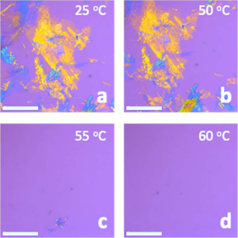
Preprints (www.preprints.org) | NOT PEER-REVIEWED | Posted: 21 June 2021 doi:10.20944/preprints202106.0516.v1 the volume. This opens up new possibilities for their application, e.g. as delivery vectors, to explore the enhanced viscosity. Gelation in DES (i.e. eutectogel formation) has been reported with 1,3:2,4-dibenzyli- dene-d-sorbitol (DBS) in reline, a DES comprising choline chloride and urea [19]. The structure of the gel was nano-fibrillar in nature, with the entanglement of these fibres causing the gelation. We have recently observed the formation of feather-like fractal ag- gregates at RT in glyceline [11], a mixture of choline chloride and glycerol in the 1:2 eu- tectic molar ratio, at SDS concentrations as low as cSDS = 0.1 wt% (~ 1 CMC). At sufficient number densities (i.e. above a critical gelation concentration cCGC > 1.9 wt%), the prevalence of the dendritic fractals led to the formation a colloidal gel, which we have termed the fracto-eutectogel. However, the effect of temperature on gel phases in DES has been relatively under studied. Such knowledge is important to understanding the mechanisms of surfactant self-assembly in DES and also to the stability and manipulation of eutectogels in practical applications. We have previously noted that, at T > ~ 45 oC, self-assembly and self-organ- isation of SDS in glyceline could be significantly affected, leading to the transition from the fracto-eutectogel phase to a fluid phase [11]. However, the nanoscopic structure un- derlying such a macroscopic phase transition was not clear. It has been reported in a re- lated system that, at T > ~ 45 oC, an SDS-in-glycerol gel underwent a lamellar gel-to-fluid transition, with the SDS aggregates transformed from lamellae, via an intermediate hex- agonal phase, to cylindrical micelle solutions [18, 20]. We note that the effect of temperature on the morphology of surfactant mesophases is well reported in aqueous media [21-23]. For instance, a family of lysine-based surfac- tants formed a range of tubule structures below their Krafft points (Tk = 25 – 49 oC depend- ing on surfactant architecture); however, a tubule-to-vesicle transition was observed as the temperature was increased [24]. Of specific relevance to gelation, the transition be- tween a lamellar liquid-crystalline phase and an -gel phase was observed at T ~ 40 oC as a ternary gemini surfactant (synthesised from oleic acid), 1-tetradecanol, and water sys- tem was cooled [25]. Here, we report a detailed structural study of morphological transformations in the dendritic SDS aggregates that accompany the fracto-eutectogel-to-fluid transition in glyceline in the temperature range 25 – 70 oC. The phase was probed microscopically using polarised light microscopy (PLM) and the nanoscopic structure was studied using small- angle neutron scattering (SANS). At temperatures below the critical gelation temperature (TGC ~ 45 oC), the SANS profiles of SDS aggregates in the fracto-eutectogel could be well described by the lamellar paracrystal stack model or the mass fractal model. Above TGC, a micellar phase was formed, similar to that observed in SDS aqueous systems at room tem- perature [26, 27]. At 70 oC, spherical SDS micelles were observed at 0.6 wt% SDS concen- tration (cSDS), which transformed to cylindrical micelles as cSDS was increased to 1.2 wt%. Such varied aggregate morphologies highlight the differences in self-assembled SDS structures in glyceline, as compared with other nonaqueous solvents and with water. This demonstrates distinct molecular interactions in DES – rich in hydrogen bonds and ions – as the driving force for molecular self-assembly, which is not currently fully understood. 2. Results and Discussion 2.1 The effect of temperature on the microscopic structure of the surfactant mesophase Polarised light microscopy (PLM) revealed an anisotropic mesophase at the room temperature (RT) for samples with SDS concentrations cSDS > 1.9 wt% in glyceline, consisting of anisotropic frac- tal-like aggregates consistent with our previous observations [11], diminishing as the temperature was increased (at a rate 0.1 oC s-1) and disappearing completely at T = 50 – 55 oC (Error! Reference source not found.). This suggests the transformation of the aggregates to an isotropic structure and/or a structure smaller than the microscope resolution. Figure S1 shows the PLM images that captured the phase transition at T = 50 – 55 oC at a finer temperature increment.
Preprints (www.preprints.org) | NOT PEER-REVIEWED | Posted: 21 June 2021 doi:10.20944/preprints202106.0516.v1

 Figure 1. PLM images of 2.5 wt% SDS in glyceline at different temperatures as indicated, showing
 the disappearance of the fractal aggregates as the temperature was increased to > ~50 oC. Images
 were taken at 4 x magnification with a 530 nm first order waveplate, and all scale bars represent
 100 μm.

 2.2 Micellar nanostructure from SANS
 Figure 2a shows the SANS profiles for h-SDS (cSDS = 5.3 wt%; 40.7 CMC) in d-glyceline
 in the temperature range T = 25 – 70 oC, with the fitting parameters for the different models
 used list in Table 1-3 and modes described in section 4.4 below. Visual and microscopic
 observations confirmed that a gel (termed a fracto-eutectogel) transitioned into a fluid
 phase at T > TCG ~ 40 – 50 oC [11]. Of the micellar models trialed, a core-shell-cylinder form
 factor, P(q), was found to best fit the data above the TCG without any structure factor, S(q),
 suggesting non-interacting cylinders in solution despite the anionic nature of SDS. This
 may be rationalized by the presence of choline and chloride ions (~33 wt %; 6.84 M choline
 chloride) which may effectively screen the interactions between the aggregates.

 Table 1. Fitting parameters for the lamellar stack paracrystal model (cf. Figure 2c) used to simu-
 late the data for cSDS = 5.3 wt% h-SDS in d-glyceline in the gel phase at 25, 35, and 40 oC (cf. Figure
 2a): SDS bilayer thickness tL, number of layers in the stack nLayers, d-spacing, polydispersity of the
 d-spacing σd, scattering length density of SDS ρSDS, scattering length density of glyceline ρGly, poly-
 dispersity of the SDS bilayer thickness σt, and chi squared value χ2.

 Model Parameters 25 oC 35 oC 40 oC
 tL (Å) 20.0 20.6 20.0
 nLayers 57.7 57.8 57.8
 d-Spacing (Å) 20.4 20.4 20.8
 σd (Å) 0.013 ~0 0.067
 ρSDS (10-6 Å-2) 0.40 0.44 0.35
 ρGly (10-6 Å-2) 5.50 5.83 5.75
 σt 1.0 1.0 1.0
 χ2 3.8 2.6 3.5
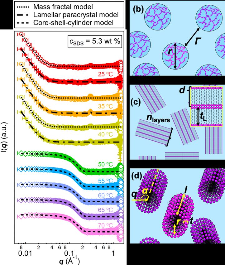
Preprints (www.preprints.org) | NOT PEER-REVIEWED | Posted: 21 June 2021 doi:10.20944/preprints202106.0516.v1 C (b) (a) b (c) 2r n layer Figure 2. (a) SANS profiles for 5.3 wt% h-SDS in d-glyceline at different temperatures (25 – 70 oC), showing the transition from the fracto-eutectogel phase to the micellar phase at a critical gelation temperature, TCG ~ 40 – 50 oC. Fits to the profiles are indicated by black lines, and different lines are used to relate to the type of model used in the fit, shown in the legend: (b) the mass fractal model where 2r = D is the building block diameter and the correlation length; (c) the lamellar stack paracrystal model with randomly oriented lamellar domains consisting of n layers of bi- layers of thickness tL, and (d) the core-shell cylinder model with cylinders of core radius r, shell thickness t, and length l. These models are described in section 4.4 below. The SANS profiles in (a) are offset on the vertical scale for clarity. Table 2. Fitting parameters for the mass fractal model (cf. Figure 2b) used to simulate the data for 5.3 wt % h-SDS in d-glyceline at 25, 35, and 40 oC (cf. Figure 2a): radius of the fractal aggregate r, fractal dimension Dm, and chi squared value χ2. Model Parameters 25 oC 35 oC 40 oC r (Å) 56.5 30.8 30.0 Dm 2.95 3.00 2.98 χ2 3.4 3.1 3.7 Table 3. Fitting parameters for the core-shell cylinder model (cf. Figure 2d) used to simulate the data for 5.3 wt% h-SDS in d-glyceline in the fluid phase at 50, 55, 60, 65, and 70 oC (cf. Figure 2a): core radius r, shell thickness t, cylinder length l, scattering length density of the core ρcore, scatter- ing length density of the shell ρshell, scattering length density of glyceline ρGly, polydispersity of the core radius σr, polydispersity of the shell thickness σt, polydispersity of the cylinder length σl, and chi squared value χ2. Model Parameters 50 oC 55 oC 60 oC 65 oC 70 oC r (Å) 17.5 17.1 16.8 16.3 16.3 t (Å) 5.0 5.0 5.0 4.8 4.8 l (Å) 26.4 25.7 25.1 23.9 23.9 ρcore (10-6 Å-2) -0.31 -0.42 -0.40 -0.38 -0.38 ρshell (10-6 Å-2) 4.98 5.03 4.95 4.80 4.80 ρGly (10-6 Å-2) 5.79 5.82 5.83 5.87 5.87 σr 0.05 0.05 0.05 0.05 0.05 σt 0.05 0.05 0.05 0.05 0.05 σl 0.05 0.05 0.05 0.05 0.05
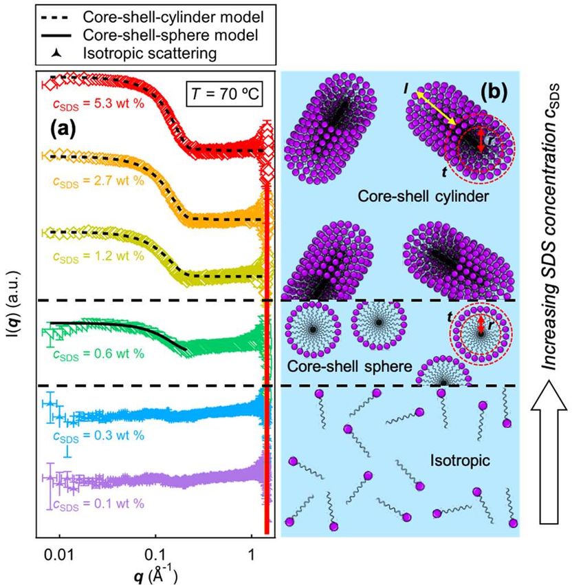
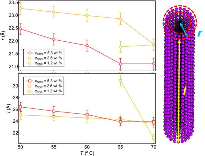
Preprints (www.preprints.org) | NOT PEER-REVIEWED | Posted: 21 June 2021 doi:10.20944/preprints202106.0516.v1

 χ2 1.87 1.88 1.82 2.28 2.45

 As also reported in our previous work [11], ordered surfactant mesophases were pre-
 sent at T < ~40-45 oC for samples at cSDS > 1.2 wt% evident from a Bragg peak at q ~ 0.30 Å
 in the SANS profile (Figure 2a). The profiles could be fitted to a lamellar paracrystal model
 with a lamellar spacing d ~ 2π/q ~ 20.9 Å. The Bragg peaks were indicative of the presence
 of the domains of solvated SDS lamellar crystals constituting the fractal dendrites[11].
 Figure 3a shows the SANS profiles at different SDS concentrations, cSDS, at a higher
 temperature (T = 70 oC). Fitting the data to various models shows a transition in the form
 factor, F(q), from isotropic scattering (cSDS = 0.1 wt% and 0.3 wt%), to a core-shell sphere
 model (cSDS = 0.6 wt %, apparent CMC at 70 oC), and then to a core-shell cylinder model
 (cSDS > 1.2 wt%) as cSDS was increased. The fitting parameters are listed in Table 4 and 5.
 This contrasts with the observation of fractal aggregates for all cSDS at T = 25 oC, where a
 critical fractal aggregate concentration (cCFC) can be defined, below which no fractal ag-
 gregates were observed. The observations of cCFC = 0.1 wt% at 25 oC and the CMC = 0.6
 wt% at 70 oC point to the different self-assembly behaviors at the RT and the elevated
 temperature. The formation of crystallites was observed at cCFC = 0.1 wt%, however, the
 CMC at 70 oC, CMC70 = 0.6 wt%. This can be rationalized by considering an effective Krafft
 temperature of SDS in glyceline, below which crystallization is favored and thus the for-
 mation of the fractal aggregates with solvated lamellae; and above TK in the fluid phase,
 SDS self-assembly dominates.
 The dimension of the cylindrical micelles varied as a function of both T and cSDS (Fig-
 ure 4) where for all cSDS both the radius, r, and length, l, of the cylinders decreased with
 increasing temperature. The length of cylindrical aggregates decreasing with increasing T
 has been observed in other systems, attributed to the greater thermal energy (kbT) which
 would encourage breakup of longer aggregates [28]. The decreasing cylinder radius, r,
 with increasing T has also been observed previously and is explained by the increased
 motions and fluidity of the hydrophobic chains at higher T, reducing steric effects and
 allowing for better packing [29]. The radius of the cylindrical micelles increased with de-
 creasing concentration in agreement with theoretical descriptions, where the micellar ra-
 dius would initially increase with concentration, but then decrease due to the increasing
 number of micelles [30]. The cylinder length here increased with cSDS, due to the increased
 number of SDS molecules incorporated in individual micelles at high surfactant concen-
 trations.
Preprints (www.preprints.org) | NOT PEER-REVIEWED | Posted: 21 June 2021 doi:10.20944/preprints202106.0516.v1

 Figure 3. (a) SANS profiles for h-SDS in d-glyceline at varying surfactant concentrations, cSDS, at T =
 70 oC. Also shown are fits to different models used (cf. Figure 3b). The profiles are offset on the
 vertical scale for clarity. (b) Schematic representation of the models used as the SDS concentration
 cSDS increased: the core-shell cylinder model (top) the core-shell sphere model (middle), and iso-
 tropic scattering (bottom).

 Table 4. Fitting parameters for the core-shell cylinder model used to simulate the data for 1.2, 2.7,
 and 5.3 wt% h-SDS in d-glyceline at 70 oC (cf. Figure 3a): core radius r, shell thickness t, cylinder
 length l, scattering length density of the core ρcore, scattering length density of the shell ρshell, scat-
 tering length density of glyceline ρGly, polydispersity of the core radius σr, polydispersity of the
 shell thickness σt, polydispersity of the cylinder length σl, and chi squared value χ2.

 Model Parameters 1.2 wt % 2.7 wt % 5.3 wt %
 r (Å) 16.8 16.9 16.3
 t (Å) 5.0 5.0 4.8
 l (Å) 23.9 23.6 21.1
 ρcore (10-6 Å-2) -0.38 -0.34 -0.39
 ρshell (10-6 Å-2) 4.80 4.80 4.80
 ρGly (10-6 Å-2) 5.87 5.86 5.88
 σr 0.05 0.05 0.05
 σt 0.05 0.05 0.05
 σl 0.05 0.05 0.05
 χ2 2.45 2.07 4.31

 Table 5. Fitting parameters for the core-shell sphere model used to simulate the data for 0.6 wt%
 h-SDS in d-glyceline at 70 oC (cf. Figure 3a): core radius r, shell thickness t, scattering length den-
 sity of the core ρcore, scattering length density of the shell ρshell, scattering length density of
 glyceline ρGly, polydispersity of the core radius σr, polydispersity of the shell thickness σt, and chi
 squared value χ2.

 Model Parameters 0.6 wt %
 r (Å) 16.4
 t (Å) 5.0
 ρcore (10-6 Å-2) -0.30
 ρshell (10-6 Å-2) 5.12
Preprints (www.preprints.org) | NOT PEER-REVIEWED | Posted: 21 June 2021 doi:10.20944/preprints202106.0516.v1

 ρGly (10-6 Å-2) 5.74
 σr 0.05
 σt 0.05
 χ2 1.35

 Figure 4. The effect of temperature, T, and cSDS on the radius, r, (top) and length, l, (bottom) of
 cylindrical aggregates formed in the glyceline bulk taken from fitting to SANS profiles, for 5.3 wt
 % (red), 2.7 wt % (orange), and 1.2 wt % (yellow) wt % SDS in glyceline. Schematic representation
 of the core-shell cylinder model shown (right).

 3. Further Discussions and Concluding Remarks
 We have probed the morphological changes that occurred upon the SDS-in-glyceline
 (a DES) fracto-eutectogel-to-fluid transition. This transition manifested in the disappear-
 ance of fractal dendrites as observed with PLM, and the change in the SANS profiles. The
 SANS profiles at the room temperature were fitted to a mass fractal model consistent with
 the dendritic aggregates observed microscopically with PLM. As discussed in [11], rheo-
 logical measurements in this system confirmed a fracto-eutectogel consisting of globular
 aggregates in solution, akin to a physical colloidal gel with interpenetrating dendritic frac-
 tal aggregates. Here we show that above the critical gelation temperature, TGC ~ 45 oC, a
 gel-to-fluid transition was observed, which was characterized nano-structurally with
 SANS. The fluid phase was shown to comprise a globular micellar fluid phase, where the
 observed morphology was concentration dependent, evident from the SANS fitting anal-
 yses.
 SANS analyses also revealed morphological transformations of SDS micelles in the
 fluid phase at T ~ 70 oC. Whilst isotropic scattering was observed below an apparent CMC,
 cSDS < 0.6 wt%, spherical micelles were observed at cSDS ~ 0.6 wt %, and cylindrical micelles
 at cSDS > 0.6 wt%. Morphological transformations of micelles have also been reported in
 aqueous SDS systems due to the addition of salt, rather than change in the SDS concen-
 tration. For instance, spherical or ellipsoidal micelles [26, 27, 31] have been observed at RT
 at similar cSDS (cSDS ~ 6.0 wt%), although micelles persisted through to very high SDS con-
 centrations – up to 40 wt% at 70 oC in water). Furthermore, upon addition of sodium chlo-
 ride (csalt = 1 – 2 M), flexible wormlike SDS micelles are observed in water [32-34]. The
 effect of salt addition on the SDS micelle shape in water could be well explained by the
 packing parameter: screening of the electrostatic headgroup repulsion would reduce the
 effective headgroup area, leading to a larger packing parameter (CPP = V0 / ael) and aggre-
 gates of a smaller curvature, accounting for the sphere-to-cylinder transition.
Preprints (www.preprints.org) | NOT PEER-REVIEWED | Posted: 21 June 2021 doi:10.20944/preprints202106.0516.v1

 This highlights the differences of self-assembly in DES, where the formation of cylin-
 drical aggregates without additional salt was observed in SDS self-assembly. This transi-
 tion from gel to a fluid phase has been reported in an SDS-in-glycerol gel [18]. However,
 the lack of structure factor, S(q), in the glyceline system (cf. Figure 3a), differs from that
 observed in glycerol [18] and also in DES systems as previously reported [14, 35]. Previous
 modelling of SANS profiles of ionic surfactants in reline and glyceline have included a
 hard sphere structure factor, S(q) [15, 35], pointing to interacting globular aggregates. The
 absence of an S(q) here suggests that the globular aggregates were non-interacting, which
 is atypical for micelles consisting of ionic surfactants. The effective concentration of cho-
 line chloride in glyceline is 6.84 M, with a corresponding Debye length κ-1 ~ 0.086 nm. This
 suggests that the presence of choline and chloride ions in the glyceline bulk screens the
 micellar aggregates from each other very effectively. It has also been suggested that the
 choline ions could actively participate in the surfactant crystallization in forming the frac-
 tal aggregates at RT [11], encouraging dissociation of choline chloride.
 The differences between the observations in glyceline and in glycerol [18] indicate
 that the self-assembly behavior is significantly influenced by the presence of choline chlo-
 ride in glyceline which dissociates and screens interactions between SDS and between
 aggregates. Our previous results of SDS in glycerol showed a microfibrillar gel phase with
 lamellar nanostructure [18] at the room temperature, transitioning to a fluid phase con-
 sisting of cylindrical aggregates interacting via a Coulombic pair potential. The elongation
 of the SDS micelles observed with increasing cSDS in glyceline suggests that either the head-
 group area decreases or the tail volume increases, to account for the reduction in curvature
 observed. Another explanation is related to the increasing number density of SDS micelles
 with increasing cSDS. For anionic SDS micelles, this would result in pockets of negative
 charges within the bulk medium, which act to screen interactions among SDS micelles.
 The positive choline ions would associate with the negative micelles with the ability to
 bridge between micelles [11]. The length of the cylinders, relatively short compared to
 SDS-in-water cylindrical aggregates (wormlike micelles, l > 1000 Å [36, 37]), is consistent
 with the choline ions bridging and linking 2 – 3 spherical micelles in forming cylindrical
 micelles.
 Our results here, in corroboration with our previous observations in glycerol [18] and
 in glyceline at RT [11], illustrate the self-assembly of surfactants in polar-nonaqueous po-
 lar DES media is complex. We are currently extending the work to investigate self-assem-
 bly of cationic surfactants and lipids in DES.

 4. Materials and Methods
 4.1 Materials
 h-Sodium dodecyl sulfate (SDS) (Sigma-Aldrich, > 98.0 %) was recrystallized three
 times from ethanol prior to use, and its purity was checked with 1H NMR. h-Glycerol
 (Fisher Scientific, > 98.0 %) and d-Glycerol (Sigma-Aldrich, > 98.0 % and > 98.0 atom % D)
 were used as received. h-Choline chloride (Sigma-Aldrich, > 98.0 %) and d9-trimethyl-cho-
 line chloride (Sigma-Aldrich, > 98.0 % and > 98.0 atom % D) were used as received. All
 glyceline-containing (both hydrogenated (h-) and deuterated (d-)) phases and controls were
 kept sealed from moisture. h-/d-Glyceline was prepared by mixing h-/d-choline chloride
 and h-/d-glycerol in a 1:2 molar ratio and leaving in a shaker incubator (Stuart SI505) for
 two hours (2 h) at 550 RPM at 60 oC until homogenous. The gel-like phase was prepared
 by adding a designated amount of h-SDS to h- or d-glyceline, then incubating the mixture
 in the shaker incubator for at 550 RPM at 60 oC for 2 h before equilibrating at room tem-
 perature overnight.
 4.2 Polarised light microscopy (PLM)
 PLM was carried out using an Olympus BX53-P microscope and a Nikon Eclipse E200
 microscope, where the polarizers were crossed at 90o with respect to each other and im-
 ages were captured using Stream and PixeLINK® Capture OEM software, respectively. 4,
Preprints (www.preprints.org) | NOT PEER-REVIEWED | Posted: 21 June 2021 doi:10.20944/preprints202106.0516.v1

 10, 20, and 40 x magnifications were used, and heating was facilitated by a Linkam stage.
 A 530 nm first order waveplate was placed into the optical patch to improve the clarity of
 image details in some cases.
 4.3 Small-angle neutron scattering (SANS)
 SANS data was obtained from samples contained in quartz cells with a 2 mm path
 length over a 0.5 h integration time on the LOQ small-angle diffractometer [38] at the ISIS
 Pulsed Neutron Source (STFC Rutherford Appleton Laboratory, Didcot, UK). LOQ uti-
 lizes neutrons with wavelengths λ = 2 – 10 Å and the data was collected in the q range of
 0.008−1.6 Å -1. The raw scattering data were corrected for the detector efficiency, sample
 transmission, and background scattering and converted to scattering cross-section data
 (∂Σ/∂Ω vs q) using MantidPlot [39]. The data were then converted to an absolute scale (cm-
 1) using the scattering intensity from a standard sample (a solid blend of hydrogenous and

 perdeuterated polystyrene) in accordance with established procedures [40].
 4.4 SANS Data Analysis
 The SANS data at 25 oC was fitted using two different models. The first was a lamellar
 paracrystal stack model (cf. Figure 2c), in which individual lamellae stacks in solution were
 treated as being independent of each other, with the SDS layer considered as a whole ra-
 ther than separate headgroup layers and a tail layer [41-44]. The general scattering inten-
 sity for lamellar systems is described as

 2 ( ) ( )
 ( ) = ( )

 where V is the scattering volume, P(q) the form factor that describes the shape of the par-
 ticles or the phase present, S(q) the structure factor that describes the interparticle inter-
 action, and d the lamellar spacing.
 The second model used to fit data at 25 oC was a mass fractal model (cf. Figure 2b)
 describing the scattering from fractal-like aggregates in solution [45]

 ( ) = ( ) ( ) ( )
 where the form factor is

 ( ) = ( ) ( )

 and the structure factor is

 Γ( − 1) sin[( − 1) tan ( )]
 ( ) = ( )
 ( )

 [1 + ( ) ]

 where r is the radius of the building block, ζ the correlation length, and Dm the mass fractal
 dimension.
 The SANS data at 70 oC was fitted using two different form factors, P(q), in SasView,
 in a core-shell-cylinder model (cf. Figure 3b) and a core-shell sphere model (cf. Figure 3b), re-
 spectively. The general scattering intensity for a core-shell-cylinder is [46-48]

 1
 ( ) = ( , ) ∙ sin ( )

 where
Preprints (www.preprints.org) | NOT PEER-REVIEWED | Posted: 21 June 2021 doi:10.20944/preprints202106.0516.v1

 1
 sin cos 2 ( sin )
 ( , ) = ( − ) 2 +
 1 sin
 cos
 2 ( )
 1
 sin + cos 2 ( ( + ) sin )
 ( − ) 2
 1 ( + ) sin
 + cos
 2

 and

 = ( + ) ( + 2 ) ( )

 Here, α is the angle between q and the cylinder axis, Vs is the total volume, Vc is the core
 volume, l is the core length, r is the core radius, t is the shell thickness, ρc is the core SLD,
 ρs is the shell SLD, ρsolv is the solvent SLD, and J1 is the first order Bessel function.
 The general scattering intensity for a core-shell-sphere model is [46]

 ( )
 ( ) = ( )

 where

 3 sin( ) − cos( ) sin( ) − cos( )
 ( ) = ( − ) + ( − ) ( )
 ( ) ( )

 where Vs is the total scattering volume of the object, Vc the scattering volume of the core,
 ρc the scattering length density (SLD) of the core, ρs the SLD of the shell, ρsolv the SLD of
 the solvent, rc the radius of the core, and rs the sum of the radius of the core and the thick-
 ness of the shell.

 Supplementary Materials: The following are available online at www.mdpi.com/xxx/s1; Figure S1:
 PLM images of 2.5 wt% SDS in glyceline between T = 50 – 55 oC at a finer temperature increment
 step; Figure S2: PLM images of 2.5 wt% SDS in glyceline at different temperatures, showing the
 disappearance of the fractal aggregates with elevated temperature; Figure S3: Fitted SANS data for
 5.3 wt % SDS in glyceline at 70 oC using different models; Figure S4: Refined fitted SANS data for
 5.3 wt % SDS in glyceline at 70 oC using a spherical and cylindrical model; Figure S5: Refined fitted
 SANS data for 0.6 wt % SDS in glyceline at 70 oC using a spherical and cylindrical model; Figure S6:
 Fitted SANS profile for 0.6 wt % SDS in glyceline at 70 oC using a spherical and cylindrical model,
 and a Hayter-MSA structure factor; Figure S7: SANS profiles for 0.1 and 0.3 wt % h-SDS in d-
 glyceline at T = 343 K with attempted fits shown by solid black lines; Figure S8: SANS profiles for
 2.7 wt % h-SDS in d-glyceline at different temperatures; Figure S9: SANS profiles for 1.2 wt % h-SDS
 in d-glyceline at different temperatures; Figure S10: SANS profiles for 0.6 wt % h-SDS in d-glyceline
 at different temperatures; Figure S11: SANS profiles for 0.3 wt % h-SDS in d-glyceline at different
 temperatures; Figure S18: SANS profiles for 0.1 wt % h-SDS in d-glyceline at different temperatures.
 Table S1: Fitting parameters for the sphere model used to simulate the data for 5.3 wt % h-SDS in d-
 glyceline at 70 oC; Table S2: Fitting parameters for the ellipsoid model used to simulate the data for
 5.3 wt % h-SDS in d-glyceline at 70 oC; Table S3: Fitting parameters for the cylinder model used to
 simulate the data for 5.3 wt % h-SDS in d-glyceline at 70 oC; Table S4: Fitting parameters for the
 parallepiped model used to simulate the data for 5.3 wt % h-SDS in d-glyceline at 70 oC; Table S5:
 Fitting parameters for the core-shell-sphere model used to simulate the data for 5.3 wt % h-SDS in
 d-glyceline at 70 oC; Table S6: Fitting parameters for the core-shell-cylinder model used to simulate
 the data for 5.3 wt % h-SDS in d-glyceline at 70 oC; Table S7: Fitting parameters for the core-shell-
 sphere model used to simulate the data for 0.6 wt % h-SDS in d-glyceline at 70 oC; Table S8: Fitting
 parameters for the core-shell-cylinder model used to simulate the data for 0.6 wt % h-SDS in d-
Preprints (www.preprints.org) | NOT PEER-REVIEWED | Posted: 21 June 2021 doi:10.20944/preprints202106.0516.v1

 glyceline at 70 oC; Table S9: Fitting parameters for the core-shell-sphere and core-shell-cylinder
 model with Hayter-MSA S(q) used to simulate the data for 0.6 wt % h-SDS in d-glyceline at 70 oC;
 Table S10: Fitting parameters for the paracrystalline lamellar stack model used to simulate the data
 for 2.7 wt % h-SDS in d-glyceline at 25, 35, and 40 oC; Table S11: Fitting parameters for the mass
 fractal model used to simulate the data for 2.7 wt % h-SDS in d-glyceline at 25, 35, and 40 oC; Table
 S12: Fitting parameters for the core-shell-cylinder model used to simulate the data for 2.7 wt % h-
 SDS in d-glyceline at 50, 55, 60, 65, and 70 oC; Table S13: Fitting parameters for the paracrystalline
 lamellar stack model used to simulate the data for 1.2 wt % h-SDS in d-glyceline at 25, and 40 oC,
 Table S14: Fitting parameters for the mass fractal model used to simulate the data for 1.2 wt % h-
 SDS in d-glyceline at 25, and 40 oC; Table S15: Fitting parameters for the core-shell-cylinder model
 used to simulate the data for 1.2 wt % h-SDS in d-glyceline at 65, and 70 oC; Table S16: Fitting pa-
 rameters for the mass fractal model used to simulate the data for 0.6 wt % h-SDS in d-glyceline at 25,
 and 40 oC; Table S17: Fitting parameters for the core-shell-sphere model used to simulate the data
 for 0.6 wt % h-SDS in d-glyceline at 65, and 70 oC; Table S18: Fitting parameters for the mass fractal
 model used to simulate the data for 0.3 wt % h-SDS in d-glyceline at 25, and 40 oC; Table S19: Fitting
 parameters for the mass fractal model used to simulate the data for 0.3 wt % h-SDS in d-glyceline at
 25 oC.
 Author Contributions: Conceptualization, P.B., R.S. and W. H. B.; methodology, L. M., S. E. R., and
 W. H. B.; validation, L. M., and W. H. B.; formal analysis, L. M., and W. H. B.; investigation, L. M.,
 S. E. S. M., and S. E. R.; resources, A. J. J., R. S., and W. H. B.; data curation, L. M., and S. E. S. M.;
 writing—original draft preparation, L. M.; writing—review and editing, W. H. B.; visualization, L.
 M.; supervision, P. B., A. J. J., R. S., and W. H. B.; project administration, A. J. J., R. S., and W. H. B.;
 funding acquisition, P. B., R. S., and W. H. B.
 Funding: L.M. was supported by a studentship jointly funded through the Engineering and Physi-
 cal Science Research Council (the Bristol Centre for Functional Nanomaterials (EPSRC,
 EP/L016648/1)) and GlaxoSmithKline.
 Data Availability Statement: SANS data is available online at https://data.isis.stfc.ac.uk/ under the
 DOI: 10.5286/ISIS.E.RB1810629.
 Acknowledgments: We acknowledge the ISIS Muon and Neutron Source for the awarded beam-
 time under experiment number: RB1810629. We thank Z ̇. Przybyłowicz, and S. Ruscigno for their
 help with the SANS experiments. SasView, originally developed under NSF award DMR-0520547,
 contains code developed with funding from the European Union’s Horizon 2020 research and in-
 novation programme under the SINE2020 project, grant agreement No 654000.
 Conflicts of Interest: The authors declare no conflicts of interest.

 References

 [1] A.P. Abbott, G. Capper, D.L. Davies, H.L. Munro, R.K. Rasheed, V. Tambyrajah, Preparation of novel, moisture-stable, Lewis-
 acidic ionic liquids containing quaternary ammonium salts with functional side chains, Chem Commun (19) (2001) 2010-2011.
 [2] A.P. Abbott, E.I. Ahmed, K. Prasad, I.B. Qader, K.S. Ryder, Liquid pharmaceuticals formulation by eutectic formation, Fluid Phase
 Equilibr 448 (2017) 2-8.
 [3] A.P. Abbott, J.C. Barron, K.S. Ryder, D. Wilson, Eutectic-Based Ionic Liquids with Metal-Containing Anions and Cations,
 Chemistry – A European Journal 13(22) (2007) 6495-6501.
 [4] E.L. Smith, A.P. Abbott, K.S. Ryder, Deep Eutectic Solvents (DESs) and Their Applications, Chem Rev 114(21) (2014) 11060-11082.
 [5] R.D. Rogers, K.R. Seddon, Ionic liquids - Solvents of the future?, Science 302(5646) (2003) 792-793.
 [6] K.R. Seddon, Ionic liquids - A taste of the future, Nat Mater 2(6) (2003) 363-365.
 [7] A.P. Abbott, G. Capper, K.J. McKenzie, K.S. Ryder, Electrodeposition of zinc-tin alloys from deep eutectic solvents based on
 choline chloride, J Electroanal Chem 599(2) (2007) 288-294.
 [8] A.P. Abbott, G. Capper, B.G. Swain, D.A. Wheeler, Electropolishing of stainless steel in an ionic liquid, T I Met Finish 83(1) (2005)
 51-53.
Preprints (www.preprints.org) | NOT PEER-REVIEWED | Posted: 21 June 2021 doi:10.20944/preprints202106.0516.v1

 [9] A.P. Abbott, K. El Ttaib, G. Frisch, K.J. McKenzie, K.S. Ryder, Electrodeposition of copper composites from deep eutectic solvents
 based on choline chloride, Phys Chem Chem Phys 11(21) (2009) 4269-4277.
 [10] A.P. Abbott, K. El Ttaib, G. Frisch, K.S. Ryder, D. Weston, The electrodeposition of silver composites using deep eutectic solvents,
 Phys Chem Chem Phys 14(7) (2012) 2443-2449.
 [11] L. Matthews, S. Ruscigno, S.E. Rogers, P. Bartlett, A.J. Johnson, R. Sochon, W.H. Briscoe, Fracto-eutectogels: SDS fractal dendrites
 via counterion condensation in a deep eutectic solvent, Phys Chem Chem Phys 23(20) (2021) 11672-11683.
 [12] J. Gorke, F. Srienc, R. Kazlauskas, Toward Advanced Ionic Liquids. Polar, Enzyme-friendly Solvents for Biocatalysis, Biotechnol
 Bioproc E 15(1) (2010) 40-53.
 [13] J. Gorke, F. Srienc, R.J. Kazlauskas, ORGN 299-Deep eutectic solvents: Alternative reaction media for hydrolase-catalyzed
 reactions, Abstr Pap Am Chem S 236 (2008).
 [14] T. Arnold, A.J. Jackson, A. Sanchez-Fernandez, D. Magnone, A.E. Terry, K.J. Edler, Surfactant Behavior of Sodium Dodecylsulfate
 in Deep Eutectic Solvent Choline Chloride/Urea, Langmuir 31(47) (2015) 12894-12902.
 [15] A. Sanchez-Fernandez, T. Arnold, A.J. Jackson, S.L. Fussell, R.K. Heenan, R.A. Campbell, K.J. Edler, Micellization of
 alkyltrimethylammonium bromide surfactants in choline chloride: glycerol deep eutectic solvent, Phys Chem Chem Phys 18(48)
 (2016) 33240-33249.
 [16] A. Sanchez-Fernandez, O.S. Hammond, A.J. Jackson, T. Arnold, J. Doutch, K.J. Edler, Surfactant-Solvent Interaction Effects on
 the Micellization of Cationic Surfactants in a Carboxylic Acid-Based Deep Eutectic Solvent, Langmuir 33(50) (2017) 14304-14314.
 [17] A. Sanchez-Fernandez, G.L. Moody, L.C. Murfin, T. Arnold, A.J. Jackson, S.M. King, S.E. Lewis, K.J. Edler, Self-assembly and
 surface behaviour of pure and mixed zwitterionic amphiphiles in a deep eutectic solvent, Soft Matter 14(26) (2018) 5525-5536.
 [18] L. Matthews, Ż. Przybyłowicz, S.E. Rogers, P. Bartlett, A.J. Johnson, R. Sochon, W.H. Briscoe, The curious case of SDS self-
 assembly in glycerol: Formation of a lamellar gel, J Colloid Interf Sci 572 (2020) 384-395.
 [19] J. Ruiz-Olles, P. Slavik, N.K. Whitelaw, D.K. Smith, Self-Assembled Gels Formed in Deep Eutectic Solvents: Supramolecular
 Eutectogels with High Ionic Conductivity, Angew Chem Int Edit 58(13) (2019) 4173-4178.
 [20] L. Matthews, M.C. Stevens, R. Schweins, P. Bartlett, A.J. Johnson, R. Sochon, W.H. Briscoe, Unexpected observation of an
 intermediate hexagonal phase upon fluid-to-gel transition: SDS self-assembly in glycerol, Colloid and Interface Science
 Communications 40 (2021) 100342.
 [21] C. Han, Y. Guo, X. Chen, M. Yao, Y. Zhang, Q. Zhang, X. Wei, Phase behaviour and temperature-responsive properties of a
 gemini surfactant/Brij-30/water system, Soft Matter 13(6) (2017) 1171-1181.
 [22] M.N. Islam, T. Kato, Temperature Dependence of the Surface Phase Behavior and Micelle Formation of Some Nonionic
 Surfactants, The Journal of Physical Chemistry B 107(4) (2003) 965-971.
 [23] Y. Zhang, X. Chen, X. Liu, Temperature-Induced Reversible-Phase Transition in a Surfactant-Free Microemulsion, Langmuir
 35(44) (2019) 14358-14363.
 [24] I.S. Oliveira, M. Lo, M.J. Araújo, E.F. Marques, Temperature-responsive self-assembled nanostructures from lysine-based
 surfactants with high chain length asymmetry: from tubules and helical ribbons to micelles and vesicles, Soft Matter 15(18) (2019)
 3700-3711.
 [25] T. Sugahara, M. Akamatsu, H. Iwase, Y. Takamatsu, K. Sakai, H. Sakai, Structural Change of an α-Gel (α-Form Hydrated Crystal)
 Induced by Temperature and Shear Flow in an Oleic Acid Based Gemini Surfactant System, Langmuir 36(17) (2020) 4695-4701.
 [26] P. Kekicheff, Phase-Diagram of Sodium Dodecyl-Sulfate Water-System .2. Complementary Isoplethal and Isothermal Phase
 Studies, J Colloid Interf Sci 131(1) (1989) 133-152.
 [27] P. Kekicheff, B. Cabane, M. Rawiso, Macromolecules Dissolved in a Lamellar Lyotropic Mesophase, J Colloid Interf Sci 102(1)
 (1984) 51-70.
 [28] L. Sepulveda, C. Gamboa, Effect of Temperature on the Viscosity of Cationic Micellar Solutions in the Presence of Added Salts,
 J Colloid Interf Sci 118(1) (1987) 87-90.
Preprints (www.preprints.org) | NOT PEER-REVIEWED | Posted: 21 June 2021 doi:10.20944/preprints202106.0516.v1

 [29] T.L. Lin, M.Y. Tseng, S.H. Chen, M.F. Roberts, Temperature-Dependence of the Growth of Diheptanoylphosphatidylcholine
 Micelles Studied by Small-Angle Neutron-Scattering, J Phys Chem-Us 94(18) (1990) 7239-7243.
 [30] L.M. Bergström, Explaining the growth behavior of surfactant micelles, J Colloid Interf Sci 440 (2015) 109-118.
 [31] P. Kekicheff, C. Grabiellemadelmont, M. Ollivon, Phase-Diagram of Sodium Dodecyl-Sulfate Water-System .1. A Calorimetric
 Study, J Colloid Interf Sci 131(1) (1989) 112-132.
 [32] L.J. Magid, Z. Li, P.D. Butler, Flexibility of Elongated Sodium Dodecyl Sulfate Micelles in Aqueous Sodium Chloride: A Small-
 Angle Neutron Scattering Study, Langmuir 16(26) (2000) 10028-10036.
 [33] M. Sammalkorpi, M. Karttunen, M. Haataja, Ionic Surfactant Aggregates in Saline Solutions: Sodium Dodecyl Sulfate (SDS) in
 the Presence of Excess Sodium Chloride (NaCl) or Calcium Chloride (CaCl2), The Journal of Physical Chemistry B 113(17) (2009)
 5863-5870.
 [34] Z. Chu, C.A. Dreiss, Y. Feng, Smart wormlike micelles, Chem Soc Rev 42(17) (2013) 7174-7203.
 [35] A. Sanchez-Fernandez, K.J. Edler, T. Arnold, R.K. Heenan, L. Porcar, N.J. Terrill, A.E. Terry, A.J. Jackson, Micelle structure in a
 deep eutectic solvent: a small-angle scattering study, Phys Chem Chem Phys 18(20) (2016) 14063-73.
 [36] M.A. da Silva, V. Calabrese, J. Schmitt, K.M.Z. Hossain, S.J. Bryant, N. Mahmoudi, J.L. Scott, K.J. Edler, Impact of wormlike
 micelles on nano and macroscopic structure of TEMPO-oxidized cellulose nanofibril hydrogels, Soft Matter 16(20) (2020) 4887-4896.
 [37] G.V. Jensen, R. Lund, J. Gummel, T. Narayanan, J.S. Pedersen, Monitoring the Transition from Spherical to Polymer-like
 Surfactant Micelles Using Small-Angle X-Ray Scattering, Angewandte Chemie International Edition 53(43) (2014) 11524-11528.
 [38] R.K. Heenan, J. Penfold, S.M. King, SANS at pulsed neutron sources: Present and future prospects, J Appl Crystallogr 30 (1997)
 1140-1147.
 [39] O. Arnold, J.C. Bilheux, J.M. Borreguero, A. Buts, S.I. Campbell, L. Chapon, M. Doucet, N. Draper, R. Ferraz Leal, M.A. Gigg,
 V.E. Lynch, A. Markvardsen, D.J. Mikkelson, R.L. Mikkelson, R. Miller, K. Palmen, P. Parker, G. Passos, T.G. Perring, P.F. Peterson,
 S. Ren, M.A. Reuter, A.T. Savici, J.W. Taylor, R.J. Taylor, R. Tolchenov, W. Zhou, J. Zikovsky, Mantid—Data analysis and
 visualization package for neutron scattering and μ SR experiments, Nuclear Instruments and Methods in Physics Research Section
 A: Accelerators, Spectrometers, Detectors and Associated Equipment 764 (2014) 156-166.
 [40] G.D. Wignall, F.S. Bates, Absolute calibration of small-angle neutron scattering data, J Appl Crystallogr 20(1) (1987) 28-40.
 [41] J. Berghausen, J. Zipfel, P. Lindner, W. Richtering, Influence of water-soluble polymers on the shear-induced structure formation
 in lyotropic lamellar phases, J Phys Chem B 105(45) (2001) 11081-11088.
 [42] M. Bergstrom, J.S. Pedersen, P. Schurtenberger, S.U. Egelhaaf, Small-angle neutron scattering (SANS) study of vesicles and
 lamellar sheets formed from mixtures of an anionic and a cationic surfactant, J Phys Chem B 103(45) (1999) 9888-9897.
 [43] A. Caille, X-Ray Scattering by Smectic-a Crystals, Cr Acad Sci B Phys 274(14) (1972) 891-893.
 [44] F. Nallet, R. Laversanne, D. Roux, Modeling X-Ray or Neutron-Scattering Spectra of Lyotropic Lamellar Phases - Interplay
 between Form and Structure Factors, J Phys Ii 3(4) (1993) 487-502.
 [45] D.F.R. Mildner, P.L. Hall, Small-Angle Scattering from Porous Solids with Fractal Geometry, J Phys D Appl Phys 19(8) (1986)
 1535-1545.
 [46] A. Guinier, G. Fournet, Small-Angle Scattering of X-Rays, John Wiley & Sons, Inc., New York, US, 1955.
 [47] S.R. Kline, Reduction and analysis of SANS and USANS data using IGOR Pro, J Appl Crystallogr 39 (2006) 895-900.
 [48] J.S. Pedersen, Analysis of small-angle scattering data from colloids and polymer solutions: modeling and least-squares fitting,
 Adv Colloid Interfac 70 (1997) 171-210.
You can also read