Myricetin ameliorates ox LDL induced HUVECs apoptosis and inflammation via lncRNA GAS5 upregulating the expression of miR 29a 3p - Nature
←
→
Page content transcription
If your browser does not render page correctly, please read the page content below
www.nature.com/scientificreports
OPEN Myricetin ameliorates
ox‑LDL‑induced HUVECs apoptosis
and inflammation via lncRNA
GAS5 upregulating the expression
of miR‑29a‑3p
Yunpeng Bai1,2,4, Xiankun Liu2,3,4, Qingliang Chen2, Tongyun Chen2, Nan Jiang2* &
Zhigang Guo2*
Oxidized low-density lipoprotein (ox-LDL)-induced endothelial cell dysfunction is a significant event
in the progression of atherosclerosis. Even Myricetin (Myr) has been exhibited strong antioxidant
potency, the effect on atherosclerosis is still elusive. HUVECs were subjected to ox-LDL, before which
cells were preconditioned with Myr. Cell Counting Kit-8 assay, flow cytometry, quantitative real-time
polymerase chain reaction and Western blot were carried out to assess the impacts of ox-LDL and
Myr on HUVECs. The expression of EndMT markers was determined by Western blot analysis and
immunocytochemistry. In addition, the relationship of GAS5 and miR-29a-3p was evaluated by RNA
Fluorescent in Situ Hybridization and RNA immunoprecipitation assay. Myr preconditioning prevented
ox-LDL-induced apoptosis, inflammatory response, and EndMT. GAS5 was upregulated in response to
ox-LDL while it was down-regulated by Myr preconditioning. GAS5 over-expression attenuates Myr
protective effects against ox-LDL–mediated HUVEC injury. Besides, miR-29a-3p is a target of GAS5
and down-regulated miR-29a-3p could further reduce the effects of GAS5 in ox-LDL–mediated HUVEC.
Furthermore, Myr inactivated the TLR4/NF-κB signalling pathway in ox-LDL-treated HUVEC by down-
regulating GAS5 or upregulating miR-26a-5p. Myr possessed an anti-inflammatory and anti-EndMT
function against ox-LDL-induced HUVEC injury by regulating the GAS5/miR-29a-3p, indicating that
Myr may have an important therapeutic function for atherosclerosis.
Atherosclerosis (AS) is a significant cause of morbidity and mortality among cardiovascular diseases, which is
initially triggered by endothelial dysfunction and characterized by an influx of atherogenic lipoprotein compo-
nents. It is the main potential factor of most fatal diseases, such as myocardial infarction, unstable angina, sudden
cardiac death, and stroke1,2. Although the etiology of AS is involved, the damage response theory that vascular
endothelial cell (EC) injury is the initiating mechanism of AS is still widely adopted3,4. Oxidative low-density
lipoprotein (ox-LDL) has been shown to be involved in the formation of atherosclerosis, and its induced vascular
endothelial injury is one of the early pathogenesis of a therosclerosis5,6. So far, controlling the development of
AS remains a major challenge because the molecular mechanisms of AS are not fully understood. Therefore,
reducing ox-LDL-induced EC injury and further explaining its mechanism may be a potential strategy for the
prevention and treatment of AS.
Myricetin (Myr) is a common natural flavonoid found in many fruits, vegetables, and herbs. Myr plays a sub-
stantial role in treating and preventing some diseases, including diabetic o steoporosis7, hepatocellular c arcinoma8,
9 10
alcoholic liver , and skin c ancer , due to its powerful iron-chelating capability, antioxidant and free-radical
scavenging activities. Recently, the cardioprotective role of Myr has attracted attention from the research com-
munity. Myr showed a protective effect on lipopolysaccharide (LPS) induced inflammatory myocardial i njury11,
cardiac hypertrophy12, and ischemia/reperfusion (I/R)-induced myocardial injury13. However, the effect of Myr
on lipid metabolism and atherosclerosis is not fully understood.
1
Chest hospital, Tianjin university, Tianjin 300222, China. 2Tianjin chest hospital, Tianjin medical university,
Tianjin 300222, China. 3Graduate School, Tianjin Medical University, Tianjin 300070, P. R. China. 4These authors
contributed equally: Yunpeng Bai and Xiankun Liu. *email: xkyyjn@sina.com; gzgtjch@126.com
Scientific Reports | (2021) 11:19637 | https://doi.org/10.1038/s41598-021-98916-7 1
Vol.:(0123456789)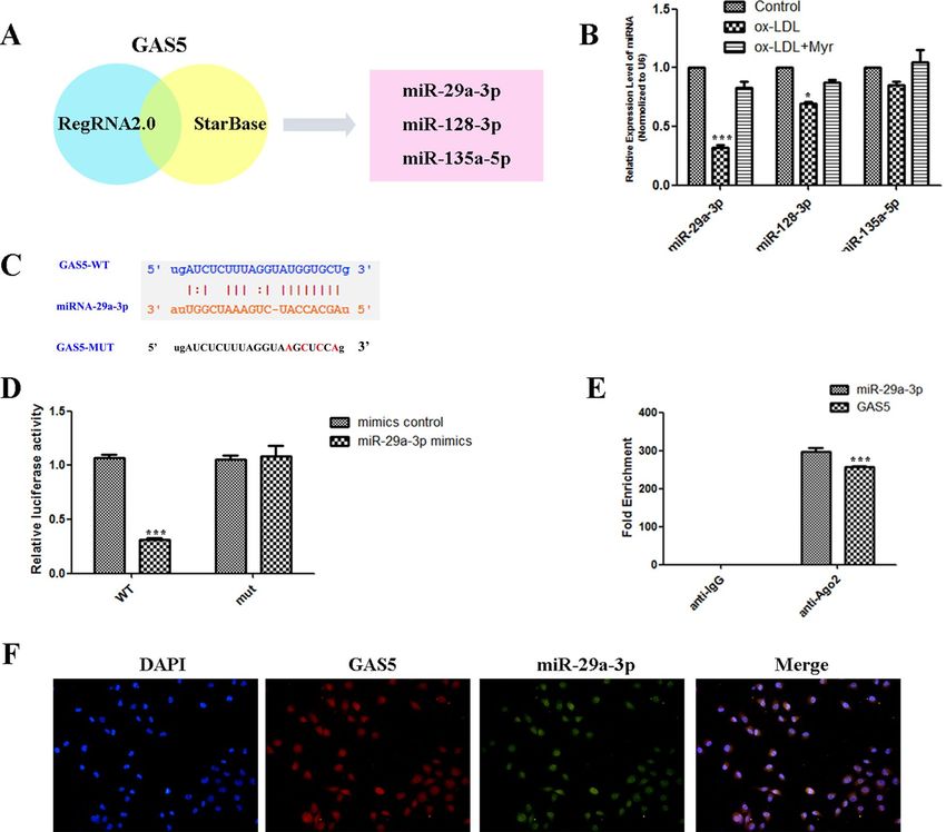
www.nature.com/scientificreports/
Long non-coding RNAs (lncRNAs) refer to a class of RNA transcripts that are more than 200 nucleotides in
length but do not encode proteins14. Increasing evidence has demonstrated that lncRNAs are involved in the onset
and development of various cardio-cerebrovascular diseases15, including hypertension, coronary artery disease,
stroke, atrial fibrillation, myocardial infarction, heart failure, and AS. Widely expressed in various tissues and
cells of humans and mice, IncRNA growth arrest-specific 5 (GAS5) has been shown to be associated with cardiac
diseases. For example, GAS5 can modulate fibroblast and fibrogenesis through TGF-β/Smad3 signalling16. Add
Wu17 et al. proved that down-regulation of GAS5 ameliorates myocardial I/R injury via the miR-335/ROCK1/
AKT/GSK-3β axis. In the present study, we aimed to explore whether GAS5 was involved in the role of Myr in
ox-LDL-induced HUVECs apoptosis and further investigated the underlying mechanism in HUVECs.
Materials and methods
Cell culture, ox‑LDL‑induced injury model, and proliferation. Human Umbilical Vein Endothelial
Cells (HUVECs) from the Cell Bank of the Chinese Academy of Sciences (Shanghai, China) were cultured in
DMEM supplemented with 10% FBS under normoxic conditions at 37 °C. The HUVECs were treated with dif-
ferent concentrations (50, 100, 200 µg/ml) of ox-LDL (Union-Bio Technology, China) and were collected at 24 h
for further measurements. According to the manufacturer’s instructions, the proliferation rates of the cells were
measured with a CCK8 assay (Solarbio, China).
Flow cytometry analysis of apoptosis with PI and annexin V double staining. Cell apoptosis
was evaluated by flow cytometry using PI and annexin V double-staining apoptosis assay kit (Solarbio, China)
following the manufacturer’s instructions.
Measurement of reactive oxygen species (ROS). The intracellular ROS was determined using the
ROS Assay Kit (Beyotime, China). Briefly, cells were incubated with 10 μM DCHF-DA for 20 min and then
fluorescence intensity was assessed under a microplate reader.
RNA immunoprecipitation (RIP) assay. RIP was performed using Magna RIP Kit (Millipore, USA)
following the manufacturer’s p rotocol18. Briefly, HUVEC cells were collected and lysed in a complete RIP lysis
buffer. The whole-cell protein extract was then incubated with RIP buffer containing magnetic beads conjugated
with human anti-AGO2 antibody or negative control normal mouse IgG. The retrieved RNA was then detected
by qRT-PCR.
RNA Fluorescent in Situ Hybridization (FISH) assay. Specific probes to the GAS5 sequence and
miR-29a-3p were designed and synthesized by RiboBio. A FISH Kit (RiboBio) was used to detect probe signals
according to the manufacturer’s instructions. Nuclei were stained with DAPI.
Dual‑luciferase reporter assay. Dual-luciferase reporter assay was used to detect the interaction between
GAS5 and miR-29a-3p promoter region. The wild-type and mutation sequences of the binding sites (WT-GAS5
and MUT-GAS5) were designed and synthesized. HUVEC cells were transfected in combination with WT (Mut)
GAS5 vector and miR-NC or miR-29a-3p using Lipofectamine 2000 Transfection Reagent (Invitrogen). After
48 h of transfection, a Dual-Luciferase reporter assay system (Promega) was used to conduct the luciferase assay.
Determination of VCAM‑1, IL‑6 and MCP‑1. The levels of supernatant monocyte chemo-attractant
protein-1 (MCP-1), interleukin-6 (IL-6), and vascular cell adhesion molecule 1 (VCAM-1) were determined by
ELISA kits. IL-6 ELISA Kit (ab178013), MCP-1 ELISA kit (ab179886), and VCAM-1 ELISA kit (ab223591) were
used according to the manufacturer’s instructions.
Real‑Time Quantified PCR (qRT‑PCR). Total RNA was isolated using Trizol reagent (Takara Bio Inc.,
Japan) according to the manufacturer’s protocol. The cDNA was generated from the total RNA (2 µg) with a
cDNA Reverse Transcription Kit (Applied Biosystems, USA), and SYBR-based real-time PCR was performed to
detect the total mRNA transcripts on an Applied Biosystems (ABI) 7300 rapid real-time PCR system. The primer
sequences were listed below.
• GAS5: forward: 5′-CTTGCCTGGACCAGCT TAAT-3′
• GAS5: reverse: 5′-CAAGCCGACTCTCCATACCT-3′
• miR-29a-3p: forward: 5′- AGCACCAUCUGAAAUCGGUUA-3′
• miR-29a-3p: reverse: 5′-GTGCAGGGTCCGAGGT-3′
• miR-128-3p: forward: 5′-AGCTAAGTAT TAGAGCGGCGGCA-3′
• miR-128-3p: reverse: 5′-GACATCAACACTCCCCTGACAAC-3′
• miR-135a-5p: forward: 5′-ACAGCCTCCATGGGAATGGAAGCAGGTTGA-3′
• miR-135a-5p: reverse: 5′-TGGAGTGTGGCGT TCG-3′
• IL-6: forward: 5′-TGGAGTCACAGAAGGAGTGGCTAAG-3′
• IL-6: reverse: 5′-TCTGACCACAGTGAGGAATGTCCAC-3′
• MCP-1: forward: 5′-TCTCTTCCTCCACCACTATGCA-3′
• MCP-1: reverse: 5′-GGCTGAGACAGCACGTGGAT-3′
• VCAM-1: forward: 5′-CTTAAAATGCCTGGGAAGATGGT-3′
• VCAM-1: reverse: 5′-GTAATGAGACGGAGTCACCAAT-3′
Scientific Reports | (2021) 11:19637 | https://doi.org/10.1038/s41598-021-98916-7 2
Vol:.(1234567890)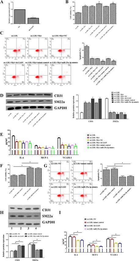
www.nature.com/scientificreports/
• GAPDH: forward: 5′-CCACATCGCTCAGACACCAT-3′
• GAPDH: reverse: 5′-CCAGGCGOCCAATACG-3′
• U6: forward: 5′-GCTTCGGCAGCACATATACTAAAT-3′
• U6: reverse: 5′-CGCT TCACGAATT TGCGTGTCAT-3′
Western blot assay. Protein expression of Bax, Bcl-2, caspase3, CD31, SM22a, p-p65, p65, p-IkBa, IkBa
and TLR4, was measured by Western blotting. Protein samples isolated from HUVEC cells (25 µg)) were
resolved using SDS-PAGE and then moved to polyvinylidene fluoride (PVDF) membrane (Millipore, USA)
with being probed with GAPDH (#2118, 1:5000, Cell Signaling), Bax (#2772S, 1:1000, Cell Signaling), Bcl-2
(#15071S, 1:1000, Cell Signaling), caspase3 (#9662S, 1:1000, Cell Signaling), CD31 (ab9498, 1:1000, Abcam),
SM22a (ab14106, 1:1000, Abcam), p-p65 (10745-1-AP, 1:1000, Proteintech), p65 (66535-1-Ig, 1:1000, Protein-
tech), p-IkBa (10268-1-AP, 1:1000, Proteintech), IkBa (66418-1-Ig, 1:1000, Proteintech) and TLR4 (66350-1-Ig,
1:1000, Proteintech) antibody followed by appropriate secondary antibody. All Western blots were repeated at
least three times. Finally, the protein expression was visualized with enhanced chemiluminescence (ECL) sys-
tem.
Immunofluorescence assay. Cells grown on slides were fixed, permeabilized, blocked, and immu-
nostained overnight with antibodies of CD31 (ab9498, 1:200, Abcam) and SM22a (ab14106, 1:200, Abcam).
After washing, specific second FITC-labeled IgG antibodies were incubated with cells continuously. Finally, the
images were obtained under a microscope (Olympus, Japan) with appropriate magnification.
Statistical analysis. All the results are shown as the means ± standard error of mean (SEM). Statistically
significant results were determined using one-way ANOVA, followed by the Student–Newman–Keuls (S–N–K)
q test. A p-value < 0.05 was considered to be significant.
Results
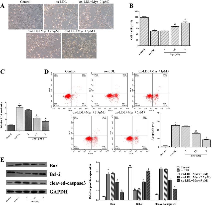
Myricetin attenuates ox‑LDL‑induced apoptosis and ROS in HUVECs. First of all, 100 ug/ml
ox-LDL incubation for 24 h led to a significant dose-dependent decrease in cell viability. Pretreatment with
1 µM Myr for 2 h had no significant influence on cell viability. However, 2.5 and 5 µM Myr caused a significant
decrease in cell viability (Fig. 1A and B). In addition, we noticed that the enhancement of ROS induced by ox-
LDL in HUVECs was reduced by Myr (Fig. 1C). We also examined the effect of Myr on apoptosis. Moreover,
flow cytometry and apoptosis-related protein result also revealed that the 100 µg/ml ox-LDL-induced HUVEC
apoptosis was dramatically dose-dependently reversed MYR-pretreated cells (Fig. 1D and E).
Myricetin inhibits the inflammatory response and EndMT in ox‑LDL‑induced HUVEC
cells. Immunofluorescence staining showed that incubation of HUVECs with ox-LDL decreased expres-
sion of the endothelial marker CD31, and increased expression of the mesenchymal marker SM22α, indicating
that the ox-LDL condition triggers EndMT. But these effects are mitigated by MYR. Consistently, western blot
showed that Myr attenuated the upregulated SM22α protein level in HUVECs induced by ox-LDL (Fig. 2A
and B). In the presence of ox-LDL, the mRNA levels of IL-6, MCP-1, VCAM-1 were obviously upregulated in
HUVECs, while Myr treatment reversed these aberrant changes (Fig. 2C). Likewise, similar results were further
confirmed by ELISA (Fig. 2D).
GAS5 over‑expression attenuates myricetin protective effects against ox‑LDL–mediated
HUVEC injury. We further detected the expression of GAS5 in HUVECs with qRT-PCR. As shown in
Fig. 3A, asignificantly higher level of GAS5 was observed in the ox-LDL group than in the control. In contrast,
the mRNA level of GAS5 was reduced in the Myr-treated group in a dose-dependent manner. These data indi-
cated that GAS5 might involve in the effect of Myr on endothelial cells. To understand the mechanisms by which
GAS5 might involve in the protective effect of Myr, we employed pcDNA-GAS5 to overexpress the expression
of GAS5 in HUVECs. The expression of GAS5 was enhanced by pcDNA-GAS5, which was initially decreased
by Myr in ox-LDL-induced HUVECs (Fig. 3B). Further experiments revealed that the viability of HUVEC cells
with GAS5 overexpression was significantly decreased, and apoptosis was markedly elevated under the treat-
ment of Myr (Fig. 3C and D). Additionally, overexpression of GAS5 in ox-LDL-treated HUVECs showed higher
content of ROS compared with matched control (Fig. 3E). These data suggest that upregulating the expression of
GAS5 attenuates Myr protective effects against ox-LDL-mediated HUVEC injury.
miR‑29a‑3p is a target of GAS5 in HUVEC. The target GAS5 was identified via RegRNA 2.0 and star-
Base, the search results were shown in Fig. 4A. Among these miRNAs, miR-29a-3p was the most significant
decrease in ox-LDL–mediated HUVEC injury (Fig. 4B). Bioinformatics analyses were carried to predict the
binding regions between GAS5 and miR-29a-3p (Fig. 4C). To validate this bioinformatics prediction, dual-lucif-
erase reporter gene assays indicated that GAS5-WT presented lower luciferase activity than the corresponding
MUT group, confirming the binding relationship of GAS5 and miR-29a-3p (Fig. 4D). Meanwhile, the level
of GAS5 enriched by Ago2 RIP was higher in the cells transfected with miR-29a-3p than that in the miR-NC
group (Fig. 4E). Meanwhile, FISH analysis in HUVEC cells showed that GAS5 was co-localized with miR-29a-3p
mainly in the nucleus (Fig. 4F). These findings suggested that GAS5 targeting miR-29a-3p.
Scientific Reports | (2021) 11:19637 | https://doi.org/10.1038/s41598-021-98916-7 3
Vol.:(0123456789)
www.nature.com/scientificreports/

                                            Figure 1.  Myr attenuates ox-LDL-induced apoptosis in HUVECs. (A) Observed under a microscope and
                                            photographed for different groups. (B) CCK8 assay of different Myr concentrations on ox-LDL-induced
                                            HUVECs cell viability. (C) The production of ROS was detected by ROS Assay Kit. (D) Representative FACS
                                            plots of Annexin V fluorescein isothiocyanate (FITC)/propidium iodide (PI) stained endothelial cells treated
                                            with ox-LDL or cotreated with ox-LDL and Myr at different concentrations for 24 h. (E) The expression of
                                            apoptosis-associated proteins was determined by western blotting. *P < 0.05 compared with control; #P < 0.05
                                            compared with ox-LDL; &P < 0.05 compared with 2.5 μM.

                                            miR‑29a‑3p mimics rescued the effects of GAS5 in ox‑LDL‑induced HUVEC treated with
                                            Myricetin. As shown in Fig. 5A, the expression of miR-29a-3p was significantly increased in the ox-
                                            LDL + Myr + pcDNA-GAS5 + miR-29a-3p mimics group compared with the ox-LDL + Myr + pcDNA-
                                            GAS5 + mimic control group (Fig. 5A). Moreover, upregulated GAS5 significantly increased the viability of
                                            ox-LDL induced HUVECs treated by Myr, while cotransfection pcDNA-GAS5 and miR-29a-3p mimics into
                                            HUVECs abolished these effects (Fig. 5B). Additionally, the ROS production and apoptosis in ox-LDL induced
                                            HUVECs treated by Myr after overexpression of GAS5, while miR-29a-3p mimics neutralized these effects
                                            (Fig. 5C and D).

                                            sh‑GAS5 or miR‑29a‑3p mimics can accelerate the protective role of Myricetin in
                                            ox‑LDL‑induced HUVECs cell. To explore whether GAS5 and miR-29a-3p were involved in Myr-medi-
                                            ated regulatory mechanism, HUVECs were transfected with NC, sh-GAS5 mimics control, miR-29a-3p mim-
                                            ics. The expression of GAS5 was significantly decreased in the sh-GAS5 group compared with the NC group
                                            (Fig. 6A). After the transfection, ox-LDL + Myr increased the viability compared with the ox-LDL group, and
                                            inhibition of GAS5 or upregulated miR-29a-3p increased the viability of ox-LDL induced HUVECs treated by
                                            Myr (Fig. 6B). Additionally, Myr restrained cell apoptosis caused by ox-LDL, which was subsequently altered
                                            by sh-GAS5 and miR-29a-3p mimics (Fig. 6C). Moreover, the protein level of CD31 and SM22a was increased

          Scientific Reports |   (2021) 11:19637 |              https://doi.org/10.1038/s41598-021-98916-7                                                 4

Vol:.(1234567890)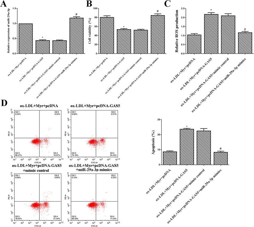
www.nature.com/scientificreports/

                                  Figure 2.  Myr inhibits the inflammatory response and EndMT in ox-LDL-induced HUVEC cells. (A) Representative
                                  immunofluorescence images of CD31 and SM22a expression in each group. Scale bar represents 50 μm. (B) Western blot
                                  analysis of CD31 and SM22a expression in HUVEC cells. (C) The mRNA levels of IL-6, MCP-1 and VCAM-1 in each group.
                                  (D) The protein levels of IL-6, MCP-1 and VCAM-1 were detected by ELISA assay. *P < 0.05 compared with control; #P < 0.05
                                  compared with ox-LDL; &P < 0.05 compared with 2.5 μM.

Scientific Reports |   (2021) 11:19637 |                https://doi.org/10.1038/s41598-021-98916-7                                                            5

                                                                                                                                                     Vol.:(0123456789)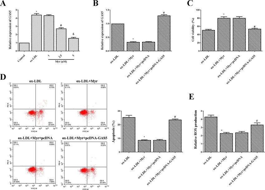
www.nature.com/scientificreports/
Figure 3. GAS5 over-expression attenuates Myr protective effects against ox-LDL–mediated HUVEC injury.
(A) GAS5 expression was determined with qRT-PCR. HUVEC cells were pre-incubated with 1/2.5/5 μM Myr
for 2 h and then stimulated with or without 100 μg/mL ox-LDL for 24 h. *P < 0.05 compared with control;
#
P < 0.05 compared with ox-LDL; &P < 0.05 compared with 2.5 μM. (B) qRT-PCR analysis of relative expression
of GAS5 in each group. (C) CCK-8 assay was conducted to evaluate the cell viability of HUVEC cells in each
group. (D) The apoptosis rate of HUVEC cells after different treatments. (E) The production of ROS was
detected by ROS Assay Kit. *P < 0.05 compared with ox-LDL; #P < 0.05 compared with ox-LDL + Myr + pcDNA.
in ox-LDL + Myr + sh-GAS5 and ox-LDL + Myr + miR-29a-3p mimics groups compared with the ox-LDL + Myr
group (Fig. 6D). Likewise, similar results of IL-6, MCP-1, VCAM-1 were detected by ELISA (Fig. 6E). What’s
more, we also detected cell viability, inflammation, apoptosis, and EndMT in sh-GAS5 alone or miR-29a-3p
mimics alone. The data indicated that sh-GAS5 and miR-29a-3p mimics improved the cell viability (Fig. 6F) and
inhibited cell apoptosis (Fig. 6G), EndMT (Fig. 6H) and inflammation (Fig. 6I) compared with the ox-LDL + NC
group.
Myricetin inactivated the TLR4/NF‑κB signalling pathway in ox‑LDL‑treated HUVEC by
down‑regulating GAS5 or upregulating miR‑29a‑3p. The TLR4/NF-κB signalling pathway is closely
involved in the process of AS. In order to explore whether Myr suppressed the TLR4/NF-κB signalling pathway,
the TLR4 and several pivotal downstream proteins, e.g., p-p65, p65, p-IkBa, IkBa, and TLR4, were identified in
HUVEC cells via western blot analysis. Figure 6 shows that Myr downregulated the expression of p-p65, p-IkBa,
and TLR4 as compared to the ox-LDL group, while the protein level of p-p65, p-IkBa, and TLR4 were decreased
in ox-LDL + Myr + sh-GAS5 and ox-LDL + Myr + miR-29a-3p mimics groups compared with the ox-LDL + Myr
group (Fig. 7). Taken together, these results suggest that GAS5/miR-29a-3p/TLR4/NF-κB pathway might be one
of the underlying mechanisms through which Myr reduced HUVEC cell inflammatory and EndMT.
Discussion
AS is a persistent inflammatory condition that is started by the deposition as well as accumulation of low-density
all19. In spite of tremendous advances in both basic and scientific study, is still among
lipoproteins in the artery w
the leading reasons for death in the w orld20. In this study, we explored the molecular mechanism of Myr on
ox-LDL-induced damage of HUVECs, revealing the crucial role of lncRNA GAS5 in this event. Our results sug-
gested that Myr regulates HUVECs cell viability, ROS, apoptosis, inflammation, and EndMT through the GAS5/
miR-29a-3p/TLR4/NF-κB pathway, clarifying a new mechanism of Myr on protecting against AS.
Ox-LDL has been shown to induce an enhanced generation of ROS, which plays a critical role in the progres-
sion of AS. Substantial evidence suggests that increased oxidative stress plays a prominent role in the pathogenesis
of vascular endothelial dysfunction along with endothelial cell EndMT, inflammation, and apoptosis. We used
ox-LDL stimulated HUVECs to simulate the occurrence of early atherosclerosis, and the inflammatory response
Scientific Reports | (2021) 11:19637 | https://doi.org/10.1038/s41598-021-98916-7 6
Vol:.(1234567890)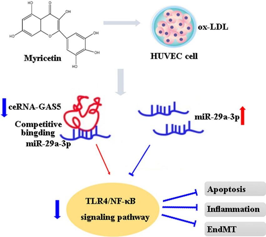
www.nature.com/scientificreports/
Figure 4. miR-29a-3p is a target of GAS5 in HUVEC. (A) Bioinformatics analyses including RegRNA2.0 and
StarBase were used for prediction. (B) Three microRNAs were identified, including miR-29a-3p, miR-128-3p
and miR-135a-5p. (C) The predicted binding between GAS5 and miR-29a-3p. (D) Dual-luciferase reporter
assay validating the interaction between GAS5 and miR-29a-3p. (E) RIP assay and qRT-PCR were conducted to
detect the enrichment of GAS5 and miR-29a-3p by using AGO2 antibodies in HUVEC cells. (F) RNA FISH for
GAS5 and miR-29a-3p was detected in HUVEC cells, miR-29a-3p was co-localized with GAS5 in the nucleus
(magnification, ×400). Nuclei were stained blue (DAPI), GAS5 was stained red, miR-29a-3p was stained green.
*P < 0.05, **P < 0.01, ***P < 0.001.
and EndMT of HUVECs were increased, and Myr significantly inhibited inflammatory response and EndMT
to HUVECs stimulated with ox-LDL. Myr is known to exert antioxidative cytoprotective effects in various cells,
including a HUVEC cell line21. A previous study found that Myr exhibited pro-proliferative and anti-apoptotic
effects in LPS-induced cardiomyocytes H9c2 cells injury22. In this research study, we checked out the result of
Myr on ox-LDL-induced atherosclerotic cell version. Our results suggested that 5 µM Myr pretreatment can
considerably turn around the ox-LDL-induced downregulation of cell viability in HUVECs. Our results dem-
onstrated that ox-LDL therapy could advertise the inflammation and EndMT of HUVECs through regulating
CD31, SM22a, and inflammatory cytokines (IL-6, MCP-1, VCAM-1), which can be reversed by Myr pretreat-
ment. Several elements established that inflammation and EndMT accompany a therosclerosis23. A previous study
found inhibition of the expression of VCAM-1 and ICAM-1 has been recognized as an essential strategy against
atherosclerosis24. Moreover, vaccarin suppressed ox-LDL-induced endothelial EndMT through downregulat-
ing endothelial marker CD31 and upregulating mesenchymal marker SM22α25. Our findings are in substantial
agreement with these researches.
The expression of GAS5 was highly expressed in both human and animal models26,27. In this study, we found
that GAS5 was upregulated and miR-29a-3p was downregulated in ox-LDL-induced HUVEC injury. Myr treat-
ment can reverse these effects. A previous study showed that Myr inhibited the HMGB1, TLR4, and MyD88
expressions in the neurons, and it restored neuronal damage and inflammation caused by activation of NF-κB
and MAPK signal p athways28. In this study, up-regulation of lncRNA GAS5 and down-regulation of miR-29a-3p
led to a decrease of cell apoptosis, inflammation, and EndMT, as well as reduction of activity of TLR4/NF-κB
Scientific Reports | (2021) 11:19637 | https://doi.org/10.1038/s41598-021-98916-7 7
Vol.:(0123456789)www.nature.com/scientificreports/
Figure 5. miR-29a-3p mimics rescued the effects of GAS5 in ox-LDL-induced HUVEC treated with Myr.
(A) miR-29a-3p expression was detected using qRT-PCR with U6 as an internal control. (B) Cell viability was
detected by MTT assay. (C)The production of ROS was detected by ROS Assay Kit. (D) Flow cytometry assay
was used to test apoptosis analysis. *P < 0.05 compared with ox-LDL + Myr + pcDNA; #P < 0.05 compared with
ox-LDL + Myr + pcDNA-GAS5 + mimic control.
signalling pathway. According to our findings, Myr reduces p-p65, p-IkBa, and TLR4 in HUVECs by regulating
GAS5/miR-29a-3p attenuating the inflammatory response and EndMT in ox-LDL-induced HUVEC injury.
Finally, our research study exposed that Myr is capable of ameliorating cell apoptosis, cell inflammation, and
EndMT via GAS5/miR-29a-3p/TLR4/NF-κB pathway (Fig. 8), suggesting Myr as a potential therapeutic agent
for AS. In addition, GAS5 was proposed to be a promising target molecule involving the pathophysiological
processes of AS.
Scientific Reports | (2021) 11:19637 | https://doi.org/10.1038/s41598-021-98916-7 8
Vol:.(1234567890)www.nature.com/scientificreports/
Figure 6. sh-GAS5 or miR-29a-3p mimics can accelerate the protective role of Myr in ox-LDL-induced HUVECs cell. (A)
The expression of GAS5 after inhibiting GAS5. (B, F) Cell viability was detected by MTT assay. (C, G) Flow cytometry assay
was used to test apoptosis analysis. (D, H) Western blot analysis of CD31 and SM22a expression in HUVEC cells. (E, I)
The protein levels of IL-6, MCP-1 and VCAM-1 were detected by ELISA assay. *P < 0.05 compared with ox-LDL; #P < 0.05
compared with ox-LDL + Myr + NC. &P < 0.05 compared with ox-LDL + Myr + mimic control.
Scientific Reports | (2021) 11:19637 | https://doi.org/10.1038/s41598-021-98916-7 9
Vol.:(0123456789)www.nature.com/scientificreports/
Figure 7. Myr inactivated the TLR4/NF-κB signalling pathway in ox-LDL-treated HUVEC by down-
regulating GAS5 or upregulating miR-29a-3p. Western blot analysis of p-p65, p65T, p-IκBα, IκBα and TLR4.
*P < 0.05 compared with Control; #P < 0.05 compared with ox-LDL + Myr + NC. &P < 0.05 compared with
ox-LDL + Myr + mimic control.
Figure 8. Scheme of the mechanisms in the protective effect of Myr on ox-LDL-induced HUVECs.
Data availability
All data generated or analyzed during this study are included in this published article.
Received: 21 January 2021; Accepted: 3 August 2021
Scientific Reports | (2021) 11:19637 | https://doi.org/10.1038/s41598-021-98916-7 10
Vol:.(1234567890)www.nature.com/scientificreports/
References
1. Kondapalli, L., Bottinor, W. & Lenneman, C. By releasing the brakes with immunotherapy, are we accelerating atherosclerosis?.
Circulation 142(24), 2312–2315 (2020).
2. Libby, P., Ridker, P. M. & Hansson, G. K. Progress and challenges in translating the biology of atherosclerosis. Nature 473(7347),
317–325 (2011).
3. Couto, N. F. et al. OxLDL alterations in endothelial cell membrane dynamics leads to changes in vesicle trafficking and increases
cell susceptibility to injury. Biochim. Biophys. Acta Biomembranes 1862, 183139 (2019).
4. Yamagata, K. Protective effect of epigallocatechin gallate on endothelial disorders in atherosclerosis. J. Cardiovasc. Pharmacol. 75,
292–298 (2019).
5. Gao, S. & Liu, J. Association between circulating oxidized low-density lipoprotein and atherosclerotic cardiovascular disease.
Chronic Dis. Transl. Med. 3(2), 89–94 (2017).
6. Torisu, K. et al. Intact endothelial autophagy is required to maintain vascular lipid homeostasis. Aging Cell 15(1), 187–191 (2016).
7. Pan, X. et al. Activation of Nrf2/HO-1 signal with Myricetin for attenuating ECM degradation in human chondrocytes and ame-
liorating the murine osteoarthritis. Int. Immunopharmacol. 75, 105742 (2019).
8. Li, M. et al. Myricetin suppresses the propagation of hepatocellular carcinoma via down-regulating expression of YAP. Cells 8(4),
358 (2019).
9. Guo, C. et al. Myricetin Ameliorates ethanol-induced lipid accumulation in liver cells by reducing fatty acid biosynthesis. Mol.
Nutr. Food Res. 63(14), e1801393 (2019).
10. Jung, S. et al. Myricetin suppresses UVB-induced skin cancer by targeting Fyn. Can. Res. 68(14), 6021–6029 (2008).
11. Zhang, N. et al. Myricetin attenuated LPS induced cardiac injury in vivo and in vitro. Phytotherapy Res. PTR 32(3), 459–470 (2018).
12. Liao, H. et al. Myricetin alleviates pathological cardiac hypertrophy via TRAF6/TAK1/MAPK and Nrf2 signaling pathway. Oxid.
Med. Cell. Longev. 2019, 6304058 (2019).
13. Tiwari, R., Mohan, M., Kasture, S., Maxia, A. & Ballero, M. Cardioprotective potential of Myricetin in isoproterenol-induced
myocardial infarction in Wistar rats. Phytotherapy Res. PTR 23(10), 1361–1366 (2009).
14. Guo, C., Xu, G. & Chen, L. Mechanisms of long noncoding RNA nuclear retention. Trends Biochem. Sci. 45(11), 947–960 (2020).
15. Zhang, H., Liu, B., Shi, X. & Sun, X. Long noncoding RNAs: Potential therapeutic targets in cardiocerebrovascular diseases. Phar-
macol. Therapeut. 221, 107744 (2020).
16. Tang, R. et al. LncRNA GAS5 attenuates fibroblast activation through inhibiting Smad3 signaling. Am. J. Physiol. Cell Physiol.
319(1), C105–C115 (2020).
17. Wu, N. et al. Down-regulation of GAS5 ameliorates myocardial ischaemia/reperfusion injury via the miR-335/ROCK1/AKT/
GSK-3β axis. J. Cell Mol. Med. 23(12), 8420–8431 (2019).
18. Huang, F. et al. LncRNA PVT1 triggers Cyto-protective autophagy and promotes pancreatic ductal adenocarcinoma development
via the miR-20a-5p/ULK1 Axis. Mol Cancer 17(1), 98–98 (2018).
19. Lee, S., Bartlett, B. & Dwivedi, G. Adaptive immune responses in human atherosclerosis. Int. J. Mol. Sci. 21(23), 9322 (2020).
20. Raggi, P. et al. Role of inflammation in the pathogenesis of atherosclerosis and therapeutic interventions. Atherosclerosis 276,
98–108 (2018).
21. Aminzadeh, A. & Bashiri, H. Myricetin ameliorates high glucose-induced endothelial dysfunction in human umbilical vein
endothelial cells. Cell Biochem. Funct. 38(1), 12–20 (2020).
22. Sun, J., Sun, J. & Zhou, X. Protective functions of Myricetin in LPS-induced cardiomyocytes H9c2 cells injury by regulation of
MALAT1. Eur. J. Med. Res. 24(1), 20–20 (2019).
23. Meng, J. et al. The dipeptidyl peptidase (DPP)-4 inhibitor trelagliptin inhibits IL-1β-induced endothelial inflammation and mono-
cytes attachment. Int. Immunopharmacol. 89, 106996 (2020).
24. Zhu, Y. et al. Astragalus polysaccharides suppress ICAM-1 and VCAM-1 expression in TNF-α-treated human vascular endothelial
cells by blocking NF-κB activation. Acta Pharmacol. Sin. 34(8), 1036–1042 (2013).
25. Gong, L. et al. Vaccarin prevents ox-LDL-induced HUVEC EndMT, inflammation and apoptosis by suppressing ROS/p38 MAPK
signaling. Am. J. Transl. Res. 11(4), 2140–2154 (2019).
26. Meng, X.-D. et al. Knockdown of GAS5 inhibits atherosclerosis progression via reducing EZH2-mediated ABCA1 transcription
in ApoE(−/−) mice. Mol. Ther. Nucl. Acids 19, 84–96 (2020).
27. Shen, S. et al. Silencing of GAS5 represses the malignant progression of atherosclerosis through upregulation of miR-135a. Biomed.
Pharmacother. 118, 109302 (2019).
28. Yang, Y. et al. Myricitrin blocks activation of NF-κB and MAPK signaling pathways to protect nigrostriatum neuron in LPS-
stimulated mice. J. Neuroimmunol. 337, 577049 (2019).
Author contributions
Research idea and study design: Y.B., Z.G., N.J.; data acquisition: Y.B., X.L., Q.C.; data analysis/interpretation:
Y.B., X.L., T.C., Q.C.; statistical analysis: X.L., T.C., Q.C.; supervision or mentorship: Z.G., N.J.
Competing interests
The authors declare no competing interests.
Additional information
Supplementary Information The online version contains supplementary material available at https://doi.org/
10.1038/s41598-021-98916-7.
Correspondence and requests for materials should be addressed to N.J. or Z.G.
Reprints and permissions information is available at www.nature.com/reprints.
Publisher’s note Springer Nature remains neutral with regard to jurisdictional claims in published maps and
institutional affiliations.
Scientific Reports | (2021) 11:19637 | https://doi.org/10.1038/s41598-021-98916-7 11
Vol.:(0123456789)www.nature.com/scientificreports/
Open Access This article is licensed under a Creative Commons Attribution 4.0 International
License, which permits use, sharing, adaptation, distribution and reproduction in any medium or
format, as long as you give appropriate credit to the original author(s) and the source, provide a link to the
Creative Commons licence, and indicate if changes were made. The images or other third party material in this
article are included in the article’s Creative Commons licence, unless indicated otherwise in a credit line to the
material. If material is not included in the article’s Creative Commons licence and your intended use is not
permitted by statutory regulation or exceeds the permitted use, you will need to obtain permission directly from
the copyright holder. To view a copy of this licence, visit http://creativecommons.org/licenses/by/4.0/.
© The Author(s) 2021
Scientific Reports | (2021) 11:19637 | https://doi.org/10.1038/s41598-021-98916-7 12
Vol:.(1234567890)You can also read