Noninvasive 3D imaging Using Optical Coherence Tomography - GEN Protocols
←
→
Page content transcription
If your browser does not render page correctly, please read the page content below
Protocol
Noninvasive 3D imaging Using Optical Coherence
Tomography
TRANSLATIONAL MEDICINE CANCER RESEARCH
Yasushi Kuromi , Biosciences & Pharmaceuticals, SCREEN Holdings., 322 Furukawa-cho, Hazukashi, Fushimi-ku, Kyoto, 612-
8486 Japan
Yuki Mori , Biosciences & Pharmaceuticals, SCREEN Holdings., 322 Furukawa-cho, Hazukashi, Fushimi-ku, Kyoto, 612-8486
Japan
Hiroki Fujimoto , Biosciences & Pharmaceuticals, SCREEN Holdings., 322 Furukawa-cho, Hazukashi, Fushimi-ku, Kyoto, 612-
8486 Japan
Ryo Hasebe , Nagahama Institute of Bioscience and Technology, 1266, Nagahama, Shiga 526-0829, Japan
Sumeer Dhar , Bioscience & Pharmaceuticals, SCREEN GP EUROPE B.V., Bouwerij 46, 1185 XX Amsterlveen, The Netherlands
Akihiro Ueda , Biosciences &Pharmaceuticals, SCREEN Holdings., 322 Furukawa-cho, Hazukashi, Fushimi-ku, Kyoto, 612-8486
Japan
Takemitsu Muira , Biosciences & Pharmaceuticals, SCREEN Holdings., 322 Furukawa-cho, Hazukashi, Fushimi-ku, Kyoto, 612-
8486 Japan
Aug 27, 2021
Abstract
This protocol describes a novel OCT-based technology for imaging and analysis of 3D structures, such as, spheroid /organoids,
tissues for growth & morphological evaluation, quantification of internal cavities, drug sensitivity testing to capture the events
leading to tumor cell death, and assessing effects of anti-angiogenic drugs.
Introduction
3D ex vivo platforms are being diligently evaluated as better predictive drug efficacy testing tools in preclinical as well as
clinical space in the quest for profiling of novel anticancer entities (as single or two-drug combinations). Within this context,
there has been growing need for improved imaging and analysis of complex 3D structures. Optical Coherent tomography
(OCT) has been widely used as one of the most important tests in ophthalmology. It is a non-invasive imaging technology that
renders high-resolution and cross-sectional images from the retina. Given its tremendous use in in vivo application, recently
the technology has been applied in 3D in vitro / ex vivo applications for performing imaging of spheroids/ organoids and large
tissues. This technology allows to perform large tissue imaging, non-invasive monitoring of macro and sprouted neo-
vasculature without the need for fluorescent staining for providing quantitative information about the vascular morphological
changes, thereby allowing for the evaluation of anti-angiogenic drugs in real-time.
Reagents and Equipment
OCT Optical System
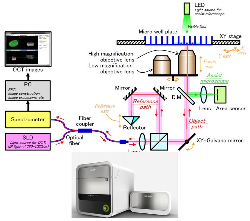
SCREEN HOLDINGS has developed a novel Optical Coherent Technology (OCT) for morphological evaluation of complex 3D
structures and tissues. The system is equipped with 850-nm light from a super luminescent diode (SLD). The OCT observation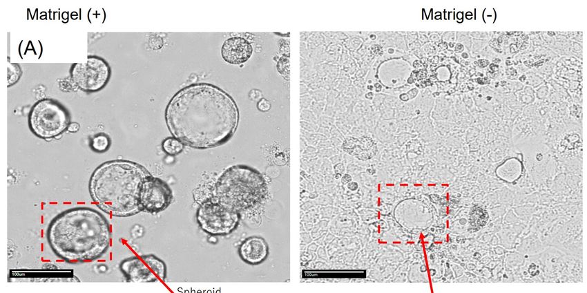
system adopts an inverted microscope, which picks up the reflected light from sample (mirror reflected or backscattered). It is
similar to a reflection microscope or confocal microscope.

   Procedure
1. All cells were cultured in a suitable manner.
2. Cells are imaged by spectrum-domain OCT (SD-OCT) system. The SD-OCT system is outlined below. The samples were
imaged from the bottom surface. Original images obtained by the OCT system contained noise (such as from the collagen gel
surrounding the sample).
3. To reduce noise, image processing was applied for the collected original OCT images using our software (SCREEN) and
ImageJ image processing software (NIH). The images were subsequently processed with filters.
4. The images were then converted into binary images so that the cell area is white and all other areas are black. Each feature
values (volume, cavities etc.) were analyzed.
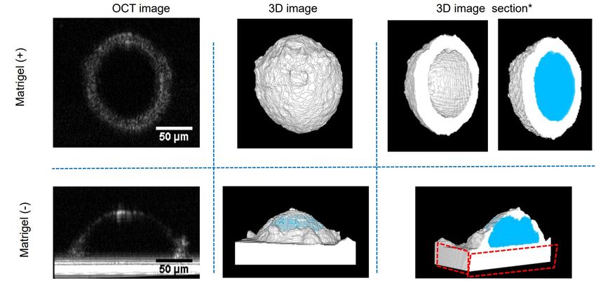
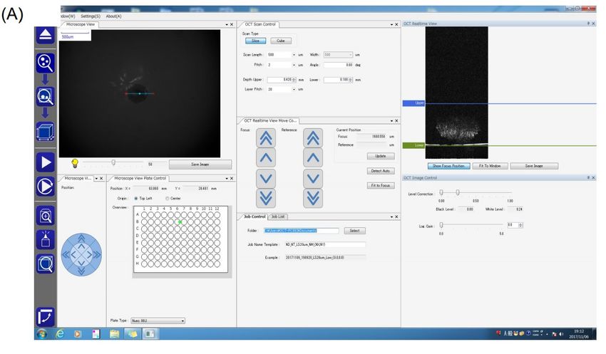
Figure 1: Panel A below depicts the interface window where the settings including selection of plate format, threshold for the
upper and lower limit of the sample (Organoid/tissue) is delineated.

Figure 1: Panel B below illustrates the sectional image of any direction: 3D view of any direction: distance between two points:
2D sectional area/3D volume and image processing
Figure 2: CaCo-2 were cultured in U-bottom plates in the presence and absence of Matrigel and the surface area, volume and
volume of cavity was discerned using inbuilt software program (A) and using Image J sofware (B)
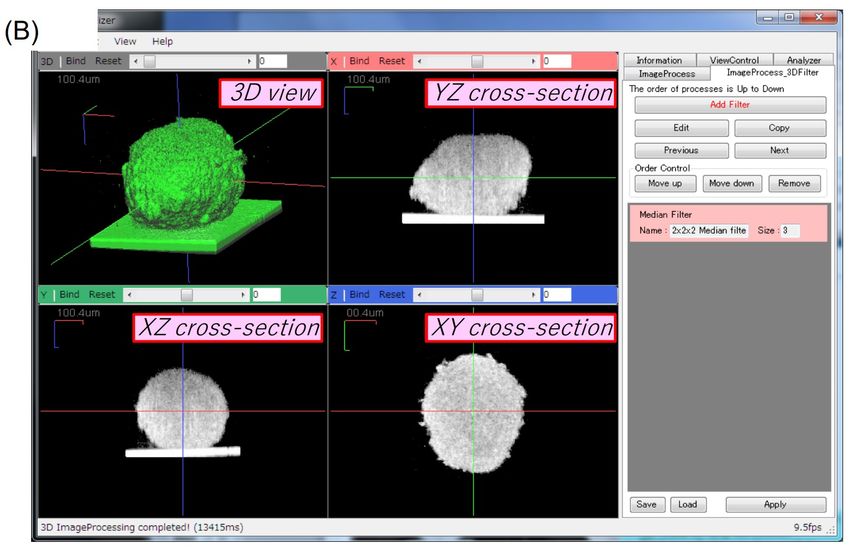
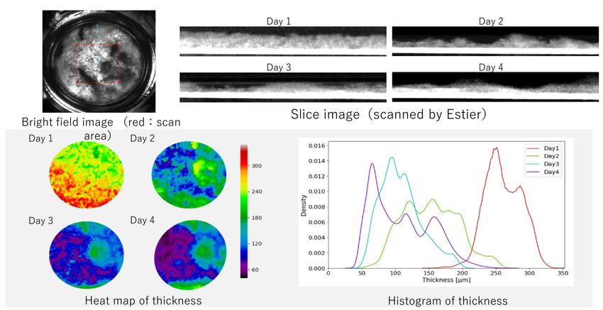
Figure 3: CaCo-2 were cultured in U-bottom plates in the presence and absence of Matrigel and the surface area, volume and
volume of cavity was discerned using in built software program and Image J software. *Blue indicates cavity Red indicates the
bottom of plate

Figure 4: Normal Fibroblast cells, NHDF were maintained in the cell culture insert, and cells were stacked to male cell sheet. The
thickness of sheet was quantitated in a label-free manner using Python. Heat map and histogram indicate thickness of sheet.
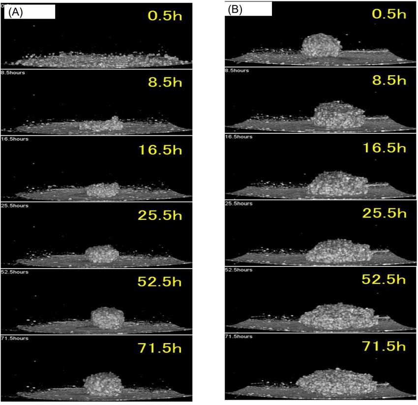
Efficacy Testing in Microtumors Derived from HEK293T cell line
HEK293T cells were prepared and seeded in 96 well plates. The cells were treated with Chetomin and incubated at 37 deg C for
up to 24-48 hr. The morphological evaluation of the spheroids derived from HEK293T cells treated and untreated was
performed in a Time-lapse measurement using OCT.
Figure 5: Illustrates the activity of Chetomin against the spheroids derived from HEK293T cells: The time course measurement
of the activity of chetomin shows that the treated spheroid gradually lose its circularity in comparison to the untreated
spheroids Panel (A) shows the spheroids cultured in the absence of Chetomin and Panel (B) represents the Chetomin-treated
spheroids.
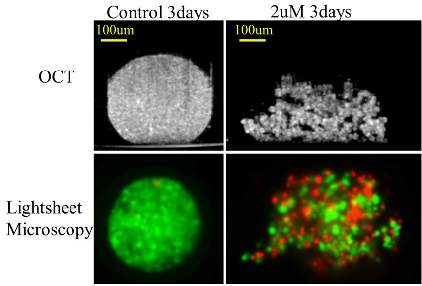
3D OCT imaging vs. Light-sheet microscopy
Figure 6: MCF-7 cells tagged with EGFP were cultured in U[1]bottom plates and treated with 2 µM of A23187 (ca ionophore).
The cells were further incubated for 3 days until the cells were organized into spheroids. Subsequently, the spheroids were
imaged by OCT as well as Light sheet microscopy to delineate the drug efficacy , shown as loss of circularity. Figure 1: CaCo-2
were cultured in U-bottom plates in the presence and absence of Matrigel and the surface area, volume and volume of cavity
was discerned using in built software program (A) and using Image J sofware (B) Assessment of Angiogenic sprouting with
OCT (A) (B) Matrigel (-) Extracted from: Tamio Mizukami et al. ( Nagahama Institute of Bio-Science and Technology, *2: Frontier
Pharma,)
Measuring Angiogenic Sprouting with OCT
Figure 7: Depicts the analysis of angiogenic sprouting using optical coherence tomography (OCT). Yukiko T. Matsunaga et. al.,
(Cent. for Int’1 Res. on Integrative Bio. Med Sys., Insti. of Ind. Sci., Univ of Tokyo, Japan)

  Time Taken
Variable
Notes and Comments
Conclusions
  1. Non-invasive IR laser technology based Cell 3imager, ESTIER can be used for True 3D imaging of complex 3D structures,
     using Spheroids/Organoids and large tissues.
  2. The time lapse measurement delineates the activity of the drugs and effects on the morphological aspects in spheroids
     when treated with Chetomin and A23187.
  3. The technology can be utilized in quantitation of size, volume and internal cavities of various complex 3D structures.
  4. High utility for multiple applications in Oncology, regenerative medicine.
  5. User friendly work flow for analysis of data with automation capability.
  6. Complementary system for exiting microscopic and high content imaging systems.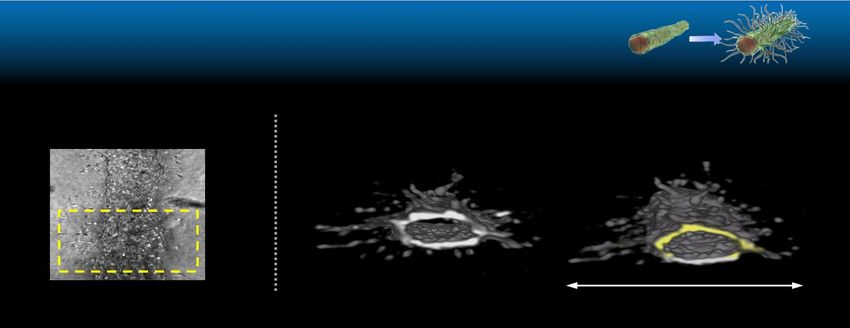
You can also read