Nuclei Segmentation via a Deep Panoptic Model with Semantic Feature Fusion - IJCAI
←
→
Page content transcription
If your browser does not render page correctly, please read the page content below
Proceedings of the Twenty-Eighth International Joint Conference on Artificial Intelligence (IJCAI-19)

    Nuclei Segmentation via a Deep Panoptic Model with Semantic Feature Fusion
            Dongnan Liu1∗ , Donghao Zhang1∗ , Yang Song2 , Chaoyi Zhang1 , Fan Zhang3 ,
                                    Lauren O’Donnell3 and Weidong Cai1
                         1
                           School of Computer Science, University of Sydney, Australia
          2
            School of Computer Science and Engineering, University of New South Wales, Australia
                       3
                         Brigham and Women’s Hospital, Harvard Medical School, USA
    {dliu5812, dzha9516}@uni.sydney.edu.au, yang.song1@unsw.edu.au, czha5168@uni.sydney.edu.au,
                        {fzhang, odonnell}@bwh.harvard.edu, tom.cai@sydney.edu.au

                              Abstract
         Automated detection and segmentation of individ-
         ual nuclei in histopathology images is important
         for cancer diagnosis and prognosis. Due to the
         high variability of nuclei appearances and numer-
         ous overlapping objects, this task still remains chal-                    (a)              (b)              (c)              (d)
         lenging. Deep learning based semantic and in-
         stance segmentation models have been proposed to                    Figure 1: Example histopathology images for nuclei segmentation
                                                                             from different organs: breast (a) and corresponding annotation (b);
         address the challenges, but these methods tend to                   bladder (c) and corresponding annotation (d); green boxes: cyto-
         concentrate on either the global or local features                  plasm which are similar to nuclei; red boxes: touching nuclei.
         and hence still suffer from information loss. In this
         work, we propose a panoptic segmentation model
         which incorporates an auxiliary semantic segmen-                    the nuclei, making it hard to differentiate cell nuclei from
         tation branch with the instance branch to integrate                 the background. Third, nuclei are often clustered with many
         global and local features. Furthermore, we design                   overlapping instances [Chen et al., 2017]. In order to find
         a feature map fusion mechanism in the instance                      the exact location and boundary for every single nucleus, fur-
         branch and a new mask generator to prevent in-                      ther processing is often required to separate the clustered or
         formation loss. Experimental results on three dif-                  overlapping nuclei.
         ferent histopathology datasets demonstrate that our                    Convolutional neural networks (CNN) are powerful for
         method outperforms the state-of-the-art nuclei seg-                 tackling image recognition tasks [He et al., 2016; Peng et
         mentation methods and popular semantic and in-                      al., 2017] by learning the features automatically. Currently,
         stance segmentation models by a large margin.                       most CNN related works for nuclei segmentation are based
                                                                             on the semantic segmentation model to separate foreground
1       Introduction                                                         and background, and involve post-processing for refinement.
                                                                             Recently, instance segmentation models have been proposed
Cell morphology in histopathology images provides critical                   for predicting the mask and region of interest (ROI) [He et
information for cancer diagnosis and prognosis. The first                    al., 2017; Liu et al., 2018b]. When utilizing them on nuclei
step in cell morphology analysis is the segmentation of in-                  segmentation tasks, each ROI represents a single nucleus.
dividual cell nuclei, which is typically performed manually
                                                                                With semantic segmentation models [Peng et al., 2017;
in current clinical practice. However, manual examination of
                                                                             Ronneberger et al., 2015], all pixels are categorized into dif-
histopathology images is labor-intensive and time-consuming
                                                                             ferent classes, which are employed for studying the uncount-
due to the large image sizes and complex cellular structures.
                                                                             able “stuff” of the image. By contrast, instance segmentation
Therefore, investigating computerized methods is necessary
                                                                             is able to assign unique labels to each object that belongs to
to reduce the workload for pathologists, and make the analy-
                                                                             the same class, and is therefore employed to study the count-
sis efficient [Veta et al., 2014].
                                                                             able “things”. The study of both stuff and things is necessary
   There are still some major challenges in nuclei segmenta-
                                                                             for image recognition because the former obtains the features
tion tasks, as illustrated in Figure 1. First, there is a high
                                                                             of the background while the latter is able to learn the features
level of heterogeneity in appearance between different types
                                                                             of different objects in the foreground. In order to reconcile
of organs or cells. Consequently, methods designed based
                                                                             the foreground and background learning, panoptic segmenta-
on prior knowledge about geometric features cannot be ap-
                                                                             tion [Kirillov et al., 2018] has been proposed to incorporate
plied directly to different images. Second, other structures
                                                                             semantic segmentation with instance segmentation, with sep-
such as cytoplasm and stroma can have similar features to
                                                                             arate training of the instance and semantic branches.
    ∗
        Authors contributed equally.                                            Different from existing methods, in our model, we propose

                                                                       861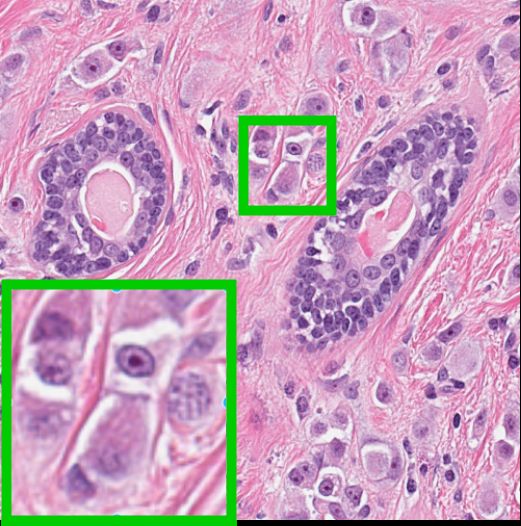
Proceedings of the Twenty-Eighth International Joint Conference on Artificial Intelligence (IJCAI-19)

an end-to-end panoptic segmentation framework incorporat-
ing an auxiliary semantic segmentation branch with an in-
stance branch which contains a dual-modal mask generator
and feature fusion mechanism. First, we consider that se-
mantic segmentation methods are only able to process global
features, which limits their capacity to separate the individ-
ual nuclei. On the other hand, instance segmentation mod-
els focus more on the local ROI-level information and lack a
global interpretation of the image. To address these limita-
tions and also utilize the advantages of both the semantic and
instance models, we propose to design a dual-branch panop-
tic segmentation model by integrating the semantic segmenta-
tion branch with the instance segmentation branch. The dual-
branch model is trained end-to-end and is able to process the
global and local features from the image at the same time.
Furthermore, we introduce a new semantic feature map from
the instance branch to further encourage the instance branch             Figure 2: Overview of our proposed framework. Refer to Section
to encode the global features along with local ones during               3.3 for the detailed loss definitions.
training. In order to prevent information loss when predict-
ing the mask on each ROI, a dual-modal mask generator is
designed with a fully connected (FC) mode and a spatial up-              to process the nuclei with different receptive fields. Beyond
scaling mode. Our main contributions can be summarized as                the standard CNN model, improvements have been made to
follows:                                                                 incorporate the contour information into the CNN architec-
                                                                         ture to facilitate segmentation between individual nuclei. For
    • A novel dual-branch panoptic model is proposed with an
                                                                         example, in [Kumar et al., 2017], nuclei boundaries are con-
      instance segmentation branch and an auxiliary semantic
                                                                         sidered as the third class for the CNN segmentation model.
      segmentation branch. The semantic branch is specially              [Oda et al., 2018] proposed a boundary enhanced module and
      designed for enlarging the receptive field of the input
                                                                         loss function based on the traditional U-Net [Ronneberger et
      object and is jointly trained with the instance branch, in
                                                                         al., 2015], and it facilitates the model’s learning of more de-
      an end-to-end fashion.
                                                                         tails about the nuclei that are hard to segment. Furthermore,
    • A feature fusion mechanism in the instance branch is               Cell R-CNN [Zhang et al., 2018a] achieves competitive per-
      designed to integrate global with local features.                  formance simply by incorporating a semantic segmentation
    • A dual-modal mask generator is designed for higher                 model with an instance model. In addition, regression-based
      mask segmentation accuracy by minimizing the infor-                models have also been proposed such as one uses the distance
      mation loss.                                                       map as the ground truth labels [Naylor et al., 2018], and an-
                                                                         other which applies general adversarial architecture [Zhang
    • Our method was proven to be effective with significant             et al., 2018b], which achieved competitive performance on
      improvements over the other state-of-the-art methods               nuclei segmentation tasks as well.
      on three public datasets: TCGA-kumar [Kumar et al.,
      2017], TNBC [Naylor et al., 2018], and MICCAI 2017                 2.2   CNN Based Image Segmentation
      Digital Pathology Challenge dataset [Vu et al., 2018].
                                                                         In the field of semantic segmentation, skip connections be-
                                                                         tween encoders and decoders are effective and prevalent
2     Related Work                                                       [Ronneberger et al., 2015; Zhang et al., 2018c; Wang et al.,
                                                                         2019]. In [Zhang et al., 2018c] and [Liu et al., 2018a], dense
2.1    Nuclei Segmentation                                               connections between the decoders in different resolutions are
Studies into nuclei segmentation in histological images have             proposed to eliminate the information loss. In addition to
been ongoing for many years. With progress in pattern                    semantic segmentation architectures, instance segmentation
recognition techniques, methods based on machine learning                models such as [He et al., 2017] and [Liu et al., 2018b] are
have shown encouraging results. These methods typically                  able to generate the masks of the image with a detection based
start with handcrafted feature extraction, such as textural fea-         architecture [Ren et al., 2015]. Beyond semantic and instance
tures [Zhang et al., 2014], Laplacian and Gaussian features              segmentation, panoptic segmentation has been proposed to
[Kong et al., 2013], and geometric features about contours               fuse the feature from things and stuff [Kirillov et al., 2018].
[Wienert et al., 2012]. Then, classification (e.g., Bayesian)            In [Zhang et al., 2018a] and [de Geus et al., 2018], both the
or clustering (e.g., K-means) techniques are employed for                instance and semantic segmentation branches are trained to-
nuclei segmentation and detection tasks [Naik et al., 2008;              gether by sharing the same set of parameters in the backbone
Chankong et al., 2014].                                                  module. Then, the losses of the two branches are summed
   With the advance of CNN, nuclei segmentation has been                 together for back propagation to optimize the parameters of
modeled as a pixel- or patch-level classification problem.               the whole framework. In [Xiong et al., 2019], a novel panop-
In [Raza et al., 2019], a multi-resolution CNN is proposed               tic segmentation head is proposed to fuse the feature about

                                                                   862
Proceedings of the Twenty-Eighth International Joint Conference on Artificial Intelligence (IJCAI-19)

Figure 3: Illustration of the instance branch. C1 and C3 represent convolutional layers with a kernel size of 1 and 3, respectively. For C3,
both the stride and padding size are 1. UP/2x and UP/4x represent a nearest unsampling layer with a scale factor of 2 and 4, respectively. We
omit the ReLU layer after each convolutional block for brevity.

things and stuff, from instance and semantic branch, respec-
tively. More recently, Panoptic Feature Pyramid Network was
proposed in [Kirillov et al., 2019] to generate a semantic out-
put in the instance segmentation branch. The model achieves
new state-of-the-art performance in several image segmenta-
tion tasks with higher memory efficiency.

3     Methods
The overall architecture of our work is illustrated in Figure 2.
During training, both the semantic and instance branches are
trained together with the same ResNet101 backbone module.
During inference, only the instance branch is used to predict
                                                                          Figure 4: Illustration of the proposed mask generator. Conv1,
the class scores, bounding box coordinates, and masks. In                 Conv2, Conv3, Conv4, and Conv5 represent five different convolu-
this section, we present the designs of our network model.                tional layers, and Conv × 3 means three consecutive convolutional
                                                                          layers. FC is a fully connected layer, Reshape1 contains reshaping
3.1    Instance Branch                                                    and channel duplication, and Reshape2 is a pixel shuffle. The final
Figure 3 is a detailed illustration of our proposed instance              fusion is a pixel-wise summation. We omit the ReLU layer after
branch for nuclei segmentation. In general, this branch is in-            each convolutional block for brevity.
spired by the structure of Mask R-CNN [He et al., 2017], but
with major differences in our design of feature fusion and the            eliminates the global information processing. In this way, the
mask generator. Specifically, after the ResNet101 backbone,               global information is fused with the local information to en-
a Feature Pyramid Network (FPN) is applied to generate fea-               able more accurate localization of the cell nuclei.
ture maps in five stages (P2, P3, P4, P5, P6). Next, with
the multiple feature maps P2 to P6, the Region Proposal Net-              Mask Generator
work (RPN) and ROI Align modules are applied to obtain the                Figure 4 illustrates our proposed mask generator, which con-
ROIs for cell nuclei. Then, each ROI passes through a box                 tains two parts. In the original Mask R-CNN, only four con-
branch for class score and bounding box coordinate predic-                secutive convolutional layers and a deconvolutional layer are
tion and our dual-modal mask generator for generating the                 employed to segment the mask. However, the size of the input
nucleus masks. In addition, the feature map at the highest                ROI of the mask generator is 14 × 14, which is small, and di-
spatial resolution of FPN (P2) passes through an upsampling               rectly applying a deconvolutional layer to such a small region
layer with a spatial factor of 4 and a convolutional layer with           is prone to cause information loss. On the other hand, an FC
a channel number of 2. Then, this feature map with the same               layer is able to learn the global information for the entire ROI,
size as the semantic segmentation ground truth is fused with              which is helpful to separate different instances with the same
the nuclei masks according to their locations with feature fu-            category label. Therefore, we incorporate the ideas of [Liu et
sion mechanism. The motivation is that in instance segmen-                al., 2018b] and [Wang et al., 2018], and design a mask gener-
tation models, only local features such as intracellular detail           ator which fuses the feature maps of dual modalities from the
and location for each single nucleus can be processed, which              FC branch and spatial upscaling branch as illustrated in Fig-

                                                                    863
Proceedings of the Twenty-Eighth International Joint Conference on Artificial Intelligence (IJCAI-19)

          Stage      Hyperparamaters            Output size                    3.2   Semantic Branch
          Input                                 256 × 14 × 14
          Conv × 3   k = (3, 3), s = 1, p = 1   256 × 14 × 14                  Even though a semantic feature map FN is rendered from the
          Conv1      k = (3, 3), s = 1, p = 1   256 × 14 × 14                  instance branch, the small kernel sizes of the convolutional
          Conv2      k = (3, 3), s = 1, p = 1   128 × 14 × 14                  layers in FPN imply that FN would still have some informa-
          FC                                    1 × 1 × 784
          Reshape1                              2 × 28 × 28                    tion loss at the global level. This is a common issue for CNN
          Conv3      k = (3, 3), s = 1, p = 1   256 × 14 × 14                  models in that as the network grows deeper, the actual recep-
          Conv4      k = (3, 3), s = 1, p = 1   1024 × 14 × 14                 tive field of the feature maps gradually becomes smaller than
          Reshape2                              256 × 28 × 28
          Conv5      k = (1, 1), s = 1, p = 0   2 × 28 × 28
                                                                               the theoretical size [Zhou et al., 2015]. By utilizing small
          Output                                2 × 28 × 28                    sizes convolutional layers [Saleh et al., 2018], it is difficult
                                                                               for the feature maps at high resolution to maintain the whole
Table 1: The parameters for each block in our proposed mask gen-               features from original images due to the limit of the receptive
erator. k, s, and p denote the kernel size, stride, and padding of the         field.
convolution operation, respectively.                                              To tackle this issue, the decoder of Global Convolutional
                                                                               Network (GCN) [Peng et al., 2017] is applied as an auxiliary
                                                                               semantic branch shown in Figure 2. By simulating a 2D large
ure 4 . The details of the parameters of each block are shown
                                                                               kernel convolutional layer with two 1D convolutional layers,
in Table 1.
                                                                               GCN is able to make the model capture a large and global re-
Feature Fusion                                                                 ceptive field with only a small portion of memory. Our model
As shown in Figure 3, after obtaining the mask predictions                     follows the original GCN architecture except the size of the
M and bounding box predictions B, the feature map F0 from                      large kernel blocks which, for memory efficiency, is fixed at
FPN is fused together with them to derive a new semantic                       5. In addition, the semantic branch is trained jointly with the
segmentation feature map FN , where N is the total number                      instance branch, by sharing the same backbone, compared to
of mask and bounding box predictions, i ∈ 1, ..., N . For the                  the traditional separate training strategy in [Saleh et al., 2018;
ith bounding box prediction, the coordinates of the bounding                   Kirillov et al., 2018]. When working on the semantic seg-
box with the highest class score are selected, which is defined                mentation task, the backbone is capable of generating the
as:                                                                            features with global information about foreground and back-
                                                                               ground, which are useful for bounding box detection and
                       Bi = (xi , yi , wi , hi )           (1)                 mask generation in the instance branch.
where xi and yi are the coordinates of the top left point of                   3.3   Loss Function
Bi in x and y axes, respectively, wi and hi are its width and                  All the losses in this work are shown in Figure 2. The total
height, respectively. When fusing the ith mask Mi with Bi                      loss is defined as:
on the feature map Fi−1 , the output feature map Fi is formu-
                                                                                   Ltotal = L(rpn−obj) + L(rpn−reg) + L(det−cls)
lated as:
                                                                                                          + L(det−reg) + L(det−mask)         (4)
    Fi = Greplace (Greshape (Mi , Bi ), sub(Fi−1 , Bi ))          (2)                                     + Lsemseg1 + Lsemseg2
                                                                                  L(rpn−obj) and L(rpn−reg) are the losses for background
where Greshape reshapes the binary mask Mi to the same size                    and foreground classification and the bounding boxes for the
as Bi . Greplace enables the new mask Greshape (Mi , Bi ) to                   anchors rendered by RPN, respectively, where L(rpn−obj) is
replace sub(Fi−1 , Bi ), which is the subset of Fi−1 according                 the cross entropy loss for classification and L(rpn−reg) is
to Bi and can be represented as:                                               the smoothed L1 loss for regression. In the instance branch,
                                                                               L(det−cls) is the cross entropy loss for object category classi-
      sub(Fi−1 , Bi ) = Fi−1 [xi : xi + wi , yi : yi + hi ]       (3)          fication, L(det−reg) is the smoothed L1 loss for bounding box
                                                                               coordinate regression, and L(det−mask) is the binary cross
Eventually, FN is the semantic feature map obtained from the                   entropy loss for instance mask segmentation. Lsemseg1 and
instance branch.                                                               Lsemseg2 are the cross entropy losses for semantic segmenta-
   For each ROI, mask prediction represents the nuclei seg-                    tion of semantic and instance branch, respectively. Although
mentation result in the small region by utilizing the local in-                adding a weight for each loss in Ltotal would be a better
formation around each single object. In addition, the bound-                   trade-off, we still chose to avoid this labor-intensive process,
ing box prediction represents the real location of this nucleus                in favor of the generalizability and reproducibility.
in the original image, which contains localization information                    In all experiments, we employed stochastic gradient de-
for the object in a global view. In order to retain more global                scent (SGD) as the optimizer with a momentum of 0.9 and a
and local information in the semantic segmentation result, we                  weight decay of 0.0001 to train our model. The learning rate
fuse the mask and bounding box predictions by passing each                     varies in each experiment with the same linear warming up in
mask prediction feature map to the large feature map from                      the first 500 iterations. Due to the small mini-batch size dur-
FPN according to its corresponding box coordinates predic-                     ing training, we had no batch normalization layers. All the
tion. Therefore, this second semantic segmentation feature                     hyperparameters for testing were fine-tuned on the validation
map (FN ) contains global and local features from the detec-                   set. We implemented our experiments using Pytorch [Paszke
tion architecture of the instance branch.                                      et al., 2017].

                                                                         864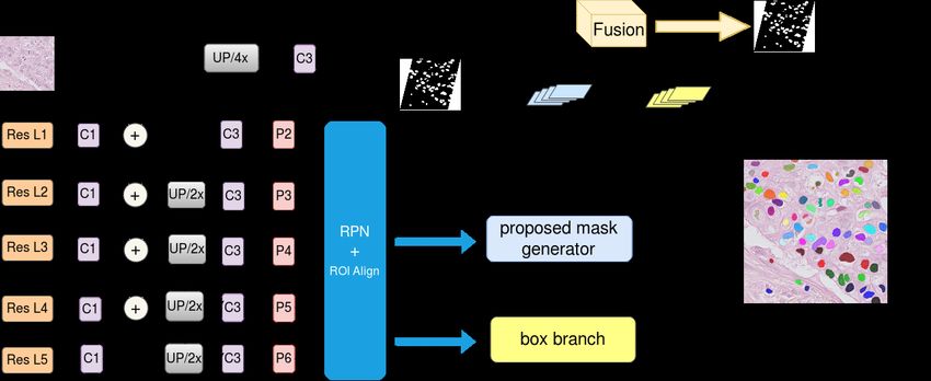
Proceedings of the Twenty-Eighth International Joint Conference on Artificial Intelligence (IJCAI-19)

4     Experiments                                                       four different organs, we randomly selected one image from
4.1    Datasets and Evaluation Metrics                                  each organ for validation and used the remaining 12 im-
                                                                        ages for training. The learning rate in this experiment was
We used three public datasets in this study. The first dataset          0.0025 and it decreased to its 1/10 at the 8640th iteration
is from The Cancer Genome Atlas (TCGA) at 40× magni-                    and 1/100 at the 12960th iteration with a total of 17280
fication [Kumar et al., 2017]. We refer to this dataset as              training iterations. When training the model, we separated
TCGA-kumar. There are 30 annotated 1000 × 1000 patches                  each 1000 × 1000 original image into four 512 × 512 patches
from 30 whole slide images of different patients. These im-             with basic data augmentation including horizontal and ver-
ages show highly varying properties since they are from 18              tical flipping and rotations of 90◦ , 180◦ , and 270◦ to avoid
different hospitals and 7 different organs (breast, liver, kid-         overfitting. In addition, we also employed advanced augmen-
ney, prostate, bladder, colon, and stomach). In contrast to the         tation including blurring, adding gaussian noise, embossing,
disease variability of TCGA-kumar, the second dataset from              sharpening, and random channel shuffle due to the noise and
[Naylor et al., 2018] focuses in particular on Triple Negative          variability of color in the histopathology images. For a fair
Breast Cancer (TNBC). In this TNBC dataset, there are 50                comparison, we applied the same data augmentation settings
annotated 512 × 512 patches at 40× magnification sampled                to all compared methods.
from 11 patients at the Curie Institute. The third dataset is
                                                                           Table 2 shows the result for each image in the testing set
the MICCAI 2017 Digital Pathology Challenge dataset [Vu et
                                                                        in comparison to 2 state-of-the-art nuclei segmentation meth-
al., 2018], also referred to as Cell17. Cell17 contains 64 an-
                                                                        ods: CNN3 in [Kumar et al., 2017] and DIST in [Naylor
notated images in total, and both the training and testing sets
                                                                        et al., 2018]. Our proposed method outperforms the others
contain 8 images from 4 different diseases: glioblastoma mul-
                                                                        based on the average AJI and F1 score. In addition, one-tailed
tiforme (GBM), lower grade glioma (LGG) tumors, head and
                                                                        paired t-test was employed for evaluation of statistically sig-
neck squamous cell carcinoma (HNSCC), and non small cell
                                                                        nificance. For object-level accuracy (AJI), the improvement
lung cancer (NSCLC). The image sizes are either 500 × 500
                                                                        of our method is significant as all the p-values of the com-
or 600 × 600 at 20× or 40× magnification.
                                                                        parison methods are under 0.1. By learning the global and
   In the experiments on TCGA-kumar and TNBC, we em-
                                                                        local features from both the semantic and instance branches,
ployed F1 score and Aggregated Jaccard Index (AJI) [Kumar
                                                                        our model is the most competitive even without any pre- or
et al., 2017] for pixel-level and object-level evaluation, re-
                                                                        post-processing compared to CNN3 and DIST. In terms of F1
spectively. AJI is an extension of the Jaccard Index that takes
                                                                        score, our improvement is significant compared to the others
false negative predictions into consideration. AJI can be de-
                                                                        except for DIST. However, DIST has a post-processing step
fined as:
                            PN                                          with two extra hyperparameters, in order to refine the distance
                                          i
                              i=1 |Gi ∩ PM |                            map from a deep regression model. If a less significant im-
          AJI = PN                                          (5)         provement is acceptable, we would prefer not to add any fur-
                                   i
                                        P
                      i=1 |Gi ∪ PM | +     F ∈U |PF |
                                                                        ther post-processing for the sake of memory efficiency and
where Gi is the ith nucleus from the ground truth with N                convenience of implementation. For Mask R-CNN, we no-
          i
nuclei. PM  means the M th connected component in predic-               tice that its AJI score is at the same level as CNN3, while the
tion which has the largest Jaccard Index with Gi , and each             F1 score is lower than all the compared semantic segmenta-
index (M ) cannot be used more than once. Set U represents              tion models. This is because Mask R-CNN processes ROI,
the connected component in the prediction without the cor-              which represents each object for instance segmentation, mak-
responding ground truth. In the experiments of Cell17, we               ing it impossible to learn the relationship between the fore-
employed the same set of evaluation metrics as the experi-              ground and background. By adding a new semantic branch,
ments of Cell R-CNN [Zhang et al., 2018a] for comparison:               an extra semantic loss, and a dual-modal mask generator, our
F1 score, object-level Dice score, and object-level Hausdorff           proposed model has a significant improvement in pixel-level
distance.                                                               accuracy.
4.2    Experimental Results and Discussion
TCGA-kumar
In this experiment, we first evaluated the performance of our
proposed model in comparison to the state-of-the-art works
from [Kumar et al., 2017; Naylor et al., 2018; He et al., 2017;
Ronneberger et al., 2015]. Then, an ablation study was em-
ployed to demonstrate the effectiveness of each module in the
overall architecture.
   With the same split for the testing set as [Kumar et
al., 2017] and [Naylor et al., 2018] (details at https://                     Baseline      Ab1        Ab2      Proposed      GT
peterjacknaylor.github.io/), we compared our work directly
to the results reported in their published works. For                   Figure 5: Visual comparison of the ablation study on the TCGA-
[He et al., 2017], we re-implemented it with officially                 kumar dataset. The first row and second row show results of images
released code from https://github.com/facebookresearch/                 from the breast and prostate, respectively. The red arrows indicate
maskrcnn-benchmark. Among the 16 training images from                   the disagreement with the ground truth.

                                                                  865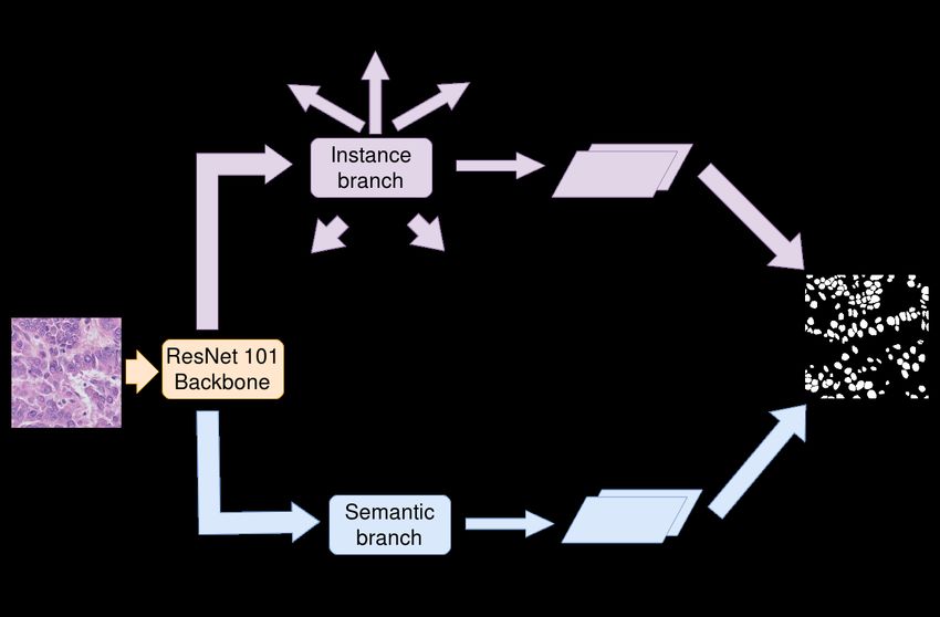
Proceedings of the Twenty-Eighth International Joint Conference on Artificial Intelligence (IJCAI-19)

                                                                       AJI                                                     F1 Score
                   Organ         Image
                                          CNN3          DIST      Mask R-CNN      U-Net     Proposed    CNN3      DIST       Mask R-CNN   U-Net    Proposed
                                  1       0.4974       0.5334       0.4960        0.3831     0.5490     0.6885   0.7761        0.7486     0.7492    0.7887
                   Breast
                                  2       0.5796       0.5884       0.5577        0.5505     0.6362     0.7476   0.8380        0.8026     0.8144    0.8478
                                  1       0.4792       0.5648       0.5794        0.5386     0.6202     0.6606   0.7805        0.7742     0.8039    0.8097
                  Kidney
                                  2       0.6672       0.5420       0.5286        0.5020     0.5902     0.7837   0.7606        0.7285     0.7786    0.7724
                                  1       0.5175       0.5466       0.5183        0.4773     0.5491     0.6726   0.7877        0.7816     0.7648    0.7907
                   Liver
                                  2       0.5148       0.4432       0.4577        0.3794     0.5179     0.7036   0.6684        0.6881     0.6313    0.7323
                                  1       0.4914       0.6273       0.5934        0.1807     0.6305     0.8306   0.8030        0.8032     0.7889    0.8090
                  Prostate
                                  2       0.3761       0.6294       0.6282        0.3118     0.6423     0.7537   0.7903        0.7937     0.7919    0.7992
                                  1       0.5465       0.6475       0.6237        0.5115     0.6749     0.9312   0.8623        0.8385     0.8258    0.8666
                  Bladder
                                  2       0.4968       0.5467       0.4677        0.4621     0.4745     0.6304   0.7768        0.6944     0.7648    0.6963
                                  1       0.4891       0.4240       0.3691        0.0786     0.4450     0.7679   0.7212        0.6251     0.7121    0.7036
                   Colon
                                  2       0.5692       0.4484       0.4354        0.1305     0.4871     0.7118   0.7360        0.6907     0.7599    0.7474
                                  1       0.4538       0.6408       0.6352        0.4096     0.6909     0.8913   0.8547        0.8323     0.8647    0.8795
                 Stomach
                                  2       0.4378       0.6550       0.6449        0.4507     0.6871     0.8982   0.8520        0.8329     0.8629    0.8668
                      Average             0.5083       0.5598       0.5382        0.3833     0.5854     0.7623   0.7863        0.7596     0.7795    0.7936
                    Significance            ??          ???          ???           ???                     ?        7           ???          ?

Table 2: Comparison with the state-of-the-art methods on TCGA-kumar dataset. For statistical significance evaluation, ? ? ? denotes p-value
under 0.01, ?? denotes p-value from 0.01 to 0.05, ? denotes p-value from 0.05 to 0.1, and 7 denotes p-value over 0.1

               Name                Baseline    Ab1        Ab3       Ab2     Proposed                         Method            GCN     Pix2Pix   MRCNN     Proposed
          feature fusion?             7         3           7        3         3                                  avg         0.1907   0.4760     0.5297    0.5865
         semantic branch?             7          7         3         3         3                         AJI      std         0.1208   0.0578     0.1513    0.1059
    dual-modal mask generator?        7          7          7         7        3
                                                                                                              significance     ???      ???        ???
                    avg            0.5382     0.5533     0.5500    0.5744    0.5854
    AJI             std            0.0851     0.0770     0.0912    0.0782    0.0820                               avg         0.3833   0.6910     0.7424      0.7792
               significance                     ??         ??       ???        ??                        F1       std         0.2175   0.0724     0.0837      0.0520
                    avg            0.7596     0.7730     0.7670    0.7881    0.7936                           significance     ???      ???        ???
    F1              std            0.0655     0.0572     0.0717    0.0545    0.0591
               significance                     ??          ?       ???         ?
                                                                                                 Table 4: The result on the TNBC testing set for different methods.
                                                                                                 ? ? ? denotes p-value under 0.01, ?? if the p-value between 0.01 and
Table 3: Ablation study on the TCGA-kumar dataset. ? ? ? is em-
                                                                                                 0.05, ? if p-value between 0.05 and 0.1.
ployed if the p-value is under 0.01, ?? for p-value from 0.01 to
0.05, ? for p-value from 0.05 to 0.1. The significance of Ab1, Ab3,
and Proposed are comparisons between Ab1 and Baseline, Ab3 and
Baseline, Proposed and Ab2, respectively. The significance of Ab2                                dition, we also compared with pixel2pixel [Isola et al., 2017]
are the comparisons between Ab2 and Ab1, Ab2 and Ab3, and both                                   to prove the effectiveness of our model without any adversar-
p-values are under 0.01.                                                                         ial based techniques. In this experiment, we either used the
                                                                                                 code from official implementation or re-implemented them in
                                                                                                 Pytorch. For each experiment, we used six cases with 30 im-
   In order to evaluate the effectiveness of each proposed                                       ages for training, two cases with seven images for validation,
module in our architecture, an ablation experiment was con-                                      and three cases with 13 images for testing. For data augmen-
ducted and the results are shown in Figure 5 and Table 3.                                        tation, we employed horizontal and vertical flipping and rota-
Figure 5 shows that the baseline Mask R-CNN tends to fail                                        tions of 90◦ , 180◦ , and 270◦ . The total training epoch was 60
in segmenting some touching or closely adjacent nuclei and                                       and the initial learning rate 0.00075 decreased to its 1/10 and
Ab1 (baseline + feature fusion) has difficulty in accurately                                     1/100 at the end of the 30th and 45th epoch, respectively.
estimating the boundary information. Compared with Ab2                                              Table 4 lists the resulting AJI and F1 score on the testing
(baseline + feature fusion + semantic branch), our method                                        set. Our proposed model outperformed all the comparison
has a higher accuracy when generating the mask for single                                        models in both average AJI and F1 score. In addition, one-
nucleus in detail. In these experiments, we used the same set-                                   tailed paired t-test was also employed to analyze whether our
tings and data as the comparison experiment. In addition to                                      improvements were statistically significant compared to the
comparing the average and standard deviation, we also calcu-                                     others. The p-values of all the comparison methods are noted
lated the p-value between the architecture with and without                                      in Table 4. For both F1 and AJI score, all the improvements
each module using one-tailed paired t-test. As shown in Ta-                                      of our method are significant (p < 0.1). For the semantic
ble 3, all the improvements after adding each module are sig-                                    segmentation model, only class labels were assigned to each
nificant (p < 0.1). Compared with Ab3 (baseline + semantic                                       pixel and it was unable to separate different instance objects
branch), Ab2 has a higher accuracy, which indicates the fea-                                     within the same category. By generating ROIs for predicting
ture fusion module is more effective than semantic branch on                                     the location and mask for each single instance object with the
TCGA-kumar dataset.                                                                              same class label, Mask R-CNN has a higher accuracy at both
                                                                                                 the pixel and object level compared to the semantic segmenta-
TNBC                                                                                             tion models. However, due to the nature of the Mask R-CNN
We were interested in whether our model with two branches                                        architecture, only the object-level information is taken into
works better than the model with a single semantic or in-                                        account, which makes it difficult for the model to process the
stance branch. For this, we designed an experiment on the                                        semantic information. By incorporating the semantic branch
TNBC dataset by comparing our proposed model with Mask                                           with the instance branch, our model is capable of processing
R-CNN [He et al., 2017] and GCN [Peng et al., 2017]. In ad-                                      the global information from the semantic branch.

                                                                                           866
Proceedings of the Twenty-Eighth International Joint Conference on Artificial Intelligence (IJCAI-19)
Method F1-score Dice Hausdorff
Pix2Pix 0.6208 ± 0.1126 0.6351 ± 0.0706 19.1441 ± 6.0933
5 Conclusion
FnsNet 0.7413 ± 0.0668 0.6165 ± 0.0839 25.9102 ± 9.5834 In this work, we propose a panoptic segmentation architec-
Mask R-CNN 0.8004 ± 0.0722 0.7070 ± 0.0598 12.6723 ± 3.4591
Cell R-CNN 0.8216 ± 0.0625 0.7088 ± 0.0564 11.3141 ± 2.6917 ture for nuclei segmentation in histopathology images. By
Proposed 0.8645 ± 0.0482 0.7506 ± 0.0491 9.5832 ± 3.6237 jointly training the semantic branch with large convolutional
kernels and instance segmentation branches with a feature fu-
Table 5: The quantitative results for the Cell17 dataset. sion mechanism, our model is able to incorporate the comple-
mentary information at both global and local levels. Results
of extensive nuclei segmentation experiments on three pub-
Cell17 lic datasets indicate that our method is highly effective and
outperforms all the compared methods by a large margin.
We designed Cell17 experiment to compare our proposed
model to Cell R-CNN from [Zhang et al., 2018a], which is Acknowledgments
also a panoptic segmentation architecture for nuclei segmen-
tation and demonstrates the state-of-the-art performance in a We gratefully acknowledge funding provided by the
recent study. In addition, classic semantic and instance seg- following National Institutes of Health (NIH) grants:
mentation models include Pix2Pix [Isola et al., 2017], Fn- P41 EB015902, P41 EB015898, R01 MH074794, R01
sNet [Johnson et al., 2016], and Mask R-CNN [He et al., MH119222, and U01 CA199459.
2017] are compared as well. Among 32 training images from
four different organs, our validation set contained one image References
randomly selected from each organ, while the remaining 28 [Chankong et al., 2014] Thanatip Chankong, Nipon Theera-
images were used for training. In this experiment, we em- Umpon, and Sansanee Auephanwiriyakul. Automatic
ployed basic data augmentation including horizontal and ver- cervical cell segmentation and classification in pap
tical flipping and rotations of 90◦ , 180◦ , and 270◦ . The to- smears. Computer Methods and Programs in Biomedicine,
tal training epoch was 100, and the initial learning rate was 113(2):539–556, 2014.
0.001, decreasing to 1/10 and 1/100 of the initial learning
rate at the end of the 50th and 75th epochs, respectively. All [Chen et al., 2017] Hao Chen, Xiaojuan Qi, Lequan Yu,
the comparison results are from [Zhang et al., 2018a]. Qi Dou, Jing Qin, and Pheng-Ann Heng. DCAN: Deep
contour-aware networks for object instance segmentation
As shown in Table 5, our proposed work outperforms all
from histology images. Medical Image Analysis, 36:135–
the compared models in all the three metrics. We noticed that
146, 2017.
the object-level Dice score of Cell R-CNN is at the same level
as Mask R-CNN. This is because only the instance branch [de Geus et al., 2018] Daan de Geus, Panagiotis Meletis, and
was used during inference, which makes the prediction tend Gijs Dubbelman. Panoptic segmentation with a joint se-
to rely on fewer global features from the foreground and mantic and instance segmentation network. arXiv preprint
background. To address the problem, we added a new se- arXiv:1809.02110, 2018.
mantic loss from the instance branch in this work so that the [He et al., 2016] Kaiming He, Xiangyu Zhang, Shaoqing
instance branch is able to learn the relationship between the Ren, and Jian Sun. Deep residual learning for image recog-
things and stuff as well. nition. In CVPR, pages 770–778, 2016.
[He et al., 2017] Kaiming He, Georgia Gkioxari, Piotr
Discussion Dollár, and Ross Girshick. Mask R-CNN. In ICCV, pages
2980–2988. IEEE, 2017.
For previous CNN based nuclei segmentation methods, the
semantic segmentation models were able to generate results [Isola et al., 2017] Phillip Isola, Jun-Yan Zhu, Tinghui Zhou,
with high accuracy at the pixel level. However, the object and Alexei A Efros. Image-to-image translation with con-
level accuracy was limited due to the inability to process lo- ditional adversarial networks. In CVPR, pages 5967–5976.
cal information inside and around the nuclei. Recently, re- IEEE, 2017.
gion based CNN such as Mask R-CNN have been prevalent [Johnson et al., 2016] Justin Johnson, Alexandre Alahi, and
for instance segmentation by processing the ROIs which con- Li Fei-Fei. Perceptual losses for real-time style transfer
tain the features for each object. Although the result of Mask and super-resolution. In ECCV, pages 694–711. Springer,
R-CNN has a high object level accuracy, failing to process 2016.
global information results in poor pixel level accuracy. In or-
[Kirillov et al., 2018] Alexander Kirillov, Kaiming He, Ross
der to address this problem, we incorporate both semantic and
instance segmentation branches. Girshick, Carsten Rother, and Piotr Dollár. Panoptic seg-
mentation. arXiv preprint arXiv:1801.00868, 2018.
In a larger perspective, the segmentation tasks for medical
[Kirillov et al., 2019] Alexander Kirillov, Ross Girshick,
image are not limited to nuclei segmentation for histopathol-
ogy images. With the significant improvement of pixel- and Kaiming He, and Piotr Dollár. Panoptic feature pyramid
object-level accuracy in the experiments of this work, we networks. arXiv preprint arXiv:1901.02446, 2019.
hope that our proposed architecture will contribute to other [Kong et al., 2013] Hui Kong, Hatice Cinar Akakin, and
medical, or even ordinary imaging applications. Sanjay E Sarma. A generalized laplacian of gaussian filter
867Proceedings of the Twenty-Eighth International Joint Conference on Artificial Intelligence (IJCAI-19)
for blob detection and its applications. IEEE Transactions Petersson, and Jose M Alvarez. Effective use of synthetic
on Cybernetics, 43(6):1719–1733, 2013. data for urban scene semantic segmentation. In ECCV,
[Kumar et al., 2017] Neeraj Kumar, Ruchika Verma, Sanuj pages 86–103. Springer, 2018.
Sharma, Surabhi Bhargava, Abhishek Vahadane, and Amit [Veta et al., 2014] Mitko Veta, Josien PW Pluim, Paul J
Sethi. A dataset and a technique for generalized nuclear Van Diest, and Max A Viergever. Breast cancer
segmentation for computational pathology. IEEE Trans- histopathology image analysis: A review. IEEE Transac-
actions on Medical Imaging, 36(7):1550–1560, 2017. tions on Biomedical Engineering, 61(5):1400–1411, 2014.
[Liu et al., 2018a] Dongnan Liu, Donghao Zhang, Siqi Liu, [Vu et al., 2018] Quoc Dang Vu, Simon Graham, Minh
Yang Song, Haozhe Jia, Dagan Feng, Yong Xia, and Nguyen Nhat To, Muhammad Shaban, Talha Qaiser,
Weidong Cai. Densely connected large kernel convolu- Navid Alemi Koohbanani, Syed Ali Khurram, Tahsin
tional network for semantic membrane segmentation in Kurc, Keyvan Farahani, Tianhao Zhao, et al. Methods for
microscopy images. In ICIP, pages 2461–2465. IEEE, segmentation and classification of digital microscopy tis-
2018. sue images. arXiv preprint arXiv:1810.13230, 2018.
[Liu et al., 2018b] Shu Liu, Lu Qi, Haifang Qin, Jianping [Wang et al., 2018] Panqu Wang, Pengfei Chen, Ye Yuan,
Shi, and Jiaya Jia. Path aggregation network for instance Ding Liu, Zehua Huang, Xiaodi Hou, and Garrison Cot-
segmentation. In CVPR, pages 8759–8768. IEEE, 2018. trell. Understanding convolution for semantic segmenta-
[Naik et al., 2008] Shivang Naik, Scott Doyle, Shannon Ag- tion. In WACV, pages 1451–1460. IEEE, 2018.
ner, Anant Madabhushi, Michael Feldman, and John [Wang et al., 2019] Heng Wang, Donghao Zhang, Yang
Tomaszewski. Automated gland and nuclei segmentation Song, Siqi Liu, Dagan Feng, Yue Wang, Hanchuan Peng,
for grading of prostate and breast cancer histopathology. and Weidong Cai. Segmenting neuronal structure in 3d
In ISBI, pages 284–287. IEEE, 2008. optical microscope images via knowledge distillation with
[Naylor et al., 2018] Peter Naylor, Marick Laé, Fabien teacher-student network. In ISBI, pages 228–231. IEEE,
Reyal, and Thomas Walter. Segmentation of nuclei in 2019.
histopathology images by deep regression of the distance [Wienert et al., 2012] Stephan Wienert, Daniel Heim, Kai
map. IEEE Transactions on Medical Imaging, 2018. Saeger, Albrecht Stenzinger, Michael Beil, Peter Huf-
[Oda et al., 2018] Hirohisa Oda, Holger R Roth, Kosuke nagl, Manfred Dietel, Carsten Denkert, and Frederick
Chiba, Jure Sokolić, Takayuki Kitasaka, Masahiro Oda, Klauschen. Detection and segmentation of cell nuclei in
Akinari Hinoki, Hiroo Uchida, Julia A Schnabel, and Ken- virtual microscopy images: a minimum-model approach.
saku Mori. Besnet: Boundary-enhanced segmentation of Scientific Reports, 2:503, 2012.
cells in histopathological images. In MICCAI, pages 228– [Xiong et al., 2019] Yuwen Xiong, Renjie Liao, Heng-
236. Springer, 2018. shuang Zhao, Rui Hu, Min Bai, Ersin Yumer, and Raquel
[Paszke et al., 2017] Adam Paszke, Sam Gross, Soumith Urtasun. Upsnet: A unified panoptic segmentation net-
Chintala, Gregory Chanan, Edward Yang, Zachary De- work. arXiv preprint arXiv:1901.03784, 2019.
Vito, Zeming Lin, Alban Desmaison, Luca Antiga, and [Zhang et al., 2014] Ling Zhang, Hui Kong, Chien Ting
Adam Lerer. Automatic differentiation in pytorch. 2017. Chin, Shaoxiong Liu, Zhi Chen, Tianfu Wang, and Siping
[Peng et al., 2017] Chao Peng, Xiangyu Zhang, Gang Yu, Chen. Segmentation of cytoplasm and nuclei of abnormal
Guiming Luo, and Jian Sun. Large kernel mat- cells in cervical cytology using global and local graph cuts.
ters—improve semantic segmentation by global convolu- Computerized Medical Imaging and Graphics, 38(5):369–
tional network. In CVPR, pages 1743–1751. IEEE, 2017. 380, 2014.
[Raza et al., 2019] Shan E Ahmed Raza, Linda Cheung, [Zhang et al., 2018a] Donghao Zhang, Yang Song, Dongnan
Muhammad Shaban, Simon Graham, David Epstein, Liu, Haozhe Jia, Siqi Liu, Yong Xia, Heng Huang, and
Stella Pelengaris, Michael Khan, and Nasir M Rajpoot. Weidong Cai. Panoptic segmentation with an end-to-end
Micro-net: A unified model for segmentation of various cell R-CNN for pathology image analysis. In MICCAI,
objects in microscopy images. Medical image analysis, pages 237–244. Springer, 2018.
52:160–173, 2019. [Zhang et al., 2018b] Donghao Zhang, Yang Song, Siqi Liu,
[Ren et al., 2015] Shaoqing Ren, Kaiming He, Ross Gir- Dagan Feng, Yue Wang, and Weidong Cai. Nuclei instance
shick, and Jian Sun. Faster R-CNN: Towards real-time segmentation with dual contour-enhanced adversarial net-
object detection with region proposal networks. In NIPS, work. In ISBI, pages 409–412. IEEE, 2018.
pages 91–99, 2015. [Zhang et al., 2018c] Zhenli Zhang, Xiangyu Zhang, Chao
[Ronneberger et al., 2015] Olaf Ronneberger, Philipp Fis- Peng, Xiangyang Xue, and Jian Sun. Exfuse: Enhancing
cher, and Thomas Brox. U-Net: Convolutional networks feature fusion for semantic segmentation. In ECCV, pages
for biomedical image segmentation. In MICCAI, pages 269–284, 2018.
234–241. Springer, 2015. [Zhou et al., 2015] Bolei Zhou, Aditya Khosla, Agata
[Saleh et al., 2018] Fatemeh Sadat Saleh, Moham- Lapedriza, Aude Oliva, and Antonio Torralba. Object de-
mad Sadegh Aliakbarian, Mathieu Salzmann, Lars tectors emerge in deep scene cnns. ICLR, 2015.
868You can also read