ORC6, Negatively Regulated by miR-1-3p, Promotes Proliferation, Migration, and Invasion of Hepatocellular Carcinoma Cells
←
→
Page content transcription
If your browser does not render page correctly, please read the page content below
ORIGINAL RESEARCH
                                                                                                                                             published: 29 July 2021
                                                                                                                                     doi: 10.3389/fcell.2021.652292

                                            ORC6, Negatively Regulated by
                                            miR-1-3p, Promotes Proliferation,
                                            Migration, and Invasion of
                                            Hepatocellular Carcinoma Cells
                                            Hu Chen 1 , Lequn Bao 1 , Jianhua Hu 2 , Dongde Wu 1 and Xianli Tong 2*
                                            1
                                             Department of Hepatobiliary and Pancreatic Surgery, Hubei Cancer Hospital, Tongji Medical College, Huazhong University
                                            of Science and Technology, Wuhan China, 2 Department of Laboratory, Hubei Cancer Hospital, Tongji Medical College,
                                            Huazhong University of Science and Technology, Wuhan, China

                                            Background: In recent years, microRNA-1-3p (miR-1-3p) has been linked to the
                                            progression of multiple cancers, whereas little is known about its role in hepatocellular
                                            carcinoma (HCC). Herein, we investigated the function of miR-1-3p in HCC, and its
                           Edited by:       regulatory function on origin recognition complex subunit 6 (ORC6).
                        Paola Costelli,
               University of Turin, Italy   Methods: Quantitative real-time polymerase chain reaction (qRT-PCR) was performed
                       Reviewed by:         for detecting the expression levels of miR-1-3p and ORC6 mRNA in HCC samples
             Maria Francesca Baietti,       and cell lines. ORC6 expression at the protein level was quantified by Western blot.
                KU Leuven, Belgium
                Rosa Maria Pascale,
                                            After gain-of-function and loss-of-function models were established, cell counting kit-8
            University of Sassari, Italy    (CCK-8) assays, Transwell assays, flow cytometry, and 5-Ethynyl-20 -deoxyuridine (EdU)
                 *Correspondence:           assay were performed for examining cell proliferation, migration, invasion, cell cycle, and
                         Xianli Tong        apoptosis. The targeting relationship between miR-1-3p and ORC6 was confirmed with
              yonglan3972@163.com
                                            bioinformatic analysis and dual-luciferase reporter assays.
                    Specialty section:
                                            Results: The expression of miR-1-3p was reduced in HCC samples and cell lines.
         This article was submitted to
    Molecular and Cellular Oncology,        Overexpression of miR-1-3p suppressed the proliferation, migration, and invasion, and
                a section of the journal    induced cell-cycle arrest and apoptosis of HCC cells, whereas the opposite effects
  Frontiers in Cell and Developmental
                                Biology     were induced by miR-1-3p inhibition. ORC6 is identified as a novel target of miR-1-
         Received: 12 January 2021          3p, the expression of which is negatively correlated with miR-1-3p expression in HCC
           Accepted: 30 June 2021           tissues. ORC6 overexpression facilitated the proliferation, migration, invasion, and cell
           Published: 29 July 2021
                                            cycle progression, and reduced apoptosis of HCC cells, whereas the opposite effects
                               Citation:
                                            were induced by ORC6 knockdown. What is more, ORC6 overexpression counteracted
     Chen H, Bao L, Hu J, Wu D and
     Tong X (2021) ORC6, Negatively         the biological functions of miR-1-3p in HCC cells.
   Regulated by miR-1-3p, Promotes
 Proliferation, Migration, and Invasion     Conclusion: MiR-1-3p targets ORC6 to suppress the proliferation, migration, invasion,
  of Hepatocellular Carcinoma Cells.        and cell cycle progression, and promote apoptosis of HCC cells.
       Front. Cell Dev. Biol. 9:652292.
      doi: 10.3389/fcell.2021.652292        Keywords: hepatocellular carcinoma, miR-1-3p, ORC6, microRNA, cells

Frontiers in Cell and Developmental Biology | www.frontiersin.org                 1                                             July 2021 | Volume 9 | Article 652292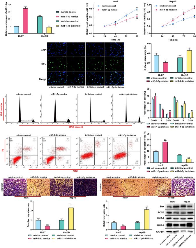
Chen et al.                                                                                                 ORC6, miR-1-3p, and Hepatocellular Carcinoma

INTRODUCTION                                                               were cultured in RPMI-1640 medium (Thermo Fisher Scientific,
                                                                           Waltham, MA, United States) supplemented with 10% fetal
Liver cancer, ranking as the fourth most common cancer                     bovine serum (FBS) (Thermo Fisher Scientific, Waltham,
worldwide, is a deadly disease. Hepatocellular carcinoma (HCC)             MA, United States), 100 µg/ml streptomycin (Thermo Fisher
is the main histological subtype of liver cancer (Villanueva, 2019).       Scientific, Waltham, MA, United States), and 100 U/ml penicillin
Currently, the advances in the treatment of HCC are limited, and           (Thermo Fisher Scientific, Waltham, MA, United States) at 37◦ C
the prognosis of HCC patients is still unsatisfactory, especially          in 5% CO2 in a humidified incubator.
for those with metastatic disease or relapse (Khemlina et al.,                MiR-1-3p mimics and its control (mimics control), miR-
2017; Greten et al., 2019; Marquardt et al., 2019; Raoul et al.,           1-3p inhibitors and its control (inhibitors control), ORC6
2019). It is necessary to explore the molecular mechanism of               overexpression plasmids (ORC6), empty vector (vector) siRNAs
HCC progression to look for novel therapeutic targets to further           for ORC6 (si-ORC6-1 and si-ORC6-2), and their negative
improve the survival time of the patients.                                 controls (si-NC) were obtained from GeneChem (Shanghai,
    More and more microRNAs (miRNAs) have been reported                    China). LipofectamineTM 3,000 (Thermo Fisher Scientific,
to participate in the tumorigenesis and progression of cancers.            Waltham, MA, United States) was used for performing the
MiR-1-3p is identified as a tumor suppressor in various tumors.            transfection. Forty-eight hours after transfection, the cells were
For instance, the expression of miR-1-3p is markedly reduced in            collected and quantitative real-time polymerase chain reaction
bladder cancer tissues; by targeting BDNF, miR-1-3p constrains             (qRT-PCR) or Western blot was performed for examining the
the proliferation and invasion and expedites the apoptosis of              transfection efficiency.
bladder cancer cells (Gao et al., 2018). Reportedly, miR-1-3p is
downregulated in HCC cell lines such as Hep3B and HCCLM3,                  qRT-PCR
and miR-1-3p overexpression represses the proliferation of HCC             Total RNA from tissue samples and cell lines was extracted
cells (Zhang et al., 2019). Nonetheless, the role of miR-1-3p in           utilizing TRIzol reagent (Invitrogen, Carlsbad, CA,
HCC requires further clarification.                                        United States). Then, total RNA was utilized as the template
    Origin recognition complex subunit 6 (ORC6) is crucial in              for the synthesis of cDNA with PrimeScriptTM RT Reagent
DNA replication initiation (Balasov et al., 2009). In colorectal           Kit (Takara, Shanghai, China). With cDNA as the template,
cancer tissues, ORC6 expression is markedly upregulated,                   qRT-PCR was performed on a 7500 Fast Real-Time PCR system
associated with the invasion depth of the tumor and the survival           (Thermo Fisher Scientific, Waltham, MA, United States), using a
time of the patients (Chesnokov et al., 2003). Additionally, ORC6          Bestar qPCR MasterMix kit (DBI Bioscience, Shanghai, China).
is associated with the tumorigenesis of prostate cancer (Wei et al.,       U6 and GAPDH were used as the endogenous control. The
2020). However, the roles of ORC6 in the development of HCC                quantification was performed with the 2−11CT method. The
require further investigation.                                             primers used in the study are listed in Table 1.
    In the present work, we report that the expression level of
miR-1-3p is markedly downregulated in HCC tissues, whereas the             Cell Counting Kit-8 (CCK-8) Assay
expression level of ORC6 is upregulated. We also demonstrate               After transfection, Huh7 and Hep3B cells were inoculated into
that in HCC, miR-1-3p is a tumor suppressor, and ORC6 can                  96-well plates with 2 × 10³ cells per well. The cells were cultured.
facilitate cancer progression. What is more, we identify ORC6 as           CCK-8 solution (10 µl) (Dojindo Molecular Technologies,
a novel target gene of miR-1-3p.                                           Kumamoto, Japan) was added to each well at 24, 48, 72, or 96 h.
                                                                           Then, the cells were further incubated for 2 h, and then the
                                                                           absorbance of the cells at 570 nm was measured.
MATERIALS AND METHODS
                                                                           Transwell Assays
Clinical Samples                                                           Transwell chambers (Millipore, Boston, MA, United States)
Forty patients with HCC who received hepatectomy in Hubei                  were used for cell migration and invasion assays. In migration
Cancer Hospital from May 2016 to May 2019 were enrolled in                 assays, cells in the upper compartment (2 × 10⁴ cells/well)
this study. HCC tissues and adjacent liver tissues were obtained.
All the enrolled patients received no chemotherapy, radiation
treatment, or target therapy before the surgery. Besides, blood            TABLE 1 | Primers for qRT-PCR.
samples from these HCC patients, along with healthy subjects
                                                                           Gene name                            Primer sequence
receiving physical examination, were collected. All tissues and
blood samples were preserved in liquid nitrogen. This work is              miR-1-3p                             F: 50 -CAGTGCGTGTCGTGGAGT-30
supported by the Ethics Committee of Hubei Cancer Hospital,                                                     R: 50 -GGCCTGGAATGTAAAGAAGT-30
and written informed consents of the patients were obtained.               U6                                   F: 50 -CTCGCTTCGGCAGCACA-30
                                                                                                                R: 50 -AACGCTTCACGAATTTGCGT-30
Cell Culture and Transfection                                              ORC6                                 F: 50 - ACAAGGAGACATATCAGAGCTGT- 30
HCC cell lines Hep3B, HepG2, HCCLM3, Huh7, and normal                                                           R: 50 - AGTGGCCTGGATAAGTCAAGAT- 30
liver cells L02 were obtained from American Type Culture                   GAPDH                                F: 50 -ACATCGCTCAGACACCATG-30
Collection (ATCC, Manassas, VA, United States). These cells                                                     R: 50 -TGTAGTTGAGGTCAATGAAGGG-30

Frontiers in Cell and Developmental Biology | www.frontiersin.org      2                                              July 2021 | Volume 9 | Article 652292
Chen et al.                                                                                                 ORC6, miR-1-3p, and Hepatocellular Carcinoma

were cultivated in serum-free RPMI-1640 medium, while the                   separated utilizing SDS-PAGE and then transferred onto the
lower compartment was supplemented with RPMI-1640 medium                    PVDF membrane (Millipore, Boston, MA, United States).
containing 10% FBS. After 24 h of culture, migrated cells across            Then, the membranes were incubated with the primary
the Transwell membrane were fixed with 4% paraformaldehyde,                 antibody (anti-ORC6 antibody, 1:1,000, ab153993; anti-
stained with 0.1% crystal violet solution, and counted in five              Bax antibody, 1:1,000, ab182733; anti-PCNA antibody,
randomly selected fields of view under an inverted microscope. In           1:1,000, ab92552; anti-MMP-2 antibody, 1:1,000, ab92536;
invasion assays, Matrigel-coated Transwell chambers were used,              anti-MMP-9 antibody, 1:1,000, ab76003, Abcam, Shanghai,
and the other procedures were the same.                                     China) at 4◦ C for 8 h. After the membranes were washed
                                                                            by TBST, the membranes were incubated with horseradish
Analysis of Apoptosis                                                       peroxidase-coupled secondary antibody (goat anti-rabbit IgG
Huh7 and Hep3B cells were resuspended in 1 × binding buffer,                H&L, 1: 2,000, Beyotime, Shanghai, China) for 1 h at room
and cell concentration was adjusted to 1 × 10⁶ /ml. In each group,          temperature. Next, the membranes were washed with TBST
100 µl of cell suspension was mixed with 5 µl of Annexin V/FITC             again. Ultimately, the protein bands were developed with an
staining solution, and 10 µl of 20 µg/ml propidium iodide (PI)              enhanced chemiluminescence substrate reaction kit (Thermo
staining solution (Beyotime, Shanghai, China), and incubated at             Fisher Scientific, Waltham, MA, United States). GAPDH was
room temperature for 15 min in the dark. Then, a flow cytometer             employed as the internal control.
(FACScan; BD Biosciences, San Jose, CA, United States) was used
to examine the apoptosis of the cells.                                      Dual-Luciferase Reporter Assay
                                                                            The binding sequence between ORC6 and miR-1-3p was co-
Analysis of Cell Cycle                                                      predicted with miRmap, microT, and miRanda. Accordingly, wild
Huh7 and Hep3B cells were harvested and rinsed with PBS. After              type ORC6 30 UTR sequence (ORC6 WT) and mutant type ORC6
the cells were fixed with 75% ethanol, the cells were stained with          30 UTR sequence (ORC6 Mut) were sub-cloned into pmirGLO
100 µg/ml PI staining solution (Beyotime, Shanghai, China) at               dual-luciferase miRNA target expression vector (Promega Corp.,
room temperature for 10 min. Then, a flow cytometer (FACScan;               Madison, WI, United States), and then, respectively, transfected
BD Biosciences, San Jose, CA, United States) was used to examine            into Huh7 cells, together with miR-1-3p mimics or control
the cell cycle distribution of the cells.                                   miRNAs. Thirty-six hours later, the luciferase activity was
                                                                            examined by a dual-luciferase reporter gene analysis system
                                                                            (Promega, Madison, WI, United States).
5-Ethynyl-20 -Deoxyuridine (EdU) Assay
The proliferation of HCC cells was also performed using                     Statistical Analysis
the Click-iT EdU Assay Kit (Invitrogen, Carlsbad, CA,
                 R

                                                                            All of the data were processed utilizing SPSS 23.0 software
United States). Huh7 and Hep3B cells in the logarithmic growth
                                                                            (SPSS Inc., Chicago, IL, United States). Data were represented
phase were harvested and trypsinized to prepare the single-cell
                                                                            as mean ± standard deviation (SD). The Student’s t-test was
suspension, and the cells were inoculated in a 96-well plate (5,000
                                                                            employed for analyzing the differences between the two groups.
cells/well). Then, 150 µl of 50 µmol/L EdU medium was added
                                                                            One-way ANOVA with Tukey’s post hoc test was utilized for
to each well, followed by the incubation for 2 h at 37◦ C. After
                                                                            analyzing the differences among multiple groups. P < 0.05
washing the cells three times with PBS, 200 µl of PBS containing
                                                                            indicated statistical significance.
4% paraformaldehyde was added to each well, with which the cells
were fixed at room temperature for 40 min. Subsequently, 200 µl
of glycine at a concentration of 2 mg/ml was added to each well             RESULTS
and incubated with cells for 5 min on a decolorizing shaker. After
discarding the solution, 300 µl of PBS was added to each to wash            Expression of miR-1-3p Was
the cells again. After PBS was discarded, 200 µl of PBS containing
0.5% Triton X-100 was added to each well, and the cells were                Downregulated in HCC
incubated for 30 min. After the cells were washed with PBS again,           We firstly analyzed the HCC miRNA expression profile data
200 µl of Apollo staining reaction solution was added to each well          from the Gene Expression Omnibus (GEO) database1 (GEO
and incubated with cells for 30 min in the dark, and then 200 µl            accession no: GSE108724), and it indicated that miR-1-3p
of DAPI solution was added to each well and incubated with cells            expression was markedly reduced in HCC samples compared
for 30 min in the dark. Finally, the pictures of the cells were taken       with adjacent normal tissues (Supplementary Figure 1A).
under a fluorescent inverted microscope, and the percentage of              Additionally, ENCORI database2 also indicated that miR-1-
EdU positive cells was calculated.                                          3p was downregulated in liver cancer tissues compared with
                                                                            normal liver tissues (Supplementary Figure 1B). Besides,
                                                                            importantly, the K-M plotter database3 suggested that HCC
Western Blot                                                                patients with high miR-1-3p expression had a favorable prognosis
RIPA lysis was used to extract the total protein from the
cells. Protein concentrations were quantified using a BCA                   1
                                                                              https://www.ncbi.nlm.nih.gov/
Protein Assay Reagent kit (Thermo Fisher Scientific, Waltham,               2
                                                                              http://starbase.sysu.edu.cn/
MA, United States). Subsequently, the protein extracts were                 3
                                                                              http://kmplot.com/analysis/index.php

Frontiers in Cell and Developmental Biology | www.frontiersin.org       3                                             July 2021 | Volume 9 | Article 652292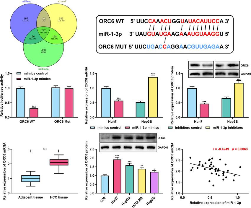
Chen et al. ORC6, miR-1-3p, and Hepatocellular Carcinoma
FIGURE 1 | MiR-1-3p was downregulated in HCC. (A) Expression of miR-1-3p in HCC tissues and adjacent tissues was detected using qRT-PCR. (B) Expression of
miR-1-3p in the serum of HCC patients and healthy controls was detected using qRT-PCR. (C) Expression of miR-1-3p in HCC cell lines and normal liver cells was
examined by qRT-PCR. *** represents p < 0.001, respectively.
(Supplementary Figure 1C). Subsequently, the expressions of 247 genes were performed using the DAVID database.7 GO
miR-1-3p in HCC tissues, HCC patients’ serum, and HCC analysis unveiled that some biological processes were enriched,
cell lines were examined by qRT-PCR. As expected, miR-1-3p including “cell division,” “mitotic nuclear division,” and “cell
expression in HCC tissue was downregulated compared with the proliferation” (Supplementary Figure 1D). KEGG analyses
adjacent liver tissues, and it was downregulated in HCC patients’ indicated that the genes were most enriched in the cell
serum (Figures 1A,B); consistently, miR-1-3p expression was cycle pathway (Supplementary Figure 1E). ORC6 is a crucial
downregulated in HCC cell lines by comparison with normal regulator in cell cycle progression (Chesnokov et al., 2003). Both
liver cell L02 (Figure 1C). Collectively, these data implied that GEPIA database and ENCORI database suggested that ORC6
miR-1-3p may function as a tumor suppressor in HCC. expression was remarkably upregulated in HCC tissues compared
with normal liver tissues (Supplementary Figures 1F,G).
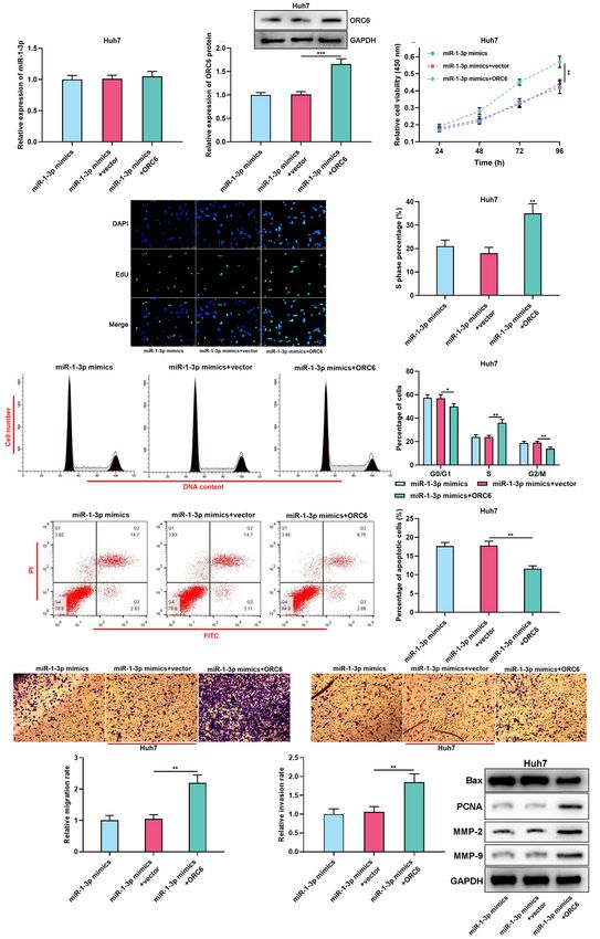
miR-1-3p Suppressed the Malignancy of Additionally, survival analysis was further performed using
HCC Cells the data of GEPIA and ENCORI databases, the samples were
For clarifying the biological functions of miR-1-3p in HCC, stratified into high and low ORC6 expression groups based on
gain-of-function and loss-of-function models were established. the median value of ORC6 expression, and it was demonstrated
Considering that miR-1-3p expression was the lowest in Huh7 that high expression of ORC6 was associated with a detrimental
cells and the highest in Hep3B cells, miR-1-3p mimics and prognosis of HCC patients (Supplementary Figures 1H,I).
miR-1-3p inhibitors were transiently transfected into Huh7 and Additionally, the data from the LinkedOmics database8 showed
Hep3B cells, respectively, and the transfection efficiency was that high ORC6 expression was linked to lymph node metastasis,
confirmed by qRT-PCR (Figure 2A). Functional experiments advanced clinical stage, and high tumor grade (Supplementary
showed that overexpression of miR-1-3p significantly repressed Figures 1J–L). What is more, the reanalysis of the HCC gene
the proliferation, cell cycle progression, migration, and invasion, expression profile data from the GEO database (GEO accession
but promoted the apoptosis of Huh7 cells, whereas miR- no: GSE84402 and GSE101685) indicated that ORC6 expression
1-3p inhibitors worked oppositely (Figures 2B–F). Western was markedly upregulated in HCC tissues, especially the HCC
blot indicated that overexpression of miR-1-3p promoted tissues in T1 and T3 stages compared with normal liver tissues
Bax expression and inhibited PCNA, MMP-2, and MMP-9 (Supplementary Figures 2A–D). Thus, we supposed that ORC6
expression, whereas miR-1-3p inhibitors exerted the opposite might be involved in the regulation of HCC progression.
effects (Figure 2G). Taken together, it was confirmed that miR- For verifying our hypothesis that ORC6 was a direct target
1-3p inhibited the malignant biological behaviors of HCC cells. of miR-1-3p, the binding site between ORC6 mRNA 30 UTR
and miR-1-3p was co-predicted utilizing miRmap, microT,
and miRanda (Figure 3B). Dual-luciferase reporter assay was
ORC6 Was Confirmed as a Direct Target conducted in Huh7 cells, the findings of which suggested
of miR-1-3p that transfection of miR-1-3p mimics remarkably repressed the
To further uncover the mechanism of miR-1-3p on HCC luciferase activity of the ORC6 WT reporter; however, when
progression, through analyzing miRmap,4 microT,5 and miRanda the binding site was mutated, miR-1-3p could not inhibit the
databases,6 we found that there were 247 potential target luciferase activity of the reporter (Figure 3C). As expected, it
genes of miR-1-3p, and interestingly, ORC6 was among them was observed that miR-1-3p overexpression repressed ORC6
(Figure 3A). Afterward, GO and KEGG analyses of these expression at both mRNA and protein levels, whereas miR-1-3p
inhibitors promoted ORC6 expression (Figures 3D,E). What is
4
https://mirmap.ezlab.org/
5 7
http://diana.imis.athena-innovation.gr/ https://david.ncifcrf.gov/
6 8
http://www.miranda.org/ http://www.linkedomics.org/login.php
Frontiers in Cell and Developmental Biology | www.frontiersin.org 4 July 2021 | Volume 9 | Article 652292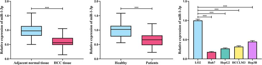
Chen et al.                                                                                                            ORC6, miR-1-3p, and Hepatocellular Carcinoma

  FIGURE 2 | MiR-1-3p inhibited the proliferation, migration, and invasion and promoted apoptosis of HCC cells. (A) The transfection efficiency of miR-1-3p mimics
  and miR-1-3p inhibitors was examined using qRT-PCR. (B) After the transfection, the proliferation of Huh7 and Hep3B cells was detected utilizing the CCK-8 assay.
  (C) After the transfection, EdU assay was performed to determine the proliferation of Huh7 and Hep3B cells. (D) After the transfection, flow cytometry was performed
  to analyze the cell cycle distribution of Huh7 and Hep3B cells. (E) After the transfection, apoptosis of Huh7 and Hep3B cells was measured using flow cytometry.
  (F) After the transfection, migration and invasion of Huh7 and Hep3B cells were measured through Transwell assay. (G) After the transfection, Western blot was used
  to detect the expressions of Bax, PCNA, MMP-2, and MMP-9 of Huh7 and Hep3B cells. *, **, *** represent p < 0.05, p < 0.01, and p < 0.001, respectively.

Frontiers in Cell and Developmental Biology | www.frontiersin.org                  5                                              July 2021 | Volume 9 | Article 652292
Chen et al.                                                                                                          ORC6, miR-1-3p, and Hepatocellular Carcinoma

  FIGURE 3 | MiR-1-3p directly targeted ORC6. (A) Venn diagram showed overlapped target genes of miR–1–3p in miRmap, microT, and miRanda databases.
  (B) The binding site between ORC6 mRNA 30 UTR and miR-1-3p was co-predicted by miRmap, microT, and miRanda databases. (C) A dual-luciferase reporter
  assay was performed to validate the binding site between miR-1-3p and ORC6. (D) The effect of miR-1-3p on ORC6 mRNA expression was measured by qRT-PCR.
  (E) The effect of miR-1-3p on ORC6 protein expression was detected by Westen blot. (F) Expression of ORC6 in HCC tissues and adjacent liver tissues was
  examined by qRT-PCR. (G) Expression of ORC6 in normal liver cells and HCC cell lines was examined utilizing Western blot. (H) The correlation between miR-1-3p
  expression and ORC6 expression in HCC tissues was analyzed. All of the experiments were performed in triplicate. *, **, and *** represent p < 0.05, p < 0.01, and
  p < 0.001, respectively.

more, qRT-PCR indicated that ORC6 expression in HCC tissues                          empty plasmids were, respectively, transiently transfected into
was significantly upregulated in HCC samples (Figure 3F).                            Hep3B cells, and si-ORC6-1, si-ORC6-2, and the si-NC were,
Similarly, Western blot showed that compared with normal liver                       respectively, transiently transfected into Huh7 cells. Western blot
cell L02, ORC6 expression in HCC cell lines was markedly                             was performed to validate the transfection efficiency (Figure 4A).
increased (Figure 3G). Besides, the expression of ORC6 mRNA                          As shown, ORC6 overexpression remarkably promoted the
was in a negative correlation with miR-1-3p in HCC tissues                           proliferation, cell cycle progression, migration, and invasion, but
(Figure 3H). These results validated that ORC6 was a target gene                     repressed the apoptosis of HCC cells, while ORC6 depletion
of miR-1-3p and could be negatively regulated by miR-1-3p.                           showed opposite effects (Figures 4B–F). Western blot results
                                                                                     indicated that overexpression of ORC6 inhibited Bax expression
ORC6 Promoted the Malignant Biological                                               and promoted PCNA, MMP-2, and MMP-9 expression, whereas
Behaviors of HCC Cells                                                               ORC6 knockdown exerted the opposite effects (Figure 4G).
For verifying whether ORC6 could promote the malignant                               These data implied that ORC6 promoted the malignant biological
phenotypes of HCC cells, ORC6 overexpression plasmids and the                        behaviors of HCC cells.

Frontiers in Cell and Developmental Biology | www.frontiersin.org                6                                              July 2021 | Volume 9 | Article 652292
Chen et al. ORC6, miR-1-3p, and Hepatocellular Carcinoma FIGURE 4 | ORC6 regulates the malignant biological behaviors of HCC cells. (A) Transfection efficiency of ORC6 overexpression plasmids and siRNAs was examined using Western blot. (B) After the transfection, the proliferation of Huh7 and Hep3B cells was detected employing a CCK-8 assay. (C) After the transfection, EdU assay was performed to determine the percentage of cells in the S phase. (D) After the transfection, flow cytometry was performed to analyze the cell cycle distribution of Huh7 and Hep3B cells. (E) After the transfection, apoptosis of Huh7 and Hep3B cells was validated using flow cytometry. (F) After the transfection, migration and invasion ability of Huh7 and Hep3B cells were measured through Transwell assay. (G) After the transfection, Western blot was used to detect the expressions of Bax, PCNA, MMP-2, and MMP-9 of Huh7 and Hep3B cells. All of the experiments were performed in triplicate. *, **, and *** represent p < 0.05, p < 0.01, and p < 0.001, respectively. Frontiers in Cell and Developmental Biology | www.frontiersin.org 7 July 2021 | Volume 9 | Article 652292
Chen et al.                                                                                                         ORC6, miR-1-3p, and Hepatocellular Carcinoma

  FIGURE 5 | MiR-1-3p/ORC6 axis regulates the malignant biological behaviors of Huh7 cells. Huh7 cells were transfected with miR-1-3p mimics, or miR-
  1-3p mimics + empty vector, or miR-1-3p mimics + ORC6 overexpression plasmids, respectively. (A) The relative expression of miR-1-3p was detected with qRT-PCR.
                                                                                                                                                        (Continued)

Frontiers in Cell and Developmental Biology | www.frontiersin.org                8                                             July 2021 | Volume 9 | Article 652292
Chen et al. ORC6, miR-1-3p, and Hepatocellular Carcinoma
FIGURE 5 | Continued
(B) The relative protein expression level of ORC6 was measured using Western blot. (C) CCK-8 assay was conducted to detect Huh7 cell proliferation. (D) EdU
assay was performed to determine the percentage of cells in the S phase. (E) Flow cytometry was performed to analyze Huh7 cell cycle distribution. (F) Apoptosis of
Huh7 cells was examined by flow cytometry. (G) Huh7 cell migration and invasion were measured using Transwell assay. (H) Western blot was used to detect the
expression of Bax, PCNA, MMP-2, and MMP-9 of Huh7 and Hep3B cells. All of the experiments were performed in triplicate. *, **, and *** represent p < 0.05,
p < 0.01, and p < 0.001, respectively.
Overexpression of ORC6 Abrogated the hypertension (GH) or preeclampsia (PE) and miR-1-3p
Effects of miR-1-3p (Hromadnikova et al., 2019a). Similarly, miR-1-3p is also
aberrantly expressed in the peripheral blood of children
For further investigating the role of the miR-1-3p/ORC6
descending from pregnancies with GH or PE (Hromadnikova
axis in HCC progression, ORC6 overexpression plasmid
et al., 2019b). Interestingly, in the present work, it was
and miR-1-3p mimics were co-transfected into Huh7 cells.
demonstrated that miR-1-3p was upregulated in both the HCC
Then, qRT-PCR and Western blot were conducted to detect
tissues and serum samples of the patients, and the expression
the expression of miR-1-3p and ORC6 after transfection
level of miR-1-3p in HCC tissues was associated with the
(Figures 5A,B). Then, “rescue experiments” were performed.
prognosis of the patients. These data suggest that miR-1-3p
As expected, ORC6 overexpression reversed the inhibitory
may be a promising biomarker for the diagnosis and prognosis
effect of miR-1-3p overexpression on Huh7 cell proliferation,
prediction of HCC. However, this should be validated with a
cell cycle progression, migration, invasion, PCNA, MMP-
larger number of patients from different medical centers in
2, and MMP-9 expression, and weakened the promotion
the following work.
of miR-1-3p overexpression on Huh7 cell apoptosis and
The current study verified that ORC6 was directly regulated
Bax expression (Figures 5C–H). Collectively, we concluded
by miR-1-3p, and ORC6 counteracted the biological effects
that miR-1-3p repressed the proliferation, migration,
of miR-1-3p on HCC cells. The origin recognition complex
and invasion and promoted the apoptosis of HCC cells
(ORC), a conserved complex consisting of six subunits, is
by targeting ORC6.
essential for the initiation of the DNA replication, serving as
the platform for the assembly of other initiation factors such
as Cdc6 and Mcm proteins (Li N. et al., 2018). ORC6 is
DISCUSSION the smallest ORC subunit. ORC6 partakes in the positioning
of ORC at the origins of DNA replication. The middle
In recent years, the relationship between miR-1-3p and human domain of human ORC6 has an overall fold similar to the
diseases has been gradually unveiled. MiR-1-3p is reported corresponding helical domain of transcription factor TFIIB,
to participate in the pathogenesis of cardiovascular diseases, and several amino acid residues of ORC6 are identified
endocrine diseases, and cancers (Gerlinger-Romero et al., 2017; to contribute to DNA binding and recognition (Prasanth
Jiao et al., 2018; Li M. et al., 2018; Ke et al., 2019; Zhang et al., 2002; Liu et al., 2011). Reportedly, upregulated ORC6
et al., 2019). In cancer biology, miR-1-3p has been identified expression is indicative of a poor clinical outcome in colorectal
as a tumor suppressor in multiple types of cancers. In lung cancer patients (Chesnokov et al., 2003). Another research
cancer, hepatocyte growth factor induces gefitinib resistance reports that depletion of ORC6 increases the sensitivity of
via repressing miR-1-3p expression in PC-9 and HCC827 cells, colonic cancer cell line HCT116 to cisplatin (Gavin et al.,
whereas miR-1-3p restoration abolishes the drug resistance 2008). In the present work, we demonstrated that ORC6 was
by inhibiting c-Met signaling (Jiao et al., 2018). MiR-1-3p significantly upregulated in HCC tissues; further experiments
expression is reported to be downregulated in gastric cancer showed that ORC6 overexpression markedly facilitated the
tissues and cell is associated with tumor size, and transfection proliferation, cell cycle, migration, and invasion of HCC cells
of miR-1-3p notably impedes the proliferation and invasion but repressed apoptosis. Our data suggest that ORC6 is a novel
of MGC-803 cells (Ke et al., 2019). In HCC, reportedly, tumor-promoting factor in HCC, and the downregulation of
miR-1-3p represses the proliferation of cancer cells through miR-1-3p contributes to its dysregulation in HCC. However,
targeting SOX9 (Zhang et al., 2019). In the present study, by the downstream mechanism by which ORC6 participates in
performing gain-of-function and loss-of-function experiments HCC progression remains unclear, which requires further
with Huh7 and Hep3B cells, we demonstrated that miR-1- investigation in the future.
3p induced cell cycle arrest, impeded proliferation, migration, All in all, in the present work, we report the expression,
and invasion, and induced apoptosis of HCC cells, which is biological functions, and regulatory mechanism of the tumor
consistent with the previous report (Zhang et al., 2019). These suppressor miR-1-3p and the tumor-promoting factor ORC6 in
findings suggest that miR-1-3p may be a promising target HCC. Our findings suggest that miR-1-3p/ORC6 axis participates
for HCC treatment. in HCC progression. This work provides novel clues for the
Interestingly, in human diseases, miR-1-3p shows the diagnosis and therapy of HCC. In the following studies, animal
potential as a biomarker. Reportedly, upregulation of miR-1-3p models are required to further validate the role of miR-1-
in peripheral blood is observed in patients with gestational 3p/ORC6 axis in HCC progression.
Frontiers in Cell and Developmental Biology | www.frontiersin.org 9 July 2021 | Volume 9 | Article 652292Chen et al. ORC6, miR-1-3p, and Hepatocellular Carcinoma
DATA AVAILABILITY STATEMENT the statistical analysis. All the authors took part in the
manuscript writing. All authors read and approved the
The original contributions presented in the study are included final manuscript.
in the article/Supplementary Material, further inquiries can be
directed to the corresponding author/s.
ACKNOWLEDGMENTS
ETHICS STATEMENT
We thank Hubei Yican Health Industry Co., Ltd., for
This work is supported by the Ethics Committee of Hubei its linguistic assistance during the preparation of this
Cancer Hospital, and written informed consents of the patients manuscript.
were obtained. The patients/participants provided their written
informed consent to participate in this study.
SUPPLEMENTARY MATERIAL
AUTHOR CONTRIBUTIONS
The Supplementary Material for this article can be found
XT conceived and designed the experiments. HC and online at: https://www.frontiersin.org/articles/10.3389/fcell.2021.
LB performed the experiments. HC and DW performed 652292/full#supplementary-material
REFERENCES a potential target and differentiate HCM from DCM. J. Transl. Med.
16:161.
Balasov, M., Huijbregts, R. P., and Chesnokov, I. (2009). Functional analysis of an Li, N., Lam, W. H., Zhai, Y., Cheng, J., Cheng, E., Zhao, Y., et al. (2018). Structure
Orc6 mutant in Drosophila. Proc. Natl. Acad. Sci. U. S. A. 106, 10672–10677. of the origin recognition complex bound to DNA replication origin. Nature 559,
doi: 10.1073/pnas.0902670106 217–222. doi: 10.1038/s41586-018-0293-x
Chesnokov, I. N., Chesnokova, O. N., and Botchan, M. (2003). A cytokinetic Liu, S., Balasov, M., Wang, H., Wu, L., Chesnokov, I. N., and Liu, Y.
function of Drosophila ORC6 protein resides in a domain distinct from its (2011). Structural analysis of human Orc6 protein reveals a homology with
replication activity. Proc. Natl. Acad. Sci. U. S. A. 100, 9150–9155. doi: 10.1073/ transcription factor TFIIB. Proc. Natl. Acad. Sci. U. S. A. 108, 7373–7378.
pnas.1633580100 doi: 10.1073/pnas.1013676108
Gao, L., Yan, P., Guo, F. F., Liu, H. J., and Zhao, Z. F. (2018). MiR-1-3p inhibits Marquardt, J. U., Saborowski, A., Czauderna, C., and Vogel, A. (2019).
cell proliferation and invasion by regulating BDNF-TrkB signaling pathway in The changing landscape of systemic treatment of advanced Hepatocellular
bladder cancer. Neoplasma 65, 89–96. doi: 10.4149/neo_2018_161128n594 Carcinoma: new targeted agents and immunotherapies. Target Oncol. 14, 115–
Gavin, E. J., Song, B., Wang, Y., Xi, Y., and Ju, J. (2008). Reduction of Orc6 123. doi: 10.1007/s11523-019-00624-w
expression sensitizes human colon cancer cells to 5-fluorouracil and cisplatin. Prasanth, S. G., Prasanth, K. V., and Stillman, B. (2002). Orc6 involved in DNA
PLoS One 3:e4054. doi: 10.1371/journal.pone.0004054 replication, chromosome segregation, and cytokinesis. Science 297, 1026–1031.
Gerlinger-Romero, F., Yonamine, C. Y., Junior, D. C., Esteves, J. V., and Machado, doi: 10.1126/science.1072802
U. F. (2017). Dysregulation between TRIM63/FBXO32 expression and soleus Raoul, J. L., Forner, A., Bolondi, L., Cheung, T. T., Kloeckner, R., and de Baere,
muscle wasting in diabetic rats: potential role of miR-1-3p, −29a/b-3p, and T. (2019). Updated use of TACE for hepatocellular carcinoma treatment: how
−133a/b-3p. Mol. Cell. Biochem. 427, 187–199. doi: 10.1007/s11010-016-2910- and when to use it based on clinical evidence. Cancer Treat Rev. 72, 28–36.
z doi: 10.1016/j.ctrv.2018.11.002
Greten, T. F., Lai, C. W., Li, G., and Staveley-O’Carroll, K. F. (2019). Targeted and Villanueva, A. (2019). Hepatocellular Carcinoma. N. Engl. J. Med. 380, 1450–1462.
Immune-Based Therapies for Hepatocellular Carcinoma. Gastroenterology 156, Wei, J., Yin, Y., Deng, Q., Zhou, J., Wang, Y., Yin, G., et al. (2020). Integrative
510–524. Analysis of MicroRNA and Gene Interactions for Revealing Candidate
Hromadnikova, I., Kotlabova, K., Dvorakova, L., and Krofta, L. (2019a). Signatures in Prostate Cancer. Front. Genet. 11:176. doi: 10.3389/fgene.2020.
Postpartum profiling of microRNAs involved in pathogenesis of 00176
cardiovascular/cerebrovascular diseases in women exposed to pregnancy- Zhang, H., Zhang, Z., Gao, L., Qiao, Z., Yu, M., Yu, B., et al. (2019). miR-1-3p
related complications. Int. J. Cardiol. 291, 158–167. doi: 10.1016/j.ijcard.2019. suppresses proliferation of hepatocellular carcinoma through targeting SOX9.
05.036 Onco. Targets Ther. 12, 2149–2157. doi: 10.2147/ott.s197326
Hromadnikova, I., Kotlabova, K., Dvorakova, L., Krofta, L., and Sirc, J. (2019b).
Postnatal Expression Profile of microRNAs Associated with Cardiovascular and Conflict of Interest: The authors declare that the research was conducted in the
Cerebrovascular Diseases in Children at the Age of 3 to 11 Years in Relation absence of any commercial or financial relationships that could be construed as a
to Previous Occurrence of Pregnancy-Related Complications. Int. J. Mol. Sci. potential conflict of interest.
20:654. doi: 10.3390/ijms20030654
Jiao, D., Chen, J., Li, Y., Tang, X., Wang, J., Xu, W., et al. (2018). miR-1-3p and miR- Publisher’s Note: All claims expressed in this article are solely those of the authors
206 sensitizes HGF-induced gefitinib-resistant human lung cancer cells through and do not necessarily represent those of their affiliated organizations, or those of
inhibition of c-Met signalling and EMT. J. Cell. Mol. Med. 22, 3526–3536. the publisher, the editors and the reviewers. Any product that may be evaluated in
doi: 10.1111/jcmm.13629 this article, or claim that may be made by its manufacturer, is not guaranteed or
Ke, J., Zhang, B. H., Li, Y. Y., Zhong, M., Ma, W., Xue, H., et al. (2019). MiR-1-3p endorsed by the publisher.
suppresses cell proliferation and invasion and targets STC2 in gastric cancer.
Eur. Rev. Med. Pharmacol. Sci. 23, 8870–8877. Copyright © 2021 Chen, Bao, Hu, Wu and Tong. This is an open-access article
Khemlina, G., Ikeda, S., and Kurzrock, R. (2017). The biology of Hepatocellular distributed under the terms of the Creative Commons Attribution License (CC BY).
carcinoma: implications for genomic and immune therapies. Mol. Cancer The use, distribution or reproduction in other forums is permitted, provided the
16:149. original author(s) and the copyright owner(s) are credited and that the original
Li, M., Chen, X., Chen, L., Chen, K., Zhou, J., and Song, J. (2018). MiR- publication in this journal is cited, in accordance with accepted academic practice. No
1-3p that correlates with left ventricular function of HCM can serve as use, distribution or reproduction is permitted which does not comply with these terms.
Frontiers in Cell and Developmental Biology | www.frontiersin.org 10 July 2021 | Volume 9 | Article 652292You can also read