Papillary Thyroid Cancer and a TERT Promotor Mutation-positive Paraganglioma in a Patient With a Germline SDHB Mutation - Oxford University Press
←
→
Page content transcription
If your browser does not render page correctly, please read the page content below
Journal of the Endocrine Society, 2022, 6, 1–9
https://doi.org/10.1210/jendso/bvac076
Advance access publication 10 May 2022
Clinical Research Article

Papillary Thyroid Cancer and a TERT Promotor
Mutation-positive Paraganglioma in a Patient With
a Germline SDHB Mutation
Ali S. Alzahrani,1,2, Meshael Alswailem,1,* Avaniyapuram Kannan Murugan,1,*
Balgees Alghamdi,1 and Hindi Al-Hindi3

                                                                                                                                                           Downloaded from https://academic.oup.com/jes/article/6/7/bvac076/6583142 by guest on 05 June 2022
1
 Department of Molecular Oncology, King Faisal Specialist Hospital & Research Centre, Riyadh, 11211, Saudi Arabia
2
 Department of Medicine, King Faisal Specialist Hospital & Research Centre, Riyadh, 11211, Saudi Arabia
3
 Department of Pathology and Laboratory Medicine, King Faisal Specialist Hospital & Research Centre, Riyadh, 11211, Saudi Arabia
*M.A. and A.K.M. contributed equally to this work.
Correspondence: Ali S. Alzahrani, MD, MBC-46, P.O. Box 3354, Department of Medicine, King Faisal Specialist Hospital & Research Centre, Riyadh, 11211,
Saudi Arabia. Email: aliz@kfshrc.edu.sa

Abstract
Purpose: About 40% of paragangliomas (PGL) are due to germline mutations in one of several susceptibility genes. These genes rarely pre-
dispose to other non-PGL tumors. Here, we describe and functionally characterize a germline SDHB mutation in a patient who developed a
BRAFV600E mutation-positive papillary thyroid cancer (PTC) and a TERT promotor mutation-positive PGL.
Experimental design: A 28-year-old asymptomatic man was discovered incidentally to have a large left-sided mid-abdominal PGL and PTC. He
underwent resection of the PGL and total thyroidectomy and neck dissection followed by I-131 adjuvant therapy for PTC. The histopathology re-
vealed a high-grade PGL and a tall cell-variant PTC with lymph node metastases (T1b N1b M0). He soon developed PGL spinal metastases that
have been rapidly progressing and is currently being treated with Lu177-dotatate therapy. Family screening revealed a positive SDHB mutation in
the mother, a son, and a brother.
Results: In addition to the heterozygous SDHB germline mutation (c.688C>T, p.Arg230Cys), molecular analysis revealed a somatic TERT pro-
motor mutation (C228T) in PGL (negative in PTC) and a somatic BRAFV600E mutation in PTC (negative in PGL). Functional studies showed a higher
proliferation rate in the mutant compared with the wild-type SDHB.
Conclusion: Germline SDHB mutations rarely occur in patients with PTC and may contribute to its aggressiveness. Somatic TERT promotor
mutations rarely occur in PGL and contribute to its aggressiveness and metastatic potential.
Key Words: papillary thyroid cancer, paraganglioma, SDHB, TERT, BRAF, mutations
Abbreviations: CS, Cowden syndrome; CSL, Cowden syndrome-like; CT, computed tomography; FDG, F-18-fluorodeoxyglucose; MRI, magnetic resonance im-
aging; PCC, pheochromocytoma; PET, positron emission tomography; PGL, paraganglioma; PPGL, paraganglioma and pheochromocytoma; PTC, papillary thyroid
cancer; SDHx, succinate dehydrogenase group; TCGA, The Cancer Genome Atlas; TERT, telomerase reverse transcriptase

Paraganglioma (PGL) and pheochromocytoma (PCC), to-                             [7, 8]. The clearest association between the risk of distant
gether abbreviated as PPGL, are neuroendocrine chromaffin                       metastases and the underlying gene mutations is with SDHB
cell-derived tumors [1, 2]. PCC arises from the adrenal me-                     [9, 10]. Patients carrying mutations in this gene have about
dulla and PGL arises from the ganglia of the autonomic ner-                     40% risk of developing distant metastases and are at an in-
vous system [2]. The most common locations for PGL are                          creased risk of death from PGL [9, 10].
the head and neck area and the retroperitoneum in the ab-                          Papillary thyroid cancer (PTC) is the most common endo-
domen, but PGL occurs in the mediastinum, pelvis, and some-                     crine malignancy [11]. Overall, its prognosis is excellent
times in the urinary bladder, lungs, and anterior abdomen                       [12, 13]. However, about 20% to 25% of cases are aggres-
[1, 3]. Over the past 2 decades, it has become clear that                       sive tumors and associated with a high risk of recurrence and
about 40% of PPGL are familial/hereditary [4, 5]. More than                     mortality [14, 15]. Factors associated with increased risk of
a dozen predisposing genes have been identified [2, 4]. The                     recurrence and mortality include the size of the tumor, histo-
most commonly mutated genes are the succinate dehydro-                          pathological subtype, vascular invasion, gross extrathyroidal
genase group (SDHx) that includes SDHA, SDHB, SDHC,                             extension, and lymph node and distant metastases [13].
SDHD, and SDHAF2 [4–6]. Other, less-frequently reported                         Many somatic mutations have also been associated with
predisposing genes include FH, MAX, RET, MDH2, VHL,                             the risk of recurrence and mortality from PTC [16, 17]. The
NF1, and TMEM127 [2, 6, 7]. Although most PPGL are                              most common genetic alteration is the BRAFV600E mutation
benign, about 10% to 40% are metastatic [8]. The risk of                        occurring in about 60% of PTC [16]. This mutation has been
metastases correlates with the underlying genetic mutations                     associated with more aggressive histopathological features

Received: 1 March 2022. Editorial Decision: 3 May 2022. Corrected and Typeset: 3 June 2022
© The Author(s) 2022. Published by Oxford University Press on behalf of the Endocrine Society.
This is an Open Access article distributed under the terms of the Creative Commons Attribution-NonCommercial-NoDerivs licence (https://
creativecommons.org/licenses/by-nc-nd/4.0/), which permits non-commercial reproduction and distribution of the work, in any medium, provided the
original work is not altered or transformed in any way, and that the work is properly cited. For commercial re-use, please contact journals.permissions@
oup.com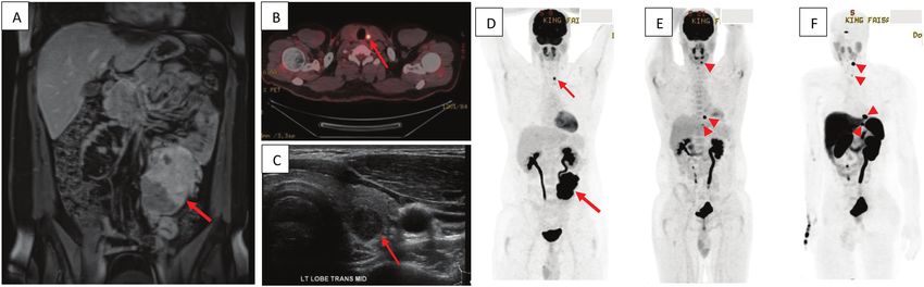
2                                                                                            Journal of the Endocrine Society, 2022, Vol. 6, No. 7

and higher risk of recurrence and mortality [18–20]. Another                  syndrome, neurofibromatosis, other types of cancer, or rare
relatively recently discovered genetic alteration is mutations                syndromes. One brother has sickle cell disease and 3 other
in the telomerase reverse transcriptase (TERT) promotor re-                   brothers have sickle cell trait. The father has end-stage
gion. TERT promotor mutations are associated with more ag-                    renal disease of an unclear cause and had renal transplants
gressive histopathological features and high risk of metastasis,              twice with a good functioning graft. The mother reported
recurrence, and mortality [21, 22]. BRAFV600E and TERT mu-                    no symptoms or significant illness.
tations, when they occur together in the same tumor, are asso-                   Laboratory investigations showed normal 24-hour
ciated with the highest risk of recurrence and mortality [23].                urine metanephrines (repeated 3 times) normetanephrine
   The occurrence of PGL and PTC in the same patient is                       1.54 µmol/L (0-3.43), metanephrine 0.78 µmol/L (0-1.49),
extremely rare and their association is not clear. In this re-                and 3-methoxytyramine 1.02 µmol/L (0-2.06). An F-18-
port, we describe an interesting case of a young man who                      fluorodeoxyglucose positron emission tomography/computed
was found to have a large abdominal PGL and PTC. Genetic                      tomography (FDG PET CT) scan showed an intense uptake in
testing revealed a germline SDHB mutation (p.Arg230Cys).                      the abdominal PGL without other foci in the abdomen, lungs,
Molecular profiling of his PGL and PTC revealed a TERT                        or skeleton, but there was a left thyroid PET incidentaloma
promotor mutation (C228T) in the PGL and BRAFV600E in                         (Fig. 1B and 1D). An ultrasound of the thyroid gland showed

                                                                                                                                                         Downloaded from https://academic.oup.com/jes/article/6/7/bvac076/6583142 by guest on 05 June 2022
PTC. Although TERT promotor mutations are more typic-                         a solid hypoechoic left mid-thyroid lobe nodule measuring
ally found in PTC [24, 25], it was negative in this case in the               1.2 × 0.9 × 0.8 cm with tiny foci of punctate calcifications
PTC but positive in the PGL. The course of the disease in                     and multiple prominent left supraclavicular lymph nodes, the
this patient is characterized by a very aggressive metastatic                 largest of which measured 0.3 cm (Fig. 1C). Fine-needle as-
PGL. This is most likely related to the underlying germline                   piration biopsy from thyroid nodule confirmed the diagnosis
SDHB mutation and the somatic TERT promotor mutation.                         of PTC.
The interplay and synergy between these different genetic al-                    In February 2020, the patient was prepared with
terations are the subject of this paper.                                      phenoxybenzamine 10 mg 3 times daily for 2 weeks and
                                                                              underwent tumor embolization followed by resection of the
                                                                              left retroperitoneal mass, with resection/anastomosis of part
Patients and Methods                                                          of lower descending colon and resection of a small part of
AA is a 28-year-old man who complained of a left scrotal                      the left ureter. The histopathological examination showed
swelling, which was diagnosed as a varicocele in September                    an 11-cm PGL with small and large vascular invasion, cap-
2019. During a workup for that complaint, a computed                          sular invasion, involvement of resection margin by the tumor,
tomography scan (CT) and magnetic resonance im-                               and adventitial ureteric invasion. The tumor has several fea-
aging (MRI) showed a large left midline abdominal mass                        tures that are associated with aggressive behavior including a
measuring 6.4 × 7.9 × 11.8 cm (Fig. 1A). It was compressing                   large size, vascular and capsular invasion, architectural and
the left ureter causing moderate hydroureteronephrosis.                       cytological atypia, and high mitotic activity. The Ki67 index
Laparoscopic biopsy of the mass showed a neuroendo-                           was also high at 40%. Immunohistochemistry showed the
crine tumor consistent with PGL. The patient was com-                         following: synaptophysin: positive; chromogranin: positive;
pletely asymptomatic at that time. Specifically, he had no                    GATA-3, positive; and S100: positive in sustentacular cells.
palpitations, sweating or headache, anxiety, panic attacks,                   The patient had an uneventful postoperative recovery.
tremors, heat or cold intolerance, fatigue, weight loss, or                      In August 2020, the patient underwent total thyroidectomy
loss of appetite. He was exercising regularly with excel-                     and bilateral central and left lateral neck dissection for PTC.
lent stamina. No history of abdominal pain or changes of                      Histopathological examination revealed a tall cell variant
bowel habits and no history of hypertension. His family                       PTC, bilateral and multifocal, largest focus 1.5 cm with no
history was negative for PPGL, Von Hippel-Lindau                              identifiable vascular invasion, and only minimal perithyroidal

Figure 1. (A) Gadolinium-enhanced coronal T1 MRI image of the abdomen showing a large lobulated heterogeneously enhancing mass with areas
of necrosis in the left lower abdomen (arrow). (B) A cross-sectional image of an FDG PET CT scan showing a left thyroid lobe focal lesion (arrow). (C)
A horizontal view of the thyroid gland showing a 1.2-cm hypoechoic suspicious nodule corresponding to the PET scan lesion (arrow). (D) A whole-body
coronal section of FDG PET scan showing an FDG-avid large left lower and mid-abdomen mass (arrow) corresponding to the mass seen on MRI and
the left thyroid lobe incidentaloma (arrow). (E) A follow-up FDG PET CT scan done 1 year after the scan in panel D, showing complete removal of the
abdominal PGL and thyroid cancer but the new appearance of spinal metastases at C4 vertebra, left 9th costovertebral junction, and T-10 vertebra
(arrowheads). (F) A gallium-68 PET CT whole-body scan done at the same time of FDG PET CT scan shown in panel E showing the same lesions
(arrowheads).
Journal of the Endocrine Society, 2022, Vol. 6, No. 7                                                                                           3

extension. There were 11 of 16 central lymph nodes involved              - Germany), and from PGL and PTC tissue using QIAamp
by PTC with extranodal extension in some. There were also                DNA FFPE Tissue Kit (cat. no. 56404, QIAGEN). All exons
3 of 25 left lateral lymph nodes involved with the tumor. The            and exon-intron boundaries of the SDHB gene were initially
8th American Joint Committee on Cancer TNM stage was                     amplified in the peripheral DNA using primers and amplifi-
stage I (T1b N1b M0).                                                    cation conditions as previously described [26]. Subsequently,
   In December 2020, after L-thyroxine withdrawal for 4                  we amplified and sequenced exon 7 looking for the same mu-
weeks, he received 109 mCi adjuvant I-131 therapy. The                   tation in PGL and PTC tumor tissue. We then amplified and
pre- and posttherapy whole-body scans showed foci of up-                 sequenced DNA from the peripheral blood, PTC and PGL
take of 3.1% in the thyroid bed only with no additional                  tissue for BRAFV600E, and TERT promotor mutations (C228T
foci outside the neck. Stimulated thyroglobulin at that time             and C250T) using previously described primers and PCR
was 20.7 µg/L, antithyroglobulin antibodies were negative                conditions [27, 28]. We also screened his family, as shown in
and TSH was 243 mU/L. In November 2020, a gallium-68                     Fig. 2.
PET CT whole-body scan revealed 2 new foci of uptake in
the pedicle of the thoracic spine 8 (T8) and the transverse
process of the cervical spine 4 (C4) that were suspicious for            Mammalian Expression Vector, Site-Directed

                                                                                                                                                    Downloaded from https://academic.oup.com/jes/article/6/7/bvac076/6583142 by guest on 05 June 2022
metastasis. A whole-spine MRI at that time did not find any              Mutagenesis, and Sequencing
corresponding abnormalities in the thoracic or cervical spine            Carboxyl-terminal (C) Myc-DDK-tagged mammalian ex-
and the whole spine was normal. However, a whole-spine                   pression vector carrying a wild-type SDHB (NM_003000)
MRI in April 2021 showed further progression with left 9th               clone (cat. no: RC203182) was purchased from OriGene
costovertebral junction-altered MRI signal with surrounding              Technologies, Inc. (Rockville, MD, USA). The SDHB mutant
soft-tissue thickening and interval development of T10 ver-              construct was made using the wild-type SDHB-Myc-DDK-
tebral body lesion concerning for metastasis. A repeat gal-              tagged plasmid as a template. To create the SDHB R230C
lium-68 and FDG PET CT whole body scans showed showed                    mutant, the mutagenesis reaction was performed in a 50-µL
new foci of increased tracer uptake increased tracer uptake              reaction using the QuickChange XL-II site-directed muta-
at T10 and C7/T1 vertebrae, which were highly suspicious                 genesis kit as recommended by the manufacturer (Agilent
for new bony metastases and progressive tracer avid lesions              Technologies, USA). The molecular size of the plasmids (mu-
in the ninth left costovertebral junction/paravertebral region,          tant clones) was checked by ethidium bromide-stained 1%
which could represent a bone metastasis. There was also a                agarose gel in a standard electrophoresis system and mutated
progressive tracer avid lesion in the left C4/C5 vertebra bony           nucleotides were confirmed by sequencing (Sanger) using
metastatic lesions (Fig. 1E and 1F). The overall findings con-           exon 7 primers with 5′ M13 tag (SDHB-F ex.7: GTA AAA
firmed disease progression. The patient is currently stable re-          CGA CGG CCA GT TCT GCC AAT CAC CTC TTT GTG;
ceiving Lu177 Dotatate radiation therapy for his metastatic              SDHB-R ex.7: CAG GAA ACA GCT ATG ACC TGA ATT
PGL. His family members are being evaluated.                             CCC TTT CCT CTG CAC) by BigDye Terminator v3.1 Cycle
                                                                         Sequencing Kit (ThermoFisher, MA USA).
Molecular Studies
After obtaining an institutional review board approval and               Plasmid Purification, Mammalian Cell Culture,
informed consents from the patient and his family members,               and Transfection
we isolated DNA from peripheral leucocytes using QIAamp                  All the plasmid constructs including C-terminal Myc-DDK-
DNA Blood Mini Kit (cat. no. 51104, QIAGEN GmbH                          tagged vector, wild-type, and mutant SDHB were purified using

Figure 2. The family pedigree shows affected and unaffected family members. All members were tested for the SDHB mutation (p.Arg230Cys). Some
of the mutation-positive individuals were evaluated for the presence of disease and some have not yet been tested.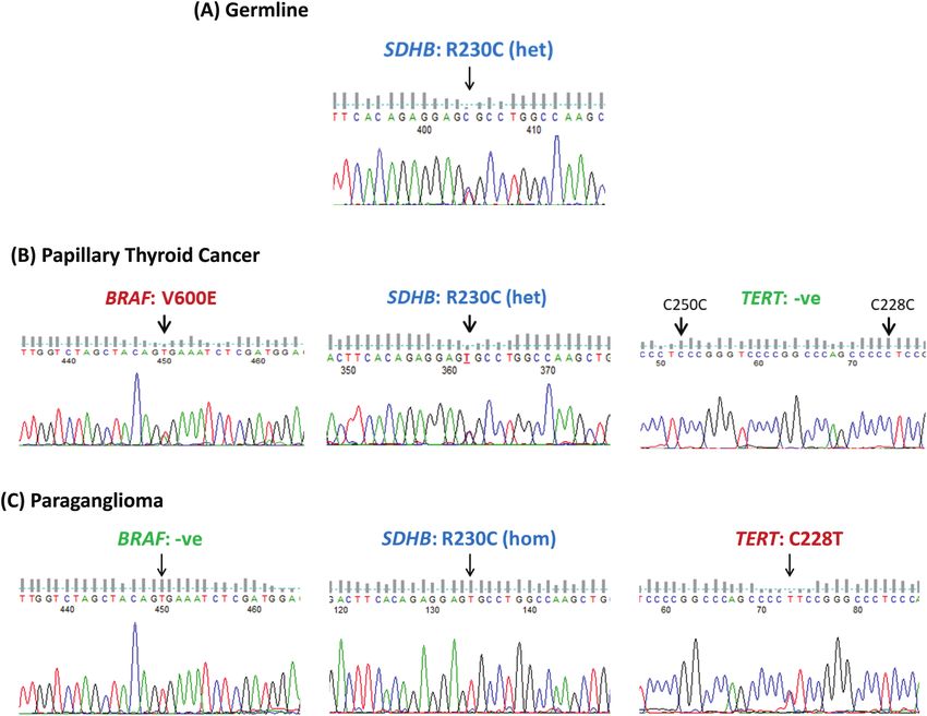
4                                                                                Journal of the Endocrine Society, 2022, Vol. 6, No. 7

EndoFree plasmid preparation kit as per the manufacturer’s           WST-1 (cat. no. 05015944001, Roche), 10 µL/well, and
instruction (Invitrogen, MA). In our experiments, we used            incubated at 37°C with 5% CO2 for 4 hours. The absorb-
HEK293T (CRL-3216) cells that were purchased from                    ance of samples was measured against a blank and absorp-
American Type Culture Collection (Manassas, VA, USA) and             tion at 440 nm by a microplate reader (xMark Microplate
Nthy-ori 3-1 cells that are an immortalized thyroid follicular       Spectrophotometer, Bio-Rad).
epithelial cell line derived from normal adult thyroid tissue
(a gift from Dr. Yufei Shi). The cells were cultured and main-       Wound-healing Assay
tained in DMEM with 10% fetal bovine serum, antibiotic, and
antimycotic per American Type Culture Collection. One day            Wound-healing assay was performed as mentioned previously
before the transfection, the HEK293T cells and the Nthy-ori          [32]. Briefly, HEK293T and Nthy-ori 3-1 cells were transi-
3-1 cells were plated in 24-well plates. The next day, after cells   ently transfected with vector, wild-type, and mutant SDHB
reached 90% confluence, we transfected these cells with equal        (R230C) constructs. After 24 hours of transfection, cells were
amounts of vector, wild-type, and mutant SDHB expression             trypsinized and an equal number of cells was plated with
constructs using Lipofectamine 2000 (Invitrogen, Carlsbad,           higher density (2 × 10⁵ cells/well) in 24-well plates. After 6
CA, USA) as per the manufacturer’s recommendation.                   hours, a scratch (0.6 mm) was made using a pipette tip on

                                                                                                                                         Downloaded from https://academic.oup.com/jes/article/6/7/bvac076/6583142 by guest on 05 June 2022
                                                                     the attached monolayer cells. The cell surface was washed 3
                                                                     times with PBS and maintained at standard cell culture con-
Determination of pH                                                  ditions. As indicated for HEK293T cells, we also performed
The pH of the cell culture medium collected from the                 the wound-healing assay in Nthy-Ori 3-1 cells with a scratch
vector, wild-type, and mutant SDHB (R230C) transfected               of 1.2 mm. Wound closure was carefully watched at 0 and 24
HEK293T and Nthy-ori 3-1 cell culture was determined as              hours from the initial scratch and photographed at ×20 mag-
mentioned previously [29]. Briefly, HEK293T and Nthy-                nification under the microscope (OPTIKA XDS-3FL4, Italy).
ori 3-1 cells were transfected with equal amounts of each
vector, wild-type, and mutant SDHB (R230C) construct as              LIMBO Score Determination
indicated earlier (24-well plate) and 24 hours after trans-
fection, the cell culture medium was replaced with a fresh           LIMBO score indicates the changes in the chaperone binding
medium and further incubated for 24 hours. Forty-eight               efficiency of the protein. LIMBO score and the indicated struc-
hours after transfection, we collected the culture medium,           ture for the wild-type and mutant SDHB were derived from
photographed, and determined the pH at room tempera-                 the SNPeffect database (https://snpeffect.switchlab.org/).
ture by SevenCompact pH/Ion S220 (Mettler-Toledo,                    The variant identification number for this SDHB R230C is
Zchwerzenbach, Switzerland).                                         VAR_054383.

Cell Lysate Preparation and Western Blotting                         The Cancer Genome Atlas Data Analysis
After collecting the cell culture medium at 48 hours                 We analyzed 184 cases of PGL and PCC and 496 cases of
posttransfection for the pH determination, the HEK293T and           papillary thyroid cancer from The Cancer Genome Atlas
Nthy-ori 3-1 cells were washed once with ice-cold PBS and            (TCGA). All the data were analyzed and visualized as de-
lysed on ice using ice-cold radioimmunoprecipitation assay           scribed previously [33].
buffer (Sigma-Aldrich). The adherent cells were scraped with
                                                                     Statistical Methods
a cell scraper, incubated on ice for 45 minutes, and gently
transferred into precooled microfuge tubes and centrifuged           All the statistical analyses were performed using GraphPad
at 12 000 rpm for 15 minutes at 4°C. Supernatants from the           Prism v.8 (GraphPad Software, CA, USA) and the con-
lysed cells were collected in precooled microfuge tubes and          tinuous data were analyzed with unpaired Student t test.
were subjected to Western blotting to determine the protein          P value < 0.05 was determined as statistically significant. The
expression of various transfected constructs (vector, wild-          statistical significance levels between the 2 compared groups
type, and mutant SDHB). We used primary antibodies anti-             are indicated by the asterisk marks (*P ≤ 0.05; **P ≤ 0.01;
myc (cat. no. sc-40, RRID:AB_627268, https://scicrunch.org/          ***P ≤ 0.001; ****P ≤ 0.0001).
resolver/AB_627268) to detect myc-tagged SDHB and anti-β-
actin (cat. no. sc-1616R, RRID:AB_630836, https://scicrunch.         Results
org/resolver/AB_630836) for loading control in the Western
blotting and was performed as earlier mentioned [30].                Germline and Somatic Mutations
                                                                     Amplification and sequencing of DNA from peripheral blood
                                                                     revealed a previously reported heterozygous SDHB mutation
Cell Proliferation Assay                                             (c.688C>T, p.R230C) (Fig. 3). The BRAFV600E mutation was
The proliferation of cells was measured as described pre-            positive in PTC but negative in PGL (Fig. 3). Unexpectedly,
viously [31]. In brief, at 24 hours, HEK293T and Nthy-ori            the C228T TERT promotor mutation was detected in PGL
3-1 cells transiently transfected with vector, wild-type, mu-        and not in PTC (Fig. 3). BARFV600E and TERT promotor mu-
tant SDHB (R230C) were washed once with PBS, trypsinized,            tations were negative in the peripheral blood (Fig. 3). Family
and seeded at a concentration of 4 × 10³ cells/well in 100 µL        screening for the same SDHB mutation revealed that the
DMEM with 10% fetal bovine serum in a 96-well tissue cul-            patient’s mother, a 17-year-old brother, and a 4-year-old son
ture microplate (Costar, cat. no. 3596, Corning Inc., NY) in         were positive (Fig. 2). The mother has not yet been evaluated,
triplicate and cells were maintained in standard conditions.         but the 17-year-old brother and the 4-year old son have been
Cell proliferation assay was conducted by using the reagent          evaluated and so far have no evidence of the disease.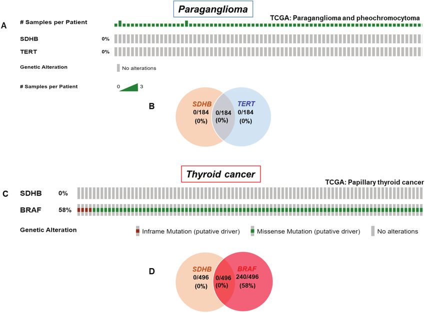
Journal of the Endocrine Society, 2022, Vol. 6, No. 7 5
Downloaded from https://academic.oup.com/jes/article/6/7/bvac076/6583142 by guest on 05 June 2022
Figure 3. Identification of the germline SDHB mutation and the somatic TERT and BRAF mutations. The upper panel (leukocyte DNA) shows the
heterozygous germline SDHB (c.688C>T p.Arg230Cys) mutation. The middle panel (papillary thyroid cancer) shows BRAFV600E mutation (left), a somatic
SDHB (p.Arg230Cys) mutation (middle), and a negative TERT promotor mutation (right). The lower panel (paraganglioma) shows a wild-type BRAF (left),
a homozygous SDHB (p.Arg230Cys) mutation (middle), and C228T TERT promotor mutation (right). All the indicated gene sequences in these panels
are sense-stranded and repeated 3 times with forward and reverse primers by independent PCR.
Functional Characterization of SDHB (p.Arg230Cys) varicocele. During the evaluation of the PGL, he was also found
Mutation to have a PTC. More interestingly is the molecular genetics of
HEK293T and Nthy-ori 3-1 cells were transiently transfected his 2 tumors. He had a previously described germline missense
with Myc-tagged vector, wild-type, and mutant SDHB. Cell SDHB mutation (pArg230Cys), which is the underlying cause
lysates from these expression constructs were subjected to for the PGL. Molecular profiling of the PTC unsurprisingly
Western blotting analyses to ensure success of transfection. revealed the somatic BRAFV600E mutation. C288T TERT pro-
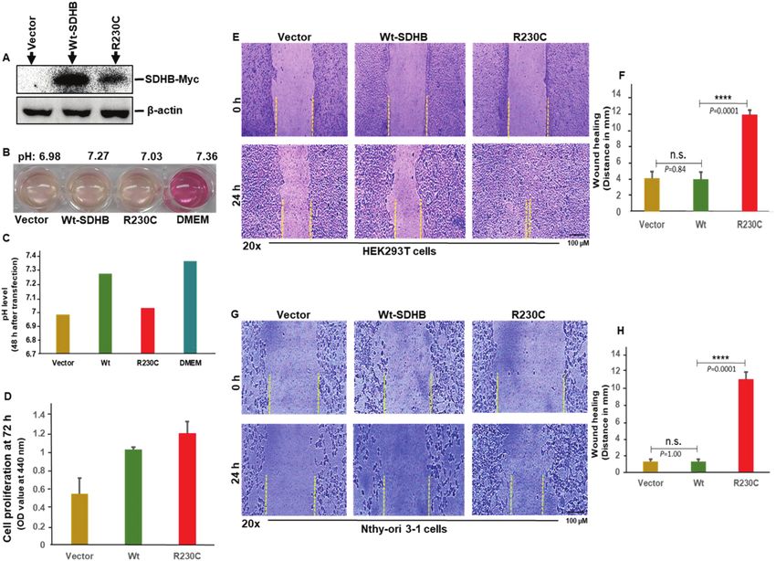
After 24 hours, pH was measured and showed a lower pH motor mutation that is more characteristic of PTC was not
in the mutant culture medium compared with wild-type and detected in the PTC, but unexpectedly was positive in the
vector alone. The proliferation index was highest in the mu- PGL. Functional characterization of the p.Arg230Cys SDHB
tant followed by wild-type and vector alone and the wound mutation confirmed its pathogenicity. Its role in the pathogen-
healing was most impressive in the mutant construct com- esis of PGL is well known [9, 34]. Its pathogenicity in PTC in
pared with the wild-type or vector alone. These experiments this patient is suggested by the functional studies that showed
were repeated 3 times and showed the same results indicating an aggressive proliferation and wound healing of the mutant
the high oncogenic effect of the p.Arg230Cys mutation (Figs. compared to the wild-type SDHB. The interactions between
4 and 5). these several germline and somatic mutations and their effects
on the pathology and course of these tumors are intriguing.
Analyses of the TCGA Data for SDHB and TERT in PTC in this patient was of an intermediate to high risk with
PGL and SDHB and BRAF in PTC multifocal tall cell variant and several positive central and lat-
As shown in Fig. 6, somatic SDHB mutations were not re- eral metastatic lymph nodes, some with extranodal extension
ported in the 496 well-differentiated PTC TCGA database. (T1b N1b M0). The PGL soon metastasized to the spine, and
Similarly, TERT promotor mutations were not reported in the these metastases were progressive over a short time. It is likely
184 pheochromocytoma and PGL TCGA. that the BRAFV600E mutation was a strong driver for PTC in
this patient and the germline SDHB mutation mutation may
have contributed to the development and aggressiveness of
Discussion PTC. The SDHB germline mutation (p.Arg230Cys) was the
In this report, we describe a young man who was found to have underlying genetic cause for PGL and is well known to be
an asymptomatic large abdominal PGL during evaluation of a associated with increased risk of metastasis, but the presence6 Journal of the Endocrine Society, 2022, Vol. 6, No. 7
Downloaded from https://academic.oup.com/jes/article/6/7/bvac076/6583142 by guest on 05 June 2022
Figure 4. Identification of SDHB mutation in paraganglioma. (A) Chromatopherogram shows the germline heterozygous mutation of SDHB gene in
exon 7 at coding nucleotide position 688 changing C to T nucleotide resulting in p.Arg230Cys. (B) Chromatopherogram shows loss of heterozygosity
(a homozygous c.688C>T SDHB mutation. (C) A Schematic diagram shows the SDHB protein and the mutated amino acid position (R230) indicated in
the C-terminal. (D) The 3-dimensional protein structure indicating wild-type SDHB. (E) The 3-dimensional protein structure showing the mutant SDHB
(R230C). (F) The bar diagram displays the LIMBO chaperone binding score of wild-type (score = 3266) and mutant protein of SDHB (score = 3318).
of the C228T TERT promoter mutation is likely to have con- 375 patients, 74 (20%) had increased manganese superoxide
tributed significantly to the rapid progression of PGL. dismutase expression, a sign of mitochondrial dysfunction.
The association of thyroid cancer with SDHx mutations Among the last group, 10 patients (13.5% of the latter sub-
has rarely been described and whether these mutations group and 2.7% of the whole group) carried germline muta-
play a part in the pathogenesis of PTC or not remains un- tions in SDHB (3/10) or SDHD (7/10). No SDHx mutations
clear [35–37]. Ni et al screened 375 PTEN-negative Cowden were found in 700 control subjects [37]. The same investiga-
syndrome (CS) or Cowden syndrome-like (CSL) patients for tors went on to test these findings in a larger group of 608
SDHB, SDHC, and SDHD mutations [37]. Among these PTEN mutation-negative CS/CSL patients, of whom 49 (8%)Journal of the Endocrine Society, 2022, Vol. 6, No. 7 7
Downloaded from https://academic.oup.com/jes/article/6/7/bvac076/6583142 by guest on 05 June 2022
Figure 5. Functional characterization of SDHB (Arg230Cys) mutation. (A) Western blotting analyses of wild-type and mutant SDHB (R230C) expression.
HEK293T cells were transiently transfected with Myc-tagged vector, wild-type, and mutant SDHB. Cell lysates from these expression constructs were
subjected to Western blotting analyses using appropriate antibodies as indicated in Materials and Methods. (B) A representative illustration shows the
color of the cell culture medium collected and photographed after 48 hours from the transiently transfected expression constructs including vector, wild-
type SDHB, and mutant SDHB (R230C) with control DMEM. (C). The bar diagram indicates the related pH of the previously indicated various transfected
constructs. (D) Cell proliferation of HEK293T cells transfected with SDHB constructs. HEK293T cells were plated in 96-well plate in triplicate 24 hours
after transient transfection, maintained in standard condition, and the proliferation assay was performed as described in Materials and Methods. (E)
Illustration shows the migrating efficiency of HEK293T cells transiently expressing SDHB constructs in wound healing assay for 0 and 24 hours. (F)
Distance of wound healing. The distance of wound healing was calculated and expressed (in millimeters) as the distance of cell coverage to the initial
cell-free zone in scratch (0 hours). (G) Illustration shows the migrating efficiency of Nthy-Ori 3-1 cells transiently expressing SDHB constructs in wound
healing assay for 0 and 24 hours. These cells were transfected and experimented exactly as indicated earlier for HEK293T cells. (H) Distance of wound
healing. The distance of wound healing of Nthy-Ori 3-1 cells was calculated and expressed (×20, in millimeters) as the distance of cell coverage to the
initial cell-free zone in scratch (0 hours). Values are presented (panels F, H) as means ± SD of 3 independent experiments. The statistical significance
shown as n.s., *, **, ***, and **** indicate not significant, P < 0.05, P < 0.01, P > 0.001, and P < 0.0001, respectively, as calculated by unpaired t test.
tested positive for germline SDHx mutations [38]. They also and heterozygosity. Immunohistochemistry of 348 tumors of
found SDHx mutations in 26 (6%) of 444 PTEN mutation- different origins, including 60 PTC, was normal with only
positive CS/CLS patients [38]. The rate of thyroid cancer partial loss of immunoreactivity for SDHB in 2 renal cell
was significantly much higher in SDHx mutation-positive cancers [35]. Absence of SDHx mutations in the 700 control
(24/47) patients compared with PTEN mutation-positive subjects in the study by Ni et al [38] and the TCGA data [16]
(27/105, P = 0.002) or PTEN mutation + SDHx mutations- and retaining of SDHB and SDHA immunoreactivity in the
positive patients (6/22, P = 0.038) [38]. The authors con- 348 control subjects in this study [35] all suggest that SDHx
cluded that SDHx mutations are associated with increased mutations are rare in sporadic PTC. Although we did not per-
risk of malignancy including breast and thyroid cancer in form SDHB immunohistochemistry, the functional studies in
patients with CS/CSL syndromes [38]. Functional studies HEK293 and Nthy-ori 3-1 thyroid cell line supports its patho-
suggested a crosstalk between PTEN and SDHx, resulting in genicity or at least contributes to the pathogenesis of PTC.
potentiation of oncogenesis [38]. Papathomas et al studied The other interesting aspect of our patient is the presence
3 SDHB-mutation- and 3 SDHD mutation-positive patients of the well-known TERT promotor mutation C228T in PGL,
for the occurrence of non-PPGL tumors including 1 PTC in which is a more characteristic mutation of thyroid cancer but
an SDHD mutation-positive patient [35]. Although some tu- was absent in the PTC in our patient. This was not reported
mors showed SDHB-negative immunohistochemistry results in the 184 patients included in the TCGA data for PPGL [4]
and loss of heterozygosity, PTC and a pituitary adenoma did (Fig. 6). However, it has been reported rarely in PPGL in
not show loss of heterozygosity for the germline SDHD mu- 2 previous studies. Using Sanger sequencing, Liu et al re-
tation that the patient had or immunoreactivity for SDHB ported C228T TERT promotor mutation in 1 benign PCC
and SDHA stains, suggesting no pathogenetic role for the and 1 metastatic PGL of 105 PCCs and 13 abdominal PGL
SDHx mutations in these cases [35]. All other 3 renal cancers examined [39]. In another study, only 2 cases of meta-
and a pituitary adenoma showed loss of immunoreactivity static extraadrenal PGL (in the urinary bladder) harbored8 Journal of the Endocrine Society, 2022, Vol. 6, No. 7
Downloaded from https://academic.oup.com/jes/article/6/7/bvac076/6583142 by guest on 05 June 2022
Figure 6. Analyses of the TCGA data for SDHB and TERT in paraganglioma and SDHB and BRAF in papillary thyroid cancer. (A) The OncoPrint tab
shows the SDHB and TERT gene promoter mutations across the paraganglioma and pheochromocytoma (TCGA). (B) Venn diagram shows the relation
between the SDHB and TERT genes across the TCGA data sets of 184 paraganglioma and pheochromocytoma cases. (C) The OncoPrint tab shows the
SDHB and BRAF gene mutations across the papillary thyroid cancer (TCGA). (D) Venn diagram shows the relation between the SDHB and BRAF genes
across the TCGA data sets of 496 papillary thyroid cancer cases.
the C228T TERT promotor mutation out of 127 PCC, 18 to determine the prevalence of SDHx mutations in PTC and its
abdominal PGL, and 37 head and neck PGL. ATRX is an exact contribution to its pathogenesis are needed. In the cur-
SWI/SNF chromatin remodelling protein. Similar to the final rent case, it is possible that the cooccurrence of PTC in this pa-
outcome of TERT, ATRX mutations contribute to telomere tient with SDHB-induced PGL was a coincident because PTC
length maintenance by promoting alternative lengthening of is quite common and SDHB mutations are rare.
telomeres. ATRX mutations have been found in 4 cases in
PPGL TCGA data and in 12.6% of 103 PPGL in another
study [40] and were clearly associated with more aggres- Acknowledgments
sive tumors [40]. In a recent study of 200 PPGL, different We thank Dr. Yufei Shi for providing us with cells and the
mechanisms of telomerase activation were investigated [41] members of the Department of Molecular Oncology for their
for TERT promotor mutations and other mechanisms of tel- support.
omerase activation [41]. One SDHC-mutated benign tumor,
1 metastatic SDHC-mutated PGL, and 5 SDHB-mutated
metastatic tumors carried the C228T mutation [41]. In the Funding
same study, ATRX mutations were found in 8 PPGL (4%), The was no specific funding for this work.
mostly in SDHx mutation-positive patients and one-half of
them have metastatic PPGL [41].
In conclusion, we presented a patient in whom an SDHB Conflict of Interest
mutation predisposed to the development of an abdominal The authors have no conflicts of interest to declare.
PGL harboring a somatic TERT promotor mutation that likely
potentiated the aggressiveness of the PGL and leading to pro-
gressive distant metastases. This suggests that, in addition to Data Availability
the primary underlying germline mutations, other genetic and All data generated or analyzed during this study are included
epigenetic alterations may contribute to the development and in this published article.
progression of distant metastases in PGL. The SDHB muta-
tion may have also contributed to the initiation steps and pro-
gression of PTC along with the somatic BRAFV600E mutation. References
However, this remains speculative because it is hard to make 1. Lenders JW, Duh Q-Y, Eisenhofer G, et al. Pheochromocytoma and
a definite conclusion based on 1 case and with no experi- paraganglioma: an Endocrine Society clinical practice guideline. J
mental work was performed to support that. Further studies Clin Endocrinol Metab. 2014;99(6):1915-1942.Journal of the Endocrine Society, 2022, Vol. 6, No. 7 9
2. Neumann HP, Young WF, Jr, Eng C. Pheochromocytoma and 23. Ren H, Shen Y, Hu D, He W, Zhou J, Cao Y, Mao Y, Dou Y,
paraganglioma. N Engl J Med. 2019;381(6):552-565. doi:10.1056/ Xiong W, Xiao Q, Zhang Y, Su X. Co-existence of BRAF(V600E)
nejmra1806651. and TERT promoter mutations in papillary thyroid carcinoma is
3. Erickson D, Kudva YC, Ebersold MJ, et al. Benign paragangliomas: associated with tumor aggressiveness, but not with lymph node me-
clinical presentation and treatment outcomes in 236 patients. J Clin tastasis. Cancer Manag Res. 2018;10:1005-1013.
Endocrinol Metab. 2001;86(11):5210-5216. 24. Alzahrani AS, Alsaadi R, Murugan AK, Sadiq BB. TERT promoter
4. Fishbein L, Leshchiner I, Walter V, et al; Cancer Genome Atlas mutations in thyroid cancer. Horm Cancer. 2016;7(3):165-177.
Research Network. Comprehensive molecular characteriza- doi:10.1007/s12672-016-0256-3.
tion of pheochromocytoma and paraganglioma. Cancer Cell. 25. Panebianco F, Nikitski AV, Nikiforova MN, Nikiforov YE.
2017;31(2):181-193. doi:10.1016/j.ccell.2017.01.001. Spectrum of TERT promoter mutations and mechanisms of ac-
5. Pang Y, Liu Y, Pacak K, Yang C. Pheochromocytomas and tivation in thyroid cancer. Cancer Med. 2019;8(13):5831-5839.
paragangliomas: from genetic diversity to targeted therapies. doi:10.1002/cam4.2467.
Cancers. 2019;11(4):436. doi:10.3390/cancers11040436. 26. Astuti D, Latif F, Dallol A, et al. Gene mutations in the succinate
6. Flores SK, Estrada-Zuniga CM, Thallapureddy K, Armaiz-Peña G, dehydrogenase subunit SDHB cause susceptibility to familial phe-
Dahia PLM. Insights into mechanisms of pheochromocytomas and ochromocytoma and to familial paraganglioma. Am J Hum Genet.
paragangliomas driven by known or new genetic drivers. Cancers. 2001;69(1):49-54. doi:10.1086/321282.
Downloaded from https://academic.oup.com/jes/article/6/7/bvac076/6583142 by guest on 05 June 2022
2021;13(18):4602. doi:10.3390/cancers13184602. 27. Cohen Y, Xing M, Mambo E, et al. BRAF mutation in papil-
7. Buffet A, Burnichon N, Favier J, Gimenez-Roqueplo A-P. An lary thyroid carcinoma. J Natl Cancer Inst. 2003;95(8):625-627.
overview of 20 years of genetic studies in pheochromocytoma doi:10.1093/jnci/95.8.625.
and paraganglioma. Best Pract. Res. Clin. Endocrinol. Metab. 28. Liu X, Bishop J, Shan Y, et al. Highly prevalent TERT promoter
2020;34(2):101416. doi:10.1016/j.beem.2020.101416. mutations in aggressive thyroid cancers. Endocr Relat Cancer.
8. Nölting S, Bechmann N, Taieb D, Beuschlein F, Fassnacht M, 2013;20(4):603-610.
Kroiss M, Eisenhofer G, Grossman A, Pacak K. Personalized man- 29. Tseng PL, Wu WH, Hu TH, et al. Decreased succinate dehydro-
agement of pheochromocytoma and paraganglioma. Endocrine genase B in human hepatocellular carcinoma accelerates tumor ma-
Reviews. 2021;43(2):199-239. lignancy by inducing the Warburg effect. Sci Rep. 2018;8(1):3081.
9. Gimenez-Roqueplo AP, Favier J, Rustin P, et al. Mutations in the doi:10.1038/s41598-018-21361-6.
SDHB gene are associated with extra-adrenal and/or malignant 30. Murugan AK, Liu R, Xing M. Identification and characteri-
phaeochromocytomas. Cancer Res. 2003;63(17):5615-5621. zation of two novel oncogenic mTOR mutations. Oncogene.
10. van Hulsteijn LT, Dekkers OM, Hes FJ, Smit JW, Corssmit EP. 2019;38(26):5211-5226. doi:10.1038/s41388-019-0787-5.
Risk of malignant paraganglioma in SDHB-mutation and SDHD- 31. Murugan AK, Al-Amr A, Al-Ansari MM, Manogaran PS,
mutation carriers: a systematic review and meta-analysis. J Med Al-Hindi H, Alzahrani AS. Single nucleotide polymorphisms in ma-
Genet. 2012;49(12):768-776. trix metalloproteinase 2 (MMP2) enhance BRAFV600E mutation-
11. Seib CD, Sosa JA. Evolving understanding of the epidemi- mediated oncogenicity and invasiveness of papillary thyroid cancer
ology of thyroid cancer. Endocrinol Metab Clin North Am. cells. Endocr Relat Cancer. 2021;28(4):273-289.
2019;48(1):23-35. 32. Murugan AK, Ihara S, Tokuda E, Uematsu K, Tsuchida N, Fukui Y.
12. Tuttle RM, Alzahrani AS. Risk stratification in differentiated thy- SWAP-70 is important for invasive phenotypes of mouse embryo
roid cancer: from detection to final follow-up. J Clin Endocrinol fibroblasts transformed by v-Src. IUBMB Life. 2008;60(4):236-
Metab. 2019;104(9):4087-4100. 240. doi:10.1002/iub.33.
13. Haugen BR. 2015. American thyroid association management 33. Gao J, Aksoy BA, Dogrusoz U, et al. Integrative analysis of com-
guidelines for adult patients with thyroid nodules and differentiated plex cancer genomics and clinical profiles using the cBioPortal. Sci
thyroid cancer: what is new and what has changed? Cancer. Signal. 2013;6(269):pl1.
2017;123(3):372-381. 34. Yang C, Matro JC, Huntoon KM, et al. Missense mutations in the
14. Tuttle RM, Tala H, Shah J, et al. Estimating risk of recurrence in human SDHB gene increase protein degradation without altering
differentiated thyroid cancer after total thyroidectomy and radioac- intrinsic enzymatic function. FASEB J. 2012;26(11):4506-4516.
tive iodine remnant ablation: using response to therapy variables to 35. Papathomas TG, Gaal J, Corssmit EP, et al. Non-pheochromocytoma
modify the initial risk estimates predicted by the new American Thyroid (PCC)/paraganglioma (PGL) tumors in patients with succinate
Association staging system. Thyroid. 2010;20(12):1341-1349. dehydrogenase-related PCC-PGL syndromes: a clinicopathological
15. Yan KL, Li S, Tseng C-H, Kim J, Nguyen DT, Dawood NB, and molecular analysis. Eur J Endocrinol. 2014;170(1):1-12.
Livhits MJ, Yeh MW, Leung AM. Rising incidence and incidence- 36. Wolf KI, Jacobs MF, Mehra R, et al. A family with a carotid body
based mortality of thyroid cancer in California, 2000-2017. Journal paraganglioma and thyroid neoplasias with a new SDHAF2
Clinical Endocrinology & Metabolism. 2020;105(6):1770-1777. germline variant. J Endocr Soc. 2019;3(11):2151-2157.
16. Cancer Genome Atlas Research N. Integrated genomic characteri- 37. Ni Y, Zbuk KM, Sadler T, et al. Germline mutations and variants
zation of papillary thyroid carcinoma. Cell. 2014;159(3):676-690. in the succinate dehydrogenase genes in Cowden and Cowden-like
17. Pak K, Suh S, Kim SJ, Kim IJ. Prognostic value of genetic mutations syndromes. Am J Hum Genet. 2008;83(2):261-268.
in thyroid cancer: a meta-analysis. Thyroid: Official J Am Thyroid 38. Ni Y, He X, Chen J, et al. Germline SDHx variants modify breast
Assoc. 2015;25(1):63-70. and thyroid cancer risks in Cowden and Cowden-like syndrome
18. Xing M. BRAF mutation in thyroid cancer. Endocr Relat Cancer. via FAD/NAD-dependant destabilization of p53. Hum Mol Genet.
2005;12(2):245-262. doi:10.1677/erc.1.0978. 2012;21(2):300-310.
19. Xing M, Alzahrani AS, Carson KA, et al. Association between 39. Liu T, Brown TC, Juhlin CC, et al. The activating TERT promoter
BRAF V600E mutation and mortality in patients with papillary mutation C228T is recurrent in subsets of adrenal tumors. Endocr
thyroid cancer. Jama. 2013;309(14):1493-1501. Relat Cancer. 2014;21(3):427-434.
20. Xing M, Alzahrani AS, Carson KA, et al. Association between 40. Fishbein L, Khare S, Wubbenhorst B, DeSloover D, D’Andrea K,
BRAF V600E mutation and recurrence of papillary thyroid cancer. Merrill S, Cho NW, Greenberg RA, Else T, Montone K, LiVolsi V,
J Clin Oncol. 2015;33(1):42-50. doi:10.1200/JCO.2014.56.8253. Fraker D, Daber R, Cohen DL, Nathanson KL. Whole-exome
21. Liu R, Xing M. TERT promoter mutations in thyroid cancer. sequencing identifies somatic ATRX mutations in pheochromocytomas
Endocr Relat Cancer. 2016;23(3):R143-R155. doi:10.1530/ and paragangliomas. Nat Commun. 2015;6:6140.
ERC-15-0533. 41. Job S, Draskovic I, Burnichon N, et al. Telomerase activation
22. Landa I, Ganly I, Chan TA, et al. Frequent somatic TERT promoter and ATRX mutations are independent risk factors for meta-
mutations in thyroid cancer: higher prevalence in advanced forms static pheochromocytoma and paraganglioma. Clin Cancer Res.
of the disease. J Clin Endocrinol Metab. 2013;98(9):E1562-E1566. 2019;25(2):760-770.You can also read