Presence of LH receptor mRNA in granulosa cells as a potential marker of oocyte developmental competence and characterization of the bovine ...
←
→
Page content transcription
If your browser does not render page correctly, please read the page content below
Reproduction (2003) 125, 437–446
                                                                                                                      Research
      Presence of LH receptor mRNA in granulosa cells as a
    potential marker of oocyte developmental competence and
         characterization of the bovine splicing isoforms
                        C. Robert1 , D. Gagné1 , J. G. Lussier2 , D. Bousquet3 ,
                                   F. L. Barnes4 and M-A. Sirard1∗
      1
          Centre de Recherche en Biologie de la Reproduction, Department of Animal Sciences, Laval University,
           QC, Canada G1K 7P4; 2 Centre de Recherche en Reproduction Animale, Biomédecine Vétérinaire,
             Université de Montréal, Saint-Hyacinthe, QC, Canada J2S 7C6; 3 L’Alliance Boviteq (LAB) Inc.,
                Saint-Hyacinthe, QC, Canada J2S 7A9; and 4 IVF Labs LLC, Salt Lake City, UT 84117, USA

As the expression of the LH receptor (LH-R) in granulosa                six of the isoforms enabled the design of specific
cells is thought to be associated with later stages of follicu-         oligonucleotides to study the presence of the isoforms in
logenesis, this study was undertaken to evaluate the pres-              different follicular cells. All LH-R transcripts studied and
ence of LH-R mRNA as a suitable marker for developmental                the 80 kDa protein product corresponding to the full length
competence of oocytes. Granulosa cells and cumulus–                     receptor were found in granulosa cells of small (< 4 mm)
oocyte complexes (COCs) were recovered from cows that                   and large (> 5 mm) follicles. When the granulosa cells were
had received ovarian stimulation. The COCs were subjec-                 cultured, the transcripts were downregulated by the culture
ted to embryo production procedures in vitro to assess the              conditions; downregulation was more acute in granulosa
embryonic potential of the oocyte, and the corresponding                cells from small follicles. The addition of LH to the cul-
granulosa cells were used to evaluate the presence of LH-R              ture media enhanced LH-R mRNA downregulation. The
mRNA by RT–PCR. The presence of LH-R transcripts in                     presence of several LH-R transcript isoforms was tissue
granulosa cells is not a key characteristic of a follicle               specific and in the theca cells LH-R mRNA was restricted
bearing a competent oocyte, although a higher propor-                   mainly to cells from larger follicles. This finding indicates
tion of oocytes reach the blastocyst stage when LH-R                    that the expression and the splicing of LH-R mRNA are
mRNA is detected in the granulosa cells. Different LH-R                 regulated in a cell-specific and follicular size-specific
isoforms were cloned and sequence discrepancies among                   manner.

                        Introduction                                    cells produce LH-R early in the follicular growth phase,
                                                                        whereas the mural granulosa cells acquire LH-R near
The physiological events involved in follicular recruit-
                                                                        the preovulatory LH surge. In support of these results,
ment, growth, selection, dominance and ovulation are
                                                                        LH-R is expressed only in the granulosa cells of one
not yet fully understood. LH plays an important role in
                                                                        follicle of  8 mm in diameter per cow (Bao et al ., 1997;
the regulation of ovarian function, as it is responsible
                                                                        Manikkam et al ., 2001). This finding implies that there
for ovulation and transformation of follicular cells into
                                                                        is a relationship between follicular dominance and LH-R
luteal cells (Smith et al ., 1994). The action of LH
                                                                        production in granulosa cells. On the contrary, Evans
is mediated by the binding to its receptor (LH-R)
                                                                        and Fortune (1997) reported that the selection of the
which is anchored on the cell membrane (Davis, 1994).
                                                                        dominant follicle occurs without observed differences
However, the events that trigger the production of
                                                                        regarding the LH-R mRNA. Even though patterns of
functional receptors that are located on the surface of
                                                                        LH-R expression in the follicular cells have not yet
the granulosa cells appear to be regulated in a complex
                                                                        been clearly demonstrated, it is generally accepted that
manner and it is still unclear whether the presence
                                                                        the receptor appears on the surface of granulosa cells in
of LH-R is associated with follicular dominance. Peng
                                                                        the late stages of folliculogenesis and that probably only
et al . (1991) reported that the temporal expression of
                                                                        in the dominant follicle does it have the capacity to
LH-R is different between follicular cells, as the theca
                                                                        ovulate.
                                                                            The competence of oocyte development is also partly
∗                                                                       associated with follicular growth, as oocytes recovered
 Correspondence
Email: marc-andre.sirard@crbr.ulaval.ca                                 from follicles of < 2 mm in diameter are meiotically

                                               c 2003 Society for Reproduction and Fertility
                                                            1470-1626/2003
                                                                                                 Downloaded from Bioscientifica.com at 11/16/2021 06:14:43PM
                                                                                                                                               via free access
438 C. Robert et al.
impaired and display poor capacities to reach the Materials and Methods
blastocyst stage, whereas most of the oocytes collected
Bovine ovarian stimulation protocols
from follicles that are 3–8 mm in diameter are meiotically
competent and 30% reach the blastocyst stage when Fourteen cyclic Holstein heifers were used. Two days
subjected to in vitro maturation, fertilization and de- before the start of the superstimulation procedures, the
velopment (Motlik and Fulka, 1986; Pavlok et al ., ovaries of heifers with palpable or functional corpora
1992; Lonergan et al ., 1994; Fair et al ., 1995). The me- lutea (days 3–15) were aspirated transvaginally to remove
chanisms by which the oocyte acquires the capacity to all large follicles. The ovaries were then stimulated for
reach the blastocyst stage during its follicular maturation 2 days with FSH (Folltropinr V, CDMV, St-Hyacinthe,
is unknown. However, it is most probable that all the QC) which was administered in four doses of 3 ml
different types of cell that compose the follicle are in (50 mg). As described by Blondin et al . (2002), at 33 or
close association with each other and that the status 48 h after the last injection, follicles from each ovary
of oocyte maturation is reflected in the surrounding were counted by ultrasonography and classified accord-
granulosa cells. Direct evidence that two types of fol- ing to diameter: small (< 4 mm) or medium to large
licular cell influence each other is the LH-R expression, (> 5 mm). Follicle collection was performed by trans-
as the mRNA is not detected in the cumulus cells vaginal aspiration of small groups of three to five follicles.
surrounding the oocyte, whereas it is present in the The recovered cumulus–oocyte complexes (COCs) were
mural granulosa cells (Peng et al ., 1991; van Tol et al ., cultured in vitro to assess their developmental capability
1996), indicating that the physical distance between the and the remaining granulosa cells were snap-frozen in
oocyte and the granulosa cells influences LH-R mRNA liquid nitrogen and stored at − 80◦ C until RNA extraction
expression. These observations are supported by Eppig to study the presence of LH-R mRNA.
et al . (1997) who showed that the mouse oocyte represses
the expression of the LH-R in cultured granulosa cells.
Embryo production in vitro
Considering these reports, it was proposed that the
expression of the LH-R in granulosa cells could be The COCs were kept in separate groups and were
associated with the presence of a mature and, hence, matured in 50 l droplets of maturation medium for 24 h.
developmentally competent oocyte, as the repressive The maturation medium included TCM-199 supplemen-
control of LH-R expression could be removed once ted with Earle’s salts (Gibco BRL Life Technologies,
the oocyte acquires the capacity for embryonic devel- Burlington, ON), bicarbonate (Sigma, St Louis, MO) and
opment, thus ensuring the ovulation of a competent 20% (v/v) heat-treated fetal calf serum (FCS), 0.5 g
oocyte. FSH-P ml−1 (NIDDK), 5.0 g LH l−1 (NIDDK), 1.0 g
The functional structure of the receptor is interesting oestradiol ml−1 (Sigma), 0.2 mmol pyruvic acid l−1 and
as several splicing isoforms have been observed in many 50.0 g gentamicin ml−1 (Sigma). The droplets were
species (Sokka et al ., 1992; VuHai-Luu Thi et al ., 1992; covered with mineral oil (Aldrich Chemical, Milwaukee,
Bacich et al ., 1994; Koo et al ., 1994; Mamluk et al ., WI) and preincubated in a humidified atmosphere
1998) and to our knowledge, no physiological function containing 5% CO2 and 95% O2 at 38.5◦ C for a
has been assigned to any of the splice forms. The LH-R minimum of 2 h before the addition of the COCs.
is composed of a large extracellular LH–CG binding After maturation, the COCs were washed twice in
domain followed by a transmembrane domain spanning Hepes-buffered Tyrode’s medium (TLH) containing 0.3%
seven times the cytoplasmic membrane that anchors (w/v) fatty acid-free BSA (Sigma), 0.2 mmol pyruvic
the receptor at the cell surface and a relatively short acid l−1 (Sigma), 2.0 g heparin ml−1 (Sigma) and
intracellular domain responsible for signal transduction 50.0 g gentamicin ml−1 (Sigma). The COCs were
(Dufau et al ., 1995). washed and transferred into 48 l fertilization medium
The present study characterizes the presence of LH-R to which 2 l of 2 mmol penicillamine l−1 , 1 mmol
mRNA in granulosa cells of superstimulated cows to eval- hypotaurine l−1 , 250 mol adrenaline l−1 (PHE, all from
uate the expression of LH-R in granulosa cells as a marker Sigma) was added followed by bovine spermatozoa.
of oocyte developmental competence. The study was ex- The fertilization procedure lasted 15–18 h. The fertiliz-
tended by characterizing the presence of LH-R isoforms ation medium contained Tyrode’s lactate medium, 0.6%
in bovine follicular cells of various sources using semi- (w/v) fatty acid-free BSA (Sigma), 0.2 mmol pyruvic
quantitative RT–PCR. The association of LH-R expression acid l−1 (Sigma), 2.0 g heparin ml−1 (Sigma) and
with follicular size was studied at the mRNA level using 50.0 g gentamicin ml−1 (Sigma). The bull spermatozoa
specific primers segregating the isoforms and at the used was kindly provided by Le Centre d’Insémination
protein level in granulosa cells from different follicular Artificielle du Québec (CIAQ) and were supplied in
size groups. Finally, RT-PCR was used to evaluate, in a frozen straws that were thawed in a 35◦ C water bath for
time-dependent manner, the effect of addition of LH on 1 min and then separated using the swim-up technique as
the presence of LH-R mRNA in cultured bovine granulosa described by Parrish and Foote (1986). An aliquot of 2 l
cells. sperm suspension was added to each of the fertilization
Downloaded from Bioscientifica.com at 11/16/2021 06:14:43PM
via free accessLH receptor splicing isoforms in bovine granulosa cells 439
droplets to a final concentration of 1 × 10⁶ spermatozoa of the extracellular LH binding domain of the LH-R
ml−1 . cDNA. Another primer was designed (LH-R G to H
The embryos were washed twice in development low) in a region located downstream and was used
medium and then transferred to 50 l of preconditioned in combination with the upstream primer LH-R A to
development droplets. The embryos were cocultured B up to find other different LH-R isoforms. First-strand
with bovine oviductal cells prepared as described by cDNA was synthesized from total RNA (5 g) using a
Blondin and Sirard (1995). ‘SuperScriptTM Preamplification System for First Strand
cDNA Synthesis’ kit (Gibco BRL Life Technologies) and
200 ng oligo(dT)12−18 primer in a total reaction volume
Culture of granulosa cells of 20 l. An aliquot of 2 l cDNA was subjected to PCR
The granulosa cells were collected from ovaries from amplification using a Programmable Thermal Controller
an abattoir; the ovaries were kept on ice during trans- (PTC-100TM , MJ Research Inc., Watertown, MA). The
portation to the laboratory and follicular dissection. Cells PCR reaction mixture contained 30 pmol of each primer,
were cultured as described by Rouiller et al . (1996) in 1 × PCR buffer (PE Applied Biosystems, Foster City,
a 24-well polystyrene dish at 1 × 10⁶ viable cells (as CA), 0.35 mmol dNTPs l−1 and water to 50 l. The
determined by trypan blue exclusion) per well in Ham’s reaction was heated to 94◦ C for 10 min (hot start)
F-12 medium supplemented with human apo-transferin after the addition of Taq Gold polymerase (PE Ap-
(10 mg l−1 ) (Sigma), ascorbic acid (17.6 mg l−1 ) (Sigma) plied Biosystems). The amplification profile comprised
and pig FSH (50 g l−1 ) (National Institute of Diabetes, 35 cycles: 94◦ C for 1 min (dissociation), 58◦ C for
Digestive and Kidney Diseases; NIDDK, Bethesda, MD) 1 min (annealing) and 72◦ C for 1 min (extension). The
in a humidified atmosphere containing 5% CO2 and 95% final cycle included a further 5 min at 72◦ C to complete
O2 at 38.5◦ C. LH (100 ng ml−1 ) (NIDDK) was added to strand extension. An aliquot of the PCR reaction was
the media to study the effect of the presence of LH on the subjected to electrophoresis on a 1% (w/v) agarose
receptor mRNA. Once the cells were collected, they were gel.
snap-frozen in liquid nitrogen and stored at − 80◦ C. The LH-R isoforms were cloned as follows: the
PCR products were purified using Qiaquick columns
(QIAGEN Inc., Argentia Road, ON) and subcloned into a
Tissue collection and RNA extraction TA cloning system (Invitrogen Corporation, Faraday Ave,
CA). One microgram of ligated DNA was transformed
Total ovarian tissue samples were collected from ovar- by heat shock in 100 l of competent DH5␣ Escherichia
ies obtained from an abattoir and stored on ice during coli cells. Colonies were grown on Luria-Bertoni (LB)
transportation. The total ovary sample included entire medium containing ampicillin for 16 h at 37◦ C. Plasmid
ovaries with corpora lutea. The granulosa and theca cells DNA of selected colonies was extracted using the
were collected by dissecting the follicles on ice under a plasmid miniprep system (QIAGEN Inc.). The preps
stereomicroscope. The cumulus cells of COCs collected were digested with EcoRI, PST I and Hind III restriction
by aspiration of follicles of 3–7 mm in diameter were enzymes to identify eight plasmids with different
dissociated mechanically. All of the different types of LH-R isoforms. The authenticity of the cloned cDNAs
cell were washed in PBS, collected by centrifugation at was verified by sequencing using T7 SequencingTM kit
900 g for 2 min and frozen at − 80◦ C. Tissue samples (Pharmacia Biotech, Baie D’Urfé, QC). Further sequen-
from liver, kidney and muscle were snap-frozen and cing was performed using an automated sequencing
stored at − 80◦ C until RNA extraction. Total RNA was service.
isolated from the tissues using Trizol reagent (Gibco
BRL Life Technologies) and quantified by measuring its
absorbancy at 260 nm. An aliquot of each sample was RT–PCR to evaluate the presence of each of the cloned
subjected to electrophoresis to verify the integrity of LH-R isoforms
RNA. The RNA samples were treated by DNase I (RNase The comparative sequence analysis of the different
free) (Ambion, Austin TX) and re-extracted by phenol– cloned LH-R isoforms revealed deleted and inserted
chloroform before the RT–PCR reactions. Genomic DNA sequences allowing the design of primers intended to
contamination in the RNA samples was evaluated by discriminate three pairs of the cloned LH-R isoforms
performing PCR reactions directly on an aliquot of the (Table 1). The RT–PCR procedure was as described
RNA samples without the reverse transcription step. above. The primers used to discriminate clones A and B
were LH-R A to B up and LH-R A to B low. The upstream
primer used to discriminate clones C and D was LH-R C
Amplification and cloning of different isoforms of the
to D up and the downstream primer was LH-R low. The
LH-R mRNA
upstream primer used to discriminate clones E and F was
Primers were designed according to the bovine LH-R up and the downstream primer was LH-R E to F
sequence found in Genbank (U20504) to amplify 713 bp low.
Downloaded from Bioscientifica.com at 11/16/2021 06:14:43PM
via free access440 C. Robert et al.
Table 1. Details of the primers used to amplify different isoforms of the bovine LH receptor mRNA
and the -actin used as internal standard
Position on Genbank
Name Primer sequence sequence U20504
LH-R up 5 -TCCCTCGGTTAAAATACCTAAGC-3 397–421
LH-R low 5 -GTAGCCCATAATGTCTTCACAGG-3 1088–1111
LH-R A–B up 5 -AACCACCATACCAAGAAATGC-3 517–537
LH-R A–B low 5 -ATCCCAGCCACTCAGTTCAC-3 1005–1024
LH-R C–D up 5 -GCCAGTTGAATCTGTTTCTGC-3 N/A
LH-R E–F low 5 -GCACACAGGTTTGATCCCTT-3 N/A
LH-R G–H low 5 -CAACAGAAAGAAATCCCTTTGG-3 1924–1945
-actin up 5 -CGTGACATTAAGGAGAAGCTGTGC-3 N/A
-actin low 5 -CTCAGGAGGAGCAATGATCTTGAT-3 N/A
Western blot analysis Table 2. Blastocyst production in relation to follicular size and
presence of LH receptor (LH-R) mRNA in bovine granulosa cells
The antibody used is specific to the first 11 amino detected by RT–PCR
acids of rat LH-R extracellular domain. The antibody
was a kind gift of P. C. Roche (Mayo Clinic, Rochester, Size of follicle Presence of Blastocyst
MN). Granulosa and theca cells were collected from (mm) LH-R∗ n Oocyte (%)
bovine ovaries from an abattoir as described above. For 5 + 8 53 31 (58.5%)a
homogenized in a modified radioimmunoprecipitation − 3 29 12 (41.4%)b
buffer containing 50 mmol Tris–HCl l−1 (pH 7.4), 1%
∗
(w/v) NP-40, 0.25% (w/v) sodium deoxycholate, The primers used can detect isoforms A to F.
n represents the number of oocyte pools used.
150 mmol NaCl l−1 , 1 mmol EDTA l−1 , 1 mmol phenyl- ab
Percentages with different superscripts are significantly different
methylsulfonyl (PMSF) l−1 , 1 g aprotinin ml−1 , 1 g (P < 0.05).
leupeptin ml−1 , 1 g pepstatin ml−1 , 1 mmol sodium
orthovanadate (Na3 VO4 ) l−1 and 1 mmol sodium fluoride
Results
l−1 (all from Sigma). Fifty micrograms of protein and a
coloured molecular size marker (Bio-Rad, Mississauga, Qualitative RT–PCR was carried out to detect the pres-
ON) were separated by 10% SDS–PAGE under reducing ence of LH-R mRNA in granulosa cells collected during
conditions. The proteins were electro-transferred on oocyte recovery, to evaluate the receptor transcript as a
to a membrane (CBS Scientific Co. Inc., Delmar, CA) potential marker of oocyte developmental competence.
and the hybridization was performed as described by The primers were designed to amplify 713 bp of the large
Han et al . (1997). In brief, non-specific binding sites extracellular domain of the LH-R cDNA spanning exon 4
were blocked with 5% (w/v) non-fat dry milk and to the beginning of the transmembrane domain (exon 11).
the blot was incubated for 2 h at room temperature The LH-R mRNA was not detected in all of the granulosa
with 1:1500 dilution of LH-R antibody. The blot was cell pools, whereas -actin, used as positive control, was
washed and reincubated for 1 h at room tempera- detected in all samples. Higher blastocyst rates were
ture with 1:3000 dilution of horseradish peroxidase- observed (P < 0.05; Table 2) when LH-R mRNA was
labelled anti-rabbit IgG (Bio-Rad). The blot was detected in the corresponding granulosa cells indicating
washed and revealed by chemiluminescence (Pharmacia a beneficial effect.
Biotech). Several bands were detected after PCR amplification,
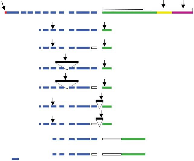
indicating the presence of multiple transcripts (Fig. 1).
The LH-R transcripts were detected in granulosa cells
collected from small (< 4 mm) and large (> 5 mm)
Statistical analysis
follicles. With this first set of primers, six different cDNAs
ANOVA was carried out to determine the effect of were cloned (Clones A to F; Fig. 2) and to find other
the follicular size and the presence of LH-R mRNA in splicing isoforms a second set of primers was designed
granulosa cells on the percentage of blastocysts obtained, to amplify a region located further downstream into
to evaluate whether the presence of LH-R transcripts exon 11. From this second set of amplifications, two
are associated with oocyte developmental competence. more isoforms were isolated (Clones G and H; Fig. 2).
All percentage data were ARCSIN transformed before Sequence analysis and comparisons between clones
analysis. indicate that clone A is identical to the targeted Genbank
Downloaded from Bioscientifica.com at 11/16/2021 06:14:43PM
via free accessLH receptor splicing isoforms in bovine granulosa cells 441
Computerized open reading frame (ORF) analysis
DNA marker Granulosa revealed that only clones A and B have an ORF over
their entire sequences, whereas the insertion in clones C
Ovary
Water
to D leads to a stop codon early in the extra sequence
< 4 mm > 5 mm
inserted between exons 7 and 8 (Fig. 2). The sequence
discrepancies in the other clones (E to H) produce a stop
codon near the transmembrane domain (Fig. 2).
713 bp The sequence discrepancies enabled the design of
632 bp primers to segregate the first six clones and semi-
LH-R quantitative RT–PCR was used to study the presence
of these LH-R isoforms in several tissues (Fig. 3). As
expected, all LH-R transcripts were detected in the ovary
and corpus luteum samples, whereas they were not
detected in liver and kidney samples. Weak signals for
isoforms A and E were observed in the muscle sample.
The cumulus cell sample showed only a very weak band
for clone E (data not shown), whereas all the transcript
isoforms were expressed in granulosa cells collected
-actin
from follicles of < 4 mm or > 5 mm in diameter. By
contrast, the isoform regulation in theca cells appears
374 bp to be related to follicular size, as the presence of
the LH-R isoforms was largely absent from theca cells
obtained from follicles of < 4 mm in diameter except
for the detection of a very faint band for clone E (data
Fig. 1. Agarose gel electrophoresis of RT–PCR amplifications of not shown). The theca cells collected from follicles of
the bovine LH receptor (LH-R) showing several bands, and of > 5 mm in diameter did not show a signal for clones C
-actin. The RT–PCR amplifications were performed on total RNA to D and only a weak band was found for clone A.
extracted from bovine granulosa cells and ovary. The sample of The same approach was used to analyse the effect
the ovary used as a positive control represents total RNA extracted of culture and the presence of LH on the presence of
from an ovarian cortex with a corpus luteum. The follicular size
the first six LH-R clones (A to F) by culturing bovine
group (< 4 mm and > 5 mm) is indicated above each lane.
granulosa cells collected from follicles with a diameter of
< 4 mm or 5 mm with or without LH supplementation.
LH-R sequence (U20504), whereas clone B is missing A fraction of the cells were harvested after a time course
82 bp. Clone A and clone B are identical to Genbank to assess the influence of culture over time (Fig. 4).
sequences U41414 and U41413, respectively. When the Follicular size had a clear impact in vitro on the presence
rat LH-R gene structure was used as a model to identify of LH-R mRNA, whereas the duration of culture resulted
the size of the exons and the splicing sites, the 82 bp gap in downregulation of the LH-R transcripts. Nearly all
corresponded to the entire exon 10 which is absent in LH-R transcripts were undetectable in granulosa cells
clones B, D, F and H. from follicles of < 4 mm in diameter after 2 h in culture,
Clones C to D and E to F differ by the insertion whereas for the follicles of > 5 mm in diameter, the
of a supplementary 323 bp and 174 bp sequence, downregulation effect was observed after 4 h of culture.
respectively (Fig. 2). The additional portions of these The addition of LH to the culture media further enhanced
clones are not homologous with any LH-R Genbank the downregulation of LH-R transcripts as shown (Fig. 4).
sequences. Furthermore, when they were submitted to Western blot analyses revealed that the reported
a BLAST analysis, the supplementary fragment of clones 80 kDa full length LH-R is translated in granulosa cells
C to D did not find any match, whereas a portion of the from follicles of < 4 mm and > 5 mm in diameter. A
additional 174 bp of clones E to F showed 91% identity doublet was also found at 45 kDa in the granulosa cells of
with the short interspersed repetitive element (SINE) both size groups and the corpus luteum, but was absent
sequences and the second exon of bovine adenylate in the theca cells of follicles of < 4 mm in diameter
kinase isozyme 2 (Genbank D90066) also showed a (Fig. 5).
92% identity over 65 nucleotides, whereas the bovine
phosphoprotein called Bucentaur (BCNT) (Genbank
AB005652) showed a 84% identity over 102 nucleotides.
Discussion
The sequence discrepancy associated to clone G was a
deletion of 265 bp at the beginning of exon 11, whereas In mice, the oocyte suppresses the expression of LH-R
clone H combined the deletion of exon 10 to the same in the granulosa cells (Eppig et al ., 1997) and in cows,
deleted portion of exon 11. LH-R mRNA is not detected in the cumulus cells but is
Downloaded from Bioscientifica.com at 11/16/2021 06:14:43PM
via free access442 C. Robert et al.
5´ UTR IC 3´ UTR
Extracellular
TM
1 2 3 4 5 6 7 8 9 10 11
Genbank
Clone A 713 bp
1
Clone B 632 bp
*
Clone C 1049 bp
*
1
Clone D 968 bp
*
Clone E 889 bp
*
1
Clone F 808 bp
* 2
Clone G 1165 bp
Clone H * 1 2
1084 bp
100 bp
Fig. 2. Illustration of the alignment for the different LH receptor (LH-R) clones. The bovine LH-R illustration is based on
Genbank sequence U20504. The limits of the exons were determined according to the splicing sites found in the rat LH-R
gene structure (Dufau et al ., 1995) (Genbank number AH004953). Specific cDNA and protein domains are indicated by
5 untranslated region (5 UTR), extracellular, transmembrane (TM), intracellular (IC) and 3 UTR. On the different LH-R
clones, boxes 1 and 2 represent missing portions, whereas for clones C, D, E and F the extra portion size and the insertion
site are represented by solid black boxes. The arrows indicate the priming sites used to discriminate the isoforms and the
stop codon is indicated by (*). The size of the fragment amplified using primers LH-R up and low (Table 1) is indicated
for each isoform.
present in the mural granulosa cells (Peng et al ., 1991; This ovarian stimulation regimen induces acquisition of
van Tol et al ., 1996). Taking these reports into consider- developmental competence of oocytes in as many as
ation together with the generally accepted contention 80% of the recovered oocytes (Blondin et al ., 2002).
that only the dominant follicle expresses LH-R in the When LH-R mRNA was detected in these granulosa cells,
granulosa cells, it was proposed that the oocyte would a higher proportion of the associated oocytes reached
allow the expression of LH-R in granulosa cells once the blastocyst stage but, unfortunately, development
it has completed its cytoplasmic maturation and awaits was normal in some groups of oocytes in which LH-R
the preovulatory LH surge. If this proposal is correct, mRNA was not detectable in the granulosa cells.
the presence of LH-R mRNA in granulosa cells could be Therefore, the presence of LH-R mRNA in the granulosa
a valuable tool for oocyte management in an in vitro cells is not by itself a reliable marker of oocyte
production programme because it would be a good developmental competence. Other unidentified factors
marker of oocyte developmental competence and the may have an important impact on oocyte developmental
technique does not destroy the gamete. competence and may act with LH-R in a synergistic
In the present study, the granulosa cells were collected manner. For instance, it has been shown that the more
in vivo from ovaries that were stimulated using a competent oocytes are recovered from follicles in the
protocol that enhances the recovery of developmentally early stage of atresia (Blondin and Sirard, 1995). During
competent oocytes (Sirard, 2001; Blondin et al ., 2002). the ovarian stimulation protocol used in the present
Downloaded from Bioscientifica.com at 11/16/2021 06:14:43PM
via free accessLH receptor splicing isoforms in bovine granulosa cells 443
Corpus luteum
DNA marker
DNA marker
G < 4 mm
G > 5 mm
T < 4 mm
T > 5 mm
Cumulus
Muscle
Kidney
Ovary
Water
Liver 507 bp
LH-R A and B
425 bp
LH-R C and D 597 bp
515 bp
623 bp
LH-R E and F 541 bp
-actin 374 bp
Fig. 3. RT–PCR discriminating the different bovine LH receptor (LH-R) transcripts cloned in diverse tissue samples of -actin. For the
negative control, water was added instead of template during reverse transcription. The total ovarian sample was extracted from several
ovaries with corpora lutea. The granulosa (G) and theca (T) cells were collected by dissecting bovine ovaries obtained from an abattoir.
The size of the follicles is indicated above the lanes. The cumulus cells were dissociated mechanically from the cumulus–oocyte
complexes collected by aspiration of follicles of 3–7 mm in diameter from bovine ovaries obtained from an abattoir.
DNA marker
DNA marker
n.c. 2h 4h 16 h
5 5 5 5
+ – + – + – + – + – + –
507 bp
LH-R A and B
425 bp
LH-R C and D 597 bp
515 bp
LH-R E and F 623 bp
541 bp
-actin
374 bp
Fig. 4. RT–PCR discriminating the different LH receptor (LH-R) transcripts in cultured bovine granulosa cells and -actin. The duration
of culture of the granulosa cells is indicated. The size of the follicles from which the granulosa cells were collected is indicated
(< 4 mm or > 5 mm in diameter). The culture media supplemented with LH is indicated with + (with LH) or − (without LH). Before
the cells were cultured, a portion of the cells was immediately frozen as indicated by n.c. (not cultured).
Downloaded from Bioscientifica.com at 11/16/2021 06:14:43PM
via free access444 C. Robert et al.
Granulosa
Theca Rat
Marker < 4 mm > 5 mm < 4 mm CL ovary
LH-R 80 kDa
45 kDa
Fig. 5. Western blot analysis of the presence of LH receptor (LH-R) in granulosa and theca cells and corpora lutea
(CL). All the cells are from bovine ovaries obtained from an abattoir except for the last lane which is the antibody
positive control. The reported molecular mass of the full length LH-R is 80 kDa for humans (Han et al ., 1997) and
rats (Al-Hader et al ., 1997).
study, this early stage of follicular atresia is purposely transcribed using an oligo dT. The genomic organization
induced by the ‘coasting’ period after the last FSH of the rat LH-R gene reveals that all the introns are
injection and it significantly increases the developmental evenly distributed in the extracellular domain and the
competence of the oocytes (Barnes and Sirard, 2000; region covered by clones A to F should include five
Sirard, 2001; Blondin et al ., 2002). However, follicular introns. If these clones were heterogeneous nuclear RNA
atresia decreases the abundance of LH-R transcripts (Tilly being processed, other isoforms containing the other
et al ., 1992), therefore diminishing the efficiency of LH-R introns should have been found. By contrast, the deleted
as a marker of oocyte developmental competence. sequences of clones G to H indicate that these isoforms
The RT–PCR detected the presence of several splicing have been ‘over spliced’ relative to clone A. It is also
isoforms of LH-R mRNA in the granulosa cells, theca interesting to note that the isoforms can be paired, so
cells and corpus luteum. Similar findings have been that one isoform in each group differs from the others by
reported in bovine corpora lutea (Mamluk et al ., 1998) the deletion of exon 10. From this finding, it is possible
and in ovarian cells of other species, such as pigs that the bovine genome has two copies of the LH-R gene,
(Sokka et al ., 1992; VuHai-Luu Thi et al ., 1992), sheep one copy of which does not contain exon 10. The number
(Bacich et al ., 1994) and rats (Wang et al ., 1991; Koo of genomic copies of LH-R in cows is still unknown.
et al ., 1994). In the present study, eight different LH-R However, species differences have been reported: the
transcripts were cloned, the sequences of clones A and rat genome has a single copy of the LH-R gene and
B of which have already been reported by van Tol the human genome has at least two copies (Tsai-Morris
et al . (1996). Furthermore, the deletion of the entire et al ., 1998).
exon 10 and partial deletion of exon 11 have also been Only clones A and B have an ORF over the entire
reported in sheep (Abdennebi et al ., 2002). By contrast, cloned sequence, indicating that they are the only
clones C to F are novel. Structure analysis demonstrated isoforms to produce functional LH-R. As the extracellular
that clones C to D and E to F have an extra portion, domain of the receptor near the transmembrane domain
whereas clones G to H have a deleted region. Similar is important for signal transduction and translocation of
sequence discrepancies have also been found in turkeys the receptor to the cell surface (Alvarez et al ., 1999),
as two LH-R sequences were isolated: in one sequence clone B may not be able to activate signal transduction
a portion was deleted, whereas the other sequence had appropriately or may not be present on the cytoplasmic
an insertion; both were near the transmembrane domain membrane. If the other LH-R isoforms are translated,
(You et al ., 2000). The inserted sequences of clones C the presence of an early stop codon indicates that they
to D and E to F are not associated with known LH-R produce truncated receptors. In pigs and sheep, respect-
sequences and as the genomic organization of the bovine ively, three and four truncated LH-R mRNA lacking the
LH-R gene is unknown, the possibility that these isoforms transmembrane domain have been found (Loosfelt et al .,
contain an unspliced intron cannot be ruled out. In 1989; Bacich et al ., 1994), and it was suggested that
the present study, sample contamination by genomic these mRNAs produce secreted soluble receptors (Bacich
DNA is unlikely because the RNA samples were treated et al ., 1994). For a complete understanding of the
with DNase I before reverse transcription. Moreover, role of these isoforms, it is necessary to localize the
PCR reactions were performed directly on RNA without product of these isoforms to evaluate whether they are
the reverse transcription step and no amplification was located on the cell surface or contained within the
detectable. Thus, the isolated clones must result from cytoplasm. In sheep, the isoforms G and H are translated
alternate splicing and must have a polyA tail to be reverse in vivo and are located in the cytosol (Bacich et al .,
Downloaded from Bioscientifica.com at 11/16/2021 06:14:43PM
via free accessLH receptor splicing isoforms in bovine granulosa cells 445
1999). Furthermore, radioimmunoassay analysis would achieved using a more global approach that compares
be useful to verify whether some of LH-R isoforms total gene expression of cells collected from follicles
are secreted in the follicular fluid. The role of these from which the developmental competence of the oocyte
secreted isoforms may be to capture LH to prevent is known (Robert et al ., 2001). Overall, these results
it from binding to the cell surface receptors and in indicate that little is known about the regulation of
turn to prevent premature cellular differentiation and temporal expression of LH-R in the different follicular
ovulation before the LH surge. A similar hormone capture cells. Although the results of the present study high-
mechanism has been demonstrated for the insulin-like light complex regulatory mechanisms associated with
growth factors (IGF) as the IGF-binding proteins secreted follicular size, cell specificity and culture conditions, the
in the follicular fluid act to keep the IGF concentrations role of these isoforms is unknown; to understand their
under a certain threshold (Schams et al ., 1999). Full physiological impact, extensive characterization of the
length and shorter LH-R isoforms were detected by transcript and protein contents in granulosa and theca
western blot analysis as a band of 80 kDa corresponding cells collected from precise follicular environment are
to the full length LH-R detected in humans (Han et al ., needed. At present, the complex nature of LH-R expres-
1997) and rats (Al-Hader et al ., 1997), whereas the other sion is reflected in the literature by the inconsistencies of
bands observed at 45 kDa could be associated with published reports.
the truncated receptors produced by clones E to H. In
the event that these isoforms are not secreted or even This project was supported by the Pacific Fertility Med-
translated, these isoforms could result from voluntary ical Center, San Francisco, CA. The authors are grateful to
splicing modification controlling the production of full H. Twagiramungu for his contribution to the assessment of the
oocytes competency by in vitro production. In addition, the authors
length LH-R. Such a mechanism involving a splicing
acknowledge S. Novak for reviewing the manuscript.
switch associated with a specific physiological state has
been proposed in ewes, in which clones G to H are
abundant during anoestrus, but are almost absent during
the breeding season (Abdennebi et al ., 2002). References
The results of the present study indicate that there are Abdennebi L, Lesport AS, Remy JJ, Grebert D, Pisselet C, Monniaux D and
tissue-specific and follicular size-specific LH-R isoforms. Salesse R (2002) Differences in splicing of mRNA encoding LH receptor
This finding may be related to follicular growth and dif- in theca cells according to breeding season in ewes Reproduction 123
ferentiation, as changes in LH-R splicing during cellular 819–826
differentiation have been reported in rat testis (Tena- Al-Hader AA, Lei ZM and Rao CV (1997) Neurons from fetal rat
brains contain functional luteinizing hormone/chorionic gonadotropin
Sempere et al ., 1994). The downregulation of LH-R receptors Biology of Reproduction 56 1071–1076
mRNA by the presence of the ligand has been reported Alvarez CA, Narayan P, Huang J and Puett D (1999) Characterization of a
by LaPolt et al . (1991) and Peegle et al . (1994), but region of the lutropin receptor extracellular domain near transmembrane
to our knowledge, the downregulation associated with helix 1 that is important in ligand-mediated signaling Endocrinology 140
follicular size observed in the present study is a novel 1775–1782
Bacich DJ, Rohan RM, Norman RJ and Rodgers RJ (1994) Characterization
finding. and relative abundance of alternatively spliced luteinizing hormone
The results of the present study demonstrate that receptor messenger ribonucleic acid in the ovine ovary Endocrinology
LH-R mRNA is expressed in granulosa cells collected 135 735–744
from all follicular size groups. This finding is in contrast to Bacich DJ, Earl CR, O’Keefe DS, Norman RJ and Rodgers RJ (1999)
Characterization of the translated products of the alternatively spliced
other studies, as it has been reported that the expression
luteinizing hormone receptor in the ovine ovary throughout the oestrous
of LH-R in granulosa cells is restricted to one follicle cycle Molecular and Cellular Endocrinology 147 113–124
of 8 mm in diameter per cow (Bao et al ., 1997; Bao B, Garverick HA, Smith GW, Smith MF, Salfen BE and Youngquist RS
Mannikam et al ., 2001). However, this result is also a (1997) Changes in messenger ribonucleic acid encoding luteinizing hor-
controversial issue as Evans and Fortune (1997) reported mone receptor, cytochrome P450-side chain cleavage, and aromatase
are associated with recruitment and selection of bovine ovarian follicles
that the selection of the dominant follicle occurs without Biology of Reproduction 56 1158–1168
concomitant LH-R mRNA differences because they did Barnes FL and Sirard MA (2000) Oocyte maturation Seminars in
not observe any LH-R mRNA hybridization in granulosa Reproductive Medicine 18 123–131
cells. Furthermore, Peegle et al . (1994) did not detect Blondin P and Sirard MA (1995) Oocyte and follicular morphology as
any LH-R mRNA hybridization in rat granulosa cells at determining characteristics for developmental competence in bovine
oocytes Molecular Reproduction and Development 41 54–62
6 h after LH administration. This absence of detectable Blondin P, Bousquet D, Twagiramungu H, Barnes F and Sirard MA (2002)
LH-R hybridization may be due to technical differences Manipulation of follicular development to produce developmentally
because all these studies used in situ hybridization, competent bovine oocytes Biology of Reproduction 66 38–43
which is less sensitive than RT–PCR. Davis JS (1994) Mechanisms of hormone action: luteinizing hormone
The presence of LH-R mRNA in granulosa cells is not receptors and second-messenger pathways Current Opinion in Obstetric
and Gynecology 6 254–261
a specific marker of oocyte developmental competence, Dufau ML, Tsai-Morris CH, Hu ZZ and Buczko EJ (1995) Structure
but it may prove to be useful in combination with other and regulation of the luteinizing hormone receptor gene Steroid
gene markers. The identification of such markers can be Biochemistry and Molecular Biology 53 283–291
Downloaded from Bioscientifica.com at 11/16/2021 06:14:43PM
via free access446 C. Robert et al.
Eppig JJ, Wigglesworth K, Pendola F and Hirao Y (1997) Murine Peng XR, Hsueh AJ, LaPolt PS, Bjersing L and Ny T (1991) Localization of
oocytes suppress expression of luteinizing hormone receptor messenger luteinizing hormone receptor messenger ribonucleic acid expression
ribonucleic acid by granulosa cells Biology of Reproduction 56 976– in ovarian cell types during follicle development and ovulation
984 Endocrinology 129 3200–3207
Eppig JJ, Pendola FL and Wigglesworth K (1998) Mouse oocytes suppress Robert C, Gagné D, Bousquet D, Barnes FL and Sirard MA (2001)
cAMP-induced expression of LH receptor mRNA by granulosa cells Differential display and suppressive subtractive hybridization used to
in vitro. Molecular Reproduction and Development 49 327–332 identify granulosa cell messenger RNA associated with bovine oocyte
Evans AC and Fortune JE (1997) Selection of the dominant follicle in cattle developmental competence Biology of Reproduction 64 1812–1820
occurs in the absence of differences in the expression of messenger Rouillier P, Matton P, Sirard MA and Guilbault LA (1996) Follicle-stimulating
ribonucleic acid for gonadotropin receptors Endocrinology 138 2963– hormone-induced estradiol and progesterone production by bovine
2971 antral and mural granulosa cells cultured in vitro in a completely defined
Fair T, Hyttel P and Greve T (1995) Bovine oocyte diameter in relation medium Journal of Animal Science 74 3012–3019
to maturational competence and transcriptional activity Molecular Schams D, Berisha B, Kosmann M, Einspanier R and Amselgruber WM
Reproduction and Development 42 437–442 (1999) Possible role of growth hormone, IGFs and IGF-binding proteins
Han SW, Lei ZM and Rao CV (1997) Homologous down-regulation of in the regulation of ovarian function in large farm animals Domestic
luteinizing hormone/chorionic gonadotropin receptors by increasing the Animal Endocrinology 17 279–285
degradation of receptor transcripts in human uterine endometrial stromal Sirard MA (2001) Resumption of meiosis: mechanism involved in
cells Biology of Reproduction 57 158–164 meiotic progression and its relation with developmental competence
Koo YB, Ji I and Ji TH (1994) Characterization of different sizes of Theriogenology 55 1241–1254
rat luteinizing hormone/chorionic gonadotropin receptor messenger Smith MF, McIntush EW and Smith GW (1994) Mechanisms associated with
ribonucleic acids Endocrinology 134 19–26 corpus luteum development Journal of Animal Science 72 1857–1872
LaPolt PS, Jia XC, Sincich C and Hsueh AJ (1991) Ligand-induced down- Sokka T, Hamalainen T and Huhtaniemi L (1992) Functional LH receptor
regulation of testicular and ovarian luteinizing hormone (LH) receptors appears in the neonatal rat ovary after changes in the alternative splicing
is preceded by tissue-specific inhibition of alternatively processed LH pattern of the LH receptor mRNA Endocrinology 130 1738–1740
receptor transcripts Molecular Endocrinology 5 397–403 Tena-Sempere M, Zhang FP and Huhtaniemi I (1994) Persistent expression
Lonergan P, Monaghan P, Rizos D, Boland MP and Gordon I (1994) of a truncated form of the luteinizing hormone receptor messenger
Effect of follicle size on bovine oocyte quality and developmental ribonucleic acid in the rat testis after selective Leydig cell destruction by
competence following maturation, fertilization, and culture in vitro. ethylene dimethane sulfonate Endocrinology 135 1018–1024
Molecular Reproduction and Development 37 48–53 Tilly JL, Kowalski KI, Schomberg DW and Hsueh AJ (1992) Apoptosis
Loosfelt H, Misrahi M, Atger M et al. (1989) Cloning and sequencing in atretic ovarian follicles is associated with selective decreases in
of porcine LH–hCG receptor cDNA: variants lacking transmembrane messenger ribonucleic acid transcripts for gonadotropin receptors and
domain Science 245 525–528 cytochrome P450 aromatase Endocrinology 131 1670–1676
Mamluk R, Chen D, Greber Y, Davis JS and Meidan R (1998) Characteriz- Tsai-Morris CH, Geng Y, Buczko E and Dufau ML (1998) A novel human
ation of messenger ribonucleic acid expression for prostaglandin F2 luteinizing hormone receptor gene Journal of Clinical Endocrinology
alpha and luteinizing hormone receptors in various bovine luteal cell and Metabolism 83 288–291
types Biology of Reproduction 58 849–856 van Tol HT, van Eijk MJ, Mummery CL, van den Hurk R and Bevers MM
Manikkam M, Calder MD, Salfen BE, Youngquist RS, Keisler DH and (1996) Influence of FSH and hCG on the resumption of meiosis of bovine
Garverick HA (2001) Concentrations of steroids and expression of oocytes surrounded by cumulus cells connected to membrana granulosa
messenger RNA for steroidogenic enzymes and gonadotropin receptors Molecular Reproduction and Development 45 218–224
in bovine ovarian follicles of first and second waves and changes in VuHai-LuuThi MT, Misrahi M, Houllier A, Jolivet A and Milgrom E
second wave follicles after pulsatile LH infusion Animal Reproduction (1992) Variant forms of the pig lutropin/choriogonadotropin receptor
Science 67 189–203 Biochemistry 31 8377–8383
Motlik J and Fulka J (1986) Factors affecting meiotic competence in pig Wang H, Ascoli M and Segaloff DL (1991) Multiple luteinizing hor-
oocytes Theriogenology 25 87–96 mone/chorionic gonadotropin receptor messenger ribonucleic acid
Parrish JJ and Foote RH (1986) Fertility of cooled and frozen rabbit sperm transcripts Endocrinology 29 133–138
measured by competitive fertilization Biology of Reproduction 35 253– You S, Kim H, Hsu CC, El Halawani ME and Foster DN (2000) Three different
257 turkey luteinizing hormone receptor (tLH-R) isoforms I: characterization
Pavlok A, Lucas-Hahn A and Niemann H (1992) Fertilization and of alternatively spliced tLH-R isoforms and their regulated expression in
developmental competence of bovine oocytes derived from different diverse tissues Biology of Reproduction 62 108–116
categories of antral follicles Molecular Reproduction and Development
31 63–67
Peegel H, Randolph J, Jr, Midgley AR and Menon KM (1994) In situ hybrid-
ization of luteinizing hormone/human chorionic gonadotropin receptor Manuscript received 3 December 2001.
messenger ribonucleic acid during hormone-induced down-regulation First decision 25 January 2002.
and the subsequent recovery in rat corpus luteum Endocrinology 135 Revised manuscript received 15 October 2002.
1044–1051 Accepted 14 November 2002.
Downloaded from Bioscientifica.com at 11/16/2021 06:14:43PM
via free accessYou can also read