Presumed Primary Bacterial Rhinosinusitis-Associated Optic Neuritis in a Cat - Frontiers
←
→
Page content transcription
If your browser does not render page correctly, please read the page content below
CASE REPORT
                                                                                                                                             published: 12 March 2020
                                                                                                                                        doi: 10.3389/fvets.2020.00122

                                             Presumed Primary Bacterial
                                             Rhinosinusitis-Associated Optic
                                             Neuritis in a Cat
                                             Rachael Moghaddam 1 , Jared A. Jaffey 1*, Eric T. Hostnik 2 , Alexandra Brower 3 and
                                             Kathryn L. Wycislo 3
                                             1
                                              Department of Specialty Medicine, College of Veterinary Medicine, Midwestern University, Glendale, AZ, United States,
                                             2
                                              Department of Veterinary Clinical Sciences, College of Veterinary Medicine, The Ohio State University, Columbus, OH,
                                             United States, 3 Department of Pathology and Population Medicine, College of Veterinary Medicine, Midwestern University,
                                             Glendale, AZ, United States

                                             Acute bacterial rhinosinusitis is a common illness in children and can lead to
                                             complications such as preseptal/orbital cellulitis, orbital/subdural/cerebral abscessation,
                                             osteomyelitis, meningitis, and optic neuritis with blindness. Primary bacterial infections
                                             leading to rhinosinusitis in cats is rare and descriptive reports are lacking. The current
                                             report describes a cat with Escherichia coli and Actinomyces spp. infections causing
                                             severe chronic rhinosinusitis and subsequent loss of vision. Treatment with antibiotics and
                                             prednisolone coincided with a complete resolution of nasal disease-related clinical signs
                             Edited by:
                                             and substantial improvement in vision. This is the first description of a cat with presumed
                          Silke Salavati,
               University of Edinburgh,      severe primary bacterial rhinosinusitis resulting in optic neuritis and loss of vision.
                      United Kingdom
                                             Keywords: Escherichia coli, Actinomyces spp., epistaxis, feline, rhinitis, optic neuritis, rhinosinusitis
                       Reviewed by:
                    Annelies Willems,
             Ghent University, Belgium
                       Ben Blacklock,
                                             BACKGROUND
              University of Edinburgh,
                     United Kingdom
                                             Chronic nasal disease is a commonly encountered disorder in cats that continues to challenge
                                             and frustrate veterinary clinicians. Chronic rhinosinusitis (CRS) is the second leading cause
                   *Correspondence:
                        Jared A. Jaffey
                                             of chronic nasal discharge and sneezing in cats (1–5). The specific etiology of CRS in cats
                jjaffe@midwestern.edu        remains unknown but primary bacterial infections are considered rare (1–6). Acute bacterial
                                             rhinosinusitis (ABS) is a common illness in children, and depending on the location of the infection,
                  Specialty section:         can result in serious orbital and intracranial complications including preseptal/orbital cellulitis,
       This article was submitted to         orbital/subdural/cerebral abscessation, osteomyelitis, meningitis, and optic neuritis with blindness
   Comparative and Clinical Medicine,        (7, 8). Delay in treatment of ABS in children can result in rapid progression of infection leading
              a section of the journal       to irreversible visual impairment or life-threatening intracranial complications (9). The paucity of
      Frontiers in Veterinary Science        literature describing primary bacterial CRS in cats can make a timely diagnosis difficult, which
       Received: 18 December 2019            can have devastating consequences. This report aims to provide a thorough description of clinical
        Accepted: 19 February 2020           features, diagnostic findings, advanced imaging, and therapeutic interventions in a cat with a
         Published: 12 March 2020            concurrent Escherichia coli and Actinomyces spp. infection causing severe CRS and loss of vision.
                            Citation:
Moghaddam R, Jaffey JA, Hostnik ET,
    Brower A and Wycislo KL (2020)
                                             CASE PRESENTATION
        Presumed Primary Bacterial
     Rhinosinusitis-Associated Optic
                                             A 7-year-old neutered male Domestic Shorthair cat weighing 3.71 kg (8.2 lb), was presented
                    Neuritis in a Cat.       to the Companion Animal Clinic at Midwestern University College of Veterinary Medicine
              Front. Vet. Sci. 7:122.        (MWU-CVM) (day 1) with a 4-month history of sneezing, intermittent bilateral epistaxis and
     doi: 10.3389/fvets.2020.00122           mucopurulent nasal discharge, progressive vision loss, as well as a 24-h onset of difficulty breathing,

Frontiers in Veterinary Science | www.frontiersin.org                              1                                                March 2020 | Volume 7 | Article 122
Moghaddam et al. Feline Primary Bacterial Rhinosinusitis Blindness anorexia, and marked weakness. The cat was housed indoors only blood transfusion to address the severe anemia. The cat was and had no contact with other animals. There was no known blood typed (type A) and was administered 9.4 mL/kg of type A trauma or access to rodenticide. Two days before presentation, packed red blood cell transfusion IV over 4 h. An hour after the the primary care veterinarian performed coagulation testing completed blood transfusion, PCV and total solids increased to (PT and PTT), serum biochemistry, and urinalysis, which were 21% and 6.4 g/dL, respectively. unremarkable. A complete blood count performed at that time Computed tomography (CT; CT scanner, Syngo VC 40 16- revealed thrombocytopenia (23 × 103 /µL; reference interval slice, Siemens Healthcare, Germany) imaging of the skull was 151–600 × 103 /µL) and a hematocrit within the reference performed on day 2. Helical images of the skull were acquired interval (39%; reference interval 30–52%). A slide review with with a slice thickness of 1.00 mm and a pitch of 0.85. Soft manual platelet count was not performed. In addition, the cat tissue filled the majority of the nasal passages bilaterally, was tested negative for feline leukemia virus (FeLV) antigen and feline non-contrast enhancing, and caused displacement of gas. The immunodeficiency virus (FIV) antibody (IDEXX Laboratories, non-contrast enhancing soft tissue contoured to the shape Inc., Westbrook, ME, USA). The cat also had a mean indirect of the turbinates. There was diffuse contrast enhancement systolic blood pressure measurement performed by Doppler of the soft tissue that lined the osseous scrolls (Figure 1A). ultrasonography that was unremarkable (149 mmHg; reference The nasal soft tissue extended from the nares caudally to the
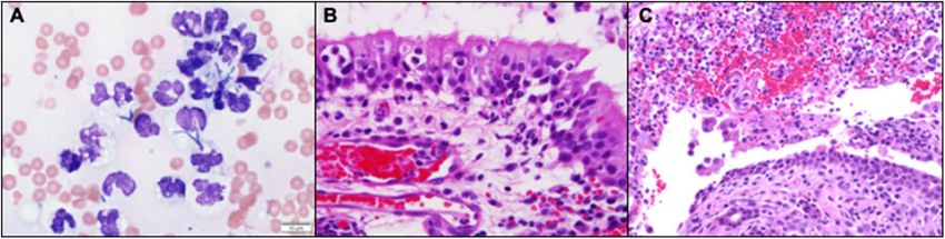
Moghaddam et al.                                                                                                        Feline Primary Bacterial Rhinosinusitis Blindness

 FIGURE 1 | (A) Transverse plane post-contrast, white arrows show the contrast enhancement of the nasal mucosal tissue. There is also soft tissue abutting the
 osseous scrolls of the caudal nasal passage. The choanae are obscured by soft tissue (#) (B) Transverse plane post-contrast, there is dependent soft tissue within the
 right frontal sinus (*). There is also a white arrow showing enhancement of the mucosal tissue in the sphenoidal sinus and non-contrast enhancing tissue in the
 nasopharynx (#). (C) Transverse plane post-contrast, white chevrons show enhancing optic nerve bilaterally. (D) Dorsal oblique plane post-contrast, white chevrons
 highlight the optic discs.

 FIGURE 2 | (A) Nasal biopsy impression smears revealed numerous markedly degenerate neutrophils with intracellular bacteria of variable, but predominantly
 rod-shaped, morphology (Wright-Giemsa, 100x). (B) Neutrophils extravasating from reactive and congested blood vessels and multifocally clustered within the
 respiratory mucosa. (Hematoxylin and Eosin stain). (C) Rounded, desquamated epithelium in a pool of suppurative exudate and hemorrhage that contains gram
 negative bacteria (Hematoxylin and Eosin stain).

total solids increased to 21% and 6.4 g/dL, respectively. On the                       susceptibility panel, the antimicrobial spectrum was expanded
day of discharge (day 4), the cat was bright, alert, responsive,                       to include amoxicillin-clavulanic acid (Clavamox, Zoetis Inc.,
afebrile, and had a normal appetite. The anemia persisted but                          Kalamazoo, MI, USA; 16.9 mg/kg, PO, q 12 h) in addition the
remained static 24 h after the blood transfusion (PCV: 22%; total                      previously prescribed pradofloxacin (7.4 mg/kg, PO, q 24 h) on
solids: 6.8 g/dL). Medical management at the time of discharge                         day 6.
included prednisolone (0.7 mg/kg, PO, q 24 h), Yunnan Baiyo (1                            The cat was presented on day 17 and had an improved
capsule, PO, q 24 h), pradofloxacin (Veraflox, Bayer HealthCare                        appetite, energy, as well as decreased frequency of sneezing and
Animal Health, Shawnee Mission, KS, USA; 7.4 mg/kg, PO,                                epistaxis. Physical examination revealed unchanged ophthalmic
q 24 h), and nebulization with 3 mls of 0.9% saline (q 12 h).                          abnormalities, absence of nasal discharge, and improved stertor.
Histopathologic sections revealed dense sheets of an almost                            A complete blood count was performed and revealed resolution
pure population of neutrophils in which there were a myriad                            of anemia (HCT: 37%). Medical management with prednisolone,
of bacterial colonies that appeared to have a short rod and                            amoxicillin-clavulinic acid, pradofloxacin, Yunnan-Baiyo, and
thin filamentous morphology (Figures 2B,C). Aerobic/anaerobic                          nebulization with 0.9% saline remained unchanged. By day 30
bacterial culture results returned on day 6 and indicated the                          the owner reported that the sneezing persisted but had decreased
presence of E. coli and Actinomyces spp. (Supplemental Table 1)                        in frequency and the epistaxis had ceased. The cat’s vision
with an absence of fungal growth (confirmed on day 33). The                            was reported to be improved but not yet normal. Physical
cat was diagnosed with severe bacterial CRS and presumed optic                         examination revealed a lack of nasal discharge and normal airflow
neuritis associated loss of vision. Based on the antimicrobial                         bilaterally. Ophthalmic examination showed bilaterally mydriatic

Frontiers in Veterinary Science | www.frontiersin.org                              3                                                March 2020 | Volume 7 | Article 122
Moghaddam et al.                                                                                       Feline Primary Bacterial Rhinosinusitis Blindness

pupils, intact pupillary light reflexes, and a menace response             of the edematous tissue and thick mucoid material. Actinomyces
could be elicited from both eyes. Indirect ophthalmoscopy was              infections have been reported to be associated with inhalation or
performed using a condensing lens and Finhoff transillumintor              ingestion of migrating grass awns and penetration of mucosa and
and revealed unremarkable optic discs and retinal vessels. Due to          soft tissue by inhaled foreign bodies (12).
the improvement of clinical signs, the amoxicillin-clavulinic acid,            Therapeutic antimicrobial use in companion animals has
pradofloxacin, and Yunnan Baiyo were discontinued. Therapy                 potential to promote the emergence of resistance in pathogenic
with prednisolone (0.7 mg/kg, PO, q 24 h) and nebulization with            as well as non-target commensal bacteria (13). Therefore, it is
0.9% sodium chloride (q 12 h) was continued.                               essential that antimicrobial therapy be aimed at known or most
   On day 70, the cat was presented for evaluation and the owner           likely pathogens (13). The decision to initiate broad spectrum
reported complete resolution of sneezing, nasal discharge, and             antimicrobial therapy in this cat with ampicillin/sublactam as
continued improvement of vision. The frequency of prednisolone             well as enrofloxacin was made because there were multiple
administration was decreased and subsequently discontinued                 different types of potential pathogens (e.g., intracellular rods and
along with nebulization of saline. Follow-up with the owner by             cocci) identified on cytology of nasal tissue and the cat fulfilled
phone at the time of this writing (day 330) revealed that the cat          the requisite criteria for a diagnosis of sepsis (14). De-escalation
had no sneezing, nasal discharge, and had improved, but not yet            of antimicrobial therapy in the cat was attempted at the time
normal vision.                                                             of hospital discharge because he was stable. Pradofloxacin was
                                                                           chosen as the sole antibiotic at that time because it provides
                                                                           a broad spectrum of pathogen coverage involving both Gram-
DISCUSSION                                                                 negative and Gram-positive aerobic and some anaerobic bacteria
                                                                           (15). In addition, pradofloxacin has a decreased resistance profile
This report documents the clinical features, diagnostic findings,          compared to other fluoroquinolones and would be expected to
advanced imaging, and therapeutic interventions in a cat with              achieve ideal tissue penetration for the deep infection in the
presumed severe primary bacterial CRS and loss of vision                   conchae (15). The bacterial culture and susceptibility diagnostic
caused by E. coli and Actinomyces spp. Chronic rhinosinusitis              results returned on day 6 and indicated that the E. coli was
is a common cause of nasal discharge in cats second only to                susceptible to the fluoroquinolone. However, fluoroquinolones
neoplasia. One small prospective study identified growth of                are not considered active against Actinomyces spp., the second
mixed bacterial organisms from biopsy specimens in 60% (6/10)              cultured bacteria (16). The antimicrobial spectrum was then
of cats with CRS (10). However, that study did not indicate if the         expanded to include amoxicillin-clavulanic acid and was chosen
cats with positive bacterial cultures were treated with antibiotics        based on the susceptibility testing results. Both pathogens
and if this resulted in complete resolution of clinical signs.             were susceptible to amoxicillin-clavulanic acid so the cat could
This is important because it is difficult to determine whether             have potentially been transitioned from pradofloxacin to only
bacteria cultured from the nasal passages are part of the normal           amoxicillin-clavulanic acid.
nasal microbiota, primary pathogens, or are secondary to a                     The cat in this report developed gradual loss of vision
primary etiology (3, 10). The absence of consistent follow-up              that coincided with the onset of sneezing, mucopurulent nasal
data in observational studies that have identified bacterial culture       discharge, and intermittent epistaxis. Rhinosinusitis in humans
positivity in cats with nasal disease limits our understanding of          has been reported to lead to orbital complications such as
the frequency and clinical characteristics of primary pathogenic           optic neuritis and loss of vision. Vision loss in these cases have
bacterial CRS. To the authors’ knowledge, this is the first                been proposed to result from four different pathophysiologic
report of a cat with presumed primary bacterial CRS with                   mechanisms including: (1) mechanical compression of the
extensive follow-up.                                                       optic nerve (e.g., abscess or mucoceles), (2) direct extension
   Evidence suggesting that E. coli, Actinomyces spp., or                  of the infection to the optic nerve, (3) secondary to orbital
both were the primary cause of nasal disease in this cat                   inflammatory changes causing optic neuritis, and (4) venous
includes: (a) there was a complete and sustained resolution                congestion of the optic nerve due to thrombophlebitis and
of all clinical signs related to nasal disease subsequent to               retinal artery occlusion due to increased pressures in the
antibiotic administration and (b) the absence of sneezing or               orbit (8, 17–20).
nasal discharge in the first 6.5 years of life. It is unclear                  The specific cause for the development of optic neuritis and
as to the reason this cat developed bacterial CRS at 7                     loss of vision in this cat is unknown but believed to be associated
years of age. The cat had no clinical history of developing                with direct extension of infection, inflammation, or both, into
other persistent infections, making primary immunodeficiency               the orbits causing optic neuritis. The initial fundic examination
unlikely. Secondary immunodeficiency cannot be ruled out,                  revealed bilaterally swollen optic discs and prominent retinal
but is also unlikely due to the negative FeLV and FIV status.              vessels, likely a result of an inflammatory process. Further,
It remains a possibility that the cat had a foreign body that              the CT revealed abnormal contrast enhancement of the optic
incited inflammation and was the source of bacterial infection.            discs and optic nerve bilaterally. This assumption could further
A non-dense object would have been difficult to identify with              be supported by the gradual restoration of the cat’s vision
CT, especially among the copious soft tissue-attenuating mucoid            after treatment with antibiotics and steroids, decreasing the
material in the nasal cavity (11). Likewise, a foreign body                inflammation, and allowing the optic nerve to recover. A vascular
would have been difficult to visualize with endoscopy because              etiology causing optic neuritis and loss of vision in this cat is

Frontiers in Veterinary Science | www.frontiersin.org                  4                                           March 2020 | Volume 7 | Article 122
Moghaddam et al. Feline Primary Bacterial Rhinosinusitis Blindness
possible but unlikely because of important anatomical differences thromboelastography parameters (24, 25). While the evidence
between cats and people. The main supply of blood to the eye supporting Yunnan Baiyo in the veterinary literature is mixed,
and orbit in cats is via the internal maxillary artery, which the drug is well-tolerated with minimal reported adverse effects
branches to give rise to the external ophthalmic artery. However, (24, 25).
the overall blood supply to the cat eye is quite diverse. In
comparison, the majority of orbital circulation in people is CONCLUSION
supplied via the internal ophthalmic artery, which traverses
within the optic nerve. Inflammation or thromboses of the The cat presented in this report demonstrates that bacterial
internal ophthalmic artery in people would likely affect both CRS should be considered in cats with severe nasal disease
the optic nerve and vision, yet it would be unlikely for a single with and without loss of vision, irrespective of age. In addition,
vascular event within the external ophthalmic artery in cats early recognition and therapeutic intervention are important in
to have the same net result (21). A limitation of this report mitigating severe sequela of infection including, but not limited
was the lack of magnetic resonance imaging, cerebrospinal fluid to, optic neuritis and vision loss.
diagnostic evaluation, or electroretinography, which could have
strengthened the supposition that the etiology of blindness was
DATA AVAILABILITY STATEMENT
optic neuritis.
Whilst the cat’s vision remained diminished, the owner The raw data supporting the conclusions of this article will be
reported a subjective improvement over time. The owner made available by the authors, without undue reservation, to any
reported that the cat was able to play with toys, navigate through qualified researcher.
the house, and jump on surfaces though he occasionally missed
the intended mark. Importantly, the cat’s vision was not assessed
objectively to definitively corroborate the owner’s subjective AUTHOR CONTRIBUTIONS
evaluation of the cat’s vision. It is unknown if the cat reported
RM, JJ, EH, AB, and KW contributed management of case,
here will continue to regain vision over time. Improvement in
collection of data, writing and editing manuscript, and review
vision of humans with rhinosinusitis associated optic neuritis
final submission.
following aggressive medical (i.e., antibiotics ± corticosteroids),
surgical intervention, or both ranges from permanent blindness
to restitution of normal vision (8). The cat in this report was ACKNOWLEDGMENTS
treated with oral Yunnan Baiyo following acquisition of nasal
biopsies in an effort to decrease hemorrhage. There are mixed We would like to thank Dr. Liz Schaefer for her assistance
results on the efficacy of Yunnan Baiyo in companion animals. in ophthalmology literature review. The authors would also
One randomized, controlled, blinded study showed that dogs like to thank Paige Hunsinger and Heather Hotchkiss for their
that had received Yunnan Baiyo the day before nasal biopsies technical expertise.
experienced significantly shorter time to cessation of bleeding
compared to control (22). Another study in dogs revealed that SUPPLEMENTARY MATERIAL
Yunnan Baiyo administration resulted in increased strength
of clots measured by thromboelastography (23). However, The Supplementary Material for this article can be found
other studies in apparently healthy dogs and cats highlighted online at: https://www.frontiersin.org/articles/10.3389/fvets.
that Yunnan Baiyo administration did not significantly alter 2020.00122/full#supplementary-material
REFERENCES 7. Sultesz M, Csakanyi Z, Majoros T, Farkas Z, Katona G. Acute bacterial
rhinosinusitis and its complications in our pediatric otolaryngological
1. Michiels L, Day MJ, Snaps F, Hansen P, Clercx C. A retrospective study of non- department between 1997 and 2006. Int J Pediatr Otorhinolaryngol. (2009)
specific rhinitis in 22 cats and the value of nasal cytology and histopathology. 73:1507–12. doi: 10.1016/j.ijporl.2009.04.027
J Feline Med Surg. (2003) 5:279–85. doi: 10.1016/S1098-612X(03)00044-5 8. Celakovsky P, Vokurka J, Skoloudik L, Ružička J. Optic neuritis
2. Henderson SM, Bradley K, Day MJ, Tasker S, Caney SMA, Moore AH, et al. and paranasal sinus diseases. Cent Eur J Med. (2010) 6:117–9.
Investigation of nasal disease in the cat – a retrospective study of 77 cases. J doi: 10.2478/s11536-010-0069-7
Feline Med Surg. (2006) 6:245–57. doi: 10.1016/j.jfms.2003.08.005 9. Wan Y, Shi G, Wang H. Treatment of orbital complications following
3. Demko JL, Cohn LA. Chronic nasal discharge in cats: 75 cases (1993-2004). J acute rhinosinusitis in children. Balkan Med J. (2016) 33:401–6.
Am Vet Med Assoc. (2007) 230:1032–7. doi: 10.2460/javma.230.7.1032 doi: 10.5152/balkanmedj.2016.141065
4. Cohn L. Feline respiratory disease complex. Vet Clin North Am Small Anim 10. Johnson LR, Foley JE, De Cock HEV, Clarke HE, Maggs DJ. Assessment of
Pract. (2011) 41:1273–89. doi: 10.1016/j.cvsm.2011.07.006 infectious organisms associated with chronicrhinosinusitis in cats. J Am Vet
5. Ferguson S, Smith KC, Welsh CE, Dobromylskyj MJ. A retrospective study Med Assoc. (2005) 227:579–85. doi: 10.2460/javma.2005.227.579
of more than 400 feline nasal biopsy samples in the UK (2006-2013). J Feline 11. Captanian N, Palma D. Limitation of computed tomography in identifying
Med Surg. (2019) doi: 10.1177/1098612X19881847. [Epub ahead of print]. intranasal porcupine quills in a dog (canis lupus familiaris). J Am Anim Hosp
6. Sharma D, Pakravan N, Pritchard JC, Hartmann FA, Young KM. Mucoid Assoc. (2019) 55:e55404. doi: 10.5326/JAAHA-MS-6697
Pseudomonas aeruginosa infection in a cat with severe chronic rhinosinusitis. 12. Schultz RM, Zwingenberger A. Radiographic, computed tomographic,
Vet Clin Pathol. (2019) 48:300–4. doi: 10.1111/vcp.12749 and ultrasonographic findings with migrating intrathoracic grass
Frontiers in Veterinary Science | www.frontiersin.org 5 March 2020 | Volume 7 | Article 122Moghaddam et al. Feline Primary Bacterial Rhinosinusitis Blindness
awns in dogs and cats. Vet Radiol Ultrasound. (2008) 49:249–55. 22. Adelman LB, Olin SJ, Egger CM, Stokes JE. The effect of oral Yunnan Baiyo on
doi: 10.1111/j.1740-8261.2008.00360.x periprocedural hemorrhage and coagulation parameters in dogs undergoing
13. Weese JS, Giguere S, Guardabassi L, Morley PS, Papich M, Ricciuto DR, nasal biopsy: a randomized, controlled, blinded study. Am J Traditional
et al. ACVIM consensus statement on therapeutic antimicrobial use in Chinese Vet Med. (2017) 12:29–38.
animals and antimicrobial resistance. J Vet Intern Med. (2015) 29:487–98. 23. Tansey C, Wiebe ML, Hybki GC, Patlogar JE, Murphy L, Bianco
doi: 10.1111/jvim.12562 D, et al. A prospective evaluation of oral Yunnan Baiyo therapy on
14. DeClue AE, Delgado C, Chang C, Sharp CR. Clinical and immunologic thromboelastographic parameters in apparently healthy dogs. J Vet Emerg Crit
assessment of sepsis and the systemic inflammatory response syndrome in Care. (2018) 28:221–5. doi: 10.1111/vec.12712
cats. J Am Vet Med Assoc. (2011) 238:890–7. doi: 10.2460/javma.238.7.890 24. Patlogar JE, Tansey C, Wiebe M, Hybki GC, Trostel T, Murphy L,
15. Spindel ME, Veir J, Radecki SV. Evaluation of pradofloxacin for et al. A prospective evaluation of oral Yunnan Baiyo therapy on
the treatment of feline rhinitis. J Feline Med Surg. (2008) 10:472–9. thromboelastographic parameters in apparently healthy cats. J Vet Emerg Crit
doi: 10.1016/j.jfms.2008.04.003 Care. (2019) 29:611–5. doi: 10.1111/vec.12895
16. Valour F, Senechal A, Dupieux C, Karsenty J, Lustig S, Breton P, 25. Frederick J, Boysen S, Wagg C, Chalhoub S. The effects of oral administration
et al. Actinomycosis: etiology, clinical features, diagnosis, treatment, and of Yunnan Baiyo of blood coagulation in beagle dogs as measured by kaolin-
management. Infect Drug Resist. (2014) 7:183–97. doi: 10.2147/IDR.S39601 activated thromboelastography and buccal mucosal bleeding times. Can J Vet
17. Nien C, Lee C, Wu P, Chen HC, Chi JCY, Sun CC, et al. The development Res. (2017) 81:41–5.
of optic neuropathy after chronic rhinosinusitis: A population-based cohort
study. PLoS ONE. (2019) 14:e0220286. doi: 10.1371/journal.pone.0220286 Conflict of Interest: The authors declare that the research was conducted in the
18. Rothstein J, Maisel RH, Berlinger NT, Wirtschafter JD. Relationship of optic absence of any commercial or financial relationships that could be construed as a
neuritis to disease of the paranasal sinuses. Laryngoscope. (1984) 94:1501–8. potential conflict of interest.
doi: 10.1288/00005537-198411000-00023
19. Awerbuch G, Labadie EL, Van Dalen JTW. Reversible optic neuritis secondary Copyright © 2020 Moghaddam, Jaffey, Hostnik, Brower and Wycislo.
to paranasal sinusitis. Eur Neurol. (1989) 29:189–93. doi: 10.1159/000116409 This is an open-access article distributed under the terms of the Creative
20. Lee L, Huang C, Lee T. Prolonged visual disturbance secondary to Commons Attribution License (CC BY). The use, distribution or reproduction
isolated sphenoid sinus disease. Laryngoscope. (2004) 114:986–90. in other forums is permitted, provided the original author(s) and the
doi: 10.1097/00005537-200406000-00006 copyright owner(s) are credited and that the original publication in this
21. Samuelson DA. Ophthalmic anatomy. In: Gelatt KN, Gilger BC, Kern TJ, journal is cited, in accordance with accepted academic practice. No use,
editors. Veterinary Ophthamology. 5th ed. Ames, IA: Wiley-Blackwell (2013). distribution or reproduction is permitted which does not comply with these
p. 39–170. terms.
Frontiers in Veterinary Science | www.frontiersin.org 6 March 2020 | Volume 7 | Article 122You can also read